Zinc is an essential trace element for spermatogenesis Sonoko Yamaguchi a , Chiemi Miura a , Kazuya Kikuchi b , Fritzie T. Celino a , Tetsuro Agusa c,1 , Shinsuke Tanabe c , and Takeshi Miura a,2 a Research Group for Reproductive Physiology, South Ehime Fisheries Research Center, Ehime University, 1289-1, Funakoshi, Ainan, Ehime 798-4131, Japan; b Graduate School of Engineering, Osaka University, Osaka 565-0871, Japan; and c Center for Marine Environmental Science, Ehime University, Matsuyama 790-8577, Japan Edited by Ryuzo Yanagimachi, University of Hawaii, Honolulu, HI, and approved May 8, 2009 (received for review January 19, 2009) Zinc (Zn) plays important roles in various biological activities but there is little available information regarding its functions in spermatogen- esis. In our current study, we further examined the role of Zn during spermatogenesis in the Japanese eel (Anguilla japonica). Human CG (hCG) was injected into the animals to induce spermatogenesis, after which the concentration of Zn in the testis increased in tandem with the progression of spermatogenesis. Staining of testicular cells with a Zn-specific fluorescent probe revealed that Zn accumulates in germ cells, particularly in the mitochondria of spermatogonia and sperma- tozoa. Using an in vitro testicular organ culture system for the Japanese eel, production of a Zn deficiency by chelation with N,N,N,N-tetrakis (2-pyridylemethyl)ethylenediamine (TPEN) caused apoptosis of the germ cells. However, this cell death was rescued by the addition of Zn to the cultures. Furthermore, an induced deficiency of Zn by TPEN chelation was found to inhibit the germ cell prolifer- ation induced by 11-ketotestosterone (KT), a fish specific androgen, 17,20-dihydroxy-4-pregnen-3-one (DHP), the initiator of meiosis in fish, and estradiol-17(E2), an inducer of spermatogonial stem-cell renewal. We also investigated the effects of Zn deficiency on sperm motility and observed that TPEN treatment of eel sperm suppressed the rate and duration of their motility but that co-treatment with Zn blocked the effects of TPEN. Our present results thus suggest that Zn is an essential trace element for the maintenance of germ cells, the progression spermatogenesis, and the regulation of sperm motility. apoptosis germ cells in vitro culture Japanese eel sperm motility Z inc (Zn) is well known as an essential trace element for a variety of biological activities. In biological systems, Zn is present in protein-bound and ionic forms, and plays important roles in mediating the function and structure of proteins, and in maintaining physiological balance. In vertebrates, Zn accumu- lates in the testis at high levels which are comparable to those in liver and kidney (1). In epidemiological studies in human, the inhibition of spermatogenesis and sperm abnormalities have been observed in patients with Crohn’s disease and nutritional disorders, both of which induce a Zn deficiency (1–3). In vivo experiments in rodents have also demonstrated that a Zn deficiency can cause severe damage to the testes such as atrophy of the testicular tubules and the inhibition of spermatid differ- entiation (4, 5). Moreover, there are some reports that exposure to Zn can alleviate testis damage by stresses such as heavy metals, fluoride, and heat (6). These findings suggest that the testes may harbor a Zn-incorporation system, and that Zn itself may exert protective effect against testicular injury and play an essential role in the maintenance of testicular functions. However, there has been no evidence reported to date that shows any direct effects of Zn upon spermatogenesis in vertebrates. In contrast to spermatogenesis, the effects of Zn on sperm motility have been examined in a number of vertebrate and invertebrate species. In humans, sperm motility declines in association with increased Zn concentrations in the seminal plasma (7). Morisawa and Yoshida have also reported that Zn in the seminal plasma of human suppresses sperm motility, and that the removal of Zn by binding to a protein named semenogelin enhances motility (6). On the other hand, in sea urchin, treat- ment with the bivalent metal ion chelator, ethylenediamine tetra acetic acid (EDTA), inhibits sperm motility that is reversed by the addition of Zn (9). These results suggest that extracellular Zn indeed affects sperm motility but whether this is inhibitory or stimulatory appears to be species-specific. Additionally, it has been reported that Zn is present in sperm mitochondria and f lagella (10, 11) but there had been no reports to date concerning the role of intracellular Zn upon sperm function. To further study the role of Zn upon spermatogenesis in our current study, we chose Japanese eel (Anguilla japonica) as our animal model. In the Japanese eel in vivo, a complete pathway of spermatogenesis, from the spermatogonia stage to sperm maturation, can be induced by the injection of human CG (hCG; 12). Furthermore, we have developed a testicular organ culture system for the Japanese eel in our laboratory, which is the only currently available system of its kind in which the induction of complete spermatogenesis can be performed in vitro by the addition of 11-ketotestosterone or hCG (13, 14). By in vivo and in vitro analyses of spermatogenesis in the Japanese eel, we have previously further clarified the regulatory mechanisms underly- ing fish spermatogenesis (15, 16). Additionally, we have revealed the inhibitory effects of 4 trace elements (lead, molybdenum, rubidium, and arsenic) on fish spermatogenesis using our in vitro testicular organ culture system (17). In our present study, we again used the Japanese eel model to investigate the concentra- tion and distribution of Zn in testis during spermatogenesis. Moreover we examined the effects of Zn addition and deficiency on spermatogenesis and sperm motility in vitro. Results Changes in the Levels and Distribution of Zinc (Zn) in the Testis of the Japanese Eel during Spermatogenesis. Before injection with hCG, the concentration of Zn in the testis of the Japanese eel was approximately 50 g/g. After injection, the Zn concentration in the testis gradually increased, and the highest levels were ob- served on day 9. Thereafter, the concentration of Zn remained at high levels until day 18 (Fig. 1). To detect the distribution of Zn in eel testes, an unfixed testicular fragment was stained with a fluorescence sensor for Zn(II), ZnAF-2DA. Strong f luorescent signals were obtained in the lobules but not in the interstitial tissue (Fig. 2A and B). We thus further investigated the distribution of Zn in testicular tissue using isolated cells. Germ cells were found to be strongly stained Author contributions: S.Y., C.M., and T.M. designed research; S.Y., C.M., F.T.C., T.A., S.T., and T.M. performed research; K.K. contributed new reagents/analytic tools; S.Y., C.M., and T.M. analyzed data; and S.Y., C.M., and T.M. wrote the paper. The authors declare no conflict of interest. This article is a PNAS Direct Submission. 1 Present address: Faculty of Medicine, Shimane University, Izumo, Shimane 693-8501, Japan. 2 To whom correspondence should be addressed. E-mail: [email protected]. www.pnas.orgcgidoi10.1073pnas.0900602106 PNAS June 30, 2009 vol. 106 no. 26 10859 –10864 PHYSIOLOGY
Transcript
Zinc is an essential trace elementfor spermatogenesisSonoko Yamaguchia, Chiemi Miuraa, Kazuya Kikuchib, Fritzie T. Celinoa, Tetsuro Agusac,1, Shinsuke Tanabec,and Takeshi Miuraa,2
aResearch Group for Reproductive Physiology, South Ehime Fisheries Research Center, Ehime University, 1289-1, Funakoshi, Ainan, Ehime 798-4131,Japan; bGraduate School of Engineering, Osaka University, Osaka 565-0871, Japan; and cCenter for Marine Environmental Science, EhimeUniversity, Matsuyama 790-8577, Japan
Edited by Ryuzo Yanagimachi, University of Hawaii, Honolulu, HI, and approved May 8, 2009 (received for review January 19, 2009)
Zinc (Zn) plays important roles in various biological activities but thereis little available information regarding its functions in spermatogen-esis. In our current study, we further examined the role of Zn duringspermatogenesis in the Japanese eel (Anguilla japonica). Human CG(hCG) was injected into the animals to induce spermatogenesis, afterwhich the concentration of Zn in the testis increased in tandem withthe progression of spermatogenesis. Staining of testicular cells witha Zn-specific fluorescent probe revealed that Zn accumulates in germcells, particularly in the mitochondria of spermatogonia and sperma-tozoa. Using an in vitro testicular organ culture system for theJapanese eel, production of a Zn deficiency by chelation withN,N,N�,N�-tetrakis (2-pyridylemethyl)ethylenediamine (TPEN) causedapoptosis of the germ cells. However, this cell death was rescued bythe addition of Zn to the cultures. Furthermore, an induced deficiencyof Zn by TPEN chelation was found to inhibit the germ cell prolifer-ation induced by 11-ketotestosterone (KT), a fish specific androgen,17�,20�-dihydroxy-4-pregnen-3-one (DHP), the initiator of meiosis infish, and estradiol-17� (E2), an inducer of spermatogonial stem-cellrenewal. We also investigated the effects of Zn deficiency on spermmotility and observed that TPEN treatment of eel sperm suppressedthe rate and duration of their motility but that co-treatment with Znblocked the effects of TPEN. Our present results thus suggest that Znis an essential trace element for the maintenance of germ cells, theprogression spermatogenesis, and the regulation of sperm motility.
apoptosis � germ cells � in vitro culture � Japanese eel � sperm motility
Z inc (Zn) is well known as an essential trace element for avariety of biological activities. In biological systems, Zn is
present in protein-bound and ionic forms, and plays importantroles in mediating the function and structure of proteins, and inmaintaining physiological balance. In vertebrates, Zn accumu-lates in the testis at high levels which are comparable to those inliver and kidney (1). In epidemiological studies in human, theinhibition of spermatogenesis and sperm abnormalities havebeen observed in patients with Crohn’s disease and nutritionaldisorders, both of which induce a Zn deficiency (1–3). In vivoexperiments in rodents have also demonstrated that a Zndeficiency can cause severe damage to the testes such as atrophyof the testicular tubules and the inhibition of spermatid differ-entiation (4, 5). Moreover, there are some reports that exposureto Zn can alleviate testis damage by stresses such as heavy metals,f luoride, and heat (6). These findings suggest that the testes mayharbor a Zn-incorporation system, and that Zn itself may exertprotective effect against testicular injury and play an essentialrole in the maintenance of testicular functions. However, therehas been no evidence reported to date that shows any directeffects of Zn upon spermatogenesis in vertebrates.
In contrast to spermatogenesis, the effects of Zn on spermmotility have been examined in a number of vertebrate andinvertebrate species. In humans, sperm motility declines inassociation with increased Zn concentrations in the seminalplasma (7). Morisawa and Yoshida have also reported that Zn inthe seminal plasma of human suppresses sperm motility, and that
the removal of Zn by binding to a protein named semenogelinenhances motility (6). On the other hand, in sea urchin, treat-ment with the bivalent metal ion chelator, ethylenediamine tetraacetic acid (EDTA), inhibits sperm motility that is reversed bythe addition of Zn (9). These results suggest that extracellular Znindeed affects sperm motility but whether this is inhibitory orstimulatory appears to be species-specific. Additionally, it hasbeen reported that Zn is present in sperm mitochondria andflagella (10, 11) but there had been no reports to date concerningthe role of intracellular Zn upon sperm function.
To further study the role of Zn upon spermatogenesis in ourcurrent study, we chose Japanese eel (Anguilla japonica) as ouranimal model. In the Japanese eel in vivo, a complete pathwayof spermatogenesis, from the spermatogonia stage to spermmaturation, can be induced by the injection of human CG (hCG;12). Furthermore, we have developed a testicular organ culturesystem for the Japanese eel in our laboratory, which is the onlycurrently available system of its kind in which the induction ofcomplete spermatogenesis can be performed in vitro by theaddition of 11-ketotestosterone or hCG (13, 14). By in vivo andin vitro analyses of spermatogenesis in the Japanese eel, we havepreviously further clarified the regulatory mechanisms underly-ing fish spermatogenesis (15, 16). Additionally, we have revealedthe inhibitory effects of 4 trace elements (lead, molybdenum,rubidium, and arsenic) on fish spermatogenesis using our in vitrotesticular organ culture system (17). In our present study, weagain used the Japanese eel model to investigate the concentra-tion and distribution of Zn in testis during spermatogenesis.Moreover we examined the effects of Zn addition and deficiencyon spermatogenesis and sperm motility in vitro.
ResultsChanges in the Levels and Distribution of Zinc (Zn) in the Testis of theJapanese Eel during Spermatogenesis. Before injection with hCG,the concentration of Zn in the testis of the Japanese eel wasapproximately 50 �g/g. After injection, the Zn concentration inthe testis gradually increased, and the highest levels were ob-served on day 9. Thereafter, the concentration of Zn remainedat high levels until day 18 (Fig. 1).
To detect the distribution of Zn in eel testes, an unfixedtesticular fragment was stained with a fluorescence sensor forZn(II), ZnAF-2DA. Strong fluorescent signals were obtained inthe lobules but not in the interstitial tissue (Fig. 2A and B). Wethus further investigated the distribution of Zn in testicular tissueusing isolated cells. Germ cells were found to be strongly stained
Author contributions: S.Y., C.M., and T.M. designed research; S.Y., C.M., F.T.C., T.A., S.T.,and T.M. performed research; K.K. contributed new reagents/analytic tools; S.Y., C.M., andT.M. analyzed data; and S.Y., C.M., and T.M. wrote the paper.
The authors declare no conflict of interest.
This article is a PNAS Direct Submission.
1Present address: Faculty of Medicine, Shimane University, Izumo, Shimane 693-8501,Japan.
2To whom correspondence should be addressed. E-mail: [email protected].
by ZnAF-2DA but Sertoli cells showed no signal (Fig. 2 C andD). When the germ cells were treated with 10 mM N,N,N�,N�-tetrakis(2-pyridylemethyl) ethylenediamine (TPEN) for 1 hbefore staining with ZnAF-2DA, fluorescence was not detected(Fig. 2 E and F). We also stained the germ cells at various stageswith ZnAF-2DA, that is, spermatogonia, spermatocytes, sper-matids, and spermatozoa. ZnAF-2DA signals were detectable inspermatogonia, most notably in the mitochondria (Fig. 3 A–C).Additionally, the mitochondria of the spermatids and sperma-tozoa also displayed strong ZnAF-2DA signals (Fig. 3 D and E).
Effects of Zn and Zn Chelators on Japanese Eel Testes in Vitro. Toinvestigate the putative key role of Zn during spermatogenesis,we analyzed the direct effects of Zn on the testis in the presenceor absence of 11-ketotestosterone (KT). After culturing for 6days, testicular fragments in the control group were found to beoccupied by type A spermatogonia. Although the histologicalstructure of the testicular fragments cultured with KT alone didnot differ from the control group, the incorporation ratio ofBrdU into the germ cells had significantly increased (Fig. 4), as
also reported in our previous study (13). Treatment of theJapanese eel testicular fragments with any level of Zn with orwithout KT did not affect the histology of the testis or the BrdUindex (Fig. 4A). Treatment with ethylenediamine-N,N,N�,N�-tetraacetic acid, calcium(II), disodium salt (Ca-EDTA), anextracellular Zn chelator, also did not affect the BrdU index ortesticular morphology after 6 days in culture (Figs. 4B and 5B).In contrast, exposure to 0.01 and 0.1 mM TPEN, an intracellularchelator of Zn, inhibited BrdU-incorporation into germ cells(Fig. 4B), and induced germ cell death (Figs. 4B and 5C).Significantly, both the cell death and the inhibition of BrdUincorporation induced by TPEN was rescued by the addition ofZn (Figs. 4B and 5D). We further investigated the type of celldeath that occurred using a TdT-mediated dUTP nick-end
Fig. 1. Changes in the Zn concentrations in the testis of the Japanese eelafter injection of human CG (hCG). The different letters indicate statisticallysignificant differences (P � 0.05).
Fig. 2. The zinc distribution in the testis of the Japanese eel determined bystaining with a Zn-specific fluorescent probe, ZnAF-2DA (A, C, and E). Bright fieldimagesarealsoshown(B,D, andF). (AandB) testicular fragmentsof theJapaneseeel at 15 days after injection of hCG; (C and D) germ cells and Sertoli cells; (E andF) TPEN-treated germ cells. (Scale bars: A and B, 100 �m; C–F, 20 �m.)
Fig. 3. Zinc distribution in germ cells of the Japanese eel. Zn was stainedusing ZnAF-2DA. Fluorescence images are shown for (A) Zn, and (B) mitochon-dria. (C) Bright field image of spermatogonia. (D) Zn fluorescence and (E)bright field image of spermatids and spermatozoa. M, mitochondria; St,spermatid; Sz, spermatozoa. (Scale bars: 10 �m.)
Fig. 4. Effects of Zn and Zn chelators on the early stages of spermatogenesisin vitro. The BrdU-labeling index was determined for germ cells in testicularfragments cultured with Zn (A) or Zn chelators (B) with or without KT. Thenumber of BrdU-positive germ cells is expressed as a percentage of the totalnumber of germ cells. C, control; Zn, ZnCl2; KT, 11- ketotestosterone; TPEN,N,N,N�,N�-tetrakis(2-pyridylmethyl)ethylenediamine; CaEDTA, ethylenedia-mine -N,N,N�,N�- tetraacetic acid, calcium(II), disodium salt, dihydrate. Resultsare given as the mean � SEM. The different letters on the columns indicatestatistically significant differences (P � 0.05).
10860 � www.pnas.org�cgi�doi�10.1073�pnas.0900602106 Yamaguchi et al.
labeling (TUNEL) assay after a 1-day culture in the presence ofTPEN. In the control and KT-treatment groups, no cell stainingwas observed (Fig. 5E). However, TUNEL-positive germ cellswere detectable after treatment with 0.01–0.1 mM TPEN withor without KT (Fig. 5F).
We also investigated the effects of 0.001 mM TPEN, a dosethat does not cause cell death, upon KT-induced spermatogen-esis using our testicular organ culture system. Treatment withthis dosage for 6 days had no effects on the histological structureof the testicular fragments. In contrast, after 15 days of thistreatment, the testicular fragments were found to only have typeA spermatogonia, although those cultured with KT alone con-tained the more progressed germ cells, type B spermatogonia(Fig. 6 A–C). Importantly, the addition of Zn led to a recoveryof spermatogenesis, such that the TPEN/KT treated culturesresembled those exposed to KT alone (Fig. 6D).
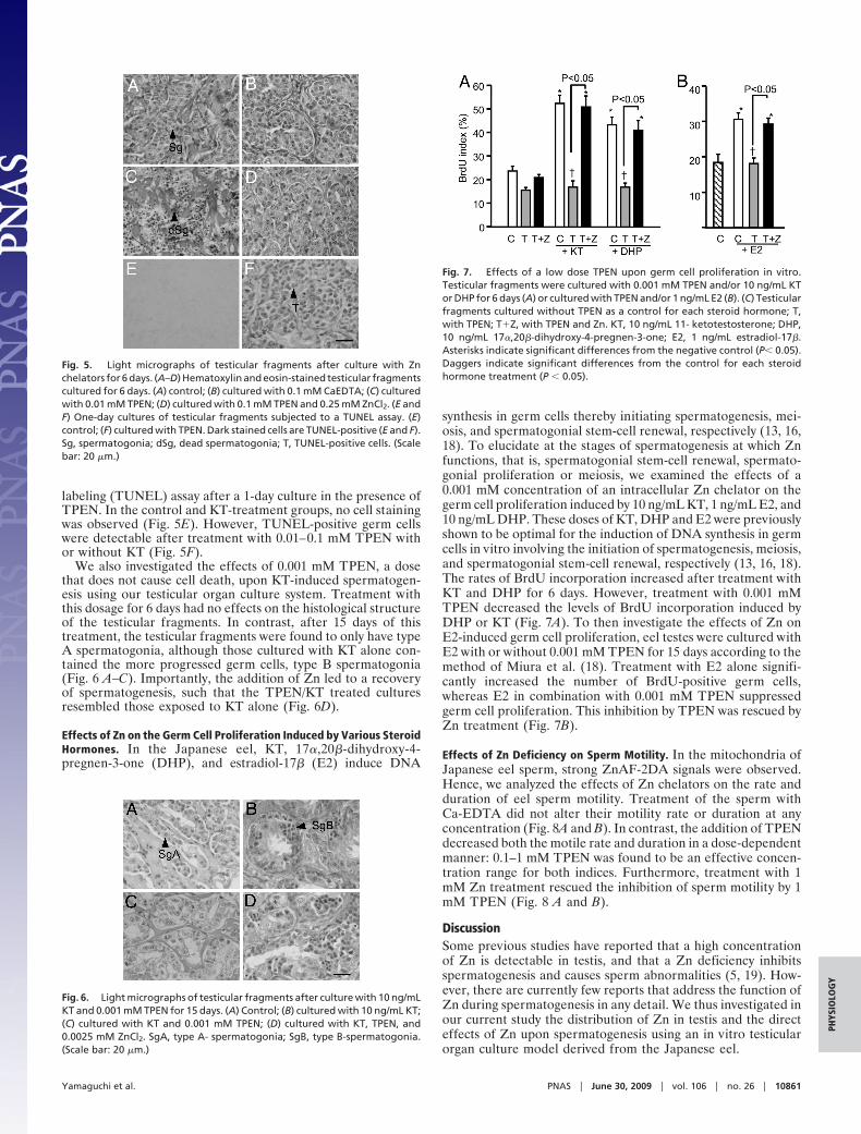
Effects of Zn on the Germ Cell Proliferation Induced by Various SteroidHormones. In the Japanese eel, KT, 17�,20�-dihydroxy-4-pregnen-3-one (DHP), and estradiol-17� (E2) induce DNA
synthesis in germ cells thereby initiating spermatogenesis, mei-osis, and spermatogonial stem-cell renewal, respectively (13, 16,18). To elucidate at the stages of spermatogenesis at which Znfunctions, that is, spermatogonial stem-cell renewal, spermato-gonial proliferation or meiosis, we examined the effects of a0.001 mM concentration of an intracellular Zn chelator on thegerm cell proliferation induced by 10 ng/mL KT, 1 ng/mL E2, and10 ng/mL DHP. These doses of KT, DHP and E2 were previouslyshown to be optimal for the induction of DNA synthesis in germcells in vitro involving the initiation of spermatogenesis, meiosis,and spermatogonial stem-cell renewal, respectively (13, 16, 18).The rates of BrdU incorporation increased after treatment withKT and DHP for 6 days. However, treatment with 0.001 mMTPEN decreased the levels of BrdU incorporation induced byDHP or KT (Fig. 7A). To then investigate the effects of Zn onE2-induced germ cell proliferation, eel testes were cultured withE2 with or without 0.001 mM TPEN for 15 days according to themethod of Miura et al. (18). Treatment with E2 alone signifi-cantly increased the number of BrdU-positive germ cells,whereas E2 in combination with 0.001 mM TPEN suppressedgerm cell proliferation. This inhibition by TPEN was rescued byZn treatment (Fig. 7B).
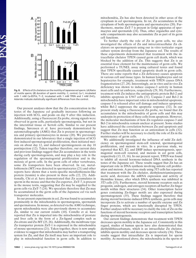
Effects of Zn Deficiency on Sperm Motility. In the mitochondria ofJapanese eel sperm, strong ZnAF-2DA signals were observed.Hence, we analyzed the effects of Zn chelators on the rate andduration of eel sperm motility. Treatment of the sperm withCa-EDTA did not alter their motility rate or duration at anyconcentration (Fig. 8A and B). In contrast, the addition of TPENdecreased both the motile rate and duration in a dose-dependentmanner: 0.1–1 mM TPEN was found to be an effective concen-tration range for both indices. Furthermore, treatment with 1mM Zn treatment rescued the inhibition of sperm motility by 1mM TPEN (Fig. 8 A and B).
DiscussionSome previous studies have reported that a high concentrationof Zn is detectable in testis, and that a Zn deficiency inhibitsspermatogenesis and causes sperm abnormalities (5, 19). How-ever, there are currently few reports that address the function ofZn during spermatogenesis in any detail. We thus investigated inour current study the distribution of Zn in testis and the directeffects of Zn upon spermatogenesis using an in vitro testicularorgan culture model derived from the Japanese eel.
Fig. 5. Light micrographs of testicular fragments after culture with Znchelators for 6 days. (A–D) Hematoxylin and eosin-stained testicular fragmentscultured for 6 days. (A) control; (B) cultured with 0.1 mM CaEDTA; (C) culturedwith 0.01 mM TPEN; (D) cultured with 0.1 mM TPEN and 0.25 mM ZnCl2. (E andF) One-day cultures of testicular fragments subjected to a TUNEL assay. (E)control; (F) cultured with TPEN. Dark stained cells are TUNEL-positive (E and F).Sg, spermatogonia; dSg, dead spermatogonia; T, TUNEL-positive cells. (Scalebar: 20 �m.)
Fig. 6. Light micrographs of testicular fragments after culture with 10 ng/mLKT and 0.001 mM TPEN for 15 days. (A) Control; (B) cultured with 10 ng/mL KT;(C) cultured with KT and 0.001 mM TPEN; (D) cultured with KT, TPEN, and0.0025 mM ZnCl2. SgA, type A- spermatogonia; SgB, type B-spermatogonia.(Scale bar: 20 �m.)
Fig. 7. Effects of a low dose TPEN upon germ cell proliferation in vitro.Testicular fragments were cultured with 0.001 mM TPEN and/or 10 ng/mL KTor DHP for 6 days (A) or cultured with TPEN and/or 1 ng/mL E2 (B). (C) Testicularfragments cultured without TPEN as a control for each steroid hormone; T,with TPEN; T�Z, with TPEN and Zn. KT, 10 ng/mL 11- ketotestosterone; DHP,10 ng/mL 17�,20�-dihydroxy-4-pregnen-3-one; E2, 1 ng/mL estradiol-17�.Asterisks indicate significant differences from the negative control (P� 0.05).Daggers indicate significant differences from the control for each steroidhormone treatment (P � 0.05).
Yamaguchi et al. PNAS � June 30, 2009 � vol. 106 � no. 26 � 10861
PHYS
IOLO
GY
Our present analyses show that the Zn concentration in thetestes of the Japanese eel gradually increases following aninjection with hCG, and peaks on day 9 after this induction.Additionally, using a f luorescent Zn probe, strong signals wereobserved in germ cells, particularly spermatogonia, but not inthe interstitial tissue or Sertoli cells. Similar to our presentfindings, Sørensen et al. have previously demonstrated byautometallography (AMG) that Zn is present in spermatogo-nia and primary spermatocytes in mouse (20). We previouslydemonstrated in our laboratory that a single injection of hCGfirst induced spermatogonial proliferation, then initiated mei-osis on about day 12, and induced spermiogenesis on day 18postinjection (12). Taken together therefore, our current dataand previous findings suggest that Zn accumulates in the testisduring early spermatogenesis, and may play a key role in theregulation of the spermatogonial proliferation and in themeiosis of germ cells. In the germ cells of other vertebrates,some Zn transporters have been observed. In rat, metal-lothionein (MT) was detected in spermatocytes (21) and otherreports have shown that a testis-specific metallothionein-likeprotein (tesmin) is also present in these cells (22, 23). Addi-tionally, Chi et al. have demonstrated that Zn accumulates insperm in the mouse and that the Zn-exporter, ZnT-7, is presentin the mouse testis, suggesting that Zn may be supplied to thegerm cells via ZnT-7 (24). We speculate therefore that Zn maybe accumulated in the germ cells of the Japanese eel via suchtransporter molecules.
In our present experiments in eel, Zn was found to accumulateprominently in the mitochondria in spermatogonia, spermatidsand spermatozoa. In mouse, as detected via the AMG technique,sperm mitochondria were also previously shown to accumulateZn, similar to our current results (10). Costello et al. havereported that Zn is imported into the mitochondria of prostateand liver cells in the form of a Zn-ligand complex such asZn-citrate and Zn-MT (25, 26). Additionally, the membrane typeZn transporter protein ZnT-1 is expressed in the mitochondriaof mouse spermatozoa (21). Taken together, there is now ampleevidence to suggest that mitochondria may harbor a transportingsystem for Zn, and that Zn itself may have an important role toplay in mitochondrial function in germ cells. In addition to
mitochondria, Zn has also been detected in other areas of thecytoplasm in eel spermatogonia. In rat, Zn accumulates in thecytoplasm of both spermatogonia and spermatocytes (20). Fur-thermore, murine ZnT-7 is present in Golgi apparatus of sper-macytes and spermatids (24). Thus, other organelles and cyto-solic compartments may also accumulate Zn as part of its germcell functions.
To further clarify the role of Zn in germ cells, we alsoinvestigated the effects of Zn and intra/extracellular Zn ch-elators on spermatogenesis using our in vitro testicular organculture system develop from the Japanese eel. The results ofthese experiments demonstrated that treatment with the in-tracellular chelator TPEN caused germ cell death, which wasblocked by the addition of Zn. This suggests that Zn is anessential trace element for the maintenance of germ cells. Weperformed a TUNEL assay using cultured testes and foundthat TPEN specifically caused apoptotic death in germ cells.There are some reports that a Zn deficiency causes apoptosisin various cell and tissue types. In human lymphocytes and rathepatocytes for example, treatment with TPEN causes DNAfragmentation (27, 28). Interestingly, an in vitro and in vivo Zndeficiency was shown to induce caspase-3 activity in humanmast cells and rat embryos, respectively (29, 30). Furthermore,treatment with Zn induces the antiapoptotic protein Bcl-2 andinhibits apoptosis in U947 cells (31). Caspase-3 and Bcl-2 inmitochondria have important roles in mitochondrial apoptosis;caspase-3 is released after cell damage and induces apoptosis,whilst Bcl-2 suppresses the apoptotic response (32). In ourpresent study using a f luorescent Zn probe, we found that Znaccumulates in the mitochondria of germ cells and this mayunderpin its protection of these cells from apoptosis. However,the molecular mechanisms of how Zn regulates caspase-3 andBcl-2 in mitochondria remain unclear at present. Some studieshave addressed the correlation between Zn and apoptosis andsuggest that Zn may function as an antioxidant in cells (32).Further studies will be necessary to clarify the role of Zn in themaintenance of germ cells.
We additionally investigated the influence of mild Zn defi-ciency on spermatogonial stem-cell renewal, spermatogonialproliferation, and meiosis in vitro. In a previous study, wereported that KT, E2, and DHP induce spermatogenesis, sper-magonial stem-cell renewal, and meiosis in eel germ cells,respectively (13, 15, 16). In our present report, TPEN was foundto inhibit all steroid hormone-induced DNA synthesis in thetestes of the Japanese eel. These results suggest that Zn has animportant role in DNA synthesis involving mitotic cell prolifer-ation and meiosis. A previous study using 3T3 cells has reportedthat treatment with the Zn chelator, diethylenetriaminepenta-acetic acid, decreases the mRNA expression and activity ofthymidine kinase, after which DNA synthesis was inhibited in3T3 cells (33). Furthermore, steroid hormone receptors such asprogestin, androgen, and estrogen receptors all harbor Zn fingermotifs within their structures (34). Other transcription factorgenes containing Zn-finger motifs are also expressed duringspermatogenesis (35). These findings suggest therefore thatduring steroid hormone-induced DNA synthesis, germ cells mayincorporate Zn to activate a number of specific enzyme and Znfinger proteins, which are functionally disrupted by TPEN.Further analyses will be necessary to clarify the role of Zn on thefunctions of steroid hormone receptors and transcription factorsduring spermatogenesis.
Our current findings demonstrate that treatment with TPENdecreases sperm motility in the Japanese eel. Consistently in thisregard, studies of human sperm have also demonstrated thatdiethyldithiocarbamate, which is an intracellular Zn chelator,inhibits sperm motility and decreases sperm velocity (36). Theseresults suggest that intracellular Zn is important for spermmotility. As mentioned above, the mitochondria in the sperm of
Fig. 8. Effects of Zn chelators on the motility of Japanese eel sperm. (A) Ratioof motile sperm; (B) duration of sperm motility. C, control; Ca-1, incubatedwith 1 mM Ca-EDTA; T�Z, incubated with 1 mM TPEN and 1 mM ZnCl2.Asterisks indicate statistically significant differences from the control.
10862 � www.pnas.org�cgi�doi�10.1073�pnas.0900602106 Yamaguchi et al.
the Japanese eel accumulate Zn. The ATP synthesized by themitochondria is required for sperm flagella motility (37). Hence,Zn may have a function in mitochondrial ATP synthesis. Addi-tionally, carbonic anhydrase (CA) is necessary for eel spermmotility, and this enzyme is expressed in the sperm membrane.CA catalyzes the reversible hydration of carbon and regulates thepH in various fluids. After spermiation, CA in the eel sperma-tozoa is activated after which it increases the pH and theninduces sperm motility (15). CA is also known to be a Zn-bindingprotein, and its activity is dependent on the Zn concentration(38). Additionally, the removal of Zn from Zn-protein com-plexes extracted from human U87 human glioblastoma-astrocytoma cells by TPEN inhibited the function of the tran-scription factor, Sp1 (39). Although there is currently noinformation on effects of TPEN on CA activity, we speculatethat TPEN may inhibit sperm motility by sequestering Zn awayfrom this enzyme in sperm.
In conclusion, the results of our present study demonstratethat the Zn concentration in testis increases during spermat-ogenesis, and that Zn accumulates mainly in germ cells but notin either interstitial tissue or Sertoli cells. Our in vitro testic-ular organ culture experiments also demonstrated that a Zndeficiency causes the inhibition of DNA synthesis in germ cells,and induces an apoptotic response. Additionally, a Zn defi-ciency was found to suppress sperm motility in the Japanese eelanimal model. These results suggest that Zn is an essentialtrace element for the maintenance and regulation of bothspermatogenesis and sperm motility. However, the detailedmechanisms of Zn action during spermatogenesis remain to beclarified in further studies.
Materials and MethodsAnimals. Cultivated male Japanese eels (180–200 g) were purchased from acommercial supplier and kept in a freshwater tank at 23 °C until use.
Measurement of Zn in Testis During Spermatogenesis. A previous report hasindicated that hCG injection of a cultivated Japanese eel induces a completecycle of spermatogenesis (11). Hence, these animals were injected with 1,000IU/eel of hCG following anesthetization by ethylbenzoate. After injection, thefish were kept in a freshwater tank at 23 °C for 1, 3, 6, 9, 12, 15, and 18 days.Thereafter, hCG-injected eels (n � 5 for each day) were anesthetized anddissected, and the testes were collected and stored at �30 °C until measure-ment of Zn concentration. Before the experiments, testicular fragments weresampled from 5 uninjected eels as an initial control group. The testicularsamples were dried for 12 h at 80 °C. For the analysis of Zn, dried testes weredigested with HNO3 in a microwave oven (ETHOS D, Milestone S.r.l.). Theconcentration of Zn was then measured using an inductively coupled plasma-mass spectrometer (ICP-MS; HP-4500, Hewlett-Packard).
Distribution of Zn in the Testis. We stained both the testicular fragments of theJapanese eel and the cells derived from these tissues with a Zn-specific probe.For this purpose, testis samples collected from the eels were cut into 100-�msections in ice-cold eel Ringer’s solution using a Vibratome 3000 (Vibratome).Testicular cells were also prepared according to Miura et al. (40, 13, 16) for Znstaining. Briefly, testes were harvested and testicular cells were isolated bycollagenase and dispase treatments. After treatment with DNase I, testicularcells were cultured in plastic culture dishes at 20 °C overnight and bothfibroblasts and interstitial cells were allowed to adhere to the bottom of thedish, thus separating these cells from germ cells and Sertoli cells. The germ cellsand Sertoli cells were then collected from the culture dishes and plated incollagen-coated dishes at 20 °C overnight. After this overnight culture, onlythe Sertoli cells adhere to the bottom of the dish. Thereafter, germ cells werecollected in a test tube, and both the germ cells and Sertoli cell preparationswere used to analyze the Zn distribution. Sertoli cells and germ cells could beidentified using a variety of distinguishing characteristics and specific markerexpression. Sertoli cells attached and spread to the bottom of the dish,
whereas germ cells did not attach and appeared spherical in shape. Further-more, only germ cells express the progenstin receptor. We separated germcells and Sertoli cells using this method previously (16).
Before staining of the germ cells, they were attached to a polyL-lysinecoated glass slide. Testicular fragments, attached germ cells and Sertoli cellswere then washed 3 times in eel Ringer’s solution, and incubated with 1 �Mof a permeable Zn-specific probe, Zn-AF 2DA (41) in eel Ringer’s solution for45 min at 20 °C. After this incubation, the cells were washed again in theRinger’s solution for 1 h at 20 °C and analyzed by fluorescence microscopy. Themitochondria of the spermatogonia were stained using MitoTracker Red(Invitrogen Co. Ltd.) according to the manufacturer’s instructions with minormodifications before staining with Zn-AF 2DA.
In Vitro Testicular Organ Cultures. Organ cultures were prepared in accordancewith the method of Miura et al. (13, 42). Male Japanese eels were dissected afteranesthetization with ethylbenzoate. The testes were then collected, placed inice-cold eel Ringer’s solution and dissected into small pieces. Testicular fragmentswere placed on nitrocellulose membranes on top of cylindrical 1.5% agarose gelsand set into a 24-well culture plate. Thereafter, 1 mL of Leibovitz’ L-15 culturemedium (Invitrogen Co. Ltd.) for eels (13) was added into each well with orwithout 0.01–1 mM ZnCl2 (Zn), 0.001–0.1 mM TPEN, or 0.001–0.1 mM Ca-EDTA,which are intracellular and extracellular chelators of Zn, respectively, in combi-nation with or without 10 ng/mL KT. The concentrations of Zn and chelators usedin the in vitro experiments were based on the results obtained from the Znmeasurement in the testis. Testicular fragments were incubated for 6 or 15 daysand then fixed Bouin’s solution for histological analysis.
Analysis of the Effects of a Mild Zn Deficiency upon Germ Cells. Testicularfragments were cultured with 0.001 mM TPEN in combination with 10 ng/mLKT, 1 ng/mL E2, and 10 ng/mL DHP for 6 or 15 days. Thereafter, testicularfragments were fixed and their histology was analyzed as described above.
Detection of Germ Cell Proliferation. The proliferation of Japanese eel germcells was analyzed by immunohistochemical detection of 5-bromo-2-deoxyuridine (BrdU, Amersham Pharmacia Biotech) incorporation into repli-cating DNA. After culture for 6 or 15 days, testicular fragments were labeledwith a 0.5 �l/mL BrdU solution for 18 h at 20 °C, and fixed in Bouin’s solution.The fixed testicular fragments were then embedded in paraffin, cut into 4-�msections, and subjected to immunohistochemistry with a mouse monoclonalanti-BrdU antibody.
TdT-Mediated dUTP Nick-End Labeling (TUNEL) Assay. For the detection ofapoptosis, the TUNEL assay was performed. One-day cultured testicular frag-ments were fixed in Bouin’s solution, cut into 5 �m-thick paraffin sections andthen analyzed using an In Situ Cell Death Detection Kit (Roche Diagnostics,Ltd.) according to the manufacturer’s instructions.
Effects of Zn on Sperm Motility. Eel sperm was collected after injection of theanimals with hCG as described by Ohta et al. (43) and diluted 1:10,000 withartificial seminal plasma (149.3 mM NaCl, 15.2 mM KCl, 1.3 mM CaCl2, 1.6 mMMgCl2, and 10 mM NaHCO3, adjusted to pH 8.2, see 43). The diluted spermwere then treated with 0.01–1 mM TPEN or 0.01–1 mM Ca-EDTA with orwithout 1 mM ZnCl2 for 12 h at 4 °C. Thereafter, the sperm motility rate inseawater was measured as described previously (43). The duration of spermmotility was measured from 15 s after dilution in seawater until all movementhad ceased completely.
Statistical Analysis. The results presented in this study are expressed as themean � SEM. In instances where the data did not distribute normally, thesevalues were converted to a logarithmic scale. Differences between themeans were analyzed by 1-way analysis of variance followed by a Bonfer-roni multicomparison test. Statistical analysis was performed using Graph-Pad Prism software (GraphPad Software Inc.). In all cases, significance wasset at P � 0.05.
ACKNOWLEDGMENTS. This study was supported by Grants-in-Aid for Sci-entific Research and for Fellows from the Japan Society for the Promotionof Science (JSPS), and by the Global COE Program from the Ministry ofEducation, Culture, Sports, Science and Technology (MEXT) of the Japanesegovernment.
1. Bedwal RS, Bahuguna A (1994) Zinc, copper, and selenium in reproduction. Cell MolLife Sci 50:624–640.
2. El-Tawil AM (2003) Zinc deficiency in men with Crohn’s disease may contribute to poorsperm function and male infertility. Andrologia 35:337–341.
3. Prasad AS (2008) Zinc deficiency. British Med J 326:409–410.4. Mason KE, Burns WA, Smith JC (1982) Testicular damage associated with zinc
deficiency in pre- and postpubertal rats: Response to zinc repletion. J Nut 112:1019 –1982.
Yamaguchi et al. PNAS � June 30, 2009 � vol. 106 � no. 26 � 10863
PHYS
IOLO
GY
5. Merker HJ, Gunther T (1997) Testis damage induced by zinc deficiency in rat. J TraceElement 11:19–22.
6. Boran C, Ozkan KU (2004) The effect of zinc therapy on damaged testis in prepubertalrats. Pediatr Surg Int 20:444–448.
7. Henkel R, et al. (2005) Molecular aspects of declining sperm motility in older man. FertSter 84:1430–1437.
8. Morisawa M, Yoshida M (2005) Activation of motility and chemotaxis in the sperma-tozoa: From invertebrates to humans. Reprod Med Biol 4:101–114.
9. Clapper DL, Davis JM, Lamothe PJ, Patton C, Epel D (1985) Involvement of zinc in theregulation of pHi, motility, and acrosome reactions in sea urchin sperm. J Cell Biol100:1817–1824.
10. Stoltenberg M, et al. (1997) Autometallographic demonstration of zinc ion in rat spermcell. Mol Hum Reprod 3:763–767.
11. Morisawa M, Mohri H (1972) Heavy metals and spermatozoan motility. I. distributionof iron, zinc and copper in sea urchin spermatozoa. Exp Cell Res 70:311–316.
12. Miura T, Yamauchi K, Nagahama Y, Takahashi H (1991) Induction of spermatogenesisin male Japanese eel, Anguilla japonica, by a single injection of human chorionicgonadotropin Zool Sci 8:63–73.
13. Miura T, Yamauchi K, Takahashi H, Nagahama Y (1991) Hormonal induction of allstages of spermatogenesis in vitro in the male Japanese eel (Anguilla japonica). ProcNatl Acad Sci USA 88:5774–5778.
14. Miura T, Yamauchi K, Takahashi H, Nagahama Y (1991) Human chorionic gonadotro-pin induced all stages of spermatogenesis in vitro in the male Japanese eel (Anuillajaponica). Dev Biol 146:258–262.
15. Miura T, Miura C (2001) Japanese eel: A model for analysis of spermatogenesis. Zool Sci18:1055–1063.
16. Miura T, Higuchi M, Ozaki Y, Ohta T, Miura C (2006) Progestin is an essential factor forthe initiation of the meiosis in spermatogenic cell of the eel. Proc Natl Acad Sci USA103:7333–7338.
17. Yamaguchi S, et al. (2007) Effects of lead, molybdenum, rubidium, arsenic, andorganochlorines on spermatogenesis in fish: Monitoring at Mekong Delta area and invitro experiment. Aquat Toxicol 83:43–51.
18. Miura T, et al. (1999) Estradiol-17� stimulated the renewal of spermatogonial stem cellin males. Biochem Biophysical Res Comm 264:230–234.
19. Hidiroglou M, Knipfel JE (1984) Zinc in mammalian sperm: A review. J Dairy Sci67:1147–1156.
20. Sørensen MB, et al. (1998) Histochemical tracing of zinc ions in the rat testis. Mol HumReprod 4:423–428.
21. Elgazar V, et al. (2005) Zinc-regulating proteins, ZnT-1, and Metallothionein I/II arepresent in different cell populations in the mouse testis. J Histochem Cytochem53:905–912.
22. Sugihara T, Wadhwa R, Kaul SC, Mitsui YA (1999) novel testis-specific metallothionein-like protein, tesmin, is an early marker of male germ cell differentiation. Genomics57:130–136.
23. Olesen C, Møller M, Byskov AG (2004) Tesmin transcription is regulated differentlyduring male and female meiosis. Mol Reprod Dev 67:116–126.
24. Chi ZH, et al. (2009) ZNT7 and Zn2� are present in different cell populations in themouse testis. Histol Histopathol 24:25–30.
25. Guan Z, et al. (2003) Kinetic indentification of a mitochondrial zinc uptake transportprocess in prostate cells. J Inorg Biochem 97:199–206.
26. Costello LC, Guan Z, Franklin RB, Feng P (2004) Metallothionein can function as achaperone for zinc uptake transport into prostate and liver mitochondria. J InorgBiochem 98:664–666.
27. Zelewski PD, Forbes IJ, Betts WH (1993) Correlation of apoptosis with change inintracellular labile Zn(II) using Zinquin [(2-methyl-8-p-toluenesulphonamido-6- quin-olyloxy)acetic acid], a new specific fluoprescent probe for Zn(II), Biochem J 296:403–408.
28. Nakatani T, Tawaramoto M, Kennedy DO, Kojima A, Matsui-Yuasa I (2000) Apoptosisinduced by chelation of intracellular zinc is associated with depletion of cellularreduced glutathione levels in rat hepatocytes. Chem-Biol Interact 125:151–163.
29. Ho LH, et al. (2004) Labile zinc and zinc transporter ZnT4 in mast cell granules: Role inregulation of caspase activation and NF-�B translocation. J Immunol 172:7750–7760.
30. Jankowski-Henning MA, Clegg MS, Daston GP, Rogers JM, Keen CL (2000) Zinc-deficient rat embryos have increased caspase 3-like activity and apoptosis. BiochemBiophysic Res Commun 271:250–256.
31. Fukamachi Y, et al. (1998) Zinc suppresses apoptosis of U937 cells induced by hydrogenperoxide through an increase of the bcl-2/bax ratio. Biochem Biophys Res Commun246:364–369.
32. Truong-Tran AQ, Carter J, Ruffin RE, Zalewski PD (2001) The role of zinc in caspaseactivation and apoptotic cell death. Biometals 14:315–330.
33. Chesters JK, Boyne R (1991) Nature of the Zn2� requirement for DNA synthesis by 3T3cells. Experiment Cell Res 192:631–634.
34. Freedman LP (1992) Anatomy of the steroid receptor zinc finger region. Endocrine Rev13:129–145.
35. Rossi P, et al. (2004). Analysis of the gene expression profile of mouse male meioticgerm cells. Gene Expr Patterns 4:267–281.267–281.
36. Sørensen MB, Stoltenberg M, Danscher G, Ernst E (1999) Chelation of intracellular zincions affects human sperm cell motility. Mol Human Reprod 5:338–341.
37. Turner RM (2006) Moving to the beat: A review of mammalian sperm motility regu-lation. Reprod Fert Devlop 18:25–38.
38. Supuran CT, Scozzafava A, Casini A (2003) Carbonic anhydrase inhibitors. Med Res Rev23:146–189.
39. Rana U, et al. (2008) Zinc binding ligands and cellular zinc trafficking: Apo- metal-lothionein, glutathione, TPEN, proteomic zinc, and Zn-sp1. J Inorg Biochem 102:489–499.
40. Miura T, Ando A, Miura C, Yamauchi K (2002) Comparative studies between in vivo andin vitro spermatogenesis of Japanese eel (Anguilla japonica). Zool Sci 19:321–329.
41. Hirano T, Kikuchi K, Urano Y, Nagano T (2002) Improvement and biological applica-tions of fluorescent probes for zinc, ZnAFs. J Am Chem Soc 124:6555–6562.
42. Miura C, Takahashi N, Michino F, Miura T (2005) The effects of para-nonylphenol onJapanese eel (Anguilla japonica) spermatogenesis. In Vitro Aquat Toxicol 71:133–141.
43. Ohta H, Izawa T (1996) Diluent for cool storage of the Japanese eel (Anguilla japonica)spermatozoa. Aquaculture 142:107–118.
10864 � www.pnas.org�cgi�doi�10.1073�pnas.0900602106 Yamaguchi et al.