55
ZOONOSES & VECTOR-BORNE DISEASES
| Date post: | 01-Jan-2016 |
| Category: |
Documents |
| Upload: | coleen-flowers |
| View: | 219 times |
| Download: | 3 times |

ZOONOSES &
VECTOR-BORNE DISEASES


SIGNALMENT: ~6mth old neutered, male DSH
PRESENTING COMPLAINT: depression, feels “hot”, looks yellow, painful abdomen, and difficulty breathing. Cat began to act strange over the last week. Poor appetite, soft stool
Hx: indoor/outdoor cat, fully vaccinated, but not against FeLV and FIV, microchipped, often brings “gifts of mice” home

PHYSICAL EXAM ◦ Temp: 104.1, HR:220, RR:40, shallow◦ Depression◦ Labored breathing◦ Icteric mm, CRT: difficult to assess, >2sec◦ Painful on abdominal palpation◦ OS: signs of inflammation/uveitis


CBC/SERUM CHEMISTRIES◦ Elevated ALT. ALP, total bilirubin◦ CBC WNL
FeLV/FIV Test◦ Neg/Neg
Thoracic radiographs◦ pneumonia
Paired titers ELISA FECAL
◦ See next slide

PNEUMONIA IS MOST COMMON IN NEONATALLY OR TRANSPLACENTALLY INFECTED CATS

THESE OOCYTSARE DIFFICULT & RARETO FIND

TRANSMISSION:◦ EATING CONTAMINATED MEAT
Ingestion of uncooked or undercooked meat is most likely the main route of infection in both cats and humans.
◦ Fecal – oral route◦ Transplacental route
Cats are the definitive host for Toxoplasma gondii, but several animal can serve as intermediate hosts

CATS ONLY SHED OOCYTSIN THE FECES FOR 1-2 WEEKSTHE OOCYTS BECOME INFECTIVEAFTER 1-5 DAYS
TACHYZOITES ARE THE RAPIDLYDIVIDING STAGE OF THIS PARASITETHAT INFECTS THE TISSUES

Clindamycin or Trimethoprim Sulfa for 2-3 weeks (may require 4 weeks treatment)
Prognosis is poor for young patients with hepatic or respiratory involvement, but good for the older cat with minimal or no signs of disease


Exposure to Toxoplasma is common – 30%-60% of adult humans are seropositive
Humans who are immunosuppressed should avoid contact with infected cats◦ Have someone else clean the litter box
Avoid getting a new cat during pregnancy Have antibody titers checked before getting
pregnant◦ Infection during the 1st or 2nd trimester can lead to
birth defects Cook all meat thoroughly DON’T PANIC


SIGNALMENT: 2yr old hound mix, intact male
PRESENTING COMPLAINT: dog is reluctant to move, has a stiff gait and seems painful, possibly ataxic, lethargic for the last week.
Hx: dog goes hunting with the owner about once month for the last 3 months. Dog is current on HW and flea preventive.

PHYSICAL EXAM◦ Temp: 103.5, HR: 116, RR:24◦ Mild mucopurulent ocular discharge◦ Mm:pale pk, CRT: 2sec◦ Animal is somewhat painful and ataxic◦ Technician finds several ticks on the head and
neck region


CBC/SERUM CHEMISTRIES◦ Anemia◦ Leukocytosis w/left shift◦ Thrombocytopenia◦ Increased liver enzymes (ALT, ALP)◦ Hypoproteinemia
SERUM TITERS – 4-fold increase between titers
TISSUE BIOPSY & FLUORESCENT STAINING

DIAGNOSIS: TICK-BORNE DISEASE◦ ROCKY MOUNTAIN SPOTTED FEVER – caused
by Rickettsia rickettsii, a gram- obligate intracellular bacterial organism.
◦ This organism is carried in the saliva of the tick
◦ Clinical signs occur secondary to vasculitis of small blood vessels throughout the body. Other clinical signs include: edema, hemorrhage, seizures, coughing, vomiting, diarrhea, and more…

TICKS MUST BE ATTACHED TO HOST FOR 5-20HOURS BEFORE TRANSMITTING INFECTIOUS ORGANISM


TREATMENT◦ Doxycycline◦ Tetracycline◦ Antibiotics only reduce the number of
organisms, the animal must have a good immune system to eliminate them.

Blood from infectious patients and from the tick can be infectious
Client should watch for signs of myalgia, headache, fever, or abdominal pain
Keep pets out of heavily infested tick areas and remove ticks quickly. Add tick prevention to the pet’s health regimen.
Incubation period is ~7days


SIGNALMENT: 2yr old mixed breed, castrated male
PRESENTING COMPLAINT: lethargy, labored breathing, swollen neck, and swollen rt rear leg for about a week that seemed to resolve. About 6 weeks later developed bleeding from the nose, dyspnea, weakness, and “red spots” on the skin
Hx: outdoor dog, vaccinations current, on HW and flea preventive.

PHYSICAL EXAM◦ Temp: 103.8, HR: 120, RR: 28◦ Mild epistaxis◦ Petechial hemorrhages◦ Edema of the extremities◦ Ticks found in the coat
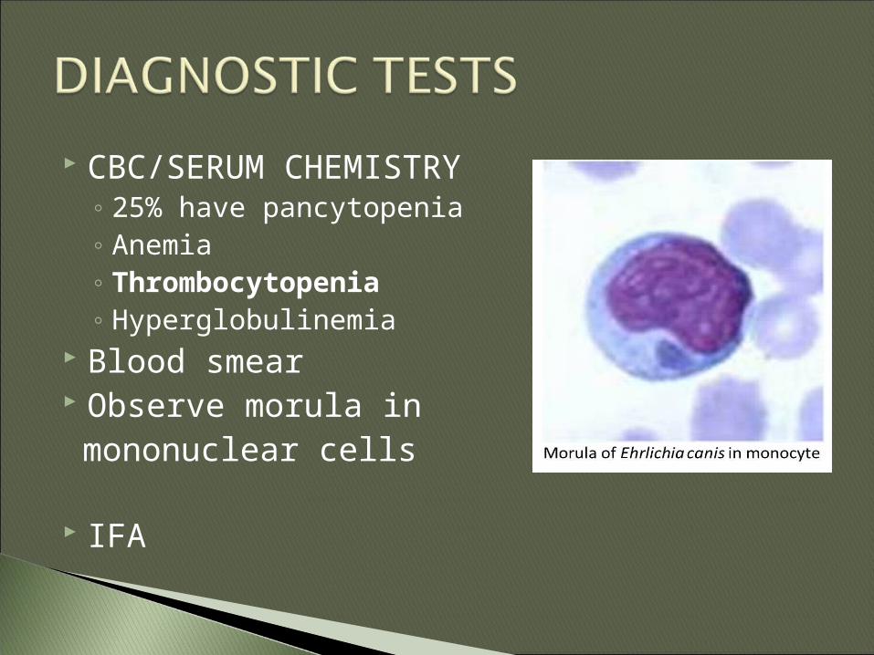
CBC/SERUM CHEMISTRY◦ 25% have pancytopenia◦ Anemia◦ Thrombocytopenia◦ Hyperglobulinemia
Blood smear Observe morula in mononuclear cells
IFA

DIAGNOSIS: TICK-BORNE DISEASE◦ CANINE MONOCYTIC EHRLICHIOSIS, caused
by Ehrlichia canis transmitted by the tick Rhipicephalus sanguineus
◦ After infection, E. canis causes acute, subclinical, and chronic stages of the disease
◦ ACUTE: lasts 2-4 weeks Organisms multiplies in mononuclear cells Mononuclear cells carry the organism to other organs
including the lungs, kidneys, and meninges. Vasculitis develops
◦ SUBCLINICAL PHASE Few clinical signs if any
◦ CHRONIC PHASE Bone marrow suppression Bleeding tendencies


CANINE GRANULOCYTIC EHRLICHIOSIS caused by Ehrlichia ewingii or Ehrlichia equi
Clinical signs associated with Ehrlichia ewingii infection:◦ Fever◦ Lethargy◦ Lameness◦ Muscle stiffness◦ CBC: Thrombocytopenia◦ Blood smear: morulae found in neutrophils
◦ Transmitted by the Amblyomma americanum tick

CANINE GRANULOCYTIC EHRLICHIOSIS caused by Ehrlichia ewingii or Ehrlichia equi
Clinical signs of Ehrlichia equi infection:◦ Fever◦ Debilitating lethargy◦ Anorexia◦ CBC: thrombocytopenia◦ Serum chemistries: Increased ALP
◦ Transmitted by the Ixodes dammini tick


ANTIBIOTICS◦ Doxycycline◦ Tetracycline◦ +/- blood transfusions
CLIENT INFO:◦ Ticks can be a threat to pets and humans
Owners should avoid exposure to the blood of the tick
◦ The prognosis is good – Improvement often seen within 48 hours
◦ Check pets frequently for ticks and remove them when found.


SIGNALMENT: 3yr old castrated male, English Setter
Hx: Moved from the northeast about 3 weeks ago. Prior to moving, owner pulled off a few ticks . Some of the areas have a red rash. In the last few days, the dogs is showing some lameness in the rear legs

PHYSICAL EXAM◦Temp:103.5, HR: 100, RR: 24◦Lethargic◦Swollen lymph nodes◦Wt. bearing lameness on the rt. Rear
limb that seems to come and go.


Radiographs◦ Would be normal
ELISA TEST◦ Lyme Positive
SYNOVIAL FLUID ANALYSIS◦ Increased nucleated cells


• LYME DISEASE is caused by the spirochete Borrelia Burgodorferi, passed by an Ixodes tick– The tick must be attached to the host for more
than 48 hours
• Other clinical signs:– Fever– Anorexia– Lymphadenopathy– Chronic flare-ups–Myocardial abnormalities– Nephritis, esp in Labs

ANTIBIOTICS◦ Doxycycline is the treatment of choice for
Borreliosis Treatment may not completely eliminate the
organism and some animals may remain permanently infected.
CLIENT INFO◦ Vaccination is effective, unless dog has already
been exposed. ◦ Animal infection should alert owners to the
possibility of human infection from ticks in the area.
◦ Use a tick preventive regularly


SIGNALMENT: 4 yr old, neutered male mixed breed.
HISTORY: owner saw dog playing with remains of a dead bat out in the back yard yesterday. The owner brings the dead bat into the clinic in a box and wants to know what to do.◦ The dog is current on all vaccinations including
rabies. He is on HW and flea prevention.

The bat should be sent to a laboratory for analysis and rabies testing◦ This requires a sample of brain tissue that has
NOT been frozen.◦ There is no antemortem test available
The dogs should be examined and handled carefully. He should be quarantined until the results from the bat are known.

Rabies virus is spread through the saliva of an infected animal◦ Bite, open wound, or mucous membranes
It travels up the nerve endings at the site of infection until it reaches the brain where it multiplies. It then enters the salivary glands where it can be transmitted through saliva.◦ This may take 3-8 weeks

RABIES IS CHARACTERIZED BY 3 STAGES:◦ PRODROMAL STAGE – people are greatest risk during
this phase. It is associated with behavior changes
◦ EXCITATIVE/FURIOUS STAGE- Infected animals may attack inanimate objects or appear hyperreactive. Some animals may appear “dumb”
◦ PARALYTIC STAGE - characterized by ascending paralysis of the hind limbs which may progress to respiratory paralysis and death.
◦ Death will occur between 2 and 10 days from the onset of clinical signs

CLINICAL SIGNS:◦ Behavioral changes◦ Difficulty swallowing◦ Voice changes◦ CNS signs (seizures, ataxia)◦ hypersalivation

CLIENT INFO:◦ Never handle wild animals that appear tame or
friendly Leave wild life in the wild
◦ Wear glove when examining a pet’s oral cavity, esp if rabies is suspected
◦ Promote vaccination against rabies◦ If your pet bites someone, it must be
quarantined for 10 days to observe for signs of clinical rabies
◦ Vaccinated animals exposed to a rabid animal should be revaccinated and observed for 90 days.


