A RESTUDY OF THE xMIDDLE CAMBRIAN BURGESS SHALE FOSSIL WORM, OTTOIA PROLIFICA WILLIAM C. BANTA* MARY E. RICE Departments of Paleobiology and Invertebrate Zoology, National Museum of Natural History Smithsonian Institution Washington, D.C. 20560 ABSTRACT Ottoia prolifica was a bilaterally symmetrical metazoan with a long, extrovertable proboscis armed with spines, a well-developed collar with about 25 rows of spines and hooks, and a finely annulated soma. There is no evidence that O. prolifica was segmented. Near the posterior end were at least 4 ventral hooks. The mouth was at the tip of the pro- boscis j the anus was at or near the posterior tip of the soma. There was a spacious body cavity. The morphology suggests the animal was a burrower in mud; paucity of sediment in the gut suggests a predatory habit. Phylogenetic affinities with the Siptmcula, Echiura, Hirudinea, Polychaeta, Oligochaeta, Kinorhyncha, Acanthocephala, Priapulida, and Tu- bilunchiis are considered. We concur with Meyer and Lang that O. prolifica is not ascrib- able to any known living phylum, but resembles members of some aschelminth and aschel- minthlike phyla, particularly the Priapulida. The Burgess Shale Member of the Middle Cambrian Stephen Formation of British Columbia (Walcott 1928 : 320, Fritz and Aitken 1959 : 887, Fritz 1970) contains a large number of soft-bodied organisms exquisitely preserved as thin organic films (compressions) on a fine-grained slate (Whittington 1969 : 901, 1970). Much of the Burgess Shale fauna has been described, largely through the efforts of Charles D. Walcott (see bibliography in Yochelson 1967 : 517). Among the remarkable Burgess Shale fossils are large numbers of compressions of what must have been completely soft-bodied marine worms. One of the most abundant single worm types in the Walcott collections of Burgess Shale organisms at the National Museum is that described by Walcott (1911 : 128) as a new genus and species, Ottoia prolifica. Into the genus Ottoia, Walcott also placed two other species, O. minor and O. tenuis, also from the Burgess Shale (Walcott 1911 : 129• •130. 1931: 6, see also Resser and Howell 1938: 215). Walcott was uncertain as to the phylogenetic position of Ottoia, but he tentatively placed it in the "Phylum Annulata, Class Gephyrea," citing Parker and Haswell's (1910) textbook for his classification. The Gephyrea was considered by Parker and Haswell to include * Present address : Department of Biology The American University, Washington, D. C. 20016 79
Transcript
A RESTUDY OF THE xMIDDLE CAMBRIAN BURGESS SHALE FOSSIL WORM, OTTOIA PROLIFICA
WILLIAM C. BANTA*
MARY E. RICE
Departments of Paleobiology and Invertebrate Zoology, National Museum of Natural History
Smithsonian Institution Washington, D.C. 20560
ABSTRACT
Ottoia prolifica was a bilaterally symmetrical metazoan with a long, extrovertable proboscis armed with spines, a well-developed collar with about 25 rows of spines and hooks, and a finely annulated soma. There is no evidence that O. prolifica was segmented. Near the posterior end were at least 4 ventral hooks. The mouth was at the tip of the pro- boscis j the anus was at or near the posterior tip of the soma. There was a spacious body cavity. The morphology suggests the animal was a burrower in mud; paucity of sediment in the gut suggests a predatory habit. Phylogenetic affinities with the Siptmcula, Echiura, Hirudinea, Polychaeta, Oligochaeta, Kinorhyncha, Acanthocephala, Priapulida, and Tu- bilunchiis are considered. We concur with Meyer and Lang that O. prolifica is not ascrib- able to any known living phylum, but resembles members of some aschelminth and aschel- minthlike phyla, particularly the Priapulida.
The Burgess Shale Member of the Middle Cambrian Stephen Formation of British Columbia (Walcott 1928 : 320, Fritz and Aitken 1959 : 887, Fritz 1970) contains a large number of soft-bodied organisms exquisitely preserved as thin organic films (compressions) on a fine-grained slate (Whittington 1969 : 901, 1970). Much of the Burgess Shale fauna has been described, largely through the efforts of Charles D. Walcott (see bibliography in Yochelson 1967 : 517).
Among the remarkable Burgess Shale fossils are large numbers of compressions of what must have been completely soft-bodied marine worms. One of the most abundant single worm types in the Walcott collections of Burgess Shale organisms at the National Museum is that described by Walcott (1911 : 128) as a new genus and species, Ottoia prolifica. Into the genus Ottoia, Walcott also placed two other species, O. minor and O. tenuis, also from the Burgess Shale (Walcott 1911 : 129• •130. 1931: 6, see also Resser and Howell 1938: 215). Walcott was uncertain as to the phylogenetic position of Ottoia, but he tentatively placed it in the "Phylum Annulata, Class Gephyrea," citing Parker and Haswell's (1910) textbook for his classification. The Gephyrea was considered by Parker and Haswell to include
* Present address : Department of Biology The American University, Washington, D. C. 20016
79
80 WILLIAM C. BANTA AND MARY E. RICE
three major groups now regarded as separate phyla: the Echiura, Sipuncula, and Priapulida. In his discussion, Walcott (1911: 128) specifically mentioned the "Or- der" Sipunculida, and cited a number of similarities and differences between sipunculans and Ottoia. It is probably because of this that when the Gephyrea was abandoned as a taxon and broken up into the presently accepted phyla, Ottoia came to be regarded as a fossil sipunculan (see, for example, Howell 1962: 169).
Meyer (1933: 527) and Lang (1953: 339), however, suggested that Walcott's placement was incorrect. These authors proposed that Ottoia was an aschelminth- -like worm with affinities close to the Acanthocephala and Priapulida. In their discussions, each contradicted Walcott's interpretation of the animal's structure, particularly his view that Ottoia prolifica was segmented, and that the mouth was located at the base of the proboscis. Their interpretations, however, were based solely on Walcott's published plates.
Preliminary studies of the large National Museum collections of Ottoia prolifica indicated not only that Meyer and Lang were correct in their interpreta- tions of structure, but that there is much detail observable in the fossils which had not been described by Walcott (see Resser in Walcott 1931: 1).
This paper is a redescription of the NMNH Burgess Shale fossils attributed by Walcott to Ottoia prolifica, with special emphasis on understanding phyloge- netic affinities of these ancient and remarkable animals.
RESULTS
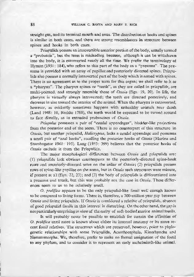
In the type collections of Burgess Shale at the National Museum of Natura Histor}'(NMNH) are 8 rock fragments, with a total of 9 animal compressions ascrib- ed by Walcott to Ottoia prolifica. Six of the specimens (NMNH catalogue num- bers 57619 through 57623) were figured by Walcott (1911: 128; pi. 19, figs. 1•5); they are designated "cotypes" (syntypes) in the Museum catalogue. One specimen (NMNH 57622) is broken into two fragments. Counterpart compressions ot two of the types (NMNH 57619 and 57623) were found in the general collection during this study. NMNH speciman 57619 (figured by Walcott, 1911, pi. 19, fig. 1; see our figs. 5•7) and its counterpart compressions display nearly all diagnostic char- acters and are here designated lectotype. The other type specimens are paralec- totypes.
The National Museum also contains a collection of about 100 Burgess Shale specimens identified by Walcott as O. prolifica and approximately 1,000 additional unlabelled specimens. The abundant material has permitted an especially detailed study of the animal's anatomy. All the material was examined, and the exception- ally well-preserved specimens sorted out for scrutiny.
The animals are preserved as thin carbonaceous films on black slate (Whitting- ton 1969: 901, 1970). The preservation is generally excellent, but observation of
SHALE FOSSIL WORM O. PROLIFIC A 81
detail requires manipulation under a dissecting microscope to see reflections from the film surface.
Walcott's photographs are good^ and generally representative of overall morphology. Really fine detail, however, especially minute spination, is not gen- erally discernible in his illustrations. Many of Walcott's photographs, moreover, were retouched by a professional artist (Walcott 1911: 111, G. A. Cooper, pers. comm., March 1970). In a few cases, detail obsers'able in the plates is virtually indiscernible on the specimens (compare Walcott 1911: 137, pi. 19, fig. 1 and our figs. 5•7). Most such discrepancies can probably be attributed to interpretive retouching, but the possibility cannot be excluded that the fossils have deterio- rated somewhat in the more than 30 years since they were first photographed.
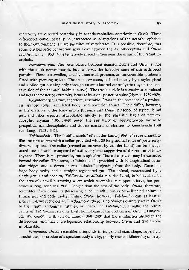
Most of the specimens closely resemble one another, and there seems to be little doubt that essentially all the animals were conspecific. It is difficult to esti- mate the size of the animals because the fossils are curved and frequently fragmen- tary. Furthermore, the animals were apparently soft-bodied and capable of consid- erable extension and contraction, so the validity of size measurements is open to question. The smallest specimens are about 3 cm long; the largest approach 25 cm. An average length in life was probably between 5 and 7 cm. The fossil compressions are elongate, between 5 and 10 times as long as wide. There arc three more or less distinct body regions, a proboscis, collar and soma.
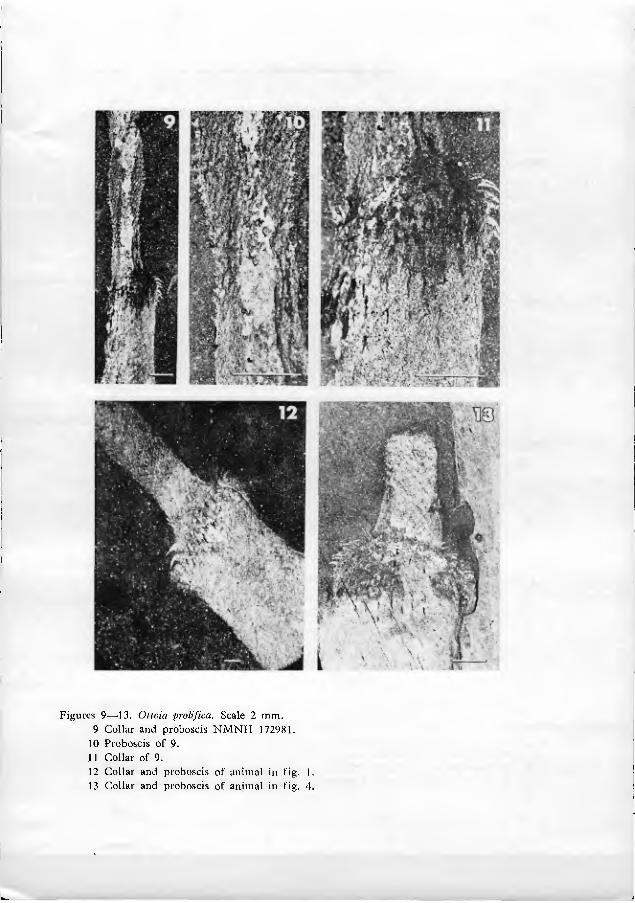
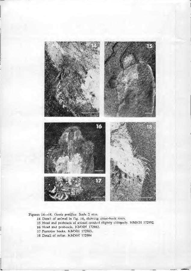
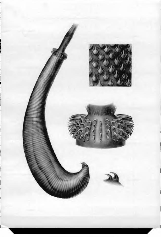
The proboscis is an elongate spinose structure which was almost certainly protrusible from the collar by evagination. Specimens occur in virtually every stage of extrusion of the proboscis, but in the vast majority of the fossils, the proboscis is almost entirely retracted (figs. 5, 15, 16). In a dozen or so exceptionally preserved specimens however, the proboscis is greatly extended (figs. 1, 9, 21). In soric spec- imens the proboscis is nearly a quarter as long as the rest of the body (fig. 1). It is not known if the living animals could extend it farther, nor is it known if proboscis extrusion was a normal part of the animals' behavior, or a pathological reaction to unfavorable stimuli just prior to preservation (see below).
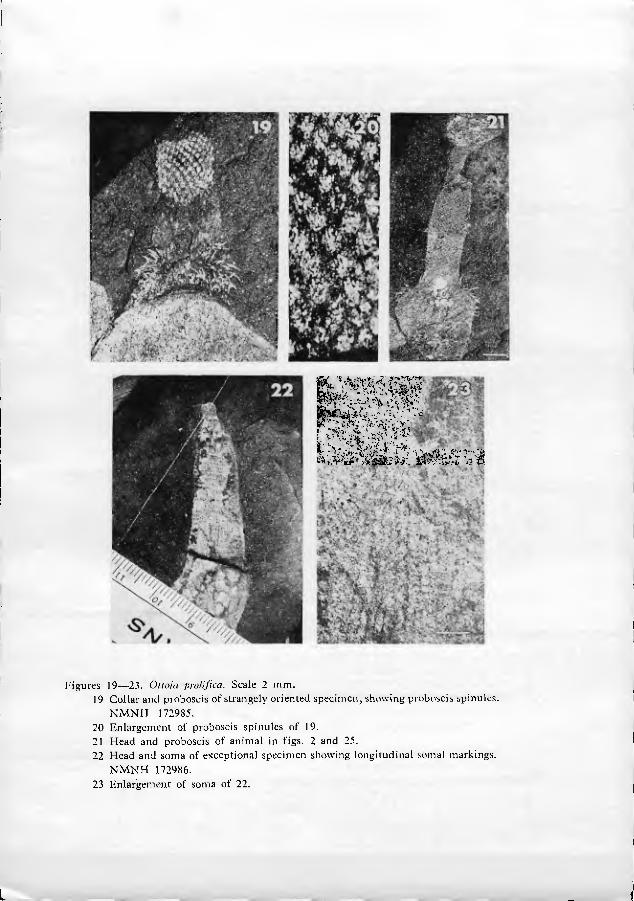
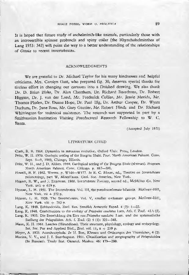
As far as can be seen, the base of the extruded proboscis is smooth and rela- tively undifferentiated (figs. 9•11, 21). Just distal to this smooth portion is a swelling, herein termed the proboscis dilation (figs. 1, 9, 21). The proboscis dila- tion is armed with a regular array of tiny plate-like spinules (figs. 6, 8•10, 19•21). In a fevvf truly remarkable specimens (figs. 19•20), the most proximal spinules can be seen to be shaped like a bear's foot-print. The palm of the print is located prox- imally, with 4 to 7 claw-like points directed distally (fig. 20). At the edge of the proboscis, the spinules appear to have a less complex structure. The central point of the spinules is larger than peripheral points in the more distal parts of the pro- boscis dilation.
Walcott (1911: 131) did not mention spines on the proboscis, but he did note that the organ was "papillose". It seems likely that he mistook poorly preserved
g2 WILLIAM C. BANTA AND MARY E. RICE
spinules for papillae. The proboscis may extend a considerable distance beyond the dilation, but there is little structure to be seen because of the generally poor preservation of this part of the animal (figs. 1, 2).
In many specimens, the gut can clearly be seen to extend well into the pro- boscis (figs. 9, 15, 16), even though its terminus is not evident. There is no evidence that the gut opens at the base of the proboscis, as suggested by Walcott (1911: 129). It seems reasonable to presume, therefore, that the animal's mouth was located at the tip of the proboscis. This conclusion concurs with the interpretations of Meyer (1933: 527) and Lang (1953: 338).
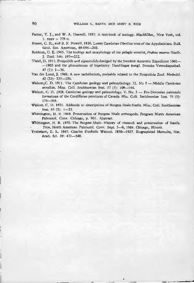
The proboscis is well demarcated from the collar by two rings of about two dozen long, fine spines, which project distally and nearly parallel to the pro- boscis (figs. 12•14, 16, 18). Immediately proximal (posterior) to these setiform spines is an array of strong-looking spines and hooks which mark the collar (figs. 9, 11•16, 18•19).
The introvertible nature of the proboscis is demonstrable in specimens in which the organ is not fully extruded. In some specimens, for example, the spinule pattern of the proboscis dilation can be seen within the soma or collar. In other cases, the proboscis dilation can be seen partly extruded (figs. 6, 13, 15, 16).
The spines and hooks of the collar are arranged in regular longitudinal rows. In several dozen specimens, the spine-hook rows are well enough preserved to be counted (figs. 12, 13). In any one specimen, between 13 and 15 spine-hook rows are visible, counting the two rows observed in silhouette. The central 11•13 rows almost certainly represent the rows on only one side of the animal, because the rows in counterpart compressions do not match exactly. The living animal, there- fore, must have possessed between 24 and 28 longitudinal spine-hook rows.
The more distal components of the spine-hook rows are long, straight, or slightly curved structures, herein termed spines (figs. 11, 14, 18). There are 3 or 4 spines in each of the rows; they intergrade proximally with collar hooks. Collar hooks are stout crescent-shaped structures oriented so that their points project proximally (posteriorly) (figs. 6, 8, 11 • 16, 18•19). The other point of the cres- cent is embedded in a fleshy base. There are 2 to 4 hooks in each spine-hook row.
The soma, which occupies about 9/10 of the length of the body (excluding the proboscis), is traversed by a series of dark lines, almost certainly representing bands or annuli of cuticular folds which circled the animal (figs. 1•5). In one spec- imen, the nature of annuli as cuticular corrugations is especially clear (Walcott 1911, pi. 19, fig. 5). There are usually about a hundred annuh located 0.2 • 1.2 mm apart. The soma is gently curved in all the specimens, and the annuli, when ob- servable, are closer together on the concave side of the curve than on the convex (figs. 2, 5). Walcott (1911 : 127) interpreted these annuli as evidence of segmenta- tion, but there is no reason to believe that they are more than superficial infoldings in the cuticle.
SHALE FOSSIL WORM O. PROLIFICA 83
In a single exceptional specimen (figs. 22•23), the distal third of the soma can be seen to bear about two dozen longitudinal dark rows with finely beaded appearance. The significance of these rows is unknown, but they may represent cuticular ornamentation of the soma.
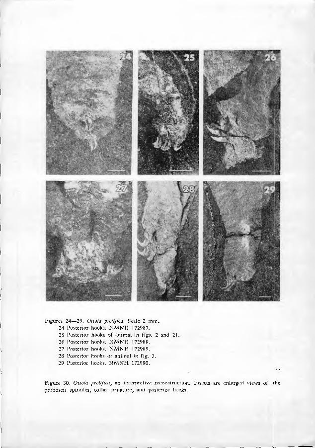
Near the presumed posterior end of the animal is a cluster of anteriorly-di- rected hooks (figs. 7, 17, 24•29). These hooks, herein nam.ed posterior hooks, are similar in shape to collar hooks, but larger in size (0.5•2 mm from one tip to the other). The posterior hooks do not seem to be located exactly at the tip of the soma, but are slightly subterminal. It is especially significant that the location of the posterior hooks correlates with the curvature of the body. Virtually all specimens show a distinct curve of the body. The posterior hooks are nearly always located on the concave side of that curve, rather than the convex (figs. 2, 3, 5). Further- more, the gut can be clearly traced in many specimens. In these cases, the presumed anus is seen to be located very near the posterior tip of the soma (that is, just posterior to the posterior hooks) (figs. 7, 29). These observations indicate that the animal was bilaterally symmetrical. The concave side of the curved body, bearing posterior hooks, is arbitrarily designated ventral; the opposite side is dorsal.
We have found it difficult to detennine the number and arrangement of posterior hooks. Slight variations in the orientation of the animal at the time of preservation greatly complicate interpretation. In occasional specimens, it can be seen that the hooks are at least sometimes arranged bilaterally (figs. 24, 27). In these cases, there appear to be at least 4 hooks in each assemblage. It seems reason- able to speculate that the hooks were associated with an unobserved structure enclosed in the circlet of hooks (a gonopore, for example). In other cases, there seem to be only two, somewhat larger hooks (fig. 26). This observation hints at the possibility of polymorphism (sexual dimorphism, for example), but until a careful study is made of many specimens and their counterpart compressions this conclusion remains speculative.
In some specimens (figs. 26, 28), a portion of the soma extends beyond the hooks, suggesting some kind of genital bursa, but its occurrence is inconsistent, and may be due to crushing to the specimen during preservation. Lang (153) re- ports that under unfavorable circumstance, Priapulus may permanently evaginate part of the gut through the anus.
The gut is frequently well preserved, and can be seen to run from near the tip of the proboscis (figs. 9•] 1) to near the tip of the soma, where it presumably ends at the anus, posterior and dorsal to the posterior hooks (figs. 1, 4, 5, 7, 25, 29). The gut is usually more prominent than any other organ, and may contain consipicuous deposits of pyrite, probably indicating that it contained a high con- centration of organic matter at the time of death. Six specimens have accumula- tions of dense material within the gut resembling the matrix of the surrounding
84 WILLIAM C. BANTA AND MARY E. RICE
rock (fig. 1). It seems reasonable to suppose that this material represents ingested sediment.
The body cavity was clearly spacious (figs. 1, 4, 5). There are occasional vague compressions ot membranous-appearing material around the gut, as though the gut may have been enclosed in a thin-walled sac (figs. 4, 5). Lang (1953: 339) interpreted faint compressions of diffuse material in the posterior part of Walcott's illustration of the lectotype (fig. 5) as remnants of an urogenital organ, but this type of structure occurs only sporadically in the fossils.
DISCUSSION
Summary of results. Let us begin the discussion with a recapitulation of the structure of Ottoia prolifica as interpreted by us (fig. 30). It must have been a bilaterally symmetrical unsegmented worm about as long as a man's finger. There were three main body parts, a proboscis, collar and soma. The proboscis was a soft, muscular organ capable of being everted a considerable distance from the animal; the mouth was located at its tip. Near the base of the proboscis was a band of anteriorly-directed, multi-pointed spinules. The proboscis was demarcated from the collar by a ring of setiform spines directed anteriorly. The collar was armed with a foi-midable array of about 25 longitudinal rows of 10 or so posteriorly di- rected spines and hooks. Behind the short collar was a cylindrical trunk, or soma, suoerficially marked by cuticular annulations. A straight gut ran from the tip of the proboscis to the tip of the soma. On the ventral surface near the posterior end, were from two to eight hooks, usally arranged bilaterally, sometimes in a circlet.
Paleoecology. The vast majority of NMNH specimens are found crushed laterally in the rock. The consistent occurrence of the posterior hooks on the inside of the body curve (¿. e., ventrally) indicates that compressions usually occurred in a plane very near the plane of bilateral symmetry. Because compression almost certainly took place horizontally as sediment accumulated, it seems necessary to conclude that the animals were lying on the bottom, rather than in it, at the time of preservation. If they had been burrowing in the substrate at the time, the plane of compression would be expected to be more or less random (but see Whittington 1969, 1970). This is not to say, however, that the animals did not burrow. They might have been physically transported to the site of final burial, or they might hjve crawled out onto the sea bottom in response to some stimulus (oxygen de- pbtion, for example) just prior to death. It seems possible, but fairly unlikely, thit the animals were pelagic, because their overall morphology is unlike that of living pelagic animals (except, perhaps, poeobioids; see Robbins 1967). Assuming th U O. prolifica was benthic, there is morphological evidence to indicate that the animal may have normally burrowed in mud. The anterior end is effectively radially symmetrical. This morphology would appear to be more suitable for a large me-
SHALE FOSSIL WORM O. PROLIFICA 85
tazoan moving through an uniform environment hke mud than crawHng over the substrate.
The spines and hooks on the collar, furthermore, are directed posteriorly. A similar arrangement is to be seen in kinorhynchs and priapulids. In these animals, cuticular projections are used in locomotion. At the start of the locomotory cycle, these mud-dwelling animals extend their bodies by contraction of circular muscles. The anterior end is expanded, presumably digging the posteriorly-directed spines into the sediment. Contraction of longitudinal muscles then pulls the animal for- ward. By repeating this cycle, the animals propel themselves through the mud (Clark 1964: 87). Assuming O. prolifica was not pelagic, there is no evident reason to suppose it did not locomote in a similar way.
The rarity of material interpreted as sediment in the guts of specimens of O./»-D/i/zca apparently indicates that either the animals did not normally ingest large quantities of sediment during feeding, or that they voided the material .shortly before death. It seems reasonable to assume that the animals' proboscis was i.x least partially involved in securing food. The occurrence of probable fossilized organic- rich remains in the gut suggests that the animals may have fed by capturing soft- bodied animals.
Affinities
Ottoia prolifica superficially resembles a number of animal groups, but in each case, detailed comparison reveals sigrificant differences. The main groups in which similarity is obvious are the Sinuncula, Echiura, Hirudinea, Polychiicta, Oligochaeta, Kinorhyncha, Acanthocephala, Nematomorpha, Priapulida, and the "tubilunchid" worms.
Sipunci/la. Ottoia prolifica resembles many sipunculans in overall L;hape, and in the possession of a muscular, introvertible anterior structure (termed a pro- boscis in Ottoia and an introvert in sipunculans). Like Ottoia, the mouth of sipun- culans opens at the distal tip of the introvert; also like Ottoia, the proboscis may be provided with cuticular, hook-like structures. But there are four important differences: (1) the gut is U-shaped in sipunculans, the anus opening near the base of the introvert, whereas in Ottoia, the anus is at or near the posterior tip of the body; (2) the gut in Ottoia is nearly straight, Vv^hereas in sipunculans, the gut is usually coiled in a characteristic way (Hj'm.an 1959: 612); (3) there is no equi\'alent in sipunculans to the collar hooks and spines of Ottoia (although some sipunculans possess papillae in a corresponding position); and (4) no sipunculan possesses posterior hooks.
Echiura. Ottoia prolifica vaguely resembles echiurans in shape and annulation, and in possessing a straight gut which opens rear the posterior tip of the soma. Furthermore, like O. prolifica, some echiurans possess posterior hooks. The pro-
85 WILLIAM C. BANTA AND MARY E. RICE
boscis of echiurans, however, is not introvertible, and the mouth is located, not at the tip of the proboscis, but near its base. No echiuran possesses obvious coun- terparts of the hooks, spines, and spinules on the proboscis and collar of O. prolifica.
Hirudinea (Leeches). Walcott (1911: HI, 127) proposed that there may be close affinities between Ottoia and leeches, and that Ottoia may "serve to link the Chaetopoda (= Polychaeta + Ologochaeta) and Hirudinea" (p. 111). Some leech- es are similar in size to Ottoia, and are similarly marked by annuli. An eversible proboscis is frequently present (Rynchobdellae), and there may be anterior hooks (Acanthobdella; see Mann, 1961). Mouth and anus are both terminal, or nearly so. Most leeches are unlike Otioîa in that they possess complex reproductive struc- tures and an elaborate gut with gastric caecae, structures which would be expected to leave some impression if a leech had been as well preserved as the Ottoia fossils. Furthermore, almost all leeches lack a well-defined body cavity; most of the space between the gut and body wall is occupied by botryoidal tissue and muscles. Ottoia prolifica evidently possessed a spacious body cavity.
One exceedingly primitive leech, however, Acanthobdella, displays less of a reduction of the body cavity than other leeches, and possesses a more simple gut than usual in the class. The anterior end, moreover, bears an array of bilaterally- -arranged hook-like setae. On the other hand, Acanthobdella lacks a probocsis, and, like all other leeches, displays conspicuous internal segmentation, serial repe- tition of organs and a posterior sucker (Mann 1961 ; 23); these features are without counterparts in O. prolifica. These latter characteristics, particularly the absence of segmentation, seem to exclude the possibility of affinities between leeches and
Ottoia. Oligochaeta and Polychaeta. Both these classes of annelids are quite diverse,
and many characters found in Ottoia are also found in various combinations of both groups. They are unlike Ottoia, however, in their possession of setae, distinct internal segmentation, and serial repetition of internal structures, particularly reproductive organs.
Kinorhyncha. Ottoia prolifica resembles some kinorhynchs in that both possess posterior hooks. Ottoia, however, is much larger than any kinorhynch, and possesses no trace of zonites, characteristic of kinorhynchs.
Acanthocephala. Ottoia prolifica shows some resemblances to some acantho- cephalid worms (for example, Polymorphus, Acanthogyrusm, Rhadinorhynchus, and others; see Hyman 1951: 9, 42). Acanthocephalids may be equivalent in size and shape, and are bilaterally symetrical. Spines and hooks are present on the in- trovertible proboscis, and sometimes also on the collar or trunk. Bilaterally arranged hooks may be present at the posterior end of the body (Hyman 1951 ; 9, fig. 4D), and large acanthocephalids are often annulated. On the other hand, there is no gut in any acanthocephalid, and all possess cement glands, lemnisci, and a lacunar system, structures with no evident counterparts in Ottoia. The proboscis spines.
SHALE FOSSIL WORM O. PROUFICA 87
moreover, are directed posteriorly in acanthocephalids, anteriorly in Ottoia. These differences could logically be interpreted as adaptations of the acanthocephalids to their environment; all are parasites of vertebrates. It is possible, therefore, that some phylogenetic connection may exist between the Acanthocephala and Ottoia prolifica. Lang (1953: 433) tentatively placed Ottoia near the origin of the Acantho- cephala.
Nematomorpha. The resemblance between nematomorphs and Ottoia is not with the adult nematomorph, but its larva, the infective state of this arthropod parasite. There is a swollen, usually annulated presoma, an introvertible probocsis fitted with piercing stylets. The trunk, or soma, is filled mostly by a stylet gland and a blind gut opening only through an anus located ventrally (that is, on the con- cave side of the animals' habitual curve). The trunk cuticle is sometimes annulated and near the posterior extremity, bears at least one posterior spine (Hyman 1959:469).
Nematomorph larvae, therefore, resemble Ottoia in the presence of a probos- cis, spinose collar, annulated body, and posterior spines. They differ, however, in the division of the body into a presoma and trunk, presence of stylets, blind gut, and other aspects, attributable mostly to the parasitic habit of nemato- morphs. Hyman (1951:469) noted the similarity of nematomorph larvae to priapulids, acanthocephalids and its less marked similarities to kinorhynchs (but see Lang, 1953: 341).
Tubilunchids. The "tubilunchids" of van der Land (1969: 249) are priapulid- like marine worms with a collar provided with 20 longitudinal rows of posteriorly- directed spines. The collar (termed an introvert by van der Land) can be invagi- nated into a "neck" composed of cuticular plates suggestive of the zonites of kino- rhynchs. There is no proboscis, but a spineless "buccal capsule" may be extruded beyond the collar. The soma, or "abdomen" is provided with 20 longitudinal cutic- ular ridges and a dozen or two "tubules" projecting from the body. There is a large body cavity and a straight regionated gut. The animal, represented by a single genus and species, Tubilunchus corallicola van der Land, is believed to be the larva of a small burrowing worm which resembles its supposed larva, but pos- sesses a long, post-anal "tail" longer than the rest of the body. Ottoia, therefore, resembles Tubilunchus in possessing a collar with posteriorly-directed spines, a similar gut and body cavity. Unlike Ottoia, however, Tubilunchus can, at least as a larva, introvert the collar. Furthermore, there is no obvious counterpart in Ottoia to the "tail", abdominal tubules, or "neck" of Tubilunchus. Finally, the buccal cavity of Tubilunchus, its only likely homologue of the proboscis of Oííoí'a, is unarm- ed. We concur with van der Land (1968: 249) that the similarities outweigh the differences, and that a phylogenetic relationship between Ottoia and Tubilunchus is plausible.
Priapulida. Ottoia resembles priapulids in its general size, shape, superficial annulations, possession of a spacious body cavity, poorly marked bilateral symmetry,
88 WILLIAM C. BANTA AND MARY E. RICE
Straight gut, and its terminal mouth and anus. The distribution ol hooks and spines is similar in both cases, and there are strong resemblances in structure between spines and hooks in both cases.
Priapulids possess an introvertible anterior portion of the body, usually termed a "proboscis", but the term is misleading because, although it can be withdrawn into the body, it is extroverted nearly all the time. We prefer the terminology of Hyman (1951: 184), who refers to this part of the body as a "presoma". The pre- soma is provided with an array of papillae and posteriorly directed spines. Priapu- lids also possess a normally introverted part of the body which is armed with spines. There is no agreement as to the proper term for this organ; we shall refer to it as a "pharjTix". The pharynx spines or "teeth", as they are called in priapulids, are multi-pointed, and strongly resemble those of Ottoia (figs. 19, 20). In life, the pharynx is virtually always introverted; the teeth are directed posteriorly, and decrease in size toward the interior of the animal. When the phaiynx is extroverted, however, as evidently sometimes happens with unhealthy animals near death (Land 1948: 10, Rathke 1788), the teeth would be expected to be turned around to face distally, as in extruded proboscises of Ottoia.
Priapulus possesses a pair of "caudal appendages", bladder-like projections from the posterior end of the soma. There is no counterpart of this structure in Ottoia, but another priapulid, Halicryptus, lacks a caudal appendage and possesses a small pair of "anal hooks", recalling the posterior hooks of Ottoia (Murina and Starobogator 1961: 195). Lang (1953: 399) believes that the posterior hooks of Ottoia exclude it from the Priapulida.
The major morphological differences between Ottoia and priapulids are: (1) priapulids lack obvious counterparts to the posteriorly-directed spine-hook rows and anteriorly-directed setae on the collar of Ottoia; (2) priapulids possess rows of spine-like papillae on the soma, but in Ottoia such structures were minute, if present at all (figs. 22, 23); and (3) the body of priapulids is diiferentiated into a prcsom.a and trunk, but this was probably not the case in Ottcia. These differ- ences seem to us to be relatively small.
O. prolifica appears to be the only priapulid-like fossil well enough known to be compared to living forms. There is, therefore, a 300-miUion year gap between Ottoia and living priapulids. If Ottoia is considered a relative of priapulids, absence of good priapulid fossils in this interval is disturbing. On the other hand, the gap is not particularly surprising in view of the rarity of soft-bodied marine animal fossils.
It will probably never be possible to establish for certain the affinities of O. prolifica until more is known about either its internal anatomy or its more re- cent fossil relatives. The structures which are preserved, however, point to phylo- genetic relationships with some Priapulida, Acanthocephala, Kinorhyncha and Nematomorpha. We, therefore, prefer to make no formal assignation of the fossil to any phylum, and to consider it to represent an early aschelminth-like animal.
6 Head of 5. Scale 2 mm. 7 Posterior end of 5. Scale 2 mm. 8 Collar and proboscis of another animal. Scale 2 mm. NMNH 172980
Figures 9•^13. Otiaia prolifica. Scale 2 mm. 9 Collar and proboscis NMNH 172981.
10 Proboscis of 9. 11 Collar of 9. 12 Collar and proboscis of animal in fig. I. 13 Collar and proboscis of animal in fig. 4.
Figures 14•18. Ottoia prolifica. Scale 2 mm. 14 Detail of animal in fig. 16, showing spine-hools rows. 15 Head and proboscis of animal crushed slightly obliquely. NMNH 172992. 16 Head and proboscis. NMNH 172982. 17 Posterior hooks. NMNH 172983. 18 Detail of collar. NMNH 172984
<^
Figures 19•23. Oiloia prolijica. Scale 2 mm. 19 Collar and proboscis of strangely oriented specimen, showing proboscis spinules.
NMNH 172985. 20 Enlargement of proboscis spinules of 19. 21 Head and proboscis of animal in figs. 2 and 25. 22 Head and soma of e.Kceptional specimen showing longitudinal somal markings.
NMNH 172986. 23 Enlargement of soma of 22.
Figures 24•29. Otioia proUfica. Scale 2 mm. 24 Posterior hool^s. NJVlNH 172987. 25 Posterior hooks of animal in figs. 2 and 21. 26 Posterior hcx)ks. NMNH 172988. 27 Posterior hooks. NMNH 172989. 28 Posterior hooks of animal in fig. 3. 29 Posterior hooks. NMNH 172990.
Figure 30. Olioia prolij'ka, an interpretive reconstruction. Inserts are enlarged views of proboscis spinules, collar armature, and posterior hooks.
the
SHALE FOSSIL WORM O. PROIJPICA 89
It is hoped that future study of aschelminth-like animals, particularly those with an introvertible spinose proboscis and spiny collar (the Rhynchohelminthes of Lang 1953: 343) will point the way to a better understanding of the relationships of Ottoia to recent invertebrates.
ACKNOWLEDGMENTS
We are grateful to Dr. Michael Taylor for his many kindnesses and helpful criticisms. Mrs. Carolyn Gast, who prepared fig. 30, deserves special thanks for tireless effort in changing our cartoons into a finished drawing. We also thank Dr. D. Brian Blake, Dr. Alan Cheetham, Dr. Richard Boardman, Dr. Robert Higgins, Dr. J. van der Land, Mr. Frederick Collier, Mr. Jessie Merida, Mr. Thomas Phalen, Dr. Duane Hope, Dr. Paul Illg, Dr. Arthur Cooper, Dr. Wyatt Durham, Dr. June Ross, Mr. Gary Gautier, Mr. Robert Hinds and Dr. Richard Whittington for technical assistance. The research was supported in part by a Smithsonian Institution Visiting Postdoctoral Research Fellowship to W. C. Banta.
(Accepted July 1971)
LITERATURE CITED
Clark, R, B. 1964. Dynamics in metazoan evolution. Oxford Univ. Press, London. Fritz, V/. H. 1970. Geologic setting of the P.u.rgess Shale. Proc. North American Paleont. Conv.
Sept. 5•9, 1969, Chicago, Illinois. Fritz, W. H.. and J. D. Aitken. 1969. Geological setting of the Burgess Shale (abstract). Program
North American Paleont. Conv. Chicago, p. 887•888. Howell, B. F. 1962. Worms, p. W144•W177. /)/ R. C. A'ioore, ed.. Treatise on In\'ertebrate
paleontology, part W, Miscellanea. Gcol. Soc. America, New York. Hcgner, R. W., and J. Engeman. 1968. Invertebrate Zoology, second ed., ivlcMillan Co. New
York, xvii -'r 619 p. Hyman, L. H. 1951. The Invertebrates. Vol. Ill, the pseudocoelom.ate bilateria, McCraH'-IIill,
New York, vii -|- 572 p. Hyman, L. H. 1959. The Invertebrates. Vol. V, smaller eoelomate groups. McGraw-Hill,
New York, viii ¡- 783 p. Lang, K. 1949. Echinoderida. Zool. Res. Swedish Antarctic Exped. 4 (2); 1•22. Lang, K. 1948. Contributions to the ecology' o{ Priapiihis caiidatus Lam. Ark. f. Zool. 41A (5). Lang, K. 1953. Die Entwicklung des Eies von i^na/)«/!« cm/rfatot Lam. und die systematische
Stellung der Priapuliden. Ark. f. Zool. (2) 5 (5): 321•348. Mann, K. H. 196L Leeches (Hirundinea). Their structure, physiologj', ecology and embryology.
Int. Ser. Pur and Applied BioL, Zool. vol. 11, x -f 210 p. Meyer, A. 1933. Acanthocephala. In H. Bon, Klassen und Ordnungen des Tierreiches, 4 (2). Murina, V. V., and J. I. Starobogator. ¡961. Classification and zoogeography of Priapuloidea
(In Russian). Trudy Inst. Okeanol. Moskva. 46; 179•200.
90 WILLIAM C. SANTA AND MARY E. RICE
Parker, T. J., and W. A. Haswell. 1897. A text-book of zoology. MacMillan, New York, vol. 1. XXXV + 779 D.
Resser, C. H., and B. F. Howell. 1938. Lower Cambrian Olenllus zone of the Appalachians. Bull. Geol. Soc. American, 49:195•248.
Robbins, D. E. 1965. The biology and morphology' of the pelagic annelid, Poebim meseres Heath. J. Zool. 146: 197•212.
Théel, H. 1911. Priapulids and sipunculids dredged by the Swedish Antarctic Expedition 1901• •1903 and the phenomenon of bipolarity. Handlingar knngl. Svenska Vetenskapsakad. 47 (1): 1•36.
Van der Land, J. 1968. A new aschelminth, probably related to the Priapulida Zool. Mededel. 42 (22): 237•250.
Walcott,C. D. 1911. The Cambrian geology and paleonthology. II. No 5 • Middle Cambrian annelids. Misc. Coll. Smithsonian Inst. 57 (5): 109•144.
Walcott, C. D. 1928. Cambrian geology and paleontology. V. No. 5 • Pre-Devonian paleozoic formations of the Cordilleran provinces of Canada. Mis. Coll. Smithsonian Inst. 75 (5): 175•368.
Walcott, C. D. 1931. Addenda to descriptions of Burgess Shale fossils. Misc. Coll. Smithsonian Inst. 85 (3): 1•23.
Whittington, H. B. 1969. Preservation of Burgess Shale arthropods. Program North American Paleontol. Conv. Chicago, p. 901. Abstract.
Whittington^ H. B. 1970. The Burgess Shale: History of research and preservation of fossils. Pros. North American Paleontol. Conv. Sept. 5•9, 1969. Chicago, Illinois.
Yochelson, E. L. 1967. Charles Doolittle Walcott. 1850•1927. Biographical Memoirs, Nat. Acad. Sei. 39: 471•540.