Amyloid Precursor Protein Regulates Brain Apolipoprotein E and Cholesterol Metabolism through Lipoprotein Receptor LRP1 Qiang Liu 1 , Celina V. Zerbinatti 1,2 , Juan Zhang 1 , Hyang-Sook Hoe 3 , Baiping Wang 4 , Sarah L. Cole 5 , Joachim Herz 6 , Louis Muglia 1 , and Guojun Bu 1,7,8,* 1Department of Pediatrics, Washington University School of Medicine, St. Louis, MO 63110, USA 3Department of Neuroscience, Georgetown University Medical Center, Washington, D. C. 20057, USA 4Huffington Center on Aging and Department of Molecular and Human Genetics, Baylor College of Medicine, Houston, TX 77030, USA 5Department of Cell and Molecular Biology, Northwestern University Feinberg School of Medicine, Chicago, IL 60611-3008, USA 6Department of Molecular Genetics, University of Texas Southwestern Medical Center, Dallas, TX 75390, USA 7Departments of Cell Biology and Physiology, Washington University School of Medicine, St. Louis, MO 63110, USA 8Hope Center for Neurological Disorders, Washington University School of Medicine, St. Louis, MO 63110, USA SUMMARY Mutations in the amyloid precursor protein (APP) cause early-onset Alzheimer's disease (AD), but the only genetic risk factor for late-onset AD is the ε4 allele of apolipoprotein E (apoE), a major cholesterol carrier. Using Cre-lox conditional knockout mice, we demonstrate that lipoprotein receptor LRP1 expression regulates apoE and cholesterol levels within the CNS. We also found that deletion of APP and its homologue APLP2, or components of the γ-secretase complex, significantly enhanced the expression and function of LRP1, which was reversed by forced expression of the APP intracellular domain (AICD). We further show that AICD, together with Fe65 and Tip60, interacts with the LRP1 promoter and suppresses its transcription. Together, our findings support that the γ- secretase cleavage of APP plays a central role in regulating apoE and cholesterol metabolism in the CNS via LRP1 and establish a biological linkage between APP and apoE, the two major genetic determinants of AD. INTRODUCTION Mounting genetic and biochemical evidence strongly supports the hypothesis that amyloid β- peptide (Aβ) accumulation in the brain is an early and toxic event in the pathogenesis of Alzheimer's disease (Hardy and Selkoe, 2002). Accordingly, reducing brain Aβ production and/or increasing its clearance have become attractive targets for AD drug development (Hardy and Selkoe, 2002). Aβ is derived from sequential proteolytic processing of amyloid precursor * Correspondence: [email protected]2 Present address: Alzheimer's Research Department, Merck & Co., Inc., West Point, PA 19486, USA Publisher's Disclaimer: This is a PDF file of an unedited manuscript that has been accepted for publication. As a service to our customers we are providing this early version of the manuscript. The manuscript will undergo copyediting, typesetting, and review of the resulting proof before it is published in its final citable form. Please note that during the production process errors may be discovered which could affect the content, and all legal disclaimers that apply to the journal pertain. NIH Public Access Author Manuscript Neuron. Author manuscript; available in PMC 2008 October 4. Published in final edited form as: Neuron. 2007 October 4; 56(1): 66–78. NIH-PA Author Manuscript NIH-PA Author Manuscript NIH-PA Author Manuscript
Transcript
Amyloid Precursor Protein Regulates Brain Apolipoprotein E andCholesterol Metabolism through Lipoprotein Receptor LRP1
Qiang Liu1, Celina V. Zerbinatti1,2, Juan Zhang1, Hyang-Sook Hoe3, Baiping Wang4, SarahL. Cole5, Joachim Herz6, Louis Muglia1, and Guojun Bu1,7,8,*
1Department of Pediatrics, Washington University School of Medicine, St. Louis, MO 63110, USA
3Department of Neuroscience, Georgetown University Medical Center, Washington, D. C. 20057, USA
4Huffington Center on Aging and Department of Molecular and Human Genetics, Baylor College of Medicine,Houston, TX 77030, USA
5Department of Cell and Molecular Biology, Northwestern University Feinberg School of Medicine, Chicago,IL 60611-3008, USA
6Department of Molecular Genetics, University of Texas Southwestern Medical Center, Dallas, TX 75390,USA
7Departments of Cell Biology and Physiology, Washington University School of Medicine, St. Louis, MO63110, USA
8Hope Center for Neurological Disorders, Washington University School of Medicine, St. Louis, MO 63110,USA
SUMMARYMutations in the amyloid precursor protein (APP) cause early-onset Alzheimer's disease (AD), butthe only genetic risk factor for late-onset AD is the ε4 allele of apolipoprotein E (apoE), a majorcholesterol carrier. Using Cre-lox conditional knockout mice, we demonstrate that lipoproteinreceptor LRP1 expression regulates apoE and cholesterol levels within the CNS. We also found thatdeletion of APP and its homologue APLP2, or components of the γ-secretase complex, significantlyenhanced the expression and function of LRP1, which was reversed by forced expression of the APPintracellular domain (AICD). We further show that AICD, together with Fe65 and Tip60, interactswith the LRP1 promoter and suppresses its transcription. Together, our findings support that the γ-secretase cleavage of APP plays a central role in regulating apoE and cholesterol metabolism in theCNS via LRP1 and establish a biological linkage between APP and apoE, the two major geneticdeterminants of AD.
INTRODUCTIONMounting genetic and biochemical evidence strongly supports the hypothesis that amyloid β-peptide (Aβ) accumulation in the brain is an early and toxic event in the pathogenesis ofAlzheimer's disease (Hardy and Selkoe, 2002). Accordingly, reducing brain Aβ productionand/or increasing its clearance have become attractive targets for AD drug development (Hardyand Selkoe, 2002). Aβ is derived from sequential proteolytic processing of amyloid precursor
*Correspondence: [email protected] address: Alzheimer's Research Department, Merck & Co., Inc., West Point, PA 19486, USAPublisher's Disclaimer: This is a PDF file of an unedited manuscript that has been accepted for publication. As a service to our customerswe are providing this early version of the manuscript. The manuscript will undergo copyediting, typesetting, and review of the resultingproof before it is published in its final citable form. Please note that during the production process errors may be discovered which couldaffect the content, and all legal disclaimers that apply to the journal pertain.
NIH Public AccessAuthor ManuscriptNeuron. Author manuscript; available in PMC 2008 October 4.
Published in final edited form as:Neuron. 2007 October 4; 56(1): 66–78.
NIH
-PA Author Manuscript
NIH
-PA Author Manuscript
NIH
-PA Author Manuscript
protein (APP), a ubiquitously expressed type I transmembrane protein that undergoes twodistinct processing pathways (Selkoe and Kopan, 2003;Zheng and Koo, 2006). In the non-amyloidogenic pathway, APP undergoes ectodomain shedding by α-secretase, identified asmembers of the ADAM metalloprotease family (Zheng and Koo, 2006). Subsequent cleavageof the APP C-terminal membrane-associated stub by γ-secretase (Selkoe and Kopan, 2003)generates a non-toxic p3 peptide, as well as the APP intracellular domain (AICD) (Selkoe andKopan, 2003). In the amyloidogenic pathway, APP is first cleaved by the β-secretase BACE1(β-site APP cleaving enzyme 1) (Vassar et al., 1999) and then by γ-secretase to generate Aβand AICD. Mutations associated with early-onset familial forms of AD (FAD) are found inthe APP gene itself or in the genes of presenilin 1 (PS1) and PS2, whose products, along withnicastrin, Pen-2 and Aph-1, are obligate components of a multi-protein complex that gives riseto γ-secretase activity (Selkoe and Kopan, 2003). A common feature of all FAD mutations isthat they increase the generation of Aβ peptides, or the proportion of the longer Aβ42 form,considered to be more amyloidogenic and pathogenic than the shorter Aβ40 due to its highlyaggregative nature (Hardy and Selkoe, 2002).
Although FAD genetics and mouse models have generated tremendous insights into ADpathogenesis, the vast majority of AD cases are sporadic with late-onset. A major genetic riskfactor that was initially discovered in 1993 and has since been validated in numerous geneticstudies is the presence of the ε4 allele of the apolipoprotein E (APOE) gene (Corder et al.,1993). ApoE is a major apolipoprotein in the brain, and exists in three isoforms in humans(apoE2, apoE3, apoE4); each differing by a single amino acid (Mahley, 1988). In the brain,apoE/lipoprotein particles are produced primarily by astrocytes and are believed to delivercholesterol and other lipids to neurons via lipoprotein receptors, namely members of the low-density lipoprotein receptor (LDLR) family (Herz and Bock, 2002;Herz and Chen, 2006).Although the mechanisms underlying the pathogenic nature of apoE4 in sporadic AD are stillpoorly understood, several models have been proposed and supported by in vitro and in vivostudies. First, apoE interacts with Aβ, and apoE4 likely possesses greater ability to promoteAβ fibrillogenesis and amyloid plaque formation (Holtzman et al., 2000). Second, apoEfacilitates Aβ clearance via apoE receptors expressed either in neurons (Zerbinatti and Bu,2005) or in the blood brain barrier (Zlokovic, 2005). ApoE4 is less functional in Aβ clearanceowing to its weaker affinity to Aβ (LaDu et al., 1994). Third, apoE4 may be toxic to neuronsindependently of Aβ aggregation and clearance (Huang, 2006). It is possible that multiplepathways contribute to the pathogenic nature of apoE4 in AD.
Cholesterol is an essential component of membranes and myelin sheathes and is crucial forsynaptic integrity and neuronal functions (Pfrieger, 2003). Interestingly, an associationbetween brain cholesterol metabolism and the risk of AD has been proposed (Shobab et al.,2005). Early studies indicated that the use of statins, which inhibit cholesterol synthesis, isassociated with a significant decrease in AD prevalence; however, several recent prospectivestudies do not support such a conclusion (Shobab et al., 2005). Additionally, the effect ofcholesterol on the amyloidogenic processing of APP to Aβ remains controversial (Ledesmaand Dotti, 2006). Intriguingly, apoE4 knock-in mice exhibit decreased brain cholesterol levelseven though the peripheral cholesterol levels are increased (Hamanaka et al., 2000). Areduction of brain cholesterol levels is also observed in AD brains (Ledesma and Dotti,2006). These disparate findings raise the need for an understanding of brain cholesterolmetabolism and its potential dysregulation in AD.
In this manuscript we present a novel linkage between APP processing and apoE/cholesterolmetabolism. Specifically, we found that lack of either APP/APLP2 or PS1/PS2 leads toincreased brain apoE/cholesterol catabolism. We show that the APP processing product, AICD,suppressed expression of the major apoE/lipoprotein receptor LRP1 by binding directly to itspromoter following association with the adaptor protein Fe65. Defective apoE/cholesterol
Liu et al. Page 2
Neuron. Author manuscript; available in PMC 2008 October 4.
NIH
-PA Author Manuscript
NIH
-PA Author Manuscript
NIH
-PA Author Manuscript
catabolism in APP/APLP2 and PS1/PS2 knockout cells was restored by a forced expressionof AICD. Together, our results reveal a novel biological function of APP in the regulation ofbrain apoE and cholesterol metabolism, offering new alternatives for the design of therapeuticstrategies to treat AD.
RESULTSAPP and APLP2 Regulate Brain ApoE and Cholesterol Metabolism
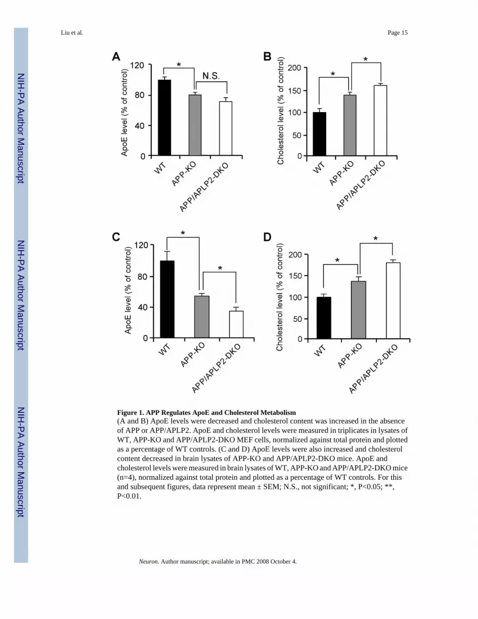
APP processing and apoE/cholesterol metabolism are two important events in the pathogenesisof AD. To examine whether these two pathways are functionally related, we analyzed cellularapoE and cholesterol levels in mouse embryonic fibroblasts (MEFs) of WT, APP-KO, or APPand APLP2 double knockout (APP/APLP2-DKO). To minimize potential clonal effects whenMEF cells were established, APP-KO MEF cells stably retransfected with APP695 cDNA wereused as WT controls. The APP-KO and APP/APLP2-DKO cells displayed a significantdecrease in apoE levels and a concomitant increase in cholesterol levels compared to WTcontrols (Figures 1A and 1B). The apoE and cholesterol levels we measured likely reflect acombination of those derived from both serum and cells. One possible explanation to thesefindings is that lack of APP or APP/APLP2 lead to increased catabolism of apoE/lipoproteinresulting in increased intracellular cholesterol levels (Mahley, 1988). To assess whether thesechanges also occur in vivo, we compared brain apoE and cholesterol levels in WT, APP-KOand APP/APLP2-DKO mouse brain. ApoE levels were decreased by ∼50% in APP-KO andfurther decreased in APP/APLP2-DKO when compared to WT littermate controls (Figure 1C).A corresponding increase in cholesterol levels was also observed in APP-KO and APP/APLP2-DKO mouse brain when compared to WT controls (Figure 1D). Together, these results raisethe possibility that apoE and cholesterol levels are modulated, either directly or indirectly, byAPP and APLP2 and/or their processing products.
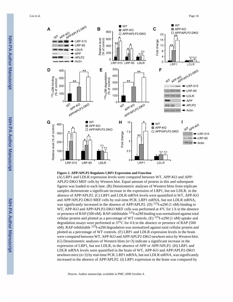
APP and APLP2 Regulate LRP1 Expression and FunctionThe increased catabolism of apoE/lipoprotein by APP-KO and APP/APLP2-DKO cells leadus to investigate the possibility that apoE receptor levels maybe up-regulated in these cells. Totest this possibility, we compared expression levels of two major brain apoE receptors LRP1and LDLR (Herz and Bock, 2002) in WT, APP-KO and APP/APLP2-DKO MEF cells. Westernblotting using two different antibodies to LRP1 showed that LRP1 levels were significantlyincreased in APP-KO MEF cells and in APP/APLP2-DKO MEF cells compared to WT MEFcells (Figures 2A and 2B). The expression of the LDLR was not altered by APP/APLP2 deletion(Figures 2A and 2B). To analyze whether changes in LRP1 expression were at thetranscriptional or post-transcriptional level, we compared LRP1 mRNA levels by real-timePCR. As shown in Figure 2C, LRP1 mRNA levels were significantly increased in both APP-KO and APP/APLP2-DKO MEF cells when compared to WT control cells. To investigatewhether changes in LRP1 expression also correlate with changes in LRP1 function, weanalyzed binding and endocytosis of α2-macroglobulin (α2M), a high affinity ligand for LRP1(Herz and Bock, 2002). Ligand binding assays using 125I-α2M demonstrated increased bindingcapacity in APP-KO and APP/APLP2-DKO MEF cells when compared to WT control cells(Figure 2D). Similar increase was seen when 125I-α2M uptake and degradation were analyzed(Figure 2E).
To examine whether our in vitro findings were reproduced in vivo, we compared LRP1expression in the brains of newborn APP-KO, APP/APLP2-DKO and littermate control mice.LRP1 expression, but not LDLR expression, was significantly increased in APP-KO and APP/APLP2-DKO mouse brains when compared to WT controls both at the protein (Figures 2Fand 2G) and mRNA levels (Figure 2H). Similar increase in LRP1 expression was also observedin the brain of APP-KO mice at 4 months of age. These results demonstrate that LRP1
Liu et al. Page 3
Neuron. Author manuscript; available in PMC 2008 October 4.
NIH
-PA Author Manuscript
NIH
-PA Author Manuscript
NIH
-PA Author Manuscript
expression and function are modulated by APP/APLP2 or their processing products and suggestthat APP/APLP2 likely modulate brain apoE and cholesterol metabolism via regulation ofLRP1 levels.
LRP1 Modulates Brain ApoE/Lipoprotein MetabolismLRP1 is abundantly expressed in the brain, but in vivo evidence that LRP1 regulates brain apoEand cholesterol metabolism is lacking. Because conventional knockout of LRP1 is earlyembryonic lethal (Herz et al., 1992), we generated conditional LRP1 forebrain knockout miceby crossing LRP1 floxP mice (Rohlmann et al., 1998) with αCamKII-Cre mice (Tsien et al.,1996). LRP1 expression was significantly decreased in the forebrain as determined byimmunobloting using antibodies directed against either the 515-kDa subunit or the 85-kDasubunit (Figures 3A and 3B). Remaining LRP1 expression detected by Western blotting likelyrepresents expression in glial cells (Moestrup et al., 1992). By double immunofluorescencestaining using LRP1-specific antibody and NeuN antibody we confirmed that LRP1 expressionwas nearly abolished in CA1 neurons of the hippocampus and in pyramidal neurons of thefrontal cortex (Figure 3C). To examine how an LRP1 deletion affects APP levels andprocessing, we compare the steady-state levels of the full-length APP and APP C-terminalfragment (CTF). We found that deletion of LRP1 slightly increased the steady-state levels offull-length APP while decreased the levels of APP-CTF both in LRP1-KO MEF cells (Figure3D) and in mouse brain (Figure 3E); likely reflecting a stabilization of APP at the cell surfacedue to reduced endocytosis (Cam et al., 2005).
To evaluate the impact of LRP1 deletion on apoE and cholesterol metabolism, we comparedapoE and cholesterol levels in the forebrain of LRP1-KO and WT littermate controls. We foundthat while apoE levels were significantly increased (Figure 3F), cholesterol levels weredecreased (Figure 3G) in the LRP1-KO mice, suggesting impaired catabolism of apoE/lipoprotein particles. The changes in apoE and cholesterol levels in the absence of LRP1 werealso observed in LRP1-KO MEF cells (Figures 4A and 4B). Interestingly, the half-life of apoEis significantly increased in LRP1-KO MEF cells (Figure 4C), suggesting that in the absenceof LRP1, apoE catabolism is decreased. ApoE mRNA levels were not changed between WTand LRP1-KO MEF cells (Figure 4D) or brain tissues (Figure 4E). Together, these resultsdemonstrate that LRP1 is a bona fide apoE/lipoprotein receptor which modulates apoE andcholesterol metabolism in the brain.
Aβ Is Not Required for LRP1 RegulationA recent study demonstrated a role for Aβ in regulating the biosynthesis of cholesterol andsphingomyelin (Grimm et al., 2005). Therefore we evaluated the possibility that Aβ may bethe processing product of APP which regulates LRP1 expression levels. Because β-secretaseBACE1 is necessary for Aβ production (Luo et al., 2001), we evaluated the effects of BACE1knockout on LRP1 expression. Deficiency of BACE1 in either MEF cells or in mouse braindid not alter LRP1 expression or function (Supplementary Figure 1). Furthermore, nodifference in LRP1 expression was found in MEF cells over-expressing a human APP bearingthe Aβ-overproducing Swedish mutation (APPsw) in either the WT background (high Aβlevels) or BACE1-KO background (no Aβ) (Supplementary Figure 1). These results indicatethat Aβ production is not required for LRP1 regulation.
γ-Secretase Activity Regulates LRP1 Expression and FunctionFollowing α- or β-cleavage, the C-terminal stub of APP is processed by γ-secretase to generateAICD, which has been previously reported to translocate to the nucleus and regulate genetranscription (Baek et al., 2002;Cao and Sudhof, 2001;Pardossi-Piquard et al., 2005). Toexamine whether γ-secretase cleavage is required for APP-mediated regulation of LRP1expression, we first compared LRP1 expression in WT MEF cells to those lacking presenilin
Liu et al. Page 4
Neuron. Author manuscript; available in PMC 2008 October 4.
NIH
-PA Author Manuscript
NIH
-PA Author Manuscript
NIH
-PA Author Manuscript
(PS), an essential component of the γ-secretase complex. Western blotting showed that LRP1expression was significantly increased in PS1/2 double knockout (PS-DKO) MEF cells(Figures 5A and 5B) when compared to WT MEF cells, while the expression of the LDLR wasnot affected. A similar increase in LRP1 expression was seen in PS1-KO MEF cells(Supplementary Figure 2). Real-time PCR analysis confirmed increase in LRP1 expression atthe mRNA levels (Figure 5C, Supplementary Figure 2). PS-DKO MEF cells also showedincreased binding and degradation of the LRP1 ligand 125I-α2M when compared to WT MEFcells (Figures 5D, 5E and Supplementary Figure 2). To test whether LRP1 expression wasaltered in the absence of the γ-secretase function in vivo, brain tissues from PS-DKO, PS1-KOand their littermate controls were analyzed for LRP1 expression by Western blotting and real-time PCR. The loss of γ-secretase function in PS-DKO mouse brains was demonstrated by anaccumulation of APP-CTF, a substrate for γ-secretase (Figure 5F). We found that expressionof LRP1 was significantly increased in PS-DKO (Figures 5F-5H) and PS1-KO (SupplementaryFigure 2) mouse brains.
The requirement of γ-secretase activity in LRP1 regulation was further verified by twoalternative approaches. MEF cells lacking nicastrin, another essential component of the γ-secretase complex (Selkoe and Kopan, 2003), also showed increased LRP1 expression andfunction compared to WT control MEF cells (Supplementary Figure 3). Furthermore, treatmentwith three distinct γ-secretase inhibitors (L685,458, DAPT, DFK) enhanced LRP1 expressionin WT MEF cells (Figures 5I and 5J). Together, our results demonstrate that γ-secretase activityregulates LRP1 expression and function both in vitro and in vivo.
AICD Rescues LRP1 Expression in the Absence of APP/APLP2 or PS ExpressionTo directly address whether the γ-secretase product, AICD, is involved in LRP1 regulation,we first analyzed the effects of AICD forced expression on LRP1 expression. Transienttransfection of AICD into U87 cells significantly suppressed LRP1 expression withoutaffecting LDLR levels (Figures 6A and 6B). The adaptor protein Fe65 has been previouslyshown to modulate AICD stability and potentiate its subsequent nuclear translocation (Caoand Sudhof, 2001). When overexpressed in U87 cells, Fe65 alone slightly suppressed LRP1expression; however, co-expression of Fe65 with AICD further enhanced the AICD-mediatedsuppression of LRP1 expression (Figures 6A and 6B).
BACE1 cleavage of APP followed by γ-secretase cleavage generates Aβ40 or Aβ42 withconcomitant production of AICD consisting of either 59 or 57 amino acids respectively(referred to as AICD C57 and C59). An additional γ-secretase cleavage, referred to as εcleavage, occurs several amino acids downstream and releases a 50 amino acid fragment(termed AICD C50). To test whether AICD can rescue LRP1 expression in cells deficient foreither PS or APP, we cloned AICD C50, C57 and C59 into a retroviral vector. Infection withretrovirus expressing AICD C50, C57, or C59 significantly suppressed LRP1 expression inMEF cells deficient for either PS1/2 or APP/APLP2 (Figures 6C, 6D, 6F and 6G). A reductionin LRP1 mRNA levels was also observed when analyzed by real-time PCR (Figures 6E and6H). Our data indicate that AICD is capable of rescuing the defective LRP1 expressionobserved in APP/APLP2-DKO and PS-DKO cells and strongly suggest that an AICD-dependent signaling pathway is crucial for the regulation of LRP1 cellular levels.
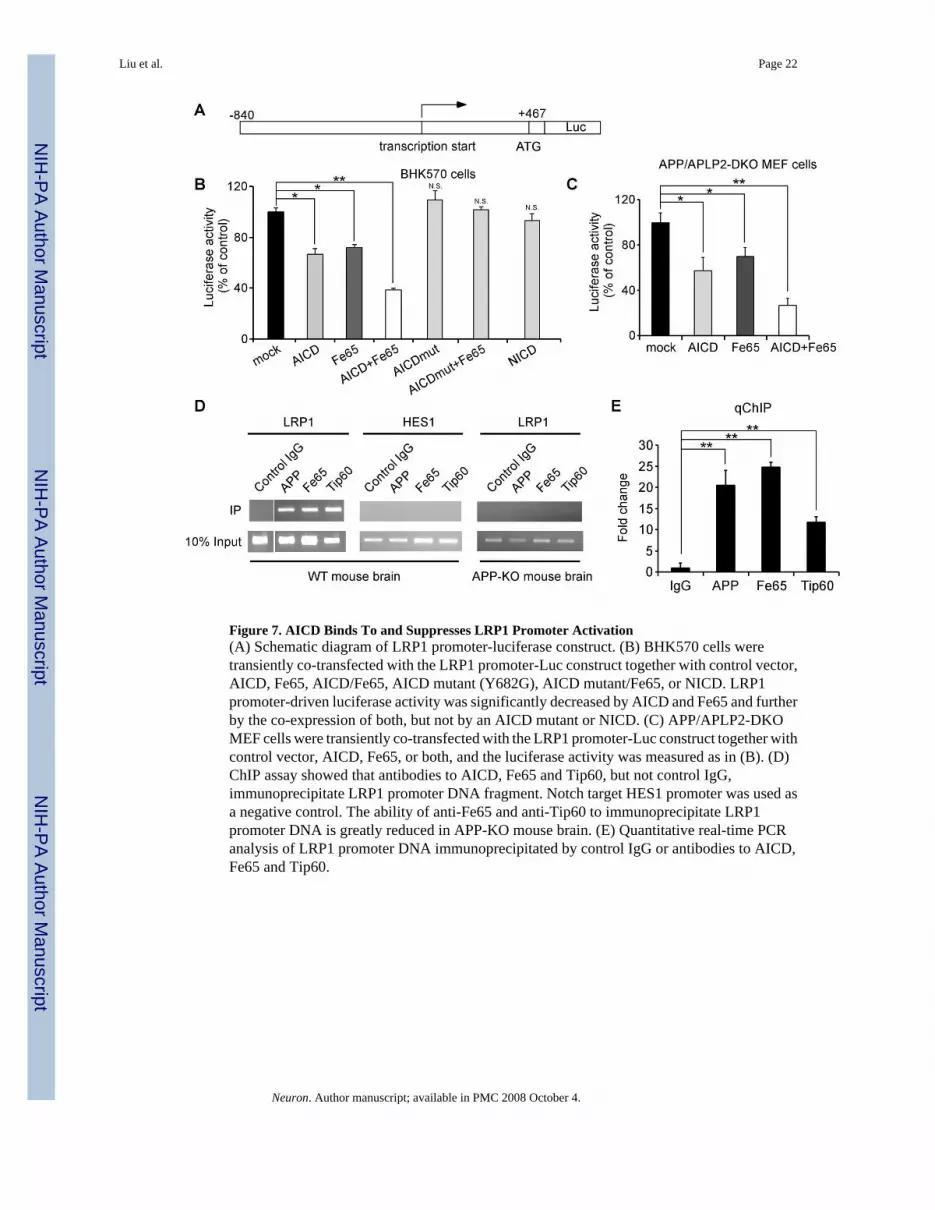
AICD Nuclear Signaling Inhibits LRP1 Promoter ActivationSince AICD down-regulates LRP1 expression at the transcriptional level and AICD has beenshown to modulate promoter function, we next tested whether LRP1 promoter activity isrepressed by AICD. The LRP1 promoter was cloned into a luciferase reporter vector pGL3(Figure 7A) and its activity was measured in BHK570 cells and APP/APLP2-DKO MEF cellsafter transfection of AICD, Fe65, or both. We found that AICD reduced LRP1 promoter activity
Liu et al. Page 5
Neuron. Author manuscript; available in PMC 2008 October 4.
NIH
-PA Author Manuscript
NIH
-PA Author Manuscript
NIH
-PA Author Manuscript
and Fe65 further potentiated this effect in both cell types (Figures 7B and 7C). An AICD mutantbearing a functional mutation in the 682YxNPxY motif (Y682G, see Borg et al., 1996) lost theability to regulate LRP1 promoter, and Notch intracellular domain (NICD) did not changeLRP1 promoter function in this assay (Figure 7B). To examine whether AICD binds directlyto the LRP1 promoter, we performed chromatin immunoprecipitation (ChIP) assay.Immunoprecipitation of mouse brain lysates with an antibody that recognizes the APP C-terminal domain showed that AICD associates with the LRP1 promoter (Figures 7D and 7E).Because biochemical and functional interactions among AICD, Fe65 and Tip60 have beendemonstrated (Baek et al., 2002;Cao and Sudhof, 2001), we also analyzed the ability ofantibodies to Fe65 and Tip60 to immunoprecipitate the LRP1 promoter. Indeed, we found thatboth Fe65 and Tip60 antibodies immunoprecipitated the LRP1 promoter (Figures 7D and 7E).The association of AICD, Fe65 and Tip60 with the LRP1 promoter was specific because normalrabbit IgG failed to immunoprecipitate the LRP1 promoter. In addition, APP, Fe65 and Tip60antibodies did not precipitate a control Notch target promoter, HES1 (Figure 7D). Further, theassociation of Fe65 and Tip60 with LRP1 promoter was greatly reduced in APP-KO mousebrain (Figure 7D). These results indicate that AICD, together with Fe65 and Tip60, binddirectly to the LRP1 promoter to suppress its activation.
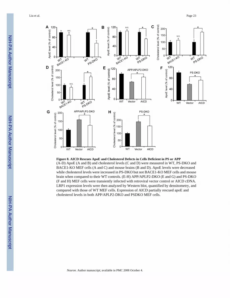
AICD Rescues ApoE and Cholesterol Defects in Cells Lacking APP/APLP2 or PSHaving demonstrated that LRP1 is a major receptor that regulates apoE and cholesterolmetabolism, and that γ-secretase activity is required for APP/APLP2-mediated regulation ofLRP1 expression, we were prompted to evaluate potential alterations in apoE and cholesterollevels in PS-DKO MEF cells and mouse brain. As expected, we found that apoE levels weredecreased while cholesterol levels were increased in PS-DKO MEF cells (Figures 8A and 8C)and mouse brain (Figures 8B and 8D) when compared to their WT controls. Normal levels ofapoE and cholesterol were found in BACE1-KO MEF cells and BACE1-KO mouse brain(Figures 8A-8D) demonstrating that BACE1 is not involved in apoE and cholesterolmetabolism. Since AICD rescued LRP1 expression in MEF cells devoid of either APP/APLP2or PS, we then analyzed whether forced expression of AICD could restore apoE and cholesterollevels in these cells. Forced expression of AICD in APP/APLP2-DKO (Figure 8E) and PS-DKO (Figure 8F) MEF cells significantly increased apoE levels when compared to MEF cellsinfected with vector alone, albeit not to the levels observed in WT control cells. Likewise,AICD, but not vector alone, partially restored cholesterol levels in APP/APLP2-DKO (Figure8G) and PS-DKO (Figure 8H) MEF cells. These results confirmed that AICD-mediated controlof LRP1 expression is a key regulatory pathway in brain apoE and cholesterol homeostasis.
DISCUSSIONIt has been postulated that the amyloidogenic processing of APP to Aβ, particularly of theaggregation-prone Aβ42, plays a central role in the pathogenesis of AD (Hardy and Selkoe,2002). Accordingly, inhibiting APP processing to Aβ is being actively explored as a therapeuticstrategy to treat AD. Here we described a novel mechanism by which the C-terminal fragmentof APP, named AICD, modulates brain apoE and cholesterol metabolism by directly regulatingthe expression and function of the lipoprotein receptor LRP1. Knockout of APP/APLP2 orcomponents of the γ-secretase complex significantly affected the expression of LRP1 as wellas apoE and cholesterol levels, and these alterations were partially restored by forcedexpression of AICD. We also show that AICD, together with the adaptor proteins Fe65 andTip60, regulates LRP1 promoter function. Our results establish, for the first time, a strongbiological relationship between APP processing and apoE/cholesterol metabolism withsignificant relevance for the pathogenesis of AD.
Liu et al. Page 6
Neuron. Author manuscript; available in PMC 2008 October 4.
NIH
-PA Author Manuscript
NIH
-PA Author Manuscript
NIH
-PA Author Manuscript
Several biological functions for APP and its processing products have been described (Zhengand Koo, 2006). The notion that APP is a cell surface receptor has long been speculated butremains unproven due to the lack of a bona fide ligand. The function of APP is furthercomplicated by the presence of two APP-related genes, APLP1 and APLP2 (Zheng and Koo,2006). Deletion of APLP2 and either APP or APLP1 results in early postnatal lethality (Zhengand Koo, 2006), suggesting redundancy between APLP2 and the other two family members.APP ectodomain has been shown to participate in cell adhesion, neurite outgrowth andsynaptogenesis (Zheng and Koo, 2006). APP intracellular domain (AICD), highlighted withan YxNPxY motif for binding of an array of interacting proteins, modulates cell migration,axonal transport and cell signaling (Zheng and Koo, 2006). The most relevant interactingprotein is Fe65, which modulates APP processing and AICD nuclear translocation (Cao andSudhof, 2001). Knockout of Fe65 and its homologues Fe65L1 results in cortical dysplasia andcompromised integrity of the pial basement membrane (Guenette et al., 2006). Intriguingly,this phenotype closely resembles that seen in triple mutant mice lacking the APP familymembers APP, APLP1 and APLP2 (Guenette et al., 2006;Herms et al., 2004), stronglysuggesting a common signaling pathway that requires the function of both Fe65 and APP. Micelacking both APP and APLP2 show defective neuromuscular synapses (Wang et al., 2005),while mice lacking APP alone exhibit increased synapses and associated synaptic function(Priller et al., 2006). In contrast, over-expression of APP in transgenic mice results in deficitsof synaptic transmission and learning (Saganich et al., 2006) and dendritic spine abnormalities(Spires et al., 2005). Whether some of these reported phenotypes relate to altered apoE andcholesterol metabolism reported herein requires further investigations.
Although the ε4 allele of the APOE gene was discovered as a strong genetic risk factor for late-onset AD over a decade ago (Corder et al., 1993), the mechanism by which apoE4 contributesto AD pathogenesis is still largely unclear. Furthermore, whether APP and apoE regulatecommon biological pathways is unknown. This study provides the first evidence that both APPand apoE participate in brain cholesterol metabolism. Cholesterol is an essential componentof the cellular membrane and plays pivotal roles in development and maintenance of neuronalplasticity and function (Ledesma and Dotti, 2006). In the adult brain, neuronal cholesterol issupplied primarily by apoE/lipoprotein particles synthesized and secreted by glial cells(Pfrieger, 2003;Puglielli et al., 2003). Uptake of apoE/lipoprotein particles by neurons ismediated by lipoprotein receptors of the LDLR family (Herz and Bock, 2002). Endocytosedcholesterol-containing lipoprotein particles are hydrolyzed in neuronal lysosomes allowingdegradation of apoE and intracellular release of free cholesterol which can be stored orincorporated into lipoprotein particles or cellular membranes. A function for LRP1 in brainapoE/cholesterol metabolism has been postulated (Pfrieger, 2003) but direct biologicalevidence had been lacking until now. Our studies provide the first evidence that deletion ofLRP1 in forebrain neurons of adult mice significantly alters brain apoE and cholesterol levels.While apoE levels were increased in LRP1 forebrain knockout mice, cholesterol levels wereconversely decreased. Consistent with these findings, our previous work has shown that over-expression of an LRP1 minireceptor in the brain results in a decrease in brain apoE level(Zerbinatti et al., 2006). Together, these results strongly support a role for LRP1 in brain apoEand cholesterol metabolism and establish LRP1 as a neuronal receptor essential for the properendocytosis and catabolism of apoE. Although the LDLR also functions as a brain apoEreceptor (Fryer et al., 2005), our results showed that its expression is not regulated by APP norγ-secretase.
The role of cholesterol in AD remains controversial (Ledesma and Dotti, 2006;Puglielli et al.,2003;Shobab et al., 2005). Several initial studies suggested a beneficial role of the cholesterol-lowering statins in reducing the risk of AD, and in vitro studies have identified a role forcholesterol in promoting Aβ production. However, several recent studies do not support theseconclusions (Shobab et al., 2005). Additionally, mice treated with lovastatin, the most brain
Liu et al. Page 7
Neuron. Author manuscript; available in PMC 2008 October 4.
NIH
-PA Author Manuscript
NIH
-PA Author Manuscript
NIH
-PA Author Manuscript
penetrant statin currently available, exhibit increased Aβ production and amyloid plaquedeposition (Park et al., 2003). Furthermore, neuronal membrane cholesterol loss was recentlyfound to enhance Aβ production (Abad-Rodriguez et al., 2004). Interestingly, a significantreduction of brain cholesterol in AD patients was observed, particularly in areas loaded withamyloid plaques (Ledesma, 2003). These findings suggest that loss of neuronal cholesterolmay contribute to synaptic dysfunction and excess Aβ production in AD. Because LRP1 levelsare also significantly reduced in AD (Kang et al., 2000), we can speculate that decreased LRP1levels in AD are directly responsible for cholesterol loss and related synaptic dysfunction.Accordingly, γ-secretase inhibitor treatment, a strategy actively explored for AD therapy, willlikely increase the expression and function of LRP1 in cholesterol metabolism, which couldin turn support synaptic integrity and function. Because LRP1 also has a role in Aβ clearanceby neurons (Zerbinatti and Bu, 2005) and across the blood brain barrier (Deane et al., 2004),restoring LRP1 expression and function in the AD brain could be explored independently as atherapeutic strategy to treat AD. Interestingly, the expression of another member of the LDLRfamily, SorLA, that regulates APP trafficking and processing to Aβ is also decreased in ADbrains (Andersen et al., 2006; Rogaeva et al., 2007) and that inherited variants in the SorLAgene, SORL1, are associated with late-onset AD (Rogaeva et al., 2007). Together, these studiespoint to multiple pathways by which LDLR family members play roles in the pathogenesis ofAD.
Our results reveal a novel role for γ-secretase-dependent APP processing in the regulation ofbrain cholesterol levels via transcriptional repression of LRP1. Presenilin-dependent cleavageof APP results in the release of AICD, which has been shown to interact with Fe65 and Tip60and has been suggested to function in nuclear signaling (Baek et al., 2002;Cao and Sudhof,2001). Subsequent work by Cao and Sudhof (Cao and Sudhof, 2004) demonstrated that nucleartranslocation of AICD may be dispensable; raising the possibility that AICD could functionby modulating activation of Fe65 rather than functioning as a transcriptional regulator itself.Several studies have reported putative target genes differentially regulated by an AICD-containing complex, including the prostate cancer anti-metastasis gene KAI1 (Baek et al.,2002), APP, GSK3β (Von Rotz et al., 2004), neprilysin (Pardossi-Piquard et al., 2005),regulators of actin dynamics (Muller et al., 2007), and the EGF receptor (Zhang et al., 2007).However, the exact role of AICD in transcriptional regulation of target genes remainscontroversial (Hebert et al., 2006;Chen and Selkoe, 2007;Pardossi-Piquard et al., 2007).Specifically, work by De Strooper and colleagues (Hebert et al., 2006) has shown thatexpression of several previously defined AICD targets genes was at best indirectly and weaklyaffected by APP processing. Further, the role of AICD in regulating neprilysin expression(Pardossi-Piquard et al., 2005;Pardossi-Piquard et al., 2007) was not reproduced by anotherreport (Chen and Selkoe, 2007). Finally, a recent study has shown that secreted APPectodomain APPsα is sufficient to rescue several anatomical, behavioral, andelectrophysiological abnormalities seen in APP-KO mice (Ring et al., 2007). The discrepancyin AICD function could be due to different experimental systems and/or approaches (Herz,2007). In line with a biological function of AICD, our results provide direct evidence that AICDbinds to the LRP1 promoter and regulates its transcriptional function. Although our studies donot exclude the possibility that APP itself or other APP processing products (e.g. soluble APPand Aβ) may also regulate LRP1 expression and function, our findings that LRP1 expressionand apoE/cholesterol metabolism are unchanged in BACE1-knockout mice argues against arole for Aβ in regulating LRP1 expression. Supporting an Aβ-independent function of γ-secretase, recent work has demonstrated that a complete loss of presenilin function in theforebrain leads to memory deficits, synaptic dysfunction, and neurodegeneration withoutgeneration of amyloid plaques (Saura et al., 2004). These results suggest that at least some ofthe neurodegenerative pathology seen in AD might result from partial loss of γ-secretaseactivity or AICD nuclear signaling functions independent of Aβ production (Shen and Kelleher,2007). It is interesting to note that a recent study demonstrates a role for Aβ in regulating
Liu et al. Page 8
Neuron. Author manuscript; available in PMC 2008 October 4.
NIH
-PA Author Manuscript
NIH
-PA Author Manuscript
NIH
-PA Author Manuscript
cholesterol biosynthesis and sphingomyelin degradation (Grimm et al., 2005). Because AICDdid not completely restore apoE and cholesterol levels in PS-DKO cells, it is possible that otherγ-secretase cleavage products may also regulate apoE/cholesterol metabolism via LRP1-dependent and/or independent mechanisms. Regulation of cholesterol synthesis by Aβ, as wellas AICD-mediated modulation of LRP1 expression, are likely to be key events in the propermaintenance of brain cholesterol levels.
In summary, this study uncovers a novel biological function of APP in modulating brain apoEand cholesterol homeostasis. We further demonstrate an important role of the APP processingproduct AICD in modulating the promoter activity of LRP1, an essential lipoprotein receptorfor brain apoE and cholesterol metabolism. Our results provide important insights into APPbiological function and its potential implications for neuronal dysfunction in AD, and may leadto the design of better therapeutic strategies to treat this devastating disease.
EXPERIMENTAL PROCEDURESMaterials
Human recombinant RAP was expressed in a glutathione S-transferase expression vector andisolated as described previously (Bu et al., 1993). All tissue culture media and serum werefrom Sigma. Anti-APP C-terminal antibody was purchased from Invitrogen; anti-Fe65 wasfrom Abcam; anti-Tip60 was from Calbiochem; anti-actin from Sigma and anti-NeuN wasfrom Chemicon. In house anti-LRP1 and anti-LDLR antibodies have been described previously(Bu et al., 1995;Li et al., 2005;Zerbinatti et al., 2004). Peroxidase-labeled anti-mouse antibodyand ECL system were from GE Healthcare. Carrier-free Na125I was purchased from PerkinElmer Lifescience. The γ-secretase inhibitors L685,458 and DAPT were from Calbiochem andDFK167 was from Enzyme Systems.
Animals and Tissue PreparationLRP1 forebrain knockout mice were generated by breeding the LRP1 loxP mice (Rohlmannet al., 1998) with (α-calcium-calmodulin-dependent kinase II-driven Cre recombinase mice[Tsien, 1996 #1326). Littermates of LRP1 forebrain knockout (LRP1flox+/+, Cre+/−) or WTcontrols (LRP1flox+/+, Cre−/−) at 11 months of age were used for Western blotting,immunofluorescence staining and apoE/cholesterol assays. APP-KO, APP/APLP2-DKO, andWT littermate control mice have been described previously and were used within 24 h afterbirth due to potential lethality of the APP/APLP2 mice. APP-KO mice were also analyzed at4 months of age. PS1/2-DKO mice were generated by Cre-lox conditional deletion of thePS1 gene in forebrain of the PS2-KO mice (Feng et al., 2004) and were used at 4 months ofage. BACE1-KO mice (Luo et al., 2001) were described in previous work and were used at 2months of age. Animals were perfused with PBS-heparin (3 units/ml) and brain tissues weredissected and kept frozen at −80°C until further analysis. All animal procedures were approvedby the Animal Study Committee at Washington University School of Medicine and inaccordance with the regulations of the American Association for the Accreditation ofLaboratory Animal Care.
Reverse Transcriptase Real-time PCRTotal RNA was isolated from tissues or cells using the SV Total RNA Isolation System(Promega) and subjected to DNase I digestion to remove contaminating genomic DNA. TotalRNA was dissolved in nuclease-free water and stored at −80°C. Reverse transcription wasperformed using a SuperScript II RNase H-reverse transcriptase (Invitrogen), and the reactionmix was subjected to quantitative real-time PCR to detect levels of the corresponding actin,LRP1, or LDLR. The set of actin primers was used as an internal control for each specific geneamplification. The relative levels of expression were quantified and analyzed by using Bio-
Liu et al. Page 9
Neuron. Author manuscript; available in PMC 2008 October 4.
NIH
-PA Author Manuscript
NIH
-PA Author Manuscript
NIH
-PA Author Manuscript
Rad iCycler iQ software. The real-time value for each sample was averaged and comparedusing the CT method, where the amount of target RNA (2−ΔΔCT) was normalized to theendogenous actin reference (ΔCT) and related to the amount of target gene in tissue cells, whichwas set as the calibrator at 1.0.
ApoE ELISAThe sandwich ELISA for mouse apoE has been described previously (Wahrle et al., 2004).Briefly, 96-well plates were coated overnight with apoE antibody (WU E4), washed with PBS,blocked with 1% milk in PBS, and then washed again. Cells or brain samples were sonicatedin 5 M guanidine HCl with 1X Complete protease inhibitor mixture (Roche Applied Science),debris was pelleted by centrifugation at 10,000 X g, and the supernatant was diluted in 0.1%BSA, 0.025% Tween-20 in PBS. Following sample incubation, the plate was washed, and 3μg/well of biotinylated goat anti-apoE (Calbiochem) was added. After incubation with thesecondary antibody, the plate was washed, and poly-horseradish peroxidase streptavidin(Pierce) was added at 1:6000 dilution and incubated. The plate was then washed, developedwith tetramethylbenzidine (Sigma), and read at 650 nm with a Biotek 600 plate reader (Bio-Tek Instruments).
Cholesterol AnalysesCells or brain samples were prepared for cholesterol analysis by sonication in PBS with 1XComplete protease inhibitor mixture. The homogenized whole cell or brain suspension wasthen subjected to enzymatic analysis for total cholesterol using the Amplex Red CholesterolKit (Invitrogen) (Wahrle et al., 2004).
Immunofluorescence StainingFrozen tissue sections were blocked with 0.1% Tween-20, 5% BSA in PBS for 30 min andstained for 2 h at room temperature with anti-LRP1 antibody. Primary antibody was thenvisualized using Alexa488-labeled goat anti-mouse secondary antibody (Invitrogen). Neuronswere counterstained with anti-NeuN (Calbiochem) and Alexa633 labeled secondary antibody(Invitrogen). Fluorescent images were captured with a confocal microscope (OlympusFluoview 500).
Chromatin Immunoprecipitation (ChIP)ChIP assays were performed using a chromatin immunoprecipitation (ChIP) assay kit (Upstate)according to the manufacturer's instructions with minor modifications. Briefly, brain tissuefrom WT C57BL/6J mice were minced into small pieces with a razor and 1% formaldehydewas added directly to the tissue mixture to cross-link proteins to DNA. Tissue was then lysedin SDS lysis buffer and sonicated to shear DNA to a size range of 200-1000 bp. Aftercentrifugation, the supernatant was diluted 10-fold in ChIP dilution buffer and incubatedovernight at 4°C with anti-APP, anti-Fe65, anti-Tip60, or normal rabbit IgG. Protein A-agarosebeads were used to immunoprecipitate the antibody/protein/DNA complexes. After washing,the complex was incubated at 65°C for 4 h to reverse the protein/DNA cross-links. The DNAwas then purified using PCR Purification kit (Qiagen) and used as template for PCRamplification. LRP1-F (5'-TCGGGTGTCCCTGTTTAC-3') and LRP1-R (5'-GAAAGCGGTCCAAGAGTG-3') primers were used to amplify the LRP1 promoter by RT-PCR. LRP1-F (5'-GGGAGCCTGAAATCCTAGAG-3') and LRP1-R (5'-GGAAAGCGGTCCAAGAGTG-3') primers were used to amplify LRP1 promoter by real-time PCR. Primers for HES1 promoter amplification are: HES1-F, 5'-CGTGTCTCTTCCTCCCATTG-3'; HES1-R, 5'- GATCCAGTGTGATCCGCAGG-3'. PCRproducts were resolved on 2% agarose gels and visualized by ethidium bromide staining.
Liu et al. Page 10
Neuron. Author manuscript; available in PMC 2008 October 4.
NIH
-PA Author Manuscript
NIH
-PA Author Manuscript
NIH
-PA Author Manuscript
Luciferase AssayBHK570 cells or MEF APP/APLP2-DKO cells were transfected with the appropriate cDNAs:empty vector (pGL3-luc), LRP1 promoter-luc, AICD, and/or Fe65. A β-gal reporter cDNAwas co-transfected to normalize data for transfection efficiency. Twenty-four hours aftertransfection, cells were rinsed, gently scraped into PBS (pH 7.4), and pelleted. Cells were thenlysed in lysis buffer, and the luciferase activity and β-gal activity were measured by theLuciferase Assay System and β-gal Assay System following the manufacturer's instructions(Promega).
Statistical AnalysisAll quantified data represent an average of at least triplicate samples. Error bars representstandard error of the mean. Statistical significance was determined by Student's t-test andP<0.05 was considered significant.
Supplementary MaterialRefer to Web version on PubMed Central for supplementary material.
ACKNOWLEDGEMENTS
We thank Jane Knisely and Alan Schwartz for the critical reading of the manuscript and members of the Bu lab forcomments and suggestions. We also thank Suzanne Wahrle, David Holtzman, Raphael Kopan, Sheila Stewart andBart De Strooper for providing valuable reagents. This work was supported by NIH grant R01 AG027924, a grantfrom the Alzheimer's Association, and a grant from the American Health Assistant Foundation to G.B.
REFERENCESAbad-Rodriguez J, Ledesma MD, Craessaerts K, Perga S, Medina M, Delacourte A, Dingwall C, De
Andersen OM, Reiche J, Schmidt V, Gotthardt M, Spoelgen R, Behlke J, von Arnim CA, et al. Neuronalsorting protein-related receptor sorLA/LR11 regulates processing of the amyloid precursor protein.Proc. Natl. Acad. Sci. USA 2005;102:13461–13466. [PubMed: 16174740]
Baek SH, Ohgi KA, Rose DW, Koo EH, Glass CK, Rosenfeld MG. Exchange of N-CoR corepressor andTip60 coactivator complexes links gene expression by NF-kappaB and beta-amyloid precursor protein.Cell 2002;110:55–67. [PubMed: 12150997]
Borg JP, Ooi J, Levy E, Margolis B. The phosphotyrosine interaction domains of X11 and FE65 bind todistinct sites on the YENPTY motif of amyloid precursor protein. Mol. Cell. Biol 1996;16:6229–6241.[PubMed: 8887653]
Bu G, Geuze HJ, Strous GJ, Schwartz AL. 39 kDa receptor-associated protein is an ER resident proteinand molecular chaperone for LDL receptor-related protein. EMBO J 1995;14:2269–2280. [PubMed:7774585]
Bu G, Maksymovitch EA, Schwartz AL. Receptor-mediated endocytosis of tissue-type plasminogenactivator by low density lipoprotein receptor-related protein on human hepatoma HepG2 cells. J. Biol.Chem 1993;268:13002–13009. [PubMed: 8389767]
Cam JA, Zerbinatti CV, Li Y, Bu G. Rapid endocytosis of the low density lipoprotein receptor-relatedprotein modulates cell surface distribution and processing of the β-amyloid precursor protein. J. Biol.Chem 2005;280:15464–15470. [PubMed: 15705569]
Cao X, Sudhof TC. A transcriptionally active complex of APP with Fe65 and histone acetyltransferaseTip60. Science 2001;293:115–120. [PubMed: 11441186]
Cao X, Sudhof TC. Dissection of amyloid-β precursor protein-dependent transcriptional transactivation.J. Biol. Chem 2004;279:24601–24611. [PubMed: 15044485]
Liu et al. Page 11
Neuron. Author manuscript; available in PMC 2008 October 4.
NIH
-PA Author Manuscript
NIH
-PA Author Manuscript
NIH
-PA Author Manuscript
Chen AC, Selkoe DJ. Response to: Pardossi-Piquard et al., “Presenilin-dependent transcriptional controlof the abeta-degrading enzyme neprilysin by intracellular domains of betaAPP and APLP.” Neuron46, 541-554. Neuron 2007;53:479–483. [PubMed: 17296549]
Corder EH, Saunders AM, Strittmatter WJ, Schmechel DE, Gaskell PC, Small GW, Roses AD, HainesJL, Pericak-Vance MA. Gene dose of apolipoprotein E type 4 allele and the risk of Alzheimer'sdisease in late onset families. Science 1993;261:921–923. [PubMed: 8346443]
Counter CM, Hahn WC, Wei W, Caddle SD, Beijersbergen RL, Lansdorp PM, Sedivy JM, WeinbergRA. Dissociation among in vitro telomerase activity, telomere maintenance, and cellularimmortalization. Proc. Natl. Acad. Sci. USA 1998;95:14723–14728. [PubMed: 9843956]
Deane R, Wu Z, Sagare A, Davis J, Du Yan S, Hamm K, Xu F, et al. LRP/amyloid beta-peptide interactionmediates differential brain efflux of Abeta isoforms. Neuron 2004;43:333–344. [PubMed: 15294142]
Feng R, Wang H, Wang J, Shrom D, Zeng X, Tsien JZ. Forebrain degeneration and ventricle enlargementcaused by double knockout of Alzheimer's presenilin-1 and presenilin-2. Proc. Natl. Acad. Sci. USA2004;101:8162–8167. [PubMed: 15148382]
Fryer JD, Demattos RB, McCormick LM, O'Dell M,A, Spinner ML, Bales KR, Paul SM, et al. The lowdensity lipoprotein receptor regulates the level of central nervous system human and murineapolipoprotein E but does not modify amyloid plaque pathology in PDAPP mice. J. Biol. Chem2005;280:25754–25759. [PubMed: 15888448]
Grimm MOW, Grimm HS, Patzold AJ, Zinser EG, Halonen R, Duering M, Tschape J-A, et al. Regulationof cholesterol and sphingomyelin metabolism by amyloid-beta and presenilin. Nat. Cell Bio2005;7:1118–1123. [PubMed: 16227967]
Guenette S, Chang Y, Hiesberger T, Richardson JA, Eckman CB, Eckman EA, Hammer RE, Herz J.Essential roles for the FE65 amyloid precursor protein-interacting proteins in brain development.EMBO J 2006;25:420–431. [PubMed: 16407979]
Hamanaka H, Katoh-Fukui Y, Suzuki K, Kobayashi M, Suzuki R, Motegi Y, Nakahara Y, et al. Alteredcholesterol metabolism in human apolipoprotein E4 knock-in mice. Hum. Mol. Genet 2000;9:353–361. [PubMed: 10655544]
Hardy J, Selkoe DJ. The amyloid hypothesis of Alzheimer's disease: progress and problems on the roadto therapeutics. Science 2002;297:353–356. [PubMed: 12130773]
Hebert SS, Serneels L, Tolia A, Craessaerts K, Derks C, Filippov MA, Muller U, De Strooper B. Regulatedintramembrane proteolysis of amyloid precursor protein and regulation of expression of putativetarget genes. EMBO Rep 2006;7:739–745. [PubMed: 16729020]
Herms J, Anliker B, Heber S, Ring S, Fuhrmann M, Kretzschmar H, Sisodia S, Muller U. Corticaldysplasia resembling human type 2 lissencephaly in mice lacking all three APP family members.EMBO J 2004;23:4106–4115. [PubMed: 15385965]
Herz J, Bock HH. Lipoprotein receptors in the nervous system. Annu. Rev. Biochem 2002;71:405–434.[PubMed: 12045102]
Herz J, Chen Y. Reelin, lipoprotein receptors and synaptic plasticity. Nat. Rev. Neurosci 2006;7:850–859. [PubMed: 17053810]
Herz J, Clouthier DE, Hammer RE. LDL receptor-related protein internalizes and degrades uPA-PAI-1complexes and is essential for embryo implantation. Cell 1992;71:411–421. [PubMed: 1423604]
Herz J. Overview: the long and winding road to understanding Alzheimer's disease. Neuron 2007;53:477–479. [PubMed: 17296548]
Holtzman DM, Bales KR, Tenkova T, Fagan AM, Parsadanian M, Sartorius LJ, Mackey B, et al.Apolipoprotein E isoform-dependent amyloid deposition and neuritic degeneration in a mouse modelof Alzheimer's disease. Proc. Natl. Acad. Sci. USA 2000;97:2892–2897. [PubMed: 10694577]
Huang Y. Molecular and cellular mechanisms of apolipoprotein E4 neurotoxicity and potentialtherapeutic strategies. Curr. Opin. Drug Discov. Devel 2006;9:627–641.
Kang DE, Pietrzik CU, Baum L, Chevallier N, Merriam DE, Kounnas MZ, Wagner SL, et al. Modulationof amyloid beta-protein clearance and Alzheimer's disease susceptibility by the LDL receptor-relatedprotein pathway. J. Clin. Invest 2000;106:1159–1166. [PubMed: 11067868]
LaDu MJ, Falduto MT, Manelli AM, Reardon CA, Getz GS, Frail DE. Isoform-specific binding ofapolipoprotein E to β-amyloid. J. Biol. Chem 1994;269:23403–23406. [PubMed: 8089103]
Liu et al. Page 12
Neuron. Author manuscript; available in PMC 2008 October 4.
NIH
-PA Author Manuscript
NIH
-PA Author Manuscript
NIH
-PA Author Manuscript
Ledesma MD, Abad-Rodriguez J, Galvan C, Biondi E, Navarro P, Delacourte A, Dingwall C, Dotti CG.Raft disorganization leads to reduced plasmin activity in Alzheimer's disease brains. EMBO Rep2003;4:1190–1196. [PubMed: 14618158]
Ledesma MD, Dotti CG. Amyloid excess in Alzheimer's disease: What is cholesterol to be blamed for?FEBS Lett 2006;580:5525–5532. [PubMed: 16814780]
Li Y, Chen J, Lu W, McCormick LM, Wang J, Bu G. Mesd binds to mature LDL-receptor-relatedprotein-6 and antagonizes ligand binding. J. Cell Sci 2005;118:5305–5314. [PubMed: 16263759]
Li Y, Marzolo MP, Kerkhof P, Strous GJ, Bu G. The YXXL motif, but not the two NPXY motifs, servesas the dominant endocytosis signal For LDL receptor-related protein (LRP). J. Biol. Chem2000;275:17187–17194. [PubMed: 10747918]
Luo Y, Bolon B, Kahn S, Bennett BD, Babu-Khan S, Denis P, Fan W, et al. Mice deficient in BACE1,the Alzheimer's beta-secretase, have normal phenotype and abolished beta-amyloid generation. Nat.Neurosci 2001;4:231–232. [PubMed: 11224535]
Mahley RW. Apolipoprotein E: cholesterol transport protein with expanding role in cell biology. Science1988;240:622–630. [PubMed: 3283935]
Moestrup SK, Gliemann J, Pallesen G. Distribution of the alpha 2-macroglobulin receptor/low densitylipoprotein receptor-related protein in human tissues. Cell Tiss. Res 1992;269:375–382.
Muller T, Concannon CG, Ward MW, Walsh CM, Tirniceriu AL, Tribl F, Kogel D, Prehn JHM,Egensperger R. Modulation of gene expression and cytoskeletal dynamics by the amyloid precursorprotein intracellular domain (AICD). Mol. Biol. Cell 2007;18:201–210. [PubMed: 17093061]
Pardossi-Piquard R, Dunys J, Kawarai T, Sunyach C, Alves da Costa C, Vincent B, Sevalle J, PimplikarS, St George-Hyslop P, Checler F. Response to Correspondence: Pardossi-Piquard et al., “Presenilin-dependent transcriptional control of the abeta-degrading enzyme neprilysin by intracellular domainsof betaAPP and APLP.” Neuron 46, 541-554. Neuron 2007;53:483–486. [PubMed: 17296550]
Pardossi-Piquard R, Petit A, Kawarai T, Sunyach C, Alves da Costa C, Vincent B, Ring S, et al. Presenilin-dependent transcriptional control of the Abeta-degrading enzyme neprilysin by intracellular domainsof betaAPP and APLP. Neuron 2005;46:541–554. [PubMed: 15944124]
Park I-H, Hwang EM, Hong HS, Boo JH, Oh SS, Lee J, Jung MW, et al. Lovastatin enhances Abetaproduction and senile plaque deposition in female Tg2576 mice. Neurobiol. Aging 2003;24:637–643. [PubMed: 12885571]
Pfrieger FW. Cholesterol homeostasis and function in neurons of the central nervous system. CMLS,Cell. Mol. Life Sci 2003;60:1158–1171.
Priller C, Bauer T, Mitteregger G, Krebs B, Kretzschmar HA, Herms J. Synapse formation and functionis modulated by the amyloid precursor protein. J. Neurosci 2006;26:7212–7221. [PubMed:16822978]
Puglielli L, Tanzi RE, Kovacs DM. Alzheimer's disease: the cholesterol connection. Nat. Neurosci2003;6:345–351. [PubMed: 12658281]
Ring S, Weyer SW, Kilian SB, Waldron E, Pietrzik CU, Filippov MA, Herms J, et al. The secreted beta-amyloid precursor protein ectodomain APPs alpha is sufficient to rescue the anatomical, behavioral,and electrophysiological abnormalities of APP-deficient mice. J. Neurosci 2007;27:7817–7826.[PubMed: 17634375]
Rogaeva E, Meng Y, Lee JH, Gu Y, Kawarai T, Zou F, Katayama T, et al. The neuronal sortilin-relatedreceptor SORL1 is genetically associated with Alzheimer disease. Nat. Genet 2007;39:168–177.[PubMed: 17220890]
Rohlmann A, Gotthardt M, Hammer RE, Herz J. Inducible inactivation of hepatic LRP gene by cre-mediated recombination confirms role of LRP in clearance of chylomicron remnants. J. Clin. Invest1998;101:689–695. [PubMed: 9449704]
Saganich MJ, Schroeder BE, Galvan V, Bredesen DE, Koo EH, Heinemann SF. Deficits in synaptictransmission and learning in amyloid precursor protein (APP) transgenic mice require C-terminalcleavage of APP. J. Neurosci 2006;26:13428–13436. [PubMed: 17192425]
Saura CA, Choi S-Y, Beglopoulos V, Malkani S, Zhang D, Rao BSS, Chattarji S, et al. Loss of presenilinfunction causes impairments of memory and synaptic plasticity followed by age-dependentneurodegeneration. Neuron 2004;42:23–36. [PubMed: 15066262]
Liu et al. Page 13
Neuron. Author manuscript; available in PMC 2008 October 4.
NIH
-PA Author Manuscript
NIH
-PA Author Manuscript
NIH
-PA Author Manuscript
Selkoe D, Kopan R. Notch and Presenilin: regulated intramembrane proteolysis links development anddegeneration. Annu. Rev. Neurosci 2003;26:565–597. [PubMed: 12730322]
Shen J, Kelleher RJ III. The presenilin hypothesis of Alzheimer's disease: Evidence for a loss-of-functionpathogenic mechanism. Proc. Natl. Acad. Sci. USA 2007;104:403–409. [PubMed: 17197420]
Spires TL, Meyer-Luehmann M, Stern EA, McLean PJ, Skoch J, Nguyen PT, Bacskai BJ, Hyman BT.Dendritic spine abnormalities in amyloid precursor protein transgenic mice demonstrated by genetransfer and intravital multiphoton microscopy. J. Neurosci 2005;25:7278–7287. [PubMed:16079410]
Tsien JZ, Chen DF, Gerber D, Tom C, Mercer EH, Anderson DJ, Mayford M, Kandel ER, Tonegawa S.Subregion- and cell type-restricted gene knockout in mouse brain. Cell 1996;87:1317–1326.[PubMed: 8980237]
Vassar R, Bennett BD, Babu-Khan S, Kahn S, Mendiaz EA, Denis P, Teplow DB, et al. Beta-secretasecleavage of Alzheimer's amyloid precursor protein by the transmembrane aspartic protease BACE.Science 1999;286:735–741. [PubMed: 10531052]
Von Rotz RC, Kohli BM, Bosset J, Meier M, Suzuki T, Nitsch RM, Konietzko U. The APP intracellulardomain forms nuclear multiprotein complexes and regulates the transcription of its own precursor.J. Cell Sci 2004;117:4435–4448. [PubMed: 15331662]
Wahrle SE, Jiang H, Parsadanian M, Legleiter J, Han X, Fryer JD, Kowalewski T, Holtzman DM. ABCA1is required for normal central nervous system ApoE levels and for lipidation of astrocyte-secretedapoE. J. Biol. Chem 2004;279:40987–40993. [PubMed: 15269217]
Wang P, Yang G, Mosier DR, Chang P, Zaidi T, Gong Y-D, Zhao N-M, et al. Defective neuromuscularsynapses in mice lacking amyloid precursor protein (APP) and APP-like protein 2. J. Neurosci2005;25:1219–1225. [PubMed: 15689559]
Zerbinatti CV, Bu G. LRP and Alzheimer's disease. Rev. Neurosci 2005;16:123–135. [PubMed:15959937]
Zerbinatti CV, Wahrle SE, Kim H, Cam JA, Bales K, Paul SM, Holtzman DM, Bu G. Apolipoprotein Eand low density lipoprotein receptor-related protein facilitate intraneuronal Abeta42 accumulationin amyloid model mice. J. Biol. Chem 2006;281:36180–36186. [PubMed: 17012232]
Zerbinatti CV, Wozniak DF, Cirrito J, Cam JA, Osaka H, Bales KR, Zhuo M, Paul SM, Holtzman DM,Bu G. Increased soluble amyloid-beta peptide and memory deficits in amyloid model miceoverexpressing the low-density lipoprotein receptor-related protein. Proc. Natl. Acad. Sci. USA2004;101:1075–1080. [PubMed: 14732699]
Zhang YW, Wang R, Liu Q, Zhang H, Liao FF, Xu H. Presenilin/γ-secretase-dependent processing ofbeta-amyloid precursor protein regulates EGF receptor expression. Proc. Natl. Acad. Sci. USA2007;104:10613–10618. [PubMed: 17556541]
Zheng H, Koo E. The amyloid precursor protein: beyond amyloid. Molecular Neurodegeneration2006;1:5. [PubMed: 16930452]
Neuron. Author manuscript; available in PMC 2008 October 4.
NIH
-PA Author Manuscript
NIH
-PA Author Manuscript
NIH
-PA Author Manuscript
Figure 1. APP Regulates ApoE and Cholesterol Metabolism(A and B) ApoE levels were decreased and cholesterol content was increased in the absenceof APP or APP/APLP2. ApoE and cholesterol levels were measured in triplicates in lysates ofWT, APP-KO and APP/APLP2-DKO MEF cells, normalized against total protein and plottedas a percentage of WT controls. (C and D) ApoE levels were also increased and cholesterolcontent decreased in brain lysates of APP-KO and APP/APLP2-DKO mice. ApoE andcholesterol levels were measured in brain lysates of WT, APP-KO and APP/APLP2-DKO mice(n=4), normalized against total protein and plotted as a percentage of WT controls. For thisand subsequent figures, data represent mean ± SEM; N.S., not significant; *, P<0.05; **,P<0.01.
Liu et al. Page 15
Neuron. Author manuscript; available in PMC 2008 October 4.
NIH
-PA Author Manuscript
NIH
-PA Author Manuscript
NIH
-PA Author Manuscript
Figure 2. APP/APLP2 Regulates LRP1 Expression and Function(A) LRP1 and LDLR expression levels were compared between WT, APP-KO and APP/APLP2-DKO MEF cells by Western blot. Equal amount of protein in this and subsequentfigures was loaded to each lane. (B) Densitometric analyses of Western blots from triplicatesamples demonstrate a significant increase in the expression of LRP1, but not LDLR, in theabsence of APP/APLP2. (C) LRP1 and LDLR mRNA levels were quantified in WT, APP-KOand APP/APLP2-DKO MEF cells by real-time PCR. LRP1 mRNA, but not LDLR mRNA,was significantly increased in the absence of APP/APLP2. (D) 125I-α2M (1 nM) binding toWT, APP-KO and APP/APLP2-DKO MEF cells was performed at 4°C for 1 h in the absenceor presence of RAP (500 nM). RAP-inhibitable 125I-α2M binding was normalized against totalcellular protein and plotted as a percentage of WT controls. (E) 125I-α2M (1 nM) uptake anddegradation assays were performed at 37°C for 4 h in the absence or presence of RAP (500nM). RAP-inhibitable 125I-α2M degradation was normalized against total cellular protein andplotted as a percentage of WT controls. (F) LRP1 and LDLR expression levels in the brainwere compared between WT, APP-KO and APP/APLP2-DKO newborn mice by Western blot.(G) Densitometric analyses of Western blots (n=3) indicate a significant increase in theexpression of LRP1, but not LDLR, in the absence of APP or APP/APLP2. (H) LRP1 andLDLR mRNA levels were quantified in the brain of WT, APP-KO and APP/APLP2-DKOnewborn mice (n=3) by real-time PCR. LRP1 mRNA, but not LDLR mRNA, was significantlyincreased in the absence of APP/APLP2. (I) LRP1 expression in the brain was compared by
Liu et al. Page 16
Neuron. Author manuscript; available in PMC 2008 October 4.
NIH
-PA Author Manuscript
NIH
-PA Author Manuscript
NIH
-PA Author Manuscript
Western blot between WT and APP-KO mice at 4 months of age. Similar increase in LRP1expression was observed in adult APP-KO mouse brain.
Liu et al. Page 17
Neuron. Author manuscript; available in PMC 2008 October 4.
NIH
-PA Author Manuscript
NIH
-PA Author Manuscript
NIH
-PA Author Manuscript
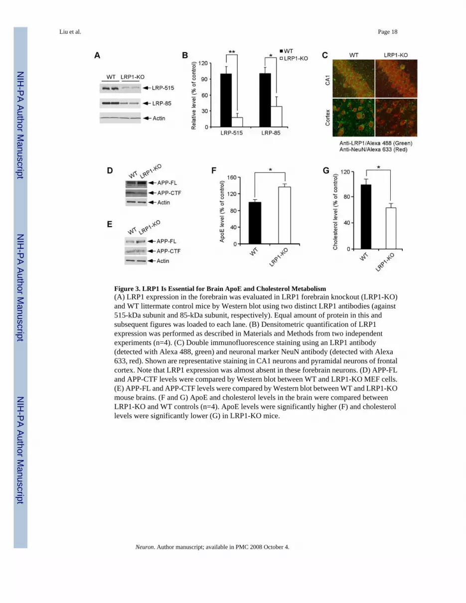
Figure 3. LRP1 Is Essential for Brain ApoE and Cholesterol Metabolism(A) LRP1 expression in the forebrain was evaluated in LRP1 forebrain knockout (LRP1-KO)and WT littermate control mice by Western blot using two distinct LRP1 antibodies (against515-kDa subunit and 85-kDa subunit, respectively). Equal amount of protein in this andsubsequent figures was loaded to each lane. (B) Densitometric quantification of LRP1expression was performed as described in Materials and Methods from two independentexperiments (n=4). (C) Double immunofluorescence staining using an LRP1 antibody(detected with Alexa 488, green) and neuronal marker NeuN antibody (detected with Alexa633, red). Shown are representative staining in CA1 neurons and pyramidal neurons of frontalcortex. Note that LRP1 expression was almost absent in these forebrain neurons. (D) APP-FLand APP-CTF levels were compared by Western blot between WT and LRP1-KO MEF cells.(E) APP-FL and APP-CTF levels were compared by Western blot between WT and LRP1-KOmouse brains. (F and G) ApoE and cholesterol levels in the brain were compared betweenLRP1-KO and WT controls (n=4). ApoE levels were significantly higher (F) and cholesterollevels were significantly lower (G) in LRP1-KO mice.
Liu et al. Page 18
Neuron. Author manuscript; available in PMC 2008 October 4.
NIH
-PA Author Manuscript
NIH
-PA Author Manuscript
NIH
-PA Author Manuscript
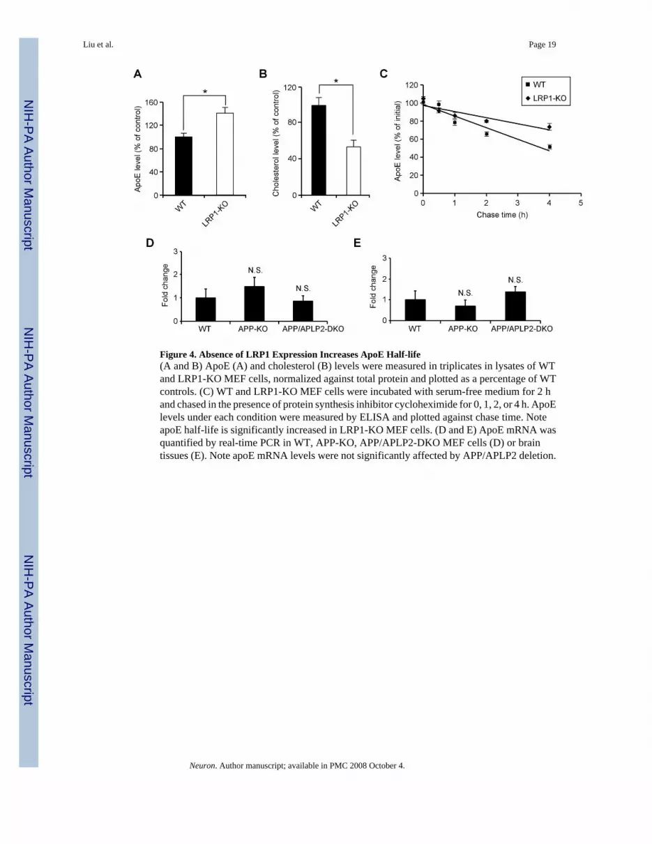
Figure 4. Absence of LRP1 Expression Increases ApoE Half-life(A and B) ApoE (A) and cholesterol (B) levels were measured in triplicates in lysates of WTand LRP1-KO MEF cells, normalized against total protein and plotted as a percentage of WTcontrols. (C) WT and LRP1-KO MEF cells were incubated with serum-free medium for 2 hand chased in the presence of protein synthesis inhibitor cycloheximide for 0, 1, 2, or 4 h. ApoElevels under each condition were measured by ELISA and plotted against chase time. NoteapoE half-life is significantly increased in LRP1-KO MEF cells. (D and E) ApoE mRNA wasquantified by real-time PCR in WT, APP-KO, APP/APLP2-DKO MEF cells (D) or braintissues (E). Note apoE mRNA levels were not significantly affected by APP/APLP2 deletion.
Liu et al. Page 19
Neuron. Author manuscript; available in PMC 2008 October 4.
NIH
-PA Author Manuscript
NIH
-PA Author Manuscript
NIH
-PA Author Manuscript
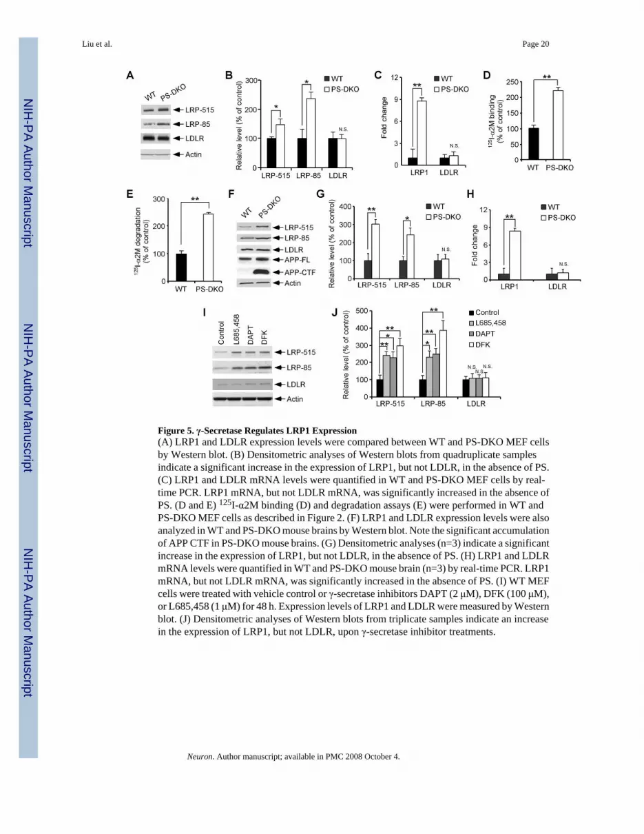
Figure 5. γ-Secretase Regulates LRP1 Expression(A) LRP1 and LDLR expression levels were compared between WT and PS-DKO MEF cellsby Western blot. (B) Densitometric analyses of Western blots from quadruplicate samplesindicate a significant increase in the expression of LRP1, but not LDLR, in the absence of PS.(C) LRP1 and LDLR mRNA levels were quantified in WT and PS-DKO MEF cells by real-time PCR. LRP1 mRNA, but not LDLR mRNA, was significantly increased in the absence ofPS. (D and E) 125I-α2M binding (D) and degradation assays (E) were performed in WT andPS-DKO MEF cells as described in Figure 2. (F) LRP1 and LDLR expression levels were alsoanalyzed in WT and PS-DKO mouse brains by Western blot. Note the significant accumulationof APP CTF in PS-DKO mouse brains. (G) Densitometric analyses (n=3) indicate a significantincrease in the expression of LRP1, but not LDLR, in the absence of PS. (H) LRP1 and LDLRmRNA levels were quantified in WT and PS-DKO mouse brain (n=3) by real-time PCR. LRP1mRNA, but not LDLR mRNA, was significantly increased in the absence of PS. (I) WT MEFcells were treated with vehicle control or γ-secretase inhibitors DAPT (2 μM), DFK (100 μM),or L685,458 (1 μM) for 48 h. Expression levels of LRP1 and LDLR were measured by Westernblot. (J) Densitometric analyses of Western blots from triplicate samples indicate an increasein the expression of LRP1, but not LDLR, upon γ-secretase inhibitor treatments.
Liu et al. Page 20
Neuron. Author manuscript; available in PMC 2008 October 4.
NIH
-PA Author Manuscript
NIH
-PA Author Manuscript
NIH
-PA Author Manuscript
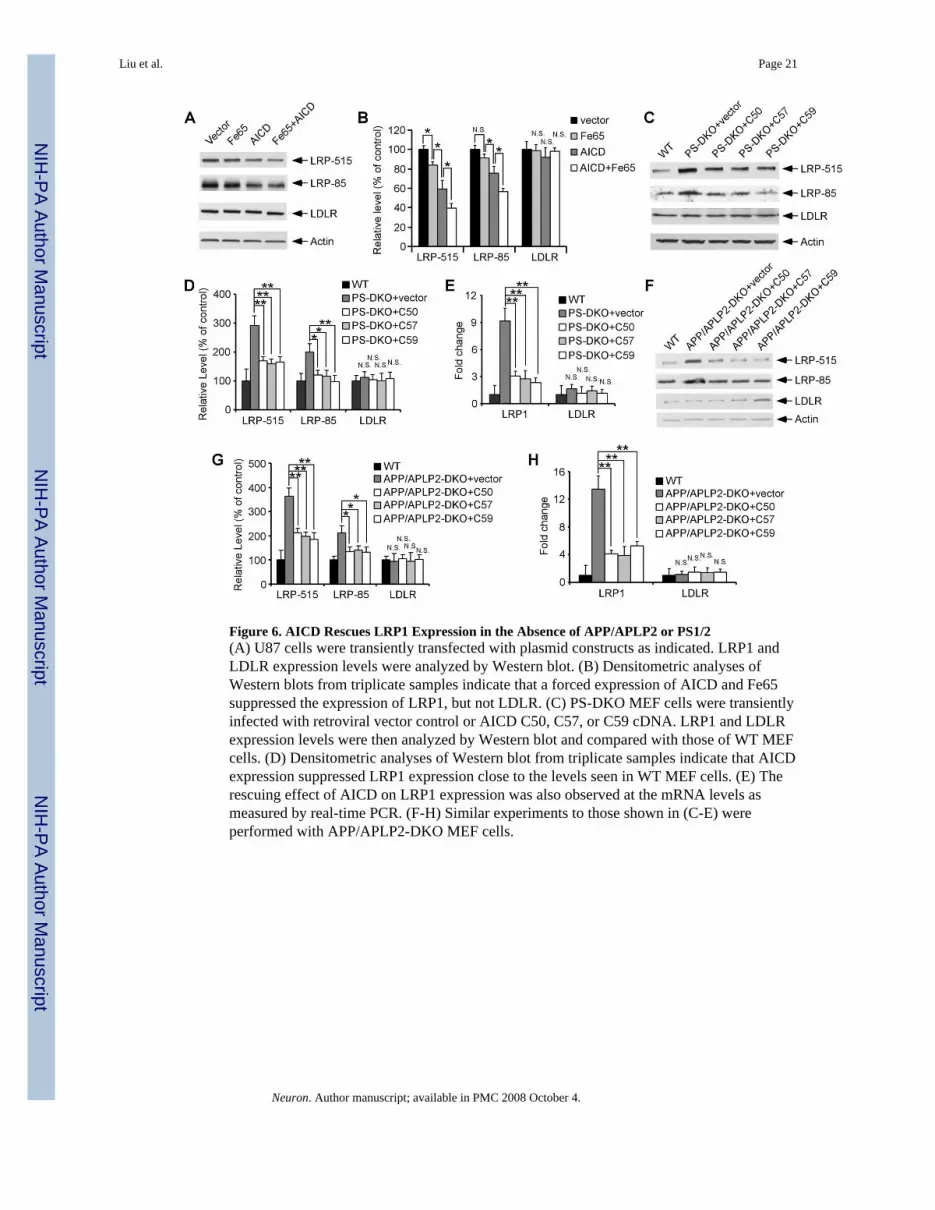
Figure 6. AICD Rescues LRP1 Expression in the Absence of APP/APLP2 or PS1/2(A) U87 cells were transiently transfected with plasmid constructs as indicated. LRP1 andLDLR expression levels were analyzed by Western blot. (B) Densitometric analyses ofWestern blots from triplicate samples indicate that a forced expression of AICD and Fe65suppressed the expression of LRP1, but not LDLR. (C) PS-DKO MEF cells were transientlyinfected with retroviral vector control or AICD C50, C57, or C59 cDNA. LRP1 and LDLRexpression levels were then analyzed by Western blot and compared with those of WT MEFcells. (D) Densitometric analyses of Western blot from triplicate samples indicate that AICDexpression suppressed LRP1 expression close to the levels seen in WT MEF cells. (E) Therescuing effect of AICD on LRP1 expression was also observed at the mRNA levels asmeasured by real-time PCR. (F-H) Similar experiments to those shown in (C-E) wereperformed with APP/APLP2-DKO MEF cells.
Liu et al. Page 21
Neuron. Author manuscript; available in PMC 2008 October 4.
NIH
-PA Author Manuscript
NIH
-PA Author Manuscript
NIH
-PA Author Manuscript
Figure 7. AICD Binds To and Suppresses LRP1 Promoter Activation(A) Schematic diagram of LRP1 promoter-luciferase construct. (B) BHK570 cells weretransiently co-transfected with the LRP1 promoter-Luc construct together with control vector,AICD, Fe65, AICD/Fe65, AICD mutant (Y682G), AICD mutant/Fe65, or NICD. LRP1promoter-driven luciferase activity was significantly decreased by AICD and Fe65 and furtherby the co-expression of both, but not by an AICD mutant or NICD. (C) APP/APLP2-DKOMEF cells were transiently co-transfected with the LRP1 promoter-Luc construct together withcontrol vector, AICD, Fe65, or both, and the luciferase activity was measured as in (B). (D)ChIP assay showed that antibodies to AICD, Fe65 and Tip60, but not control IgG,immunoprecipitate LRP1 promoter DNA fragment. Notch target HES1 promoter was used asa negative control. The ability of anti-Fe65 and anti-Tip60 to immunoprecipitate LRP1promoter DNA is greatly reduced in APP-KO mouse brain. (E) Quantitative real-time PCRanalysis of LRP1 promoter DNA immunoprecipitated by control IgG or antibodies to AICD,Fe65 and Tip60.
Liu et al. Page 22
Neuron. Author manuscript; available in PMC 2008 October 4.
NIH
-PA Author Manuscript
NIH
-PA Author Manuscript
NIH
-PA Author Manuscript
Figure 8. AICD Rescues ApoE and Cholesterol Defects in Cells Deficient in PS or APP(A-D) ApoE (A and B) and cholesterol levels (C and D) were measured in WT, PS-DKO andBACE1-KO MEF cells (A and C) and mouse brains (B and D). ApoE levels were decreasedwhile cholesterol levels were increased in PS-DKO but not BACE1-KO MEF cells and mousebrain when compared to their WT controls. (E-H) APP/APLP2-DKO (E and G) and PS-DKO(F and H) MEF cells were transiently infected with retroviral vector control or AICD cDNA.LRP1 expression levels were then analyzed by Western blot, quantified by densitometry, andcompared with those of WT MEF cells. Expression of AICD partially rescued apoE andcholesterol levels in both APP/APLP2-DKO and PSDKO MEF cells.
Liu et al. Page 23
Neuron. Author manuscript; available in PMC 2008 October 4.