Journal of Agricultural Technology 301 Arbuscular mycorrhizal fungi from Iran K.H. Kariman 1 , E. Mohammadi Goltapeh 1* and V. Minassian 2 1 Department of Plant Pathology, Faculty of Agriculture, Tarbiat Modarres University, P.O. Box: 14155-336, Tehran, Iran 2 Department of Plant Protection, Faculty of Agriculture, Shahid Chamran University, Ahvaz, Iran Kariman, K.H., Goltapeh, E.M. and Minassian, V. (2005). Arbuscular mycorrhizal fungi from Iran. Journal of Agricultural Technology 1(2): 301-313. Samples of root and attached soil were collected from sugar cane fields of Khuzestan and Mazandaran Provinces to identify arbuscular mycorrhizal fungi in Iran. Trap cultures were established to obtain healthy spores for identification. To confirm symbiosis, roots of host plants (sugarcane) and trap plant (corn) were stained with 0.05% aniline blue in lactophenol. Spores and sporocarps of AM fungi were extracted by wet sieving, decanting and centrifugation in 55% sucrose solution. In this study 17 species of AM fungi belonging to the genera Glomus and Acaulospora were identified. Acaulospora rugosa, Glomus ambisporum, G. lamellosum, G. viscosum, G. luteum and G. versiforme are reported from Iran for the first time. Key words: arbuscular mycorrhizal fungi, identification, Iran, sugarcane Introduction Arbuscular mycorrhizal fungi are a plant symbionts belonging to four orders: Glomerales, Archaeosporales, Paraglomales and Diversisporales in the division Glomeromycota (Schussler et al., 2001). They play an important role in plant growth and development (Powell, 1982; Jensen, 1984). The potential for increasing crop growth by effective management of AM strains reinforces the need to determine species composition in cultivated soils of different locations. This study was conducted to identify AM species in Iranian sugarcane fields. The plant family Poaceae including sugar cane, is regarded as one of the best hosts of AM fungi (Powell and Bagyaraj, 1984). In Iran, sugarcane is cultivated in north (Mazandaran Province) and south (Khuzestan Province). Mazandaran Province is a tropical area located in the south east shore of the Caspian sea, soils are sandy and with a relatively high organic content. Khuzestan Province is a semi arid region in south west of Iran and * Corresponding author: E. Mohammadi Goltapeh; e-mail: [email protected]
Transcript
Journal of Agricultural Technology
301
Arbuscular mycorrhizal fungi from Iran K.H. Kariman1, E. Mohammadi Goltapeh1* and V. Minassian2 1Department of Plant Pathology, Faculty of Agriculture, Tarbiat Modarres University, P.O. Box: 14155-336, Tehran, Iran 2Department of Plant Protection, Faculty of Agriculture, Shahid Chamran University, Ahvaz, Iran Kariman, K.H., Goltapeh, E.M. and Minassian, V. (2005). Arbuscular mycorrhizal fungi from Iran. Journal of Agricultural Technology 1(2): 301-313. Samples of root and attached soil were collected from sugar cane fields of Khuzestan and Mazandaran Provinces to identify arbuscular mycorrhizal fungi in Iran. Trap cultures were established to obtain healthy spores for identification. To confirm symbiosis, roots of host plants (sugarcane) and trap plant (corn) were stained with 0.05% aniline blue in lactophenol. Spores and sporocarps of AM fungi were extracted by wet sieving, decanting and centrifugation in 55% sucrose solution. In this study 17 species of AM fungi belonging to the genera Glomus and Acaulospora were identified. Acaulospora rugosa, Glomus ambisporum, G. lamellosum, G. viscosum, G. luteum and G. versiforme are reported from Iran for the first time. Key words: arbuscular mycorrhizal fungi, identification, Iran, sugarcane Introduction
Arbuscular mycorrhizal fungi are a plant symbionts belonging to four orders: Glomerales, Archaeosporales, Paraglomales and Diversisporales in the division Glomeromycota (Schussler et al., 2001). They play an important role in plant growth and development (Powell, 1982; Jensen, 1984). The potential for increasing crop growth by effective management of AM strains reinforces the need to determine species composition in cultivated soils of different locations. This study was conducted to identify AM species in Iranian sugarcane fields. The plant family Poaceae including sugar cane, is regarded as one of the best hosts of AM fungi (Powell and Bagyaraj, 1984). In Iran, sugarcane is cultivated in north (Mazandaran Province) and south (Khuzestan Province). Mazandaran Province is a tropical area located in the south east shore of the Caspian sea, soils are sandy and with a relatively high organic content. Khuzestan Province is a semi arid region in south west of Iran and
*Corresponding author: E. Mohammadi Goltapeh; e-mail: [email protected]
soils are generally clay. Spores of AMF collected directly from a field soil have many problems: 1) they appear healthy but are not viable and are not suitable for use in trap cultures as inoculum; 2) they lose or change the appearance of their structures because of soil conditions, age of spores or parasitism; and 3) they represent only those arbuscular fungi with enough activity and biomass to sporulate. Because of these problems trap cultures with corn were established in the green house. Materials and methods
Sixty one samples of soil and root fragments were collected from sugarcane fields of Mazandaran and Khuzestan Provinces during December, January and February 2002. For this purpose 3-5 samples of root and attached soil collected from different sites of fields were mixed and a sample of 500 grams selected. To establish trap cultures, a subsample of 100 grams from each sample was mixed with autoclaved sand and soil (1:2:1 v/v), placed in 15cm plastic pots, and corn seeds were planted. The pots were maintained in a green house at 25-30°C with 16hour photo period for four months and 15 days. Pot contents were harvested and stored at 4°C until examination. To confirm symbiosis with the host plant (sugarcane) and the trap plant (corn), roots were stained following the protocol of Philips and Haymann (1970). Roots were boiled in 10% KOH for 20 minutes (clearing), then stained with 0.05% aniline blue in lactophenol solution (20-30 minutes in hot water bath) and destained in lactophenol. Spores were obtained by wet sieving (25-80-400 mesh) and a decanting method (Gerdemann and Nicolson, 1963) followed by centrifugation in 55% sucrose solution (Jenkins, 1964). A spore suspension was made in distilled water, distributed onto two 0.8 µm gridded milipore filters, and transferred using a needle to 1:1 mixture of polyvinyle alcohol lacto glycerol, [(PVLG), Koske and Tessier, 1983] and Melzer`s reagent (Hall, 1984) using. To identify species morphological and morphometerical characteristics of spores and subtending hyphae were examined and measured using a stereomicroscope (Olympus ZSH10) and a bright field microscope (Olympus BH-2). Results and discussion
Spores of arbuscular fungi were obtained from all 61 samples, the number varying from 4 to 11, with an average of 7. In this study 17 species of arbuscular mycorrhizal fungi belonging to the genera Glomus and Acaulospora were identified including Glomus geosporum (86%), G. claroideum (77%),
Journal of Agricultural Technology
303
G. fasciculatum (75%), G. macrocarpum (72%), G. lamellosum (68%), G. luteum (46%), G. ambisporum (24), G. sinousum (23%), G. liquidambaris (17%), G. coremioides (16%), G. rubiformis (10%), G. versiforme (10%), G. viscosum (2%), G. etunicatum (57%),G. intraradices (51%) and Acaulospora rugosa (10%).
Ten species: G. geosporum, G. claroideum, G. fasciculatum, G. macrocarpum, G. lamellosum, G. luteum, G. sinuosum, G. mosseae, G. etunicatum and G. intraradices were found in sugar cane fields of both Khuzestan and Mazandaran Provinces. Four species: G. ambisporum, G. coremioides, G. rubiformis, G. liquidambaris were only found in fields of Mazandaran Province and three species Acaulospora rugosa, G. versiforme and G. viscosum in sugar cane fields in Khuzestan Province.
Eleven of the identified species have been reported previously from grape and some grains (wheat, barley, maize and sorghum) respectively by Sedaghati (2002) and Sadravi (1999) from Iran. The six species including Acaulospora rugosa, Glomus ambisporum, G. lamellosum, G. viscosum, G. luteum and G. versiforme are recorded in Iran for the first time. Acaulospora rugosa Morton, Mycologia 78: 641, 1986 .
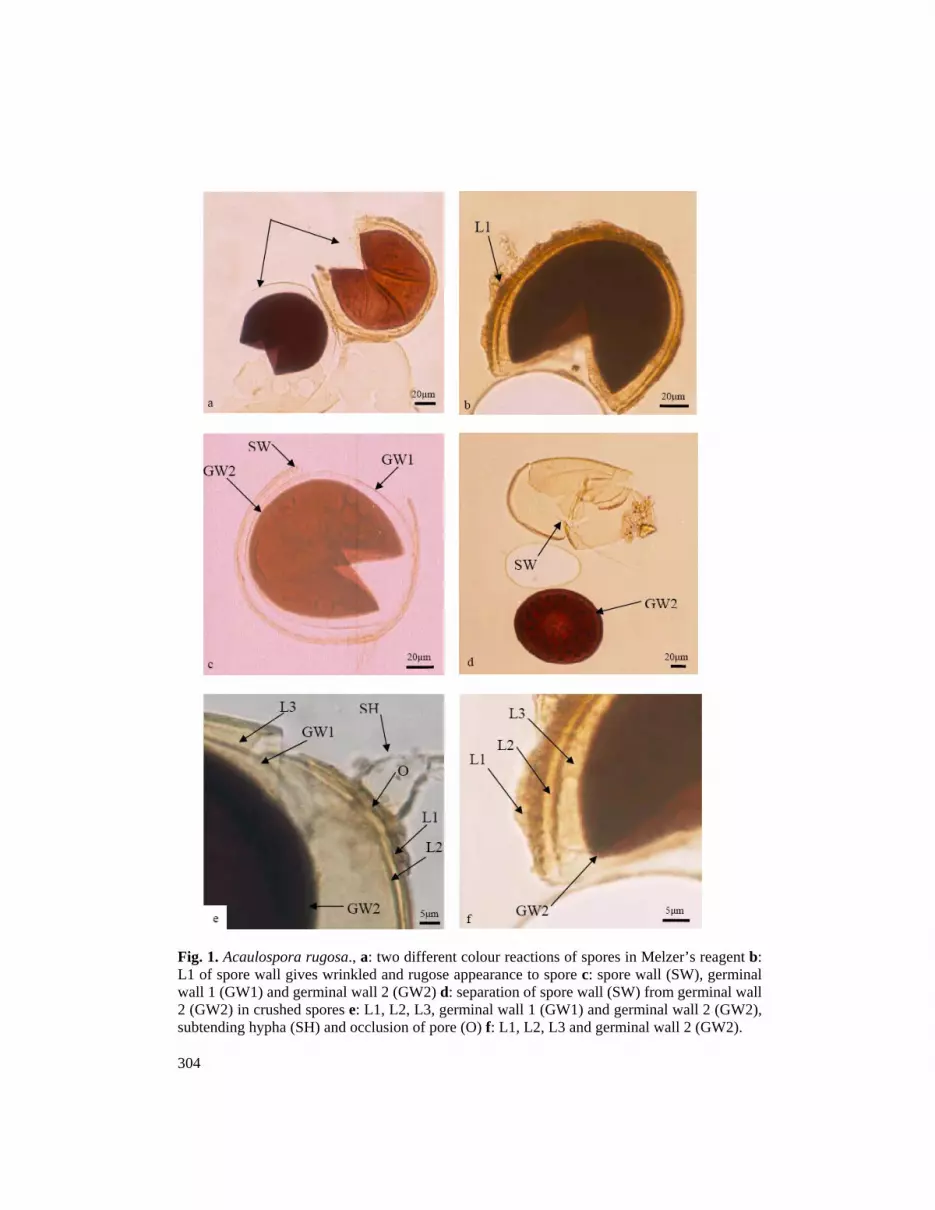
Spores formed singly in soil, sub-hyaline to straw-coloured, mostly globose to sub-globose (70-) 102 (-128) µm diameter. Spore wall consisting of 5 layers in 3 groups (A, B, C). Group A consisting of 2 layers (L1, L2). L1: hyaline layer 1-1.5 µm thick, this layer degrades in mature spores and may be completely absent (especialy those collected from field soils). This layer appears wrinkled or rugose (Figs 1b,d). L2, laminated, pale yellow (1-) 1.5(-2) µm thick (Figs 1e,f). Group B consists of L3, a semi rigid hyaline layer 1-1.3 µm thick (Fig. 1e). Group C consisting of two germinal walls (GW1, GW2). Germinal wall1 (GW1) is membraneous with weakly beaded appearance and composed of two sublayers hardly adherent together (Figs 1c,e). Germinal wall 2 (GW2), rigid, 1-2.5 µm thick, becoming dark purple (Figs 1a,c,d,e,f) or pink orange (Fig. 1a) in Melzer`s reagent, composed of two sublayers seen as a layer. Hypha at the point of spore attachment (9-)13.6(-18) µm wide (Fig. 1e) and pore is occluded (Fig. 1e). In some crushed spores, layers of spore wall separate and GW2 is visible as a dark purple core (Fig. 1d).
Morton (1986) isolated this species from rhizosphere of Fragaria rubra and F. arondinacea in West Virginia for the first time. In extracted spores, the subtending hypha at the point of attachment was wider than for the published description (10-14 µm) (Morton, 1986).
304
Fig. 1. Acaulospora rugosa., a: two different colour reactions of spores in Melzer’s reagent b: L1 of spore wall gives wrinkled and rugose appearance to spore c: spore wall (SW), germinal wall 1 (GW1) and germinal wall 2 (GW2) d: separation of spore wall (SW) from germinal wall 2 (GW2) in crushed spores e: L1, L2, L3, germinal wall 1 (GW1) and germinal wall 2 (GW2), subtending hypha (SH) and occlusion of pore (O) f: L1, L2, L3 and germinal wall 2 (GW2).
Journal of Agricultural Technology
305
Distribution: Found in 6 samples (10%) collected from sugarcane fields, Khuzestan Province (Amirkabir and Shoaibyeh Sugar cane and Byproducts Development Co.).
Glomus ambisporum Smith & Schenck, Mycologia 77: 566, 1985. Spores produced singly or aggregated around roots, dark brown to black,
predominantly globose, (85-)149.4(-193) µm diameter to occassionaly sub-globose, (109-)147.8(-173) × (96-)133.5(-150) µm (Figs 2a,b). Spore wall composed of three layers. L1: ephemeral, reticulate, 2-4 µm thick (Fig. 2c). L2: laminated, red brown to dark brown, (3-)6.6(-9) µm thick confluent with hyphal attachment (Fig. 2d). L3: membranous less than 1 µm thick. Subtending hyphae cylindrical, rarely with two adjacent hyphae,observed only in one spore (Fig. 2c). Hypha at the point of attachment (11-)18.5(-26) µm wide. Hyphal wall at the point of attachment (2-)4.7(-7) µm thick. Subtending hyphae are frequently branched in (16-)50.6(-98) µm from the point of spore attachment (Figs 2a,b,c,d). Pores of spores occluded by innermost sublayers of spore wall.
Smith and Schenck (1985) isolated and described this species from unknown grass in a garden at Gainesrila in Florida. Sizes of spores are a little larger than those in published description. Some spores parasitized by Glomus claroideum (Fig. 2e) or Pythium sp (Fig. 2f).
Distribution: Found in 15 samples (24%) collected from sugar cane fields, Mazandaran province. Glomus lamellosum Dalpe, Koske & Tews, Mycotaxon 43: 289, 1992.
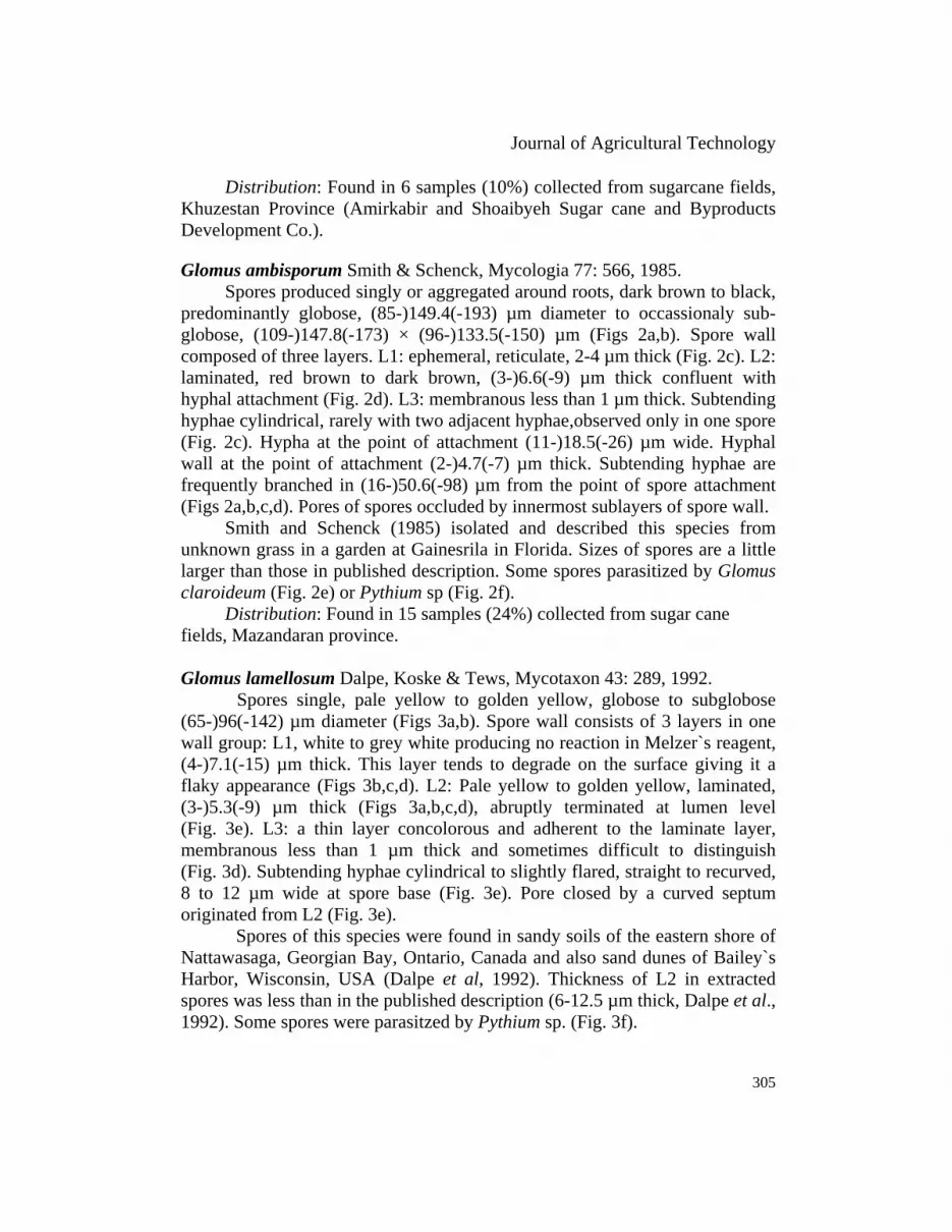
Spores single, pale yellow to golden yellow, globose to subglobose (65-)96(-142) µm diameter (Figs 3a,b). Spore wall consists of 3 layers in one wall group: L1, white to grey white producing no reaction in Melzer`s reagent, (4-)7.1(-15) µm thick. This layer tends to degrade on the surface giving it a flaky appearance (Figs 3b,c,d). L2: Pale yellow to golden yellow, laminated, (3-)5.3(-9) µm thick (Figs 3a,b,c,d), abruptly terminated at lumen level (Fig. 3e). L3: a thin layer concolorous and adherent to the laminate layer, membranous less than 1 µm thick and sometimes difficult to distinguish (Fig. 3d). Subtending hyphae cylindrical to slightly flared, straight to recurved, 8 to 12 µm wide at spore base (Fig. 3e). Pore closed by a curved septum originated from L2 (Fig. 3e).
Spores of this species were found in sandy soils of the eastern shore of Nattawasaga, Georgian Bay, Ontario, Canada and also sand dunes of Bailey`s Harbor, Wisconsin, USA (Dalpe et al, 1992). Thickness of L2 in extracted spores was less than in the published description (6-12.5 µm thick, Dalpe et al., 1992). Some spores were parasitzed by Pythium sp. (Fig. 3f).
306
Fig. 2. Glomus ambisporum., a and b: spores with branched hyphae (BH) c: spores with 2 subtending hyphae (SH) and degrading L1 d: L2 of spore wall and branched hypha (BH) e: spores of Glomus ambisporum (G.A) parasitized by Glomus claroideum (G.C) f: spore of Glomus ambisporum parasitized by Pythium sp. (oospore: OO).
Journal of Agricultural Technology
307
Fig. 3. Glomus lamellosum., a: globose spore, L1 & L2 of spore wall b: brokenspore and degrading L1 c: L1 and L2 of spore wall d: L1, L2 and L3 of spore wall in broken spore e: L1 absent (degraded), subtending hypha (SH) and occlusion of pore (O) f: spore of Glomus lamellosum parasitized by Pythium sp. (oospores: OO).
308
Distribution: Known from 43 samples (68%) collected from sugar cane fields, Mazandaran (Behnemir) and Khuzestan Provinces (Sugarcane & Byproducts Development Co., Amirkabir, Karoon, Shoaibyeh and Mianab). Glomus viscosum T.H. Nicolson, Mycological Research. 99: 1500, 1995.
Spores formed in loose clusters in the soil (Fig. 4a), subhyaline to pale straw, globose (79-) 98 (-113) µm or sub-globose, (81-)86.6(-92) × (65-)76.4 (-83) µm diameter. A mucilaginous-like substance, presumably a polysaccharide is produced by the outer layer of spore wall. As a result organic matters and soil particles are attached to spores and surface of spore appears dull and rough (Figs 4a,b,c,d,f). Spore wall consisted of two layers. L1: A semi flexible hyaline layer, less than 1 µm thick, without evident laminations, often difficult to discern under the light microscope, thickness of attached soil particles up to 23 µm thick (Figs 4b,c,d,f). L2: laminated layer, hyaline to straw, (1.2)1.5(-2) µm thick (Figs 4d,e,f). Subtending hypha cylindrical, occassionally slightly constricted, (7-)8.5(-10) µm wide. Hyphal wall at the point of attachment (1.5)1.8(-2) µm thick (Fig. 4c). Extracted spores always lacked an occlusion (Fig. 4e).
This species was described from a potted Magnolia (Magnolia solangena) from a nursery in Pesica, Tuscany, Italy (Walker et al., 1995). No occlusion was found in the extracted spores in this study, and pores were generally open (Fig. 4e).
Distribution: Found only in one sample (2%) collected from sugarcane fields, Khuzestan Province, Shaeibieh Sugarcane & Byproducts Development Co. Glomus luteum Kennedy, Stutz & Morton, Mycologia 91: 1083, 1999.
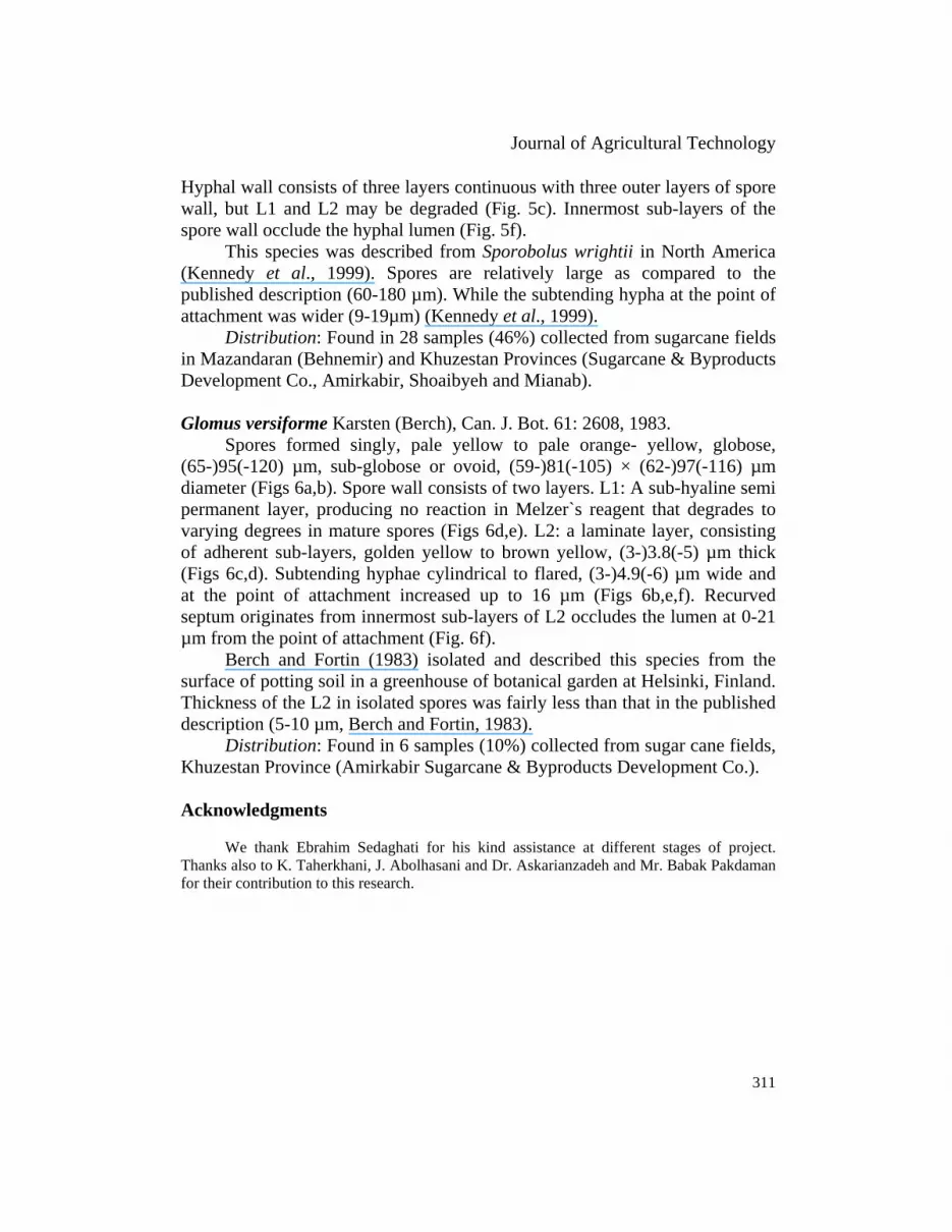
Spores formed singly in soil, pale yellow to dark yellow with a brownish tint, globose (81-)142.3(-208) µm or sub-globose, (76-)119.7(-158) × (85-)129(-170) µm diameter (Figs 5a,b). Spore wall composed of four layers. L1: hyaline mucilaginous layer, 1.3 to 5 µm thick, degrading as the spore matures and may be completely absent in mature spores. L2: hyaline, semi rigid layer, (2-)3.4(-6) µm thick, producing no reaction in Melzer`s reagent, degrading as the spore matures and in older spores may be absent (Figs 5c,d,e). L3: rigid permanent layer consisted of fine adherent sub-layers, pale yellow to brownish yellow, (4-)8(-12.5) µm thick (Figs 5c,d,e,f). L4: A thin flexible layer concolorous with the laminat layer less than 0.5 µm thick (Figs 5d,e). Subtending hypha cylindrical to slightly flared, (9-)15.1(-25) µm wide (Figs 5a,c,f). Hyphal wall at the point of attachment (2-)4.5(-8) µm thick (Figs 5c,f).
Fig. 4. Glomus viscosum., a: Clusters of spores. b and c: soil particles (SP) attached to spore wall and surface d: L2 of spore wall (laminated layer) in crushed spore e: subtending hypha (SH) hyphal wall (HW), L2 of spore wall and open spore (P) f: Soil particles (SP) attached to spore wall up to 23 µm, L2 of spore wall.
310
Fig. 5. Glomus luteum., a: Globose spore with short broken hypha (SH) b: Spore with long subtending hypha (SH) c: L2 & L3 of spore wall continous with subtending hypha d: L2, L3 and L4 of spore wall e: L2, L3 and L4 of spore wall in crushed spore f: Hyphal wall (HW) and occlusion of pore (O).
Journal of Agricultural Technology
311
Hyphal wall consists of three layers continuous with three outer layers of spore wall, but L1 and L2 may be degraded (Fig. 5c). Innermost sub-layers of the spore wall occlude the hyphal lumen (Fig. 5f).
This species was described from Sporobolus wrightii in North America (Kennedy et al., 1999). Spores are relatively large as compared to the published description (60-180 µm). While the subtending hypha at the point of attachment was wider (9-19µm) (Kennedy et al., 1999).
Distribution: Found in 28 samples (46%) collected from sugarcane fields in Mazandaran (Behnemir) and Khuzestan Provinces (Sugarcane & Byproducts Development Co., Amirkabir, Shoaibyeh and Mianab). Glomus versiforme Karsten (Berch), Can. J. Bot. 61: 2608, 1983.
Spores formed singly, pale yellow to pale orange- yellow, globose, (65-)95(-120) µm, sub-globose or ovoid, (59-)81(-105) × (62-)97(-116) µm diameter (Figs 6a,b). Spore wall consists of two layers. L1: A sub-hyaline semi permanent layer, producing no reaction in Melzer`s reagent that degrades to varying degrees in mature spores (Figs 6d,e). L2: a laminate layer, consisting of adherent sub-layers, golden yellow to brown yellow, (3-)3.8(-5) µm thick (Figs 6c,d). Subtending hyphae cylindrical to flared, (3-)4.9(-6) µm wide and at the point of attachment increased up to 16 µm (Figs 6b,e,f). Recurved septum originates from innermost sub-layers of L2 occludes the lumen at 0-21 µm from the point of attachment (Fig. 6f).
Berch and Fortin (1983) isolated and described this species from the surface of potting soil in a greenhouse of botanical garden at Helsinki, Finland. Thickness of the L2 in isolated spores was fairly less than that in the published description (5-10 µm, Berch and Fortin, 1983).
Distribution: Found in 6 samples (10%) collected from sugar cane fields, Khuzestan Province (Amirkabir Sugarcane & Byproducts Development Co.). Acknowledgments
We thank Ebrahim Sedaghati for his kind assistance at different stages of project. Thanks also to K. Taherkhani, J. Abolhasani and Dr. Askarianzadeh and Mr. Babak Pakdaman for their contribution to this research.
Fig. 6. Glomus versiforme., a: Spore (S) and hyphal swollen (HS) b: funnel-shaped subtending hypha at the point of attachment c: Broken spore with subtending hypha (SH) d: L1 and L2 (laminated layer) of spore wall e: L1 degrading and sloughing away f: hyphal wall (HW) and occlusion (O) of spore by a thin curved septum.
Journal of Agricultural Technology
313
References
Berch, S.M. and Fotin, J.A. (1983). Lectotypification of Glomus macrocarpum and proposal of a new combinations: Glomus austerales, Glomus versiforme, and Glomus tenebrosum (Endogonaceae). Canadian Journal of Botany 61: 2608-2617.
Dalpe, Y., Koske, R.E. and Tews, L.L. (1992). Glomus lamellosum sp. nov. : A new Glomaceae associated with Beach Grass. Mycotaxon 43: 289-293
Gerdmann, G.W. and Nicolson, T.H. (1963). Spore of mycorrhizal Endogone species extracted from soil by wet sieving and decanting. Transactions of the British Mycological Society 46: 235-244.
Hall, I.R. (1984). Taxonomy of VA mycorrhizal fungi. In: VA Mycorrhiza (eds. C.L. Powell, and D.J. Bagyaraj,). VA Mycorrhiza. CRC Press, Boca raton, FL: 57-94.
Jenkins, W.R. (1964). A rapid centrifugal-technique for separating nematodes from soil. Plant Disease Reporter 48: 692.
Jensen, A. (1984). Responces of barely, pea and maize to inoculation with different vesicular arbuscular mycorrhizal fungi in irradiated soils. Plant Soil 78: 315-323.
Kennedy, L.J., Stutz, J.C. and Morton, J.B. (1999). Glomus eburneum and Glomus luteum two new species of arbuscular mycorrhizal fungi with emendation of Glomus spurcum. Mycologia 91: 1083-1093.
Koske, R.E. and Tessier, B. (1983). A convenient, permanent slide mounting medium. Mycological Society of American Newsletter 34: 59.
Morton, J.B. (1986). Three new species of Acaulospora (Endogonaceae) from high alluminium low PH soils in west Virginia. Mycologia 78: 641-648.
Philips, J.M. and Hayman, D.S. (1970). Improved procedures clearing root and staining parasitic and vesicular arbuscular mycorrhizal fungi for rapid assessment of infections. Transaction of the British Mycological Society 55: 158-161.
Powell, C.L.I. (1982). Selection of efficient VA Mycorrhizal fungi. Plant Soil 68: 3-9. Powell, C.l. and Bagyaraj, D.J. (1984). VA Mycorrhiza. CRC Press. 234pp. Sadravi, M. (1999). Identification of vesicular arbuscular mycorrhizal fungi on wheat, barley,
corn and sorghum in Tehran and Khuzestan Provinces and study on their possible propagation by tissue culture. Ph. D thesis in Plant Pathology, Faculty of Agriculture, Tarbiat Modarres University. 222p.
Sedaghati, E. (2002). Isolation and identification of arbuscular mycorrhizal fungi on grape in Khorasan and Ghazvin Provinces. M.Sc. Thesis in Plant Pathology, Faculty of Agriculture, Tarbiat Modarres University. 130p.
Smith, G.S. and Schenck, N.C. (1985). Two new dimorphic species in the endogonaceae: Glomus ambisporum and Glomus heterosporum. Mycologia 77: 566-574.
Schussler, A., Schwarzott, D. and Walker, C. (2001). A new phylum, the Glomeromycota: phylogeny and evolution. Mycological Research 105: 1413-1421.
Walker, C., Giovanetti, M., Avio, L., Siternesi, A.S., and Nicolson, T.H. (1995). A new fungal species forming arbuscular mycorrhizas: Glomus viscosum, Mycological Research 99: 1500-1506.
(Received 13 September 2005; accepted 5 December 2005)