Boosting TiO 2 -anatase antimicrobial activity: Polymer-oxide thin films Anna Kubacka a , Manuel Ferrer a , Marı ´a L. Cerrada b, *, Cristina Serrano b , Manuel Sa ´ nchez-Chaves b , Marta Ferna ´ ndez-Garcı ´a b , Alicia de Andre ´s c , Rafael J. Jime ´ nez Riobo ´o c , Fernando Ferna ´ ndez-Martı ´n d , Marcos Ferna ´ ndez-Garcı ´a a, ** a Instituto de Cata ´lisis y Petroleoquı´mica, Consejo Superior de Investigaciones Cientı´ficas, C/Marie Curie 2, 28049-Madrid, Spain b Instituto de Ciencia y Tecnologı´a de Polı´meros, Consejo Superior de Investigaciones Cientı´ficas, C/Juan de la Cierva 3, 28006-Madrid, Spain c Instituto Ciencia de Materiales de Madrid, Consejo Superior de Investigaciones Cientı´ficas, C/Sor Juan Ine ´s de la Cruz 3, 28049-Madrid, Spain d Instituto del Frı´o, Consejo Superior de Investigaciones Cientı´ficas, C/Jose ´ Antonio Novais 10, 28040-Madrid, Spain 1. Introduction The use of photocatalytic semiconductor oxides emerges as a successful technology to struggle against biological risks. TiO 2 - anatase is by far the most widely used photocatalyst, being a wide band-gap (3.2 eV) semiconductor that under UV illumination generates energy-rich electron–hole pairs able to degrade cell components of microorganisms rendering innocuous products. Moreover, none weakness with respect to the microorganism nature (bacteria, virus, fungus, etc.) is nowadays known [1,2]. Consequently, its incorporation as a constituent in polymeric multicomponent materials could be a future alternative within goods packaging or biomedical fields, and would open the current playground for photocatalysis application. The important indus- trial success of isotactic polypropylene (iPP) and the wide range of its applications are basically due to the combination of outstanding physical properties, as the low density, the high tensile strength, stiffness, hardness, thermal and good environmental resistance, ease of processability and recycling at a moderate cost [3]. The discovery of single-site metallocene catalysts has determined great improvements and advantages in polymerization and copolymerization of olefins [4,5]. Taking into account their good transparency, their aesthetic and mechanical properties, metallo- cene-catalyzed polypropylene resins provide a replacement for glass in some critical uses, as packaging of species keeping their organoleptic qualities, and for other plastics, as polyvinyl chloride in medical devices. These novel fields as well as the current general uses of PP, including automotive components, stationery and textiles (e.g. ropes, thermal underwear and carpets), among others, demand for improved polyolefins and, in particular, an important research area concerns the possibility of adding bactericidal properties to such polymeric matrices [6]. Modification of these polymer-based systems to prevent growth or reduce adhesion of detrimental microorganisms appears thus as a highly desired objective. A point of relevance within the incorporation of the biocidal oxide into polymeric matrixes is the control of the TiO 2 polymorphism ensuring the presence of the anatase form, the one with biocidal capability, as well as to control primary particle size in the nanometer range, a fact that would limit scattering Applied Catalysis B: Environmental 89 (2009) 441–447 ARTICLE INFO Article history: Received 21 November 2008 Received in revised form 24 December 2008 Accepted 11 January 2009 Available online 19 January 2009 Keywords: Titania Anatase Nanocomposites Thin films Biocide Germicide Disinfection ABSTRACT TiO 2 incorporation into an isotactic polypropylene (iPP) polymeric matrix was achieved via a straightforward and cost-effective melting process using laboratory-made nanometric anatase-TiO 2 and an industrial polymer. The structural characteristics of the resulting nanocomposite thin films as a function of the inorganic component content were examined using wide and small angle X-ray scattering (WAXS/SAXS) and vibrational Raman spectroscopy. Electron scanning and transmission microscopy (SEM/TEM) studies were also performed to provide evidence of the nanometric dispersion of the oxide within the polymer matrix, showing the presence of average aggregates of ca. 80 nm. TiO 2 incorporation into the iPP renders self-sterilized nanocomposite films upon light excitation, the activity of which was tested against Gram negative (P. aeruginosa) and positive (E. faecalis) bacteria. TiO 2 displays maximum activity for a sample containing a 2 wt.% of anatase-TiO 2 irrespective of the microorganism nature. The antimicrobial activity of the nanocomposite films is significantly enhanced with respect to that of the oxide alone. This key fact is interpreted on physical basis with the help of a complete optical (UV–vis and photoluminescence) and electron paramagnetic resonance (EPR) characterization. ß 2009 Elsevier B.V. All rights reserved. * Corresponding author. ** Corresponding author. Tel.: +34 915 85 54 75; fax: +34 915 85 47 60. E-mail addresses: [email protected](M.L. Cerrada), [email protected](M. Ferna ´ ndez-Garcı ´a). Contents lists available at ScienceDirect Applied Catalysis B: Environmental journal homepage: www.elsevier.com/locate/apcatb 0926-3373/$ – see front matter ß 2009 Elsevier B.V. All rights reserved. doi:10.1016/j.apcatb.2009.01.002
Boosting TiO2-anatase antimicrobial activity: Polymer-oxide thin films
Anna Kubacka a, Manuel Ferrer a, Marıa L. Cerrada b,*, Cristina Serrano b, Manuel Sanchez-Chaves b,Marta Fernandez-Garcıa b, Alicia de Andres c, Rafael J. Jimenez Rioboo c,Fernando Fernandez-Martın d, Marcos Fernandez-Garcıa a,**a Instituto de Catalisis y Petroleoquımica, Consejo Superior de Investigaciones Cientıficas, C/Marie Curie 2, 28049-Madrid, Spainb Instituto de Ciencia y Tecnologıa de Polımeros, Consejo Superior de Investigaciones Cientıficas, C/Juan de la Cierva 3, 28006-Madrid, Spainc Instituto Ciencia de Materiales de Madrid, Consejo Superior de Investigaciones Cientıficas, C/Sor Juan Ines de la Cruz 3, 28049-Madrid, Spaind Instituto del Frıo, Consejo Superior de Investigaciones Cientıficas, C/Jose Antonio Novais 10, 28040-Madrid, Spain
A R T I C L E I N F O
Article history:
Received 21 November 2008
Received in revised form 24 December 2008
Accepted 11 January 2009
Available online 19 January 2009
Keywords:
Titania
Anatase
Nanocomposites
Thin films
Biocide
Germicide
Disinfection
A B S T R A C T
TiO2 incorporation into an isotactic polypropylene (iPP) polymeric matrix was achieved via a
straightforward and cost-effective melting process using laboratory-made nanometric anatase-TiO2 and
an industrial polymer. The structural characteristics of the resulting nanocomposite thin films as a
function of the inorganic component content were examined using wide and small angle X-ray
scattering (WAXS/SAXS) and vibrational Raman spectroscopy. Electron scanning and transmission
microscopy (SEM/TEM) studies were also performed to provide evidence of the nanometric dispersion of
the oxide within the polymer matrix, showing the presence of average aggregates of ca. 80 nm. TiO2
incorporation into the iPP renders self-sterilized nanocomposite films upon light excitation, the activity
of which was tested against Gram negative (P. aeruginosa) and positive (E. faecalis) bacteria. TiO2 displays
maximum activity for a sample containing a 2 wt.% of anatase-TiO2 irrespective of the microorganism
nature. The antimicrobial activity of the nanocomposite films is significantly enhanced with respect to
that of the oxide alone. This key fact is interpreted on physical basis with the help of a complete optical
(UV–vis and photoluminescence) and electron paramagnetic resonance (EPR) characterization.
� 2009 Elsevier B.V. All rights reserved.
Contents lists available at ScienceDirect
Applied Catalysis B: Environmental
journa l homepage: www.e lsev ier .com/ locate /apcatb
1. Introduction
The use of photocatalytic semiconductor oxides emerges as asuccessful technology to struggle against biological risks. TiO2-anatase is by far the most widely used photocatalyst, being a wideband-gap (3.2 eV) semiconductor that under UV illuminationgenerates energy-rich electron–hole pairs able to degrade cellcomponents of microorganisms rendering innocuous products.Moreover, none weakness with respect to the microorganismnature (bacteria, virus, fungus, etc.) is nowadays known [1,2].Consequently, its incorporation as a constituent in polymericmulticomponent materials could be a future alternative withingoods packaging or biomedical fields, and would open the currentplayground for photocatalysis application. The important indus-trial success of isotactic polypropylene (iPP) and the wide range ofits applications are basically due to the combination of outstandingphysical properties, as the low density, the high tensile strength,
0926-3373/$ – see front matter � 2009 Elsevier B.V. All rights reserved.
doi:10.1016/j.apcatb.2009.01.002
stiffness, hardness, thermal and good environmental resistance,ease of processability and recycling at a moderate cost [3]. Thediscovery of single-site metallocene catalysts has determinedgreat improvements and advantages in polymerization andcopolymerization of olefins [4,5]. Taking into account their goodtransparency, their aesthetic and mechanical properties, metallo-cene-catalyzed polypropylene resins provide a replacement forglass in some critical uses, as packaging of species keeping theirorganoleptic qualities, and for other plastics, as polyvinyl chloridein medical devices. These novel fields as well as the current generaluses of PP, including automotive components, stationery andtextiles (e.g. ropes, thermal underwear and carpets), among others,demand for improved polyolefins and, in particular, an importantresearch area concerns the possibility of adding bactericidalproperties to such polymeric matrices [6]. Modification of thesepolymer-based systems to prevent growth or reduce adhesion ofdetrimental microorganisms appears thus as a highly desiredobjective.
A point of relevance within the incorporation of the biocidaloxide into polymeric matrixes is the control of the TiO2
polymorphism ensuring the presence of the anatase form, theone with biocidal capability, as well as to control primary particlesize in the nanometer range, a fact that would limit scattering
A. Kubacka et al. / Applied Catalysis B: Environmental 89 (2009) 441–447442
events among other things [1,7]. The adequate handling of themorphological properties just mentioned would drive to thecorrect (UV) light–matter interaction and would ultimately lead toan optimized photoactivity in the elimination of microorganisms.This reasoning line implies, on the other hand, that the control ofphase purity and morphological properties of the titania compo-nent would claim for the use of inorganic materials previouslyobtained to the nanocomposite preparation, ensuring in this waytheir physico-chemical properties. A side but important point tomention is the concomitant degradation of the polymer matrix byeffect of the charge carriers; this has been proved to be limited byaddition of small amounts of titania, typically below 5 wt.%.Moreover, inclusion of titania in moderate amounts opens a newway to solve the general problem of disposal after completion ofthe polymer lifetime [7].
In this work, iPP–TiO2 nanoparticulated composite thin filmsare characterized and described in the context of the application ofnovel biocidal capabilities resulting from a nanometer anatase-TiO2 component [8]. As well known, the addition of an inorganicmodifier can alter iPP structure and crystallinity, affecting in thisway physico-chemical properties and demanding for a completestructural characterization of the whole system [9]. Through amultitechnique approach, the study aims to show that adequateincorporation of TiO2 into a polymer-based nanocomposite filmleads up to a powerful antimicrobial system, with a biocidalpotential higher than that of the oxide alone. An efficient organo-inorganic contact changes the nature of the TiO2 agent erasing therequirement of a close proximity with the pathogen, making theoxide in the nanocomposite a non-contact agent.
2. Experimental
The TiO2 component was prepared using a microemulsionsynthetic route by addition of titanium (IV) isopropoxide (Aldrich)to an inverse emulsion containing an aqueous phase dispersed inn-heptane (Panreac), using Triton X-100 (Aldrich) as surfactant and1-hexanol (Aldrich) as cosurfactant. The resulting mixture wasstirred for 24 h, centrifuged, decanted, rinsed under stirring fiveconsecutive times with methanol (2), water (2) and acetone (1), inorder to eliminate any portion from the organic and surfactantmedia, dried at 110 8C for 24 h and calcined at 500 8C for 2 h. Thesynthesis gives pure anatase with a primary particle size of 10 nm[8]. A commercially available metallocene-catalyzed isotacticpolypropylene, iPP (Basell Metocene X50081: melt flow index of60 g/10 min at 230 8C/2.16 kg, ASTM D1238), meeting FDArequirements (Federal Regulations, 21 CFR 177.1520) for foodcontact, has been used as polymeric matrix in the preparation ofiPP–TiO2 nanocomposites with different TiO2 nanoparticle con-tents: 0.5, 1, 2 and 5 wt.%, labeled as Ti0.5, Ti1, Ti2 and Ti5,respectively. The Licomont1 AR 504 fine grain from Clariant (apolypropylene wax partially modified with maleic anhydride,PPgMA) was added as a compatibilizer agent to improve theinterfacial adhesion between the iPP and TiO2. Use of PPgMA as anorganic–inorganic compatibilizer is well established [10] andpreliminary tests have evidenced that the best ratio of interfacialagent to be added is the 80 wt.% related to the TiO2 nanoparticlesweight content at a given composition. These novel three-component materials were prepared through melt processing inan internal mixer with a volumetric capacity of 3 cm3 at 160 8C andat 60 rpm for 5 min. After blending and homogenization of thedifferent components, specimens were obtained as films bycompression molding in a Collin press between hot plates(175 8C) at a pressure of 1.5 MPa for 5 min. A quench from themelt to room temperature was applied to the different samples.
The crystal lattice characteristics of the nanocomposites wereexamined by wide angle X-ray scattering, WAXS, in the reflection
mode at room temperature using a Rigaku Rotaflex RTP300diffractometer connected to a computer having a Geiger counterand using a Ni-filtered Cu Ka radiation. The diffraction scans werecollected at a rate of 18/min between 2u values from 58 to 308, usinga sampling rate of 1 Hz. The goniometer was calibrated with astandard of silicon. The X-ray determinations of the degree ofcrystallinity were performed by subtraction of the correspondingamorphous component [11] comparing to the totally amorphousprofile of an elastomeric PP sample. In addition, the transitionassociated with the melting process was investigated by real-timeX-ray diffraction experiments with synchrotron radiation. Thesynchrotron studies were performed in the soft-condensed matterbeamline A2 at Hasylab (Hamburg, Germany), working at awavelength of 0.150 nm. Two different detectors were used in thesetup. The first one, a MAR CCD detector, at a distance of 230 cmfrom the sample (covering the small angle scattering, SAXS, region)and the other, linear one, at around 17 cm from the sample andcovering the approximate 2u range from 118 to 288 (WAXS region).This WAXS detector was calibrated with the diffractions of acrystalline PET sample, and the SAXS detector, with the differentorders of the long spacing of rat-tail cornea (L = 65 nm). A heatingrate of 8 8C/min was employed, acquiring profiles every 15 s. Thetwo-dimensional X-ray patterns were processed with the FIT2Dprogram (ESRF) and converted into one-dimensional arrays afternormalization for the intensity of the primary beam andsubtraction of the scattering of an empty sample.
Raman and photoluminescence measurements were performedat room temperature with different laser lines of an Ar+–Kr+ laser:333 nm + 365 nm, and 514 nm. A home-made micro-Ramansystem was utilized; it consists on a Jobin-Yvon HR 460monochromator, a N2 cooled CCD and a Kaiser Super-Notch-Plusfilters (the latter used to suppress the elastic scattered light at514 nm). The excitation light was focused on the sample with anOlympus microscope (except for 333 and 365 nm UV laser lines)which is also used to collect the scattered light. Spectra arecorrected by the instrumental function recorded with a calibratedwhite source and a CaF2 pellet. For intensity measurements, Ramanspectra are normalized using the total intensity of the C–H rockingvibrations [12]. The EPR measurements were done with a BrukerER200D spectrometer operating in the X-band and calibrated witha DPPH standard.
Scanning electron microscopy experiments for cross-sectionmaterial analysis were carried out at room temperature in a XL30ESEM PHILIPS equipment working at 25 kV. Samples were in situcryofractured prior observations of the film cross-section. Thesamples were coated with gold–palladium (80:20) with a SputterCoater (Polaron SC7640) working at 800 V and 5 mA. Transmis-sion electron microscopy experiments were carried out at roomtemperature in a 200 kV JEM-2000 FX JEOL microscope. Sampleswere embedded in Spurr resine (low viscosity epoxi, cured at 60 8Cfor 48 h) to obtain parallel cuts of the films surface in thin (80 mm)sections by ultramicrotomy (Reichert-Jung Ultracut E), whichwere picked up on cooper grids and coated with a thin layer ofcarbon graphite (MED 010 Balzers evaporator) to improve heatconductivity.
The microorganisms used in this study included two clinicalisolates: P. aeruginosa PAO clinical isolate PBCLOp11 from burnwound infections and E. faecalis clinical isolate brs30 from humanbiliary, both classified according to 16S rRNA (unpublished).Bacterial cells were streaked from a glycerol stock onto a LB agarplate, grown overnight at 37 8C (P. aeruginosa: OD600 �6.0) (noantibiotics) and subsequently used. To study the antimicrobialactivity of the films, a suspension containing 10 ml of microbialcells (ca. 109 cfu ml�1) suspended in 1 ml broth solution was madeas described elsewhere [13]. Aliquots of 1 ml from thesesuspensions were added to a 4 ml quartz cubic cell containing
Table 1Characteristics of the iPP crystalline phase for the different Tix nanocomposite thin
films and for the neat iPP homopolymer: f WAXSc (crystallinity degree determined by
WAXS at room temperature); LSAXS (long spacing estimated by SAXS at room
temperature), and lc (most probable crystal size calculated assuming a two-phase
model: lc = LSAXS�lc ¼ LSAXS � f WAXSc Þ.
Sample f WAXSc LSAXS (nm) lc (nm)
iPP 0.70 10.3 7.2
Ti05 0.66 11.6 7.7
Ti1 0.66 12.5 8.3
Ti2 0.65 15.8 10.3
Ti5 0.67 22.7 15.2
f WAXSc standard error: �4%; L and lc standard error: �0.5 nm.
A. Kubacka et al. / Applied Catalysis B: Environmental 89 (2009) 441–447 443
1 ml of sterilized water and the corresponding film undercontinuous stirring. The film–cell slurry was placed in the UVspectrometer chamber (UVIKON 930) and irradiated with a UVlight of 280 nm for different periods of time. Care was put of using asub-lethal, maximum radiation energy fluence of ca. 1 kJ m�2
throughout the study [14]. This was further confirmed with thehelp of blank tests. After irradiation and for different time intervals,aliquots of 100 ml were transferred to a 10 ml Luria-Bertani brothtest tube. The order of cell dilution at this stage was 10�2. Loss ofviability after each exposure time was determined by the viablecount procedure on LB agar plates after serial dilution (10�2 to10�5). All plates were incubated at 37 8C for 24 h prior toenumeration. A minimum of three experimental runs was carriedout to determine antimicrobial activity.
3. Results and discussion
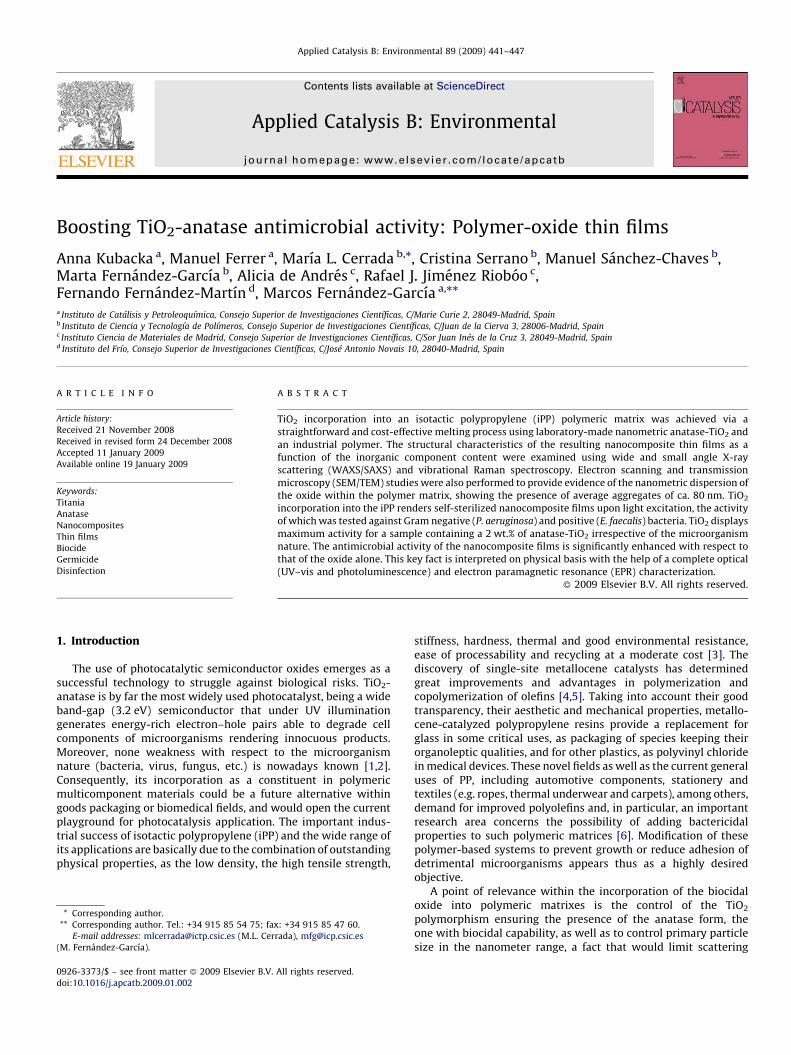
The iPP matrix and all of the Tix nanocomposites are semicrystal-line as their (reflection mode) WAXS profiles at room temperature,depicted in Fig. 1, indicate. These patterns display the five maindiffractions characteristic of the a iPP modification [15,16]. Threedifferent polymorphic modifications, a, b and g, all sharing a three-fold conformation [17–19], have been reported. In addition, a fastquenching of iPP leads to a phase of intermediate or mesomorphicorder [17,19,20]. Concentrations of the g polymorph exceeding 80%have been found in metallocene-type polypropylenes prepared atconditions approaching those used in industrial processes [21].However, no significant evidence of this g modification, through apeak at 19.78 corresponding to its characteristic (1 1 7) reflection[22], is detected either in the iPP polymer or in the different Tixnanocomposites. This fact might be associated with the processingconditions imposed during the film preparation (e.g. cooling fromthe melt between water plates) since the proportion of g crystals isdependent on defects that interrupt isotactic sequences as well as ongeneral crystallization conditions, diminishing at low crystallizationtemperatures [23] or with high cooling rates [24]. Estimation of theamorphous component content at room temperature, obtained fromthe totally amorphous profile of an elastomeric PP sample, allowsdetermination of the crystallinity degree ð f WAXS
c Þ from WAXS
Fig. 1. WAXS profiles of different Tix nanocomposite thin films and iPP reference at
room temperature. Inner plot: WAXS profile of amorphous polypropylene.
experiments by simple subtraction. The f WAXSc values obtained are
listed in Table 1 for the different films, rather constant values ofcrystallinity being observed independently of TiO2 incorporation.However, effect of TiO2 composition is mainly seen in relativevariations of the intensity exhibited by several diffractions ((0 4 0),(1 1 1) and (1 3 1, 0 4 1)) as deduced from Fig. 1.
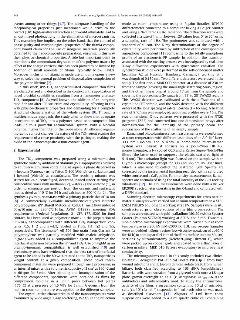
The WAXS/SAXS structural characterization is summarized inTable 1 and indicates the relatively limited structural modificationof the iPP matrix by effect of the anatase component presence; thesingle noticeable effect is the growth of the most probable crystalsize (lc), particularly in the Ti5 film. The essential constancy of theiPP structural characteristics is also evidenced with Raman (Fig. 2)which, on the other hand, provides also information about theinorganic component state on the nanocomposites. As judged by
Fig. 2. Normalized Raman spectra of iPP (A), and width and intensity (B) of the
anatase-TiO2 Eg peak at 149 cm�1. Starts in plot A indicate anatase peaks.
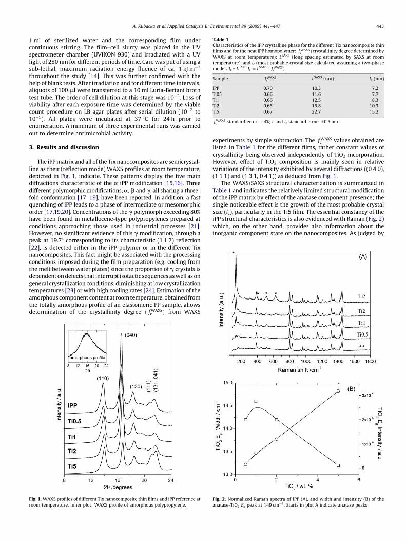
Fig. 3. TEM image of the Ti2 nanocomposite thin film and different details showing nanometer scale oxide agglomerates.
A. Kubacka et al. / Applied Catalysis B: Environmental 89 (2009) 441–447444
the invariant characteristics of the stronger Eg anatase peak at ca.150 cm�1 (Fig. 2 bottom displays the peak width and intensity), theTiO2 component morphological characteristics seem to smoothlyvary within the Tix series. The width informs about the expectedconstancy of the primary particle size of the inorganic componentafter the melt compounding while the linear behavior of theintensity would indicate that scattering events are similar in allsamples, suggesting the probable similar aggregation state of theinorganic component. This latter was further studied with SEM;the cross-section SEM photographs (result not shown) showed nomicrosized aggregated nuclei, indicating that TiO2 nanoparticleshave been highly dispersed within the iPP matrix and theirpractical absence at the surface of the material. This contrast withthe microsized aggregates typically observed in other polyolefin-based nanocomposites with similar incorporations to the onesused in this study [25]. The absence of the oxide at the surface ofthe materials was further confirmed using the sensitive EPRtechnique with the assist of O2 as a probe molecule (see below).
ThenoticeablehomogeneityoftheTix materialsatthenanometerscale indicated by the SEM study is only possible because of the use ofa coupling agent as well as the correct surface/size characteristics ofthe nano-oxide. To confirm this aspect, a TEM study of the Ti2 samplebulk (Fig. 3) was performed. In this case, the oxide is dispersed in thepolymeric matrix exhibiting nanometer-scale aggregates rangingfrom 10 (the oxide primary particle size) to 200 nm, with an averagesize (Feret diameter) [26] of 80 nm (�20 nm). Considering that thetitania preparation makes use of an oxide previously calcined at hightemperature (500 8C) to ensure the exclusive presence of the anatasepolymorph (e.g. absence of an amorphous contribution) and the strictcontrol of its biocidal capabilities, the nanometric dispersion of theoxide reached for loadings below 5 wt.% is significant, particularly withrespect tomicro-sizedor bulk titaniaspecimens [9c], orcomparing with
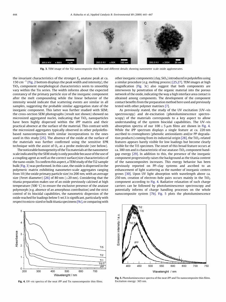
Fig. 4. UV–vis spectra of the neat iPP and Tix nanocomposite thin films.
other inorganic components (clay, SiO2) introduced in polyolefins usinga similar procedure (e.g. melting process) [25,27]. TEM images at highmagnification (Fig. 3c) also suggest that both components areinterwoven by penetration of the organic material into the porousnetwork of the oxide, indicating the way a high interface area contact isobtained among components. The development of the componentcontact benefits from the preparation method here used and previouslytested with other polymer matrixes [7].
As previously stated, the study of the UV excitation (UV–visspectroscopy) and de-excitation (photoluminescence spectro-scopy) of the materials corresponds to a key aspect to allowunderstanding of the system biocidal capabilities. The UV–visabsorption spectra of our 100 � 5 mm films are shown in Fig. 4.While the iPP spectrum displays a single feature at ca. 220 nmascribed to cromophores (phenolic antioxidants and/or PP degrada-tion products) coming from its industrial origin [28], the TiO2-relatedfeatures appears barely visible for low loadings but become clearlyvisible for the Ti5 specimen. The onset of this broad feature occurs atca. 380 nm and is characteristic of our anatase-TiO2 component band-gap energy [29]. In addition to this, the presence of the inorganiccomponent progressively raises the background as the titania contentof the nanocomposites increases. This energy behavior has beenpreviously reported on PP–clay systems and ascribed to anenhancement of light scattering as the number of inorganic centersgrows [30]. Upon UV light absorption with wavelength above ca.250 nm, creation of electron–hole pairs occurs mainly in the TiO2
component according to Fig. 4. Radiative relaxation of such chargecarriers can be followed by photoluminescence spectroscopy andpotentially informs of charge handling processes on the wholenanocomposite system [7b]. Fig. 5 plots the photoluminescence
Fig. 5. Photoluminescence spectra of the neat iPP and Tix nanocomposite thin films.
Excitation energy: 365 nm.
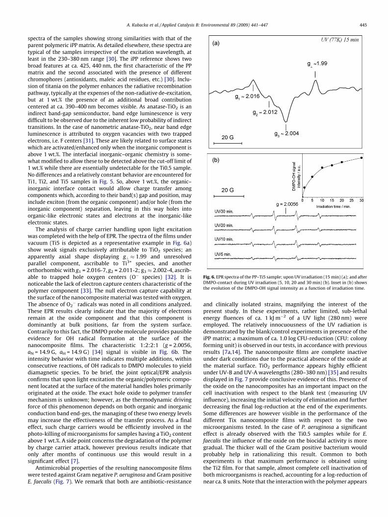
Fig. 6. EPR spectra of the PP–Ti5 sample; upon UV irradiation (15 min) (a); and after
DMPO-contact during UV irradiation (5, 10, 20 and 30 min) (b). Inset in (b) shows
the evolution of the DMPO-OH signal intensity as a function of irradiation time.
A. Kubacka et al. / Applied Catalysis B: Environmental 89 (2009) 441–447 445
spectra of the samples showing strong similarities with that of theparent polymeric iPP matrix. As detailed elsewhere, these spectra aretypical of the samples irrespective of the excitation wavelength, atleast in the 230–380 nm range [30]. The iPP reference shows twobroad features at ca. 425, 440 nm, the first characteristic of the PPmatrix and the second associated with the presence of differentchromophores (antioxidants, maleic acid residues, etc.) [30]. Inclu-sion of titania on the polymer enhances the radiative recombinationpathway, typically at the expenses of the non-radiative de-excitation,but at 1 wt.% the presence of an additional broad contributioncentered at ca. 390–400 nm becomes visible. As anatase-TiO2 is anindirect band-gap semiconductor, band edge luminescence is verydifficult to be observed due to the inherent low probability of indirecttransitions. In the case of nanometric anatase-TiO2, near band edgeluminescence is attributed to oxygen vacancies with two trappedelectrons, i.e. F centers [31]. These are likely related to surface stateswhich are activated/enhanced only when the inorganic component isabove 1 wt.%. The interfacial inorganic–organic chemistry is some-what modified to allow these to be detected above the cut-off limit of1 wt.% while there are essentially undetectable for the Ti0.5 sample.No differences and a relatively constant behavior are encountered forTi1, Ti2, and Ti5 samples in Fig. 5. So, above 1 wt.%, the organic–inorganic interface contact would allow charge transfer amongcomponents which, according to their band(s) gap and position, mayinclude exciton (from the organic component) and/or hole (from theinorganic component) separation, leaving in this way holes intoorganic-like electronic states and electrons at the inorganic-likeelectronic states.
The analysis of charge carrier handling upon light excitationwas completed with the help of EPR. The spectra of the films undervacuum (Ti5 is depicted as a representative example in Fig. 6a)show weak signals exclusively attributable to TiO2 species; anapparently axial shape displaying g? � 1.99 and unresolvedparallel component, ascribable to Ti3+ species, and anotherorthorhombic with g1 = 2.016-7, g2 = 2.011-2; g3 � 2.002-4, ascrib-able to trapped hole oxygen centers (O� species) [32]. It isnoticeable the lack of electron capture centers characteristic of thepolymer component [33]. The null electron capture capability atthe surface of the nanocomposite material was tested with oxygen.The absence of O2
� radicals was noted in all conditions analyzed.These EPR results clearly indicate that the majority of electronsremain at the oxide component and that this component isdominantly at bulk positions, far from the system surface.Contrarily to this fact, the DMPO probe molecule provides pausibleevidence for OH radical formation at the surface of thenanocomposite films. The characteristic 1:2:2:1 (g = 2.0056,aN = 14.9 G, aH = 14.9 G) [34] signal is visible in Fig. 6b. Theintensity behavior with time indicates multiple additions, withinconsecutive reactions, of OH radicals to DMPO molecules to yielddiamagnetic species. To be brief, the joint optical/EPR analysisconfirms that upon light excitation the organic/polymeric compo-nent located at the surface of the material handles holes primarilyoriginated at the oxide. The exact hole oxide to polymer transfermechanism is unknown; however, as the thermodynamic drivingforce of this phenomenon depends on both organic and inorganicconduction band end-ges, the managing of these two energy levelsmay increase the effectiveness of the transfer process. As a finaleffect, such charge carriers would be efficiently involved in thephoto-killing of microorganisms for samples having a TiO2 contentabove 1 wt.%. A side point concerns the degradation of the polymerby charge carrier attack, however previous results indicate thatonly after months of continuous use this would result in asignificant effect [7].
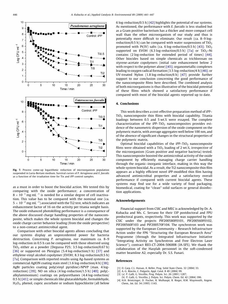
Antimicrobial properties of the resulting nanocomposite filmswere tested against Gram negative P. aeruginosa and Gram positiveE. faecalis (Fig. 7). We remark that both are antibiotic-resistance
and clinically isolated strains, magnifying the interest of thepresent study. In these experiments, rather limited, sub-lethalenergy fluences of ca. 1 kJ m�2 of a UV light (280 nm) wereemployed. The relatively innocuousness of the UV radiation isdemonstrated by the blank/control experiments in presence of theiPP matrix; a maximum of ca. 1.0 log CFU-reduction (CFU: colonyforming unit) is observed in our tests, in accordance with previousresults [7a,14]. The nanocomposite films are complete inactiveunder dark conditions due to the practical absence of the oxide atthe material surface. TiO2 performance appears highly efficientunder UV-B and UV-A wavelengths (280–380 nm) [35] and resultsdisplayed in Fig. 7 provide conclusive evidence of this. Presence ofthe oxide on the nanocomposites has an important impact on thecell inactivation with respect to the blank test (measuring UVinfluence), increasing the initial velocity of elimination and furtherdecreasing the final log-reduction at the end of the experiments.Some differences are however visible in the performance of thedifferent Tix nanocomposite films with respect to the twomicroorganisms tested. In the case of P. aeruginosa a significanteffect is already observed with the Ti0.5 samples while for E.
faecalis the influence of the oxide on the biocidal activity is moregradual. The thicker wall of the Gram positive bacterium wouldprobably help in rationalizing this result. Common to bothexperiments is that maximum performance is obtained usingthe Ti2 film. For that sample, almost complete cell inactivation ofboth microorganisms is reached, accounting for a log-reduction ofnear ca. 8 units. Note that the interaction with the polymer appears
Fig. 7. Process come-up logarithmic reduction of microorganism population
suspended in Luria-Bertani medium. Survival curves of P. Aeruginosa and E. faecalis
as a function of the irradiation time for Tix and iPP control samples.
A. Kubacka et al. / Applied Catalysis B: Environmental 89 (2009) 441–447446
as a must in order to boost the biocidal action. We tested this bycomparing with the oxide performance; a concentration of8 � 10�1 mg mL�1 is needed for a similar degree of cell inactiva-tion. This value has to be compared with the nominal one (ca.5 � 10�2 mg mL�1) associated with the Ti2 test, which indicates anenhancement factor of 16 on the activity per titania weight basis.The oxide enhanced photokilling performance is a consequence ofthe above discussed charge handling properties of the nanocom-posite, which makes the whole system biocidal and changes theoxide charge carrier behavior leading (from the oxide perspective)to a non-contact antimicrobial agent.
Comparison with other biocidal agents allows concluding thatour systems display an unprecedented power for bacteriadestruction. Concerning P. aeruginosa, our maximum ca. 8–9log-reduction in 0.5 h can be compared with those observed usingTiO2 either as a powder (Degussa P25; 3.5 log-reduction/0.67 h)[36] or supported on Plexiglas (5.4 log-reduction/1 h) [37] andethylene-vinyl alcohol copolymer (EVOH; 8.3 log-reduction/0.5 h)[7a]. Comparison with reported results using Ag-based systems ascommercial AgION coating stain steel (1.6 log-reduction/4 h) [38];AgBr particles coating poly(vinyl pyridine)-NPVP (max. 4 log-reduction) [39]; NO on silica (4 log-reduction/1.5 h) [40]; poly(-alkylammonium) coatings on polyurethanes (4.4 log-reduction/0.5 h) [41]; or simple chemicals like glutaraldehyde, formaldehyde,H2O2, phenol, cupric ascorbate or sodium hypochlorite (all below
6 log-reduction/0.5 h) [42] highlights the potential of our systems.As mentioned, the performance with E. faecalis is less studied butas a Gram positive bacterium has a thicker and more compact cellwall than the other microorganism of our study and thus ispotentially more difficult to eliminate. Our result (ca. 8–9 log-reduction/0.5 h) can be compared with water suspensions of TiO2
promoted with Pt(IV) salts (ca. 6 log-reduction/0.5 h) [43]; TiO2
supported on EVOH (6.3 log-reduction/0.5 h) [7a] or TiO2–Nicontains (2 log-reduction for extended period of times) [44].Other biocides based on simple chemicals as trichlorosan onstyrene-acetate copolymers (initial rate enhancement below 2with respect to the polymer alone) [45]; organometallic complexesleading to oxygen radical formation (3.5 log-reduction/4 h) [46]; orUV-treated Nylon (1.8 log-reduction/6 h) [47] provide furthersupport to our conclusion concerning the good performance ofthe nanocomposite films here described. The combined analysisof both microorganisms is thus illustrative of the biocidal potentialof these films which showed a satisfactory performance ifcompared with most of the biocidal agents reported up to date.
4. Conclusions
This work describes a cost-effective preparation method of iPP–TiO2 nanocomposite thin films with biocidal capability. Titanialoadings between 0.5 and 5 wt.% were essayed. The completecharacterization of the iPP–TiO2 nanocomposites provides evi-dence of the nanometric dispersion of the oxide component on thepolymeric matrix, with average aggregates well below 100 nm, andof the absence of significant changes in the structural properties ofthe polymeric matrix.
Optimal biocidal capabilities of the iPP–TiO2 nanocompositefilms were obtained with a TiO2 loading of 2 wt.%, irrespective ofthe microorganism (Gram positive and negative bacteria) tested.This nanocomposite boosted the antimicrobial activity of the oxidecomponent by efficiently managing charge carrier handlingthrough the organic–inorganic interface, making in this way thewhole system biocidal. As a result, the Ti2 nanocomposite thin filmappears as a highly efficient novel iPP-modified thin film havingadvanced antimicrobial properties and a satisfactory overallperformance if compared with current biocidal agents. Thesesystems may find use for a wide variety of food packaging,biomedical, coating for ‘‘clean’’ solid surfaces or general disinfec-tion applications.
Acknowledgements
Financial support from CSIC and MEC is acknowledged by Dr. A.Kubacka and Ms. C. Serrano for their I3P postdoctoral and FPUpredoctoral grants, respectively. This work was supported by theCSIC under the projects PIF200580F0101, PIF200560F0102,PIF200560F103 and PIF200570F104. The synchrotron work wassupported by the European Community – Research InfrastructureAction under the FP6 ‘‘Structuring the European Research Area’’Programme (through the Integrated Infrastructure Initiative‘‘Integrating Activity on Synchrotron and Free Electron LaserScience’’), contract RII3-CT-2004-506008 (IA-SFS). We thank thecollaboration of the Hasylab personnel in the soft-condensedmatter beamline A2, especially Dr. S.S. Funari.
References
[1] O. Carp, C.L. Huisan, A. Reller, Prog. Solid State Chem. 32 (2004) 33.[2] A.-G. Rincon, C. Pulgarın, Appl. Catal. B 49 (2004) 99.[3] (a) P. Galli, G. Vecellio, Prog. Polym. Sci. 26 (2001) 1287;
(b) P. Galli, G. Vecellio, J. Polym. Sci. Polym. Chem. Ed. 42 (2004) 396.[4] H.H. Brintzinger, D. Fischer, R. Mulhaupt, B. Rieger, R.M. Waymouth, Angew.
Chem., Int. Ed. 34 (1995) 1143.
A. Kubacka et al. / Applied Catalysis B: Environmental 89 (2009) 441–447 447
[5] L. Resconi, L. Cavallo, A. Fait, F. Piemontesi, Chem. Rev. 100 (2000) 1253.[6] P. Appendini, J.N. Hotchkiss, Innovative Food Sci. Technol. 3 (2002) 113.[7] (a) A. Kubacka, C. Serrano, M. Ferrer, H. Lundsford, P. Bieleck, M.L. Cerrada, M.
Fernandez-Garcıa, M. Fernandez-Garcıa, Nano Letters 7 (2007) 2529;(b) A. Kubacka, M.L. Cerrada, C. Serrano, M. Fernandez-Garcıa, M. Ferrer, M.Fernandez-Garcıa, J. Nanosci. Nanotechnol. 8 (2008) 3241;(c) M.L. Cerrada, C. Serrano, M. Sanchez-Chaves, M. Fernandez-Garcıa, A. deAndres, R.J. Jimenez-Rioboo, A. Kubacka, M. Ferrer, M. Fernandez-Garcıa, Adv.Funct. Mater. 18 (2008) 1949.
[8] M. Fernandez-Garcıa, X. Wang, C. Belver, J.C. Hanson, J.A. Rodrıguez, J. Am. Chem.Soc. 129 (2007) 13604.
[9] (a) X.S. Fan, J. Vinyl Add. Technol. 13 (2007) 65;(b) P. Supaphol, P. Thanomkiat, J. Jonkesen, R. Dangtungee, Polym. Test. 26 (2007) 2;(c) D. Garcıa-Lopez, O. Picazo, J.C. Marino, J.M. Pastor, Eur. Polym. J. 39 (2003) 945.
[10] D.N. Bikiaris, A. Vassilou, E. Pavlidou, G.P. Karayannidis, Eur. Polym. J. 41 (2005)1965.
[11] S. Mansel, E. Perez, R. Benavente, J.M. Perena, A. Bello, W. Roll, R. Kirsten, S. Beck,H.-H. Brintzinger, Macromol. Chem. Phys. 200 (1999) 1292.
[12] A.S. Nielsen, D.N. Batcheler, R. Pryz, Polymer 43 (2002) 2671.[13] M. Ferrer, J. Soliveri, F.J. Plou, N. Lopez-Cortes, D. Reyes-Duarte, M. Christensen, J.L.
Copa-Patino, A. Ballesteros, Enzyme Microb. Technol. 36 (2005) 391.[14] J. Jagger, Photochem. Photobiol. 34 (1981) 761.[15] G. Natta, P. Corradini, Nuovo Cimento Suppl. 15 (1960) 40.[16] A. Turner-Jones, J.M. Aizlewood, D.R. Becket, Makromol. Chem. 75 (1964) 134.[17] S. Bruckner, S.V. Meille, V. Petraccone, B. Pirozzi, Prog. Polym. Sci. 16 (1991) 361.[18] B. Lotz, J.C. Wittmann, A.J. Lovinger, Polymer 37 (1996) 4979.[19] P.J. Phillips, K. Mezghani, in: J.C. Salamone (Ed.), The Polymeric Materials
Encyclopedia, vol. 9, CRC Press, Boca Raton, 1996, p. 6637.[20] J. Arranz-Andres, R. Benavente, E. Perez, M.L. Cerrada, Polym. J. 35 (2003) 766.[21] R.G. Alamo, M.-H. Kim, M.J. Galante, J.R. Isasi, L. Mandelkern, Macromolecules 32
(1999) 4050.[22] C. De Rosa, F. Auriemma, T. Circelli, Macromolecules 35 (2002) 3622.[23] C. De Rosa, F. Aurienma, M. Paolillo, L. Resconi, I. Camurati, Macromolecules 38
(2005) 9143.[24] E. Perez, D. Zucchi, M.C. Sacchi, F. Forlini, A. Bello, Polymer 40 (1999) 675.
[25] D. Ma, Y.A. Akpalu, Y. Li, R.W. Siegel, L.S. Schadler, Polym. J. Sci. Part B: Polym.Phys. 43 (2005) 488.
[26] R.J. Matyi, L.H. Schartz, J.B. Butt, Catal. Rev. 29 (1987) 41.[27] V. Vladimirov, C. Betcher, A. Vassilou, A. Papageonigiou, J. Bikiaris, Comp. Sci.
Technol. 66 (2006) 2935.[28] S. Morlat-Therias, B. Mailhot, D. Gonzales, J.L. Gardette, Chem. Mater. 16 (2004)
377.[29] A. Kubacka, M. Ferrer, A. Martınez-Arias, M. Fernandez-Garcıa, Appl. Catal. B 84
(2008) 87.[30] M. Ambid, G. Teyssedre, D. Mary, C. Laurent, G.C. Montanari, IEEJ Trans. FM 126
(2006) 1097.[31] N. Serpone, D. Lowless, A. Khairutdinov, J. Phys. Chem. 99 (1996) 16646.[32] J.M. Coronado, A.J. Maira, J.C. Conesa, K.L. Yeung, V. Augugliaro, J. Soria, Langmuir
17 (2001) 5368.[33] S. Esnouf, E. Balanzat, Polymer 48 (2007) 7531, and references therein.[34] D. Dvoranova, V. Brezova, M. Mazur, M.A. Malati, Appl. Catal. B 37 (2002) 91.[35] A.K. Benabbou, Z. Derriche, C. Felix, C. Guillard, Appl. Catal. B 76 (2007) 257.[36] J.A. Ibanez, M.I. Litter, R.A. Pizarro, J. Photochem. Photobiol. A 157 (2003) 81.[37] K.P. Kuhn, I.F. Chaberny, K. Massholder, M. Stickler, V.W. Benz, H.-G. Sonnatg, L.
M.H. Schoenfisch, ACS Nano 2 (2008) 235.[41] P. Kurt, L. Wood, D.E. Ohman, K.J. Wynne, Langmuir 23 (2007) 4719.[42] J.C. Sagripanti, A. Bonifacio, J. AOAC Int. 83 (2000) 1415.[43] D. Mitoraj, A. Janczyk, M. Strus, G. Stochel, P.B. Heczko, W. Macyk, Photochem.
Photobiol. Sci. 6 (2007) 642.[44] Z. Zhi-hong, Y. Sakagami, T. Osaka, Chem. Lett. 90 (1997) 9.[45] D. Chung, S.E. Papadakis, K.L. Yam, Int. J. Food Sci. Technol. 38 (2003) 165.[46] L. Villen, F. Majon, D. Garcıa-Resnadillo, G. Orellana, Appl. Catal. B 69 (2006) 1.[47] A.E.H. Shearer, J.S. Park, D.G. Hoover, S.L. Hayne, Biotechnol. Bioeng. 67 (2000)