Date post: | 30-Nov-2023 |
Category: |
Documents |
Upload: | independent |
View: | 0 times |
Download: | 0 times |
Luigi CanulloRoberto CocchettoFabio MarinottiDavid Pe~narrocha OltraMar�ıa Pe~narrocha DiagoIgnazio Loi
Clinical evaluation of an improvedcementation technique for implant-supported restorations: a randomizedcontrolled trial
Authors’ affiliations:Luigi Canullo, Private Practice and IndependentResearcher, Rome, ItalyLuigi Canullo, Department of Oral Surgery andImplantology, Valencia University Medical andDental School, Valencia, SpainRoberto Cocchetto, Private Practice, Verona, ItalyFabio Marinotti, Laboratory Technician, Rome,ItalyDavid Pe~narrocha Oltra, Mar�ıa Pe~narrocha Diago,Department of Oral Surgery, Valencia UniversityMedical and Dental School, Valencia, SpainIgnazio Loi, Private Practice, Cagliari, Italy
Corresponding author:Luigi CanulloVia Nizza, 46, 00198 Rome, ItalyTel.: +39 06 8411980Fax: +39 06 8411980e-mail: [email protected]
Key words: abutment design, cement remnants, chamfer, feather-edge abutment, peri-
implantitis, shoulderless abutment
Abstract
Background: Cement remnants were frequently associated with peri-implantitis. Recently, a
shoulderless abutment was proposed, raising some concern about cement excess removal.
Aim: To compare different cementation techniques for implant-supported restorations assessing
the amount of cement remnants in the peri-implant sulcus. Additional aim was to compare the
effect of these cementation techniques using two different abutment designs.
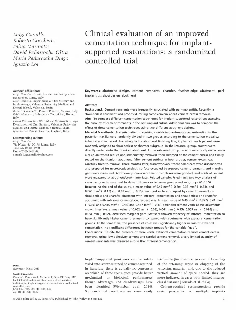
Material & methods: Forty-six patients requiring double implant-supported restoration in the
posterior maxilla were randomly divided in two groups according to the cementation modality:
intraoral and extraoral. According to the abutment finishing line, implants in each patient were
randomly assigned to shoulderless or chamfer subgroup. In the intraoral group, crowns were
directly seated onto the titanium abutment. In the extraoral group, crowns were firstly seated onto
a resin abutment replica and immediately removed, then cleansed of the cement excess and finally
seated on the titanium abutment. After cement setting, in both groups, cement excess was
carefully tried to remove. Three months later, framework/abutment complexes were disconnected
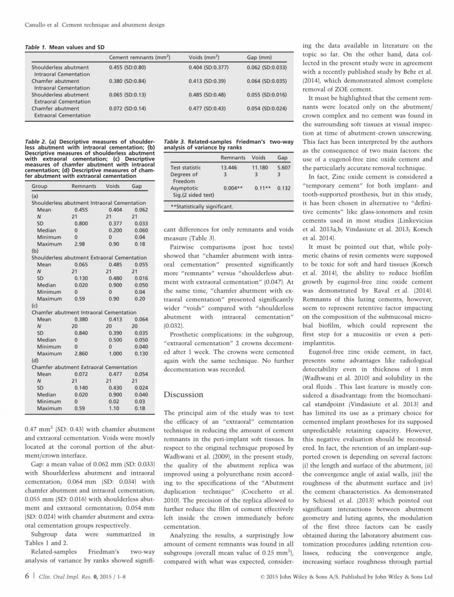
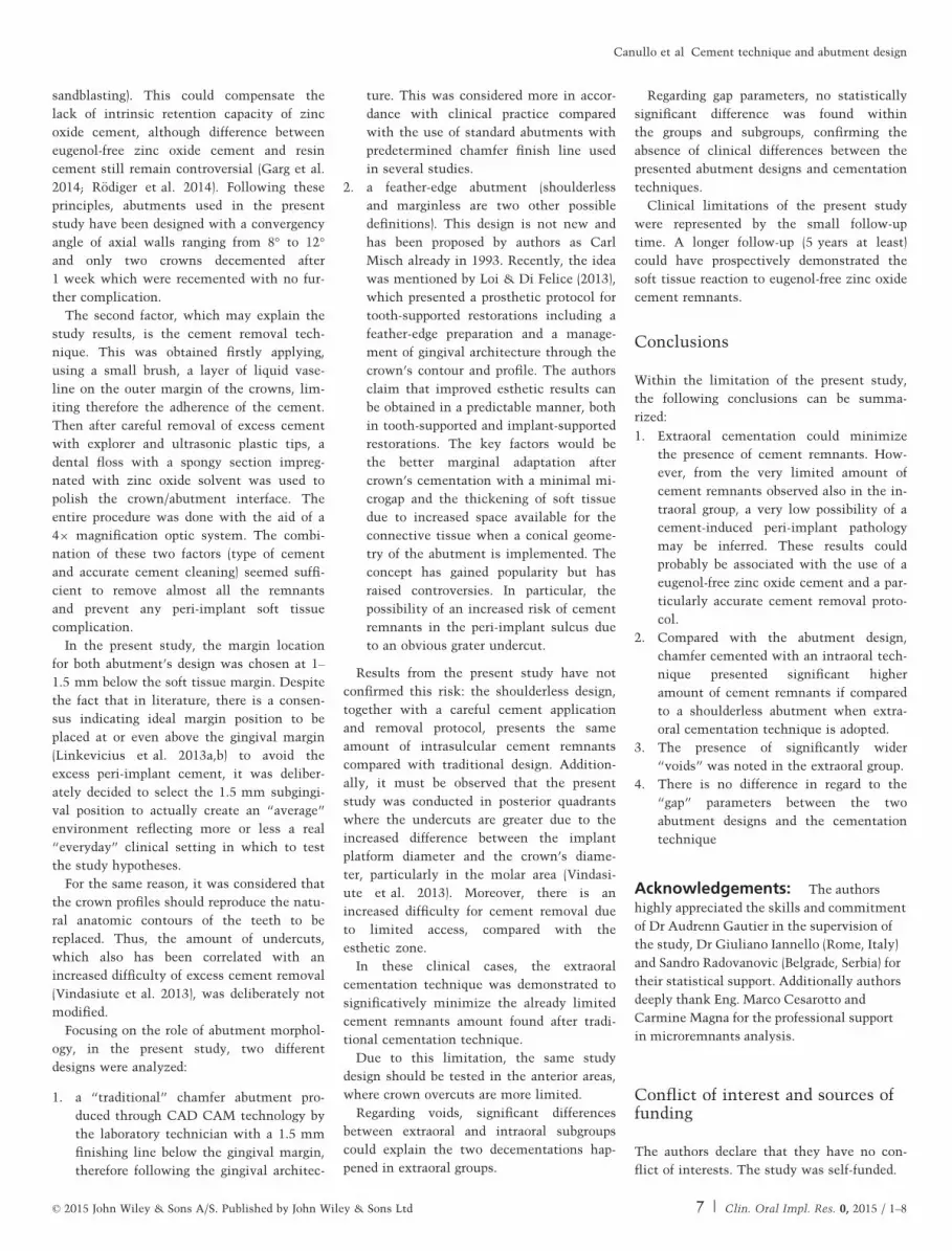
and prepared for microscopic analysis: surface occupied by exposed cement remnants and marginal
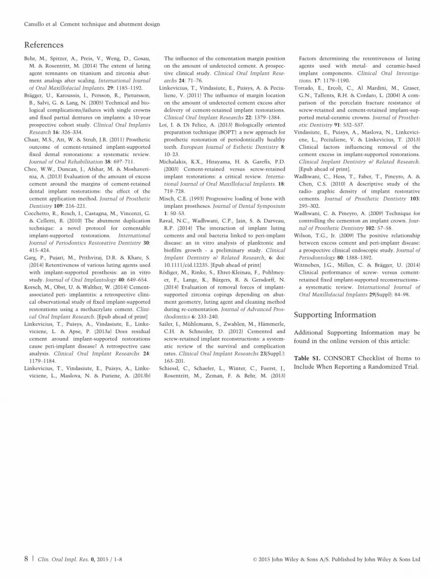
gaps were measured. Additionally, crown/abutment complexes were grinded, and voids of cement
were measured at abutment/crown interface. Related-samples Friedman’s two-way analysis of
variance by ranks was used to detect differences between groups and subgroups (P ≤ 0.5).
Results: At the end of the study, a mean value of 0.45 mm2 (�0.80), 0.38 mm2 (�0.84), and
0.065 mm2 (�0.13) and 0.07 mm2 (�0.15) described surface occupied by cement remnants in
shoulderless and chamfer abutment with intraoral cementation and shoulderless and chamfer
abutment with extraoral cementation, respectively. A mean value of 0.40 mm2 (�0.377), 0.41 mm2
(�0.39) and 0.485 mm2 (�0.47) and 0.477 mm2 (�0.43) described cement voids at the abutment/
crown interface; a mean value of 0.062 mm (�0.03), 0.064 mm (�0.35), 0.055 mm (�0.016) and
0.054 mm (�0.024) described marginal gaps. Statistics showed tendency of intraoral cementation to
have significantly higher cement remnants compared with abutments with extraoral cementation
groups. At the same time, the presence of voids was significantly higher in case of extraoral
cementation. No significant differences between groups for the variable “gap”.
Conclusions: Despite the presence of more voids, extraoral cementation reduces cement excess.
However, using low adhesivity cement and careful cement removal, a very limited quantity of
cement remnants was observed also in the intraoral cementation.
Implant-supported prostheses can be subdi-
vided into screw-retained or cement-retained.
In literature, there is actually no consensus
on which of these techniques provide better
mechanical or biological performances
though advantages and disadvantages have
been identified (Wittneben et al. 2014).
Screw-retained prostheses are more easily
retrievable (for instance, in case of loosening
of the retaining screw or chipping of the
veneering material) and, due to the reduced
vertical amount of space needed, they are
more indicated in cases with limited interoc-
clusal distance (Torrado et al. 2004).
Cement-retained reconstructions provide
easier passivation on multiple implants
Date:Accepted 4 March 2015
To cite this article:Canullo L, Cocchetto R, Marinotti F, Oltra DP, Diago MP,Loi I. Clinical evaluation of an improved cementationtechnique for implant-supported restorations: a randomizedcontrolled trial.Clin. Oral Impl. Res. 00, 2015; 1–8.doi: 10.1111/clr.12589
© 2015 John Wiley & Sons A/S. Published by John Wiley & Sons Ltd 1
splinted frameworks, better esthetics (due to
the lack of occlusal openings) and lower costs
(Michalakis et al. 2003).
On the other hand, literature suggests that,
even after a careful removal procedure, some
residual cement may remain in the peri-
implant sulcus (Chaar et al. 2011; Sailer
et al. 2012). The risk of such event increases
as the restorations’ margins are located dee-
per subgingivally (Linkevicius et al. 2011,
2013a,b). Cement remnants have been fre-
quently associated with peri-implant disease
(Wilson 2009; Linkevicius et al. 2013a,
Korsch et al. 2014), and for this reason,
screw-retained restorations have been sug-
gested as a first choice in daily practice by
some authors (Br€agger et al. 2005).
An original cementation technique for
implant-supported restorations has been pro-
posed (Wadhwani et al. 2009) as a mean to
reduce cement remnants in peri-implant sul-
cus: a silicon abutment replica is used as an
initial “extraoral “cementation with the pur-
pose to extrude excess cement from the
crown, clean it and then place it onto the
abutment intraorally to complete setting. An
in vitro study (Chee et al. 2013) seemed to
confirm the efficacy of the method compared
with three other cementation methods.
The abutment and crown design may play
a role in increasing the risk of cement rem-
nants in the peri-implant sulcus. In fact, to
minimize cement remnants, undercuts
should be reduced to a minimum (Vindasiute
et al. 2013). Recently, a new prosthetic proto-
col aiming to improve esthetic outcomes of
prosthetic restorations on natural teeth was
presented (Loi & Di Felice 2013). This
approach is based on a feather-edge prepara-
tion of abutment teeth and the gingival adap-
tation to crowns contours. The same
principles and concepts are suggested by the
authors for cemented implant restorations
through the use of a shoulderless abutment
design. However, such geometry presents
increased undercuts, raising some concern
about the difficulty to completely remove the
excess cement.
The aim of the present study was to com-
pare in vivo two different cementation tech-
niques for implant-supported restorations in
relation to the amount of cement remnants
in the peri-implant sulcus. Additional aim
was to compare the effect of these cementa-
tion techniques using two different abutment
design, chamfer and shoulderless.
The article was written following the
CONSORT statement for improving the
quality of RCTs.
Material and methods
The present randomized controlled prospec-
tive study was performed following the prin-
ciples outlined in the Declaration of Helsinki
on experimentation involving human sub-
jects. All procedures and materials in the
present prospective study were approved by
the Ethical Committee of the University of
Valencia (# H1406287295470).
Patients were required to sign a consent
form after being informed about the study.
Between Sept 2013 and Sept 2014, accord-
ing to the power analysis, 46 consecutive
patients needing two adjacent implant-sup-
ported restorations in the posterior maxilla
were selected. Ninety implants (Premium SP;
Sweden & Martina, Padua, Italy) were
inserted in a two-stage modality.
In all patients, Premium SP implants (Swe-
den & Martina) were used (Fig. 1).
For every case, after implant osseointegra-
tion was achieved (2 months), healing abut-
ments were connected (Fig. 2). After soft
tissue healing, a silicon impression was taken
at implant level. The working cast was
scanned in laboratory (Echo, Sweden & Marti-
na), and prosthetic components were designed
and constructed with CAD CAM technology
(Echo, Sweden &Martina) (Fig. 3).
Abutment randomization
Every implant in each case was randomly
assigned to receive either an abutment with a
chamfer finish line or an abutment with a
shoulderless (feather edge) design: if the first
implant received one kind of abutment design,
the second would receive the other design.
Random assignment was performed using a
free dedicated software (www.examplerand
omxxx.com).
(a)
(f) (g) (h) (i)
(b) (c) (d) (e)
Fig. 1. Surgical phases: pre-operative buccal and occlusal overview (a, b); flap elevation, surgical guide and pre-operative CBT analysis (c, d, e); implant insertion with buccal
and occlusal overview (f, g); control X-ray (h); sutures (i).
2 | Clin. Oral Impl. Res. 0, 2015 / 1–8 © 2015 John Wiley & Sons A/S. Published by John Wiley & Sons Ltd
Canullo et al �Cement technique and abutment design
For each implant, two identical titanium
abutments and Cr/Co metal frameworks
were fabricated: one for the study, the sec-
ond for the definitive restoration by CAD
CAM technology (Echo, Sweden & Martina).
The crowns for both the chamfer and the
feather-edge abutments were designed to
have a subgingival margin of maximum
1.5 mm.
An occlusal opening was made in the first
metal framework in order to have access to
the abutment screw after cementation. This
was necessary to ensure the retrievability of
the abutment–restoration complex for the
study. However, to prevent venting of luting
agent during cementation, the crown was
veneered with composite material.
Cementation technique randomization
Eugenol-free zinc oxide cement (Temp Bond,
Kerr, US) was selected as a luting agent for
this study. Before cementation, the screw
access of each abutment was filled with poly-
tetrafluoroethylene tape and sealed with pro-
visional restorative material – Cavit (3 M
UNITEK, Monrovia, CA, USA).
A thin layer of Vaseline was applied over
the external marginal contour of every crown
to reduce cement adhesion over the external
surface of the crowns and facilitate removal
excess cement. The cement was mixed
according to the manufacturer’s instructions;
a thin layer was applied to all internal sur-
faces of the crowns using microbrush (Micro-
brush International, Waterford, Ireland).
In the subgroup “intraoral cementation”,
crown was directly seated without any addi-
tional procedures (Fig. 4).
In the subgroup “extraoral cementation”, a
custom-made polyurethane resin replica of
the abutment had been previously prepared
according to ADT technique (Cocchetto et al.
2010). Every crown, after cement application,
was fully seated onto the abutment replica,
then rapidly removed, cleansed of the
extruded excess cement and finally seated on
the titanium abutment until cement setting
was complete (Fig. 5).
Random assignment was performed using a
free dedicated software (www.examplerando
mxxx.com).
After final cement setting, in all sub-
groups, a stainless steel explorer (Dentsply
International Inc., Milford, DE, USA), and
ultrasound plastic tip were used until the
researcher was confident to have removed
any eventual excess cement. Thereafter,
super floss (Curaprox, Kriens, Switzerland)
imbibed of Bio Orange Solvent (Ogna Lab-
oratori Farmaceutici, Milano, Italia) was
used to polish the abutment/crown com-
plex (Fig. 6). The cement removal proce-
dure was performed using magnifying
loupes.
Three months later, clinical measurements
were performed, and implant stability and
(a) (b) (c) (d)
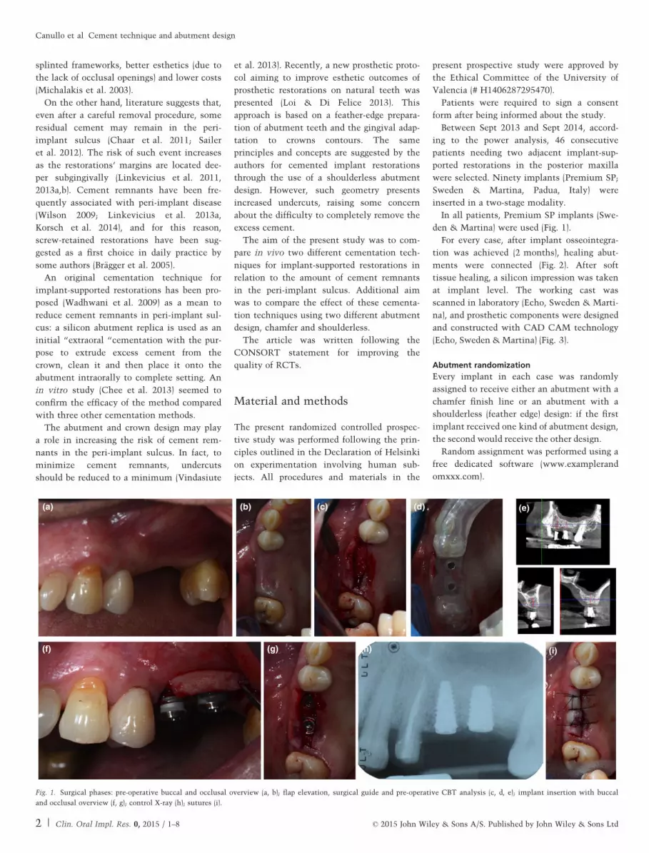
Fig. 2. Second surgical phase: tissues after healing (a); healing abutments connection, occlusal and buccal overview (b, c); control X-ray (d).
(a)
(b)
(c)
(d) (e) (f)
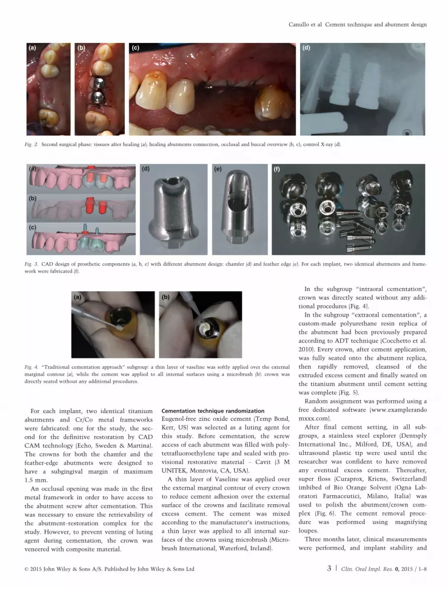
Fig. 3. CAD design of prosthetic components (a, b, c) with different abutment design: chamfer (d) and feather edge (e). For each implant, two identical abutments and frame-
work were fabricated (f).
(a) (b)
Fig. 4. “Traditional cementation approach” subgroup: a thin layer of vaseline was softly applied over the external
marginal contour (a), while the cement was applied to all internal surfaces using a microbrush (b): crown was
directly seated without any additional procedures.
© 2015 John Wiley & Sons A/S. Published by John Wiley & Sons Ltd 3 | Clin. Oral Impl. Res. 0, 2015 / 1–8
Canullo et al �Cement technique and abutment design
the presence of complications were recorded.
Then, the connection screw was accessed,
the abutment screw unscrewed and the
suprastructure disconnected for assessment.
Occlusal photographs of the implants’ plat-
forms and the surrounding soft tissues were
taken. The peri-implant sulcus was probed to
evaluate depth and BOP.
Immediately after, the second metal cera-
mic crown/abutment complex was defini-
tively placed (Fig. 7). Before ceramic
veneering, the two metallic frameworks had
been laser soldered.
Outcome measures
The trial tested two hypotheses. The first
one was that no difference in cement
remnants between the use of a tradi-
tional “intraoral” and an improved “extra-
oral” cementation technique would be
observed. The second one was that no dif-
ference in cement remnants between the
use of a traditional “chamfer“ and an
improved “shoulderless” abutment would
be observed.
Outcome measures were as follows:
1. cement remnants at the abutment/crown
margin
2. void of cement at the implant/abutment
interface
3. gap at the abutment/crown level
Microscopic analysis
Optical light microscope (Stemi DV 4, Carl
Zeiss, Oberkochen, Germany) was used to
measure the presence of eventual cement
remnants or gaps.
For every abutment/crown complex, the
following pictures were taken:
1. one picture (109) of the complex from
apical to coronal
2. four pictures (329) of the abutment/
crown finishing line with the abutment
laying on the interproximal aspect
3. four pictures (329) of the abutment/
crown finishing line with the abutment
inclined at 45° on the occlusal aspect
(Figs 8 and 9)
To measure eventual voids of cement at
the abutment/crown interface, removed
abutment/crown complex, after photo-
graphic analysis, was grinded and under-
went a microscopic analysis. Two pictures
for each sample were taken at 109 and 209
(Fig. 10).
Using a dedicated software (Solidworks
Premium 2012; DASSAULT SYST�EMES,
VelizVillacoublay, France), the surface
(squared microns) occupied by exposed
cement remnants was measured.
Measurements were performed by other
two blinded calibrated examiners (M.C,
C.M). The mean value of the two measure-
ments was used for the analysis.
Prosthetic failures (exposed margins and
crown decementation) were recorded.
Statistical analysis
Descriptive statistics, including mean values
and standard deviations, were calculated.
Statistical analysis of the data was con-
ducted using related-samples Friedman’s two-
way analysis of variance by ranks. The level
of statistical significance was set at P ≤ 0.05.
Results
Between Sept 2013 and Sept 2014, 62 patients
were screened for inclusion and 46 (29 male
and 17 females) participated in the study
(mean age 60.58 years, SD: 10.91). Reasons
for exclusion were as follows: not meeting
the inclusion criteria (n.12) and refused to
participate (n.4).
All surgical interventions and post-opera-
tive healing period were without any serious
complication or side effect for all patients.
At the end of the study, four patients
dropped out.
After microscopic analysis, the following
data were found:
Cement remnants: a mean value of
0.45 mm2 (SD: 0.80) was recorded with shoul-
derless abutment and intraoral cementation;
(a) (b)
(e) (f) (g)
(c) (d)
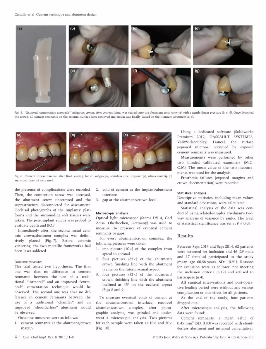
Fig. 5. “Extraoral cementation approach” subgroup: crown, after cement lying, was seated onto the abutment resin copy (a) with a gentle finger pressure (b, c, d). Once detached
the crown, all cement remnants on the external surface were removed and crown was finally seated on the titanium abutment (e, f).
(a) (b) (c)
Fig. 6. Cement excess removal after final seating: for all subgroups, stainless steel explorer (a), ultrasound tip (b)
and super floss (c) were used.
4 | Clin. Oral Impl. Res. 0, 2015 / 1–8 © 2015 John Wiley & Sons A/S. Published by John Wiley & Sons Ltd
Canullo et al �Cement technique and abutment design
0.38 mm2 (SD: 0.84) with shoulderless
abutment and extraoral cementation;
0.065 mm2 (SD: 0.13), 0.07 mm2 (SD: 0.15)
with chamfer abutment and intraoral cemen-
tation, 0.072 mm2 (SD:0.14) with chamfer
abutment and extraoral cementation. Rem-
nants were mostly located below the IAJ.
Voids: a mean value of 0.40 mm2 (SD: 0.38)
was recorded with Shoulderless abutment and
intraoral cementation; 0.41 mm2 (SD: 0.40)
with shoulderless abutment and extraoral
cementation, 0.48 mm2 (SD: 0.48) with cham-
fer abutment and intraoral cementation;
(a) (b)
(c)
(d)
(e)
(f)
(g)
(h)
(i)
Fig. 8. Optical light microscope analysis on “chamfer” group: apical (a) and marginal [90°(b, c, d, e) and 45° (f, g, h, i)] overview of the implant/abutment complex.
(a) (b)
(c)
(d)
(e)
(f)
(g)
(h)
(i)
Fig. 9. Optical light microscope analysis on “feather-edge” group: apical 109 (a) and marginal 329 [90°(b, c, d, e) and 45° (f, g, h, i)] overview of the implant/abutment complex.
(a) (b) (c) (d)
Fig. 10. Optical light microscope analysis: voids of cement at the abutment/crown interface were evaluated after
crown/abutment complex grinding both in the chamfer (a, b) and shoulderless (c, d) group.
(a) (c) (d) (e) (f) (h)
(g)(b)
Fig. 7. Crown removal: after 3 months of loading (a, b, c), access to connection screw was obtained (d) and crown/abutment complexes removed (e). Immediately after, defini-
tive restoration was seated (f, g, h).
© 2015 John Wiley & Sons A/S. Published by John Wiley & Sons Ltd 5 | Clin. Oral Impl. Res. 0, 2015 / 1–8
Canullo et al �Cement technique and abutment design
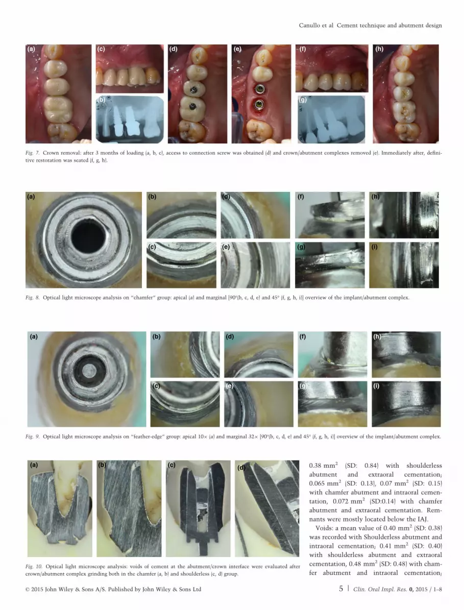
0.47 mm2 (SD: 0.43) with chamfer abutment
and extraoral cementation. Voids were mostly
located at the coronal portion of the abut-
ment/crown interface.
Gap: a mean value of 0.062 mm (SD: 0.033)
with Shourlderless abutment and intraoral
cementation; 0.064 mm (SD: 0.034) with
chamfer abutment and intraoral cementation;
0.055 mm (SD: 0.016) with shoulderless abut-
ment and extraoral cementation; 0.054 mm
(SD: 0.024) with chamfer abutment and extra-
oral cementation groups respectively.
Subgroup data were summarized in
Tables 1 and 2.
Related-samples Friedman’s two-way
analysis of variance by ranks showed signifi-
cant differences for only remnants and voids
measure (Table 3).
Pairwise comparisons (post hoc tests)
showed that “chamfer abutment with intra-
oral cementation” presented significantly
more “remnants” versus “shoulderless abut-
ment with extraoral cementation” (0.047). At
the same time, “chamfer abutment with ex-
traoral cementation” presented significantly
wider “voids” compared with “shoulderless
abutment with intraoral cementation”
(0.032).
Prosthetic complications: in the subgroup,
“extraoral cementation” 2 crowns decement-
ed after 1 week. The crowns were cemented
again with the same technique. No further
decementation was recorded.
Discussion
The principal aim of the study was to test
the efficacy of an “extraoral” cementation
technique in reducing the amount of cement
remnants in the peri-implant soft tissues. In
respect to the original technique proposed by
Wadhwani et al. (2009), in the present study,
the quality of the abutment replica was
improved using a polyurethane resin accord-
ing to the specifications of the “Abutment
duplication technique” (Cocchetto et al.
2010). The precision of the replica allowed to
further reduce the film of cement effectively
left inside the crown immediately before
cementation.
Analyzing the results, a surprisingly low
amount of cement remnants was found in all
subgroups (overall mean value of 0.25 mm2),
compared with what was expected, consider-
ing the data available in literature on the
topic so far. On the other hand, data col-
lected in the present study were in agreement
with a recently published study by Behr et al.
(2014), which demonstrated almost complete
removal of ZOE cement.
It must be highlighted that the cement rem-
nants were located only on the abutment/
crown complex and no cement was found in
the surrounding soft tissues at visual inspec-
tion at time of abutment–crown unscrewing.
This fact has been interpreted by the authors
as the consequence of two main factors: the
use of a eugenol-free zinc oxide cement and
the particularly accurate removal technique.
In fact, Zinc oxide cement is considered a
“temporary cement” for both implant- and
tooth-supported prosthesis, but in this study,
it has been chosen in alternative to “defini-
tive cements” like glass-ionomers and resin
cements used in most studies (Linkevicius
et al. 2013a,b; Vindasiute et al. 2013; Korsch
et al. 2014).
It must be pointed out that, while poly-
meric chains of resin cements were supposed
to be toxic for soft and hard tissues (Korsch
et al. 2014), the ability to reduce biofilm
growth by eugenol-free zinc oxide cement
was demonstrated by Raval et al. (2014).
Remnants of this luting cements, however,
seem to represent retentive factor impacting
on the composition of the submucosal micro-
bial biofilm, which could represent the
first step for a mucositis or even a peri-
implantitis.
Eugenol-free zinc oxide cement, in fact,
presents some advantages like radiological
detectability even in thickness of 1 mm
(Wadhwani et al. 2010) and solubility in the
oral fluids . This last feature is mostly con-
sidered a disadvantage from the biomechani-
cal standpoint (Vindasiute et al. 2013) and
has limited its use as a primary choice for
cemented implant prostheses for its supposed
unpredictable retaining capacity. However,
this negative evaluation should be reconsid-
ered. In fact, the retention of an implant-sup-
ported crown is depending on several factors:
(i) the length and surface of the abutment, (ii)
the convergence angle of axial walls, (iii) the
roughness of the abutment surface and (iv)
the cement characteristics. As demonstrated
by Schiessl et al. (2013) which pointed out
significant interactions between abutment
geometry and luting agents, the modulation
of the first three factors can be easily
obtained during the laboratory abutment cus-
tomization procedures (adding retention cou-
lisses, reducing the convergence angle,
increasing surface roughness through partial
Table 1. Mean values and SD
Cement remnants (mm2) Voids (mm2) Gap (mm)
Shoulderless abutmentIntraoral Cementation
0.455 (SD:0.80) 0.404 (SD:0.377) 0.062 (SD:0.033)
Chamfer abutmentIntraoral Cementation
0.380 (SD:0.84) 0.413 (SD:0.39) 0.064 (SD:0.035)
Shoulderless abutmentExtraoral Cementation
0.065 (SD:0.13) 0.485 (SD:0.48) 0.055 (SD:0.016)
Chamfer abutmentExtraoral Cementation
0.072 (SD:0.14) 0.477 (SD:0.43) 0.054 (SD:0.024)
Table 2. (a) Descriptive measures of shoulder-less abutment with intraoral cementation; (b)Descriptive measures of shoulderless abutmentwith extraoral cementation; (c) Descriptivemeasures of chamfer abutment with intraoralcementation; (d) Descriptive measures of cham-fer abutment with extraoral cementation
Group Remnants Voids Gap
(a)Shoulderless abutment Intraoral Cementation
Mean 0.455 0.404 0.062N 21 21 21SD 0.800 0.377 0.033Median 0 0.200 0.060Minimum 0 0 0.04Maximum 2.98 0.90 0.18
(b)Shoulderless abutment Extraoral Cementation
Mean 0.065 0.485 0.055N 21 21 21SD 0.130 0.480 0.016Median 0.020 0.900 0.050Minimum 0 0 0.04Maximum 0.59 0.90 0.20
(c)Chamfer abutment Intraoral Cementation
Mean 0.380 0.413 0.064N 20 20 20SD 0.840 0.390 0.035Median 0 0.500 0.050Minimum 0 0 0.040Maximum 2.860 1.000 0.130
(d)Chamfer abutment Extraoral Cementation
Mean 0.072 0.477 0.054N 21 21 21SD 0.140 0.430 0.024Median 0.020 0.900 0.040Minimum 0 0.02 0.03Maximum 0.59 1.10 0.18
Table 3. Related-samples Friedman’s two-wayanalysis of variance by ranks
Remnants Voids Gap
Test statistic 13.446 11.180 5.607Degrees ofFreedom
3 3 3
AsymptoticSig.(2 sided test)
0.004** 0.11** 0.132
**Statistically significant.
6 | Clin. Oral Impl. Res. 0, 2015 / 1–8 © 2015 John Wiley & Sons A/S. Published by John Wiley & Sons Ltd
Canullo et al �Cement technique and abutment design
sandblasting). This could compensate the
lack of intrinsic retention capacity of zinc
oxide cement, although difference between
eugenol-free zinc oxide cement and resin
cement still remain controversial (Garg et al.
2014; R€odiger et al. 2014). Following these
principles, abutments used in the present
study have been designed with a convergency
angle of axial walls ranging from 8° to 12°
and only two crowns decemented after
1 week which were recemented with no fur-
ther complication.
The second factor, which may explain the
study results, is the cement removal tech-
nique. This was obtained firstly applying,
using a small brush, a layer of liquid vase-
line on the outer margin of the crowns, lim-
iting therefore the adherence of the cement.
Then after careful removal of excess cement
with explorer and ultrasonic plastic tips, a
dental floss with a spongy section impreg-
nated with zinc oxide solvent was used to
polish the crown/abutment interface. The
entire procedure was done with the aid of a
49 magnification optic system. The combi-
nation of these two factors (type of cement
and accurate cement cleaning) seemed suffi-
cient to remove almost all the remnants
and prevent any peri-implant soft tissue
complication.
In the present study, the margin location
for both abutment’s design was chosen at 1–
1.5 mm below the soft tissue margin. Despite
the fact that in literature, there is a consen-
sus indicating ideal margin position to be
placed at or even above the gingival margin
(Linkevicius et al. 2013a,b) to avoid the
excess peri-implant cement, it was deliber-
ately decided to select the 1.5 mm subgingi-
val position to actually create an “average”
environment reflecting more or less a real
“everyday” clinical setting in which to test
the study hypotheses.
For the same reason, it was considered that
the crown profiles should reproduce the natu-
ral anatomic contours of the teeth to be
replaced. Thus, the amount of undercuts,
which also has been correlated with an
increased difficulty of excess cement removal
(Vindasiute et al. 2013), was deliberately not
modified.
Focusing on the role of abutment morphol-
ogy, in the present study, two different
designs were analyzed:
1. a “traditional” chamfer abutment pro-
duced through CAD CAM technology by
the laboratory technician with a 1.5 mm
finishing line below the gingival margin,
therefore following the gingival architec-
ture. This was considered more in accor-
dance with clinical practice compared
with the use of standard abutments with
predetermined chamfer finish line used
in several studies.
2. a feather-edge abutment (shoulderless
and marginless are two other possible
definitions). This design is not new and
has been proposed by authors as Carl
Misch already in 1993. Recently, the idea
was mentioned by Loi & Di Felice (2013),
which presented a prosthetic protocol for
tooth-supported restorations including a
feather-edge preparation and a manage-
ment of gingival architecture through the
crown’s contour and profile. The authors
claim that improved esthetic results can
be obtained in a predictable manner, both
in tooth-supported and implant-supported
restorations. The key factors would be
the better marginal adaptation after
crown’s cementation with a minimal mi-
crogap and the thickening of soft tissue
due to increased space available for the
connective tissue when a conical geome-
try of the abutment is implemented. The
concept has gained popularity but has
raised controversies. In particular, the
possibility of an increased risk of cement
remnants in the peri-implant sulcus due
to an obvious grater undercut.
Results from the present study have not
confirmed this risk: the shoulderless design,
together with a careful cement application
and removal protocol, presents the same
amount of intrasulcular cement remnants
compared with traditional design. Addition-
ally, it must be observed that the present
study was conducted in posterior quadrants
where the undercuts are greater due to the
increased difference between the implant
platform diameter and the crown’s diame-
ter, particularly in the molar area (Vindasi-
ute et al. 2013). Moreover, there is an
increased difficulty for cement removal due
to limited access, compared with the
esthetic zone.
In these clinical cases, the extraoral
cementation technique was demonstrated to
significatively minimize the already limited
cement remnants amount found after tradi-
tional cementation technique.
Due to this limitation, the same study
design should be tested in the anterior areas,
where crown overcuts are more limited.
Regarding voids, significant differences
between extraoral and intraoral subgroups
could explain the two decementations hap-
pened in extraoral groups.
Regarding gap parameters, no statistically
significant difference was found within
the groups and subgroups, confirming the
absence of clinical differences between the
presented abutment designs and cementation
techniques.
Clinical limitations of the present study
were represented by the small follow-up
time. A longer follow-up (5 years at least)
could have prospectively demonstrated the
soft tissue reaction to eugenol-free zinc oxide
cement remnants.
Conclusions
Within the limitation of the present study,
the following conclusions can be summa-
rized:
1. Extraoral cementation could minimize
the presence of cement remnants. How-
ever, from the very limited amount of
cement remnants observed also in the in-
traoral group, a very low possibility of a
cement-induced peri-implant pathology
may be inferred. These results could
probably be associated with the use of a
eugenol-free zinc oxide cement and a par-
ticularly accurate cement removal proto-
col.
2. Compared with the abutment design,
chamfer cemented with an intraoral tech-
nique presented significant higher
amount of cement remnants if compared
to a shoulderless abutment when extra-
oral cementation technique is adopted.
3. The presence of significantly wider
“voids” was noted in the extraoral group.
4. There is no difference in regard to the
“gap” parameters between the two
abutment designs and the cementation
technique
Acknowledgements: The authors
highly appreciated the skills and commitment
of Dr Audrenn Gautier in the supervision of
the study, Dr Giuliano Iannello (Rome, Italy)
and Sandro Radovanovic (Belgrade, Serbia) for
their statistical support. Additionally authors
deeply thank Eng. Marco Cesarotto and
Carmine Magna for the professional support
in microremnants analysis.
Conflict of interest and sources offunding
The authors declare that they have no con-
flict of interests. The study was self-funded.
© 2015 John Wiley & Sons A/S. Published by John Wiley & Sons Ltd 7 | Clin. Oral Impl. Res. 0, 2015 / 1–8
Canullo et al �Cement technique and abutment design
References
Behr, M., Spitzer, A., Preis, V., Weng, D., Gosau,
M. & Rosentritt, M. (2014) The extent of luting
agent remnants on titanium and zirconia abut-
ment analogs after scaling. International Journal
of Oral Maxillofacial Implants. 29: 1185–1192.
Br€agger, U., Karoussis, I., Persson, R., Pjetursson,
B., Salvi, G. & Lang, N. (2005) Technical and bio-
logical complications/failures with single crowns
and fixed partial dentures on implants: a 10-year
prospective cohort study. Clinical Oral Implants
Research 16: 326–334.
Chaar, M.S., Att, W. & Strub, J.R. (2011) Prosthetic
outcome of cement-retained implant-supported
fixed dental restorations: a systematic review.
Journal of Oral Rehabilitation 38: 697–711.
Chee, W.W., Duncan, J., Afshar, M. & Moshaveri-
nia, A. (2013) Evaluation of the amount of excess
cement around the margins of cement-retained
dental implant restorations: the effect of the
cement application method. Journal of Prosthetic
Dentistry 109: 216–221.
Cocchetto, R., Resch, I., Castagna, M., Vincenzi, G.
& Celletti, R. (2010) The abutment duplication
technique: a novel protocol for cementable
implant-supported restorations. International
Journal of Periodontics Restorative Dentistry 30:
415–424.
Garg, P., Pujari, M., Prithviraj, D.R. & Khare, S.
(2014) Retentiveness of various luting agents used
with implant-supported prosthesis: an in vitro
study. Journal of Oral Implantology 40: 649–654.
Korsch, M., Obst, U. & Walther, W. (2014) Cement-
associated peri- implantitis: a retrospective clini-
cal observational study of fixed implant-supported
restorations using a methacrylate cement. Clini-
cal Oral Implant Research. [Epub ahead of print]
Linkevicius, T., Puisys, A., Vindasiute, E., Linke-
viciene, L. & Apse, P. (2013a) Does residual
cement around implant-supported restorations
cause peri-implant disease? A retrospective case
analysis. Clinical Oral Implant Researchs 24:
1179–1184.
Linkevicius, T., Vindasiute, E., Puisys, A., Linke-
viciene, L., Maslova, N. & Puriene, A. (2013b)
The influence of the cementation margin position
on the amount of undetected cement. A prospec-
tive clinical study. Clinical Oral Implant Rese-
archs 24: 71–76.
Linkevicius, T., Vindasiute, E., Puisys, A. & Peciu-
liene, V. (2011) The influence of margin location
on the amount of undetected cement excess after
delivery of cement-retained implant restorations.
Clinical Oral Implant Researchs 22: 1379–1384.
Loi, I. & Di Felice, A. (2013) Biologically oriented
preparation technique (BOPT): a new approach for
prosthetic restoration of periodontically healthy
teeth. European Journal of Esthetic Dentistry 8:
10–23.
Michalakis, K.X., Hirayama, H. & Garefis, P.D.
(2003) Cement-retained versus screw-retained
implant restorations: a critical review. Interna-
tional Journal of Oral Maxillofacial Implants. 18:
719–728.
Misch, C.E. (1993) Progressive loading of bone with
implant prostheses. Journal of Dental Symposium
1: 50–53.
Raval, N.C., Wadhwani, C.P., Jain, S. & Darveau,
R.P. (2014) The interaction of implant luting
cements and oral bacteria linked to peri-implant
disease: an in vitro analysis of planktonic and
biofilm growth - a preliminary study. Clinical
Implant Dentistry & Related Research, 6: doi:
10.1111/cid.12235. [Epub ahead of print]
R€odiger, M., Rinke, S., Ehret-Kleinau, F., Pohlmey-
er, F., Lange, K., B€urgers, R. & Gersdorff, N.
(2014) Evaluation of removal forces of implant-
supported zirconia copings depending on abut-
ment geometry, luting agent and cleaning method
during re-cementation. Journal of Advanced Pros-
thodontics 6: 233–240.
Sailer, I., M€uhlemann, S., Zwahlen, M., H€ammerle,
C.H. & Schneider, D. (2012) Cemented and
screw-retained implant reconstructions: a system-
atic review of the survival and complication
rates. Clinical Oral Implant Researchs 23(Suppl.):
163–201.
Schiessl, C., Schaefer, L., Winter, C., Fuerst, J.,
Rosentritt, M., Zeman, F. & Behr, M. (2013)
Factors determining the retentiveness of luting
agents used with metal- and ceramic-based
implant components. Clinical Oral Investiga-
tions. 17: 1179–1190.
Torrado, E., Ercoli, C., Al Mardini, M., Graser,
G.N., Tallents, R.H. & Cordaro, L. (2004) A com-
parison of the porcelain fracture resistance of
screw-retained and cement-retained implant-sup-
ported metal-ceramic crowns. Journal of Prosthet-
etic Dentistry 91: 532–537.
Vindasiute, E., Puisys, A., Maslova, N., Linkevici-
ene, L., Peciuliene, V. & Linkevicius, T. (2013)
Clinical factors influencing removal of the
cement excess in implant-supported restorations.
Clinical Implant Dentistry & Related Research.
[Epub ahead of print].
Wadhwani, C., Hess, T., Faber, T., Pineyro, A. &
Chen, C.S. (2010) A descriptive study of the
radio- graphic density of implant restorative
cements. Journal of Prosthetic Dentistry 103:
295–302.
Wadhwani, C. & Pineyro, A. (2009) Technique for
controlling the cementon an implant crown. Jour-
nal of Prosthetic Dentistry 102: 57–58.
Wilson, T.G., Jr. (2009) The positive relationship
between excess cement and peri-implant disease:
a prospective clinical endoscopic study. Journal of
Periodontology 80: 1388–1392.
Wittneben, J.G., Millen, C. & Br€agger, U. (2014)
Clinical performance of screw- versus cement-
retained fixed implant-supported reconstructions–
a systematic review. International Journal of
Oral Maxillofacial Implants 29(Suppl): 84–98.
Supporting Information
Additional Supporting Information may be
found in the online version of this article:
Table S1. CONSORT Checklist of Items to
Include When Reporting a Randomized Trial.
8 | Clin. Oral Impl. Res. 0, 2015 / 1–8 © 2015 John Wiley & Sons A/S. Published by John Wiley & Sons Ltd
Canullo et al �Cement technique and abutment design