Behavior Genetics, Vol. 27, No. 6, 1997 Corticotropin-Releasing Hormone Modulation of a Conditioned Stress Response in the Central Amygdala of Roman High (RHA/Verh)-Avoidance and Low (RLA/Verh)-Avoidance Rats A. Wiersma, 1,3 S. Knollema, 2 J. P. Konsman, 1,2 B. Bohus, 1 and J. M. Koolhaas 1 Roman high (RHA/Verh)- and low (RLA/Verh)-avoidance rats are selected and bred for rapid versus nonacquisition of two-way, active avoidance behavior in the shuttle box. RHA/Verh rats generally show a more active coping style than do their RLA/Verh coun- terparts when exposed to various environmental challenges. The central nucleus of the amygdala (CeA) is known to be involved in the regulation of autonomic, neuroendocrine, and behavioral responses to stress. Its involvement in the selection of coping strategies has also been suggested. Corticotropin-releasing hormone (CRH) seems to be one of the key neurohormones in the control of CeA output. Neuroanatomical studies have revealed that the majority of CRH fibers from the CeA have direct connections with autonomic regu- latory nuclei in the brain-stem, e.g. lateral parabrachial nucleus (1PB). The effects of CRH (30 ng) on modulating CeA activity were studied by infusion of CRH into the CeA during conditioned stress (inescapable foot-shocks) in RHA/Verh and RLA/Verh male rats. Heart- rate responses after CRH treatment were not changed in either line. However, distinctly different behavioral responses were seen after CRH infusion into the CeA of both rat lines. A decrease in immobility responses was seen in both RHA/Verh and RLA/Verh rats, while an increase in exploration was observed in RHA/Verh rats only in the conditioned stress situation. Rearing levels were increased in the RHA/Verh rats, whereas they were decreased in the RLA/Verh animals. As a result of CRH infusion, the number of FOS immunoreactive cells in the 1PB of RLA/Verh rats was decreased, whereas an opposite response was found in RHA/Verh rats. These results indicate that the CRH system of the CeA connected with output brain-stem areas is differentially involved in the cardiovascular and behavioral re- sponses of these rats having different coping styles. KEY WORDS: Corticotropin-releasing hormone; conditioned stress response; Roman high-avoid- ance rats; Roman low-avoidance rats; central amygdala. INTRODUCTION Corticotropin-releasing hormone (CRH) is a neu- ropeptide that has been widely associated with sev- 1 Department of Animal Physiology, Center for Behavioral and Cognitive Neurosciences, University of Groningen, P.O. Box 14, 9750 AA Haren, The Netherlands. 2 Department of Biological Psychiatry, Center for Behavioral and Cognitive Neurosciences, University of Groningen, P.O. Box 14, 9750 AA Haren, The Netherlands. 3 To whom correspondence should be addressed at AKZO NOBEL, N.V. Organon, Department of Endocrinology, Room RE 2211, P.O. Box 20, 5340 BH Oss, The Netherlands. Tele- phone: 31-412-661954. Fax: 31-412-662542. e-mail: a.wiersma@ organon.oss.akzonobel.nl and [email protected]. eral behavioral and physiological aspects of stress (Dunn and Berridge, 1990; Owens and Nemeroff, 1991). In addition to its activating effects on the hypothalamus-pituitary-adrenal axis, intracere- broventricular (icv) injection of CRH produces elevations of heart rate and blood pressure, rises in plasma catecholamine and glucose levels (Bakke et al., 1990; Brown and Fisher, 1985; Korte et al., 1993; Kurosawa et al., 1986), and behavioral changes mimicking stress responses (Britton et al., 1982; Diamant and DeWied, 1991; Korte et al., 1993; Sherman and Kalin, 1988; Sparado et al., 547 0001-8244/97/1100-0547$12.50/0 C 1997 Plenum Publishing Corporation
Transcript
Behavior Genetics, Vol. 27, No. 6, 1997
Corticotropin-Releasing Hormone Modulation of aConditioned Stress Response in the Central Amygdala ofRoman High (RHA/Verh)-Avoidance and Low(RLA/Verh)-Avoidance RatsA. Wiersma,1,3 S. Knollema,2 J. P. Konsman,1,2 B. Bohus,1 and J. M. Koolhaas1
Roman high (RHA/Verh)- and low (RLA/Verh)-avoidance rats are selected and bred forrapid versus nonacquisition of two-way, active avoidance behavior in the shuttle box.RHA/Verh rats generally show a more active coping style than do their RLA/Verh coun-terparts when exposed to various environmental challenges. The central nucleus of theamygdala (CeA) is known to be involved in the regulation of autonomic, neuroendocrine,and behavioral responses to stress. Its involvement in the selection of coping strategies hasalso been suggested. Corticotropin-releasing hormone (CRH) seems to be one of the keyneurohormones in the control of CeA output. Neuroanatomical studies have revealed thatthe majority of CRH fibers from the CeA have direct connections with autonomic regu-latory nuclei in the brain-stem, e.g. lateral parabrachial nucleus (1PB). The effects of CRH(30 ng) on modulating CeA activity were studied by infusion of CRH into the CeA duringconditioned stress (inescapable foot-shocks) in RHA/Verh and RLA/Verh male rats. Heart-rate responses after CRH treatment were not changed in either line. However, distinctlydifferent behavioral responses were seen after CRH infusion into the CeA of both rat lines.A decrease in immobility responses was seen in both RHA/Verh and RLA/Verh rats, whilean increase in exploration was observed in RHA/Verh rats only in the conditioned stresssituation. Rearing levels were increased in the RHA/Verh rats, whereas they were decreasedin the RLA/Verh animals. As a result of CRH infusion, the number of FOS immunoreactivecells in the 1PB of RLA/Verh rats was decreased, whereas an opposite response was foundin RHA/Verh rats. These results indicate that the CRH system of the CeA connected withoutput brain-stem areas is differentially involved in the cardiovascular and behavioral re-sponses of these rats having different coping styles.
KEY WORDS: Corticotropin-releasing hormone; conditioned stress response; Roman high-avoid-ance rats; Roman low-avoidance rats; central amygdala.
INTRODUCTIONCorticotropin-releasing hormone (CRH) is a neu-ropeptide that has been widely associated with sev-1 Department of Animal Physiology, Center for Behavioral and
Cognitive Neurosciences, University of Groningen, P.O. Box14, 9750 AA Haren, The Netherlands.
2 Department of Biological Psychiatry, Center for Behavioraland Cognitive Neurosciences, University of Groningen, P.O.Box 14, 9750 AA Haren, The Netherlands.
3 To whom correspondence should be addressed at AKZONOBEL, N.V. Organon, Department of Endocrinology, RoomRE 2211, P.O. Box 20, 5340 BH Oss, The Netherlands. Tele-phone: 31-412-661954. Fax: 31-412-662542. e-mail:[email protected] and [email protected].
eral behavioral and physiological aspects of stress(Dunn and Berridge, 1990; Owens and Nemeroff,1991). In addition to its activating effects on thehypothalamus-pituitary-adrenal axis, intracere-broventricular (icv) injection of CRH produceselevations of heart rate and blood pressure, rises inplasma catecholamine and glucose levels (Bakke etal., 1990; Brown and Fisher, 1985; Korte et al.,1993; Kurosawa et al., 1986), and behavioralchanges mimicking stress responses (Britton et al.,1982; Diamant and DeWied, 1991; Korte et al.,1993; Sherman and Kalin, 1988; Sparado et al.,
5470001-8244/97/1100-0547$12.50/0 C 1997 Plenum Publishing Corporation
548 Wiersma, Knollema, Konsman, Bohus, and Koolhaas
1990; Sutton et al., 1982; Veldhuis and DeWied,1984). The central nucleus of the amygdala (CeA)has been implicated as an important locus amongthe brain sites responsible for the behavioral andcardiovascular effects of icv CRH. Administrationof CRH icv results in FOS labeling in the CeA(Andreae and Herbert, 1993). Microinfusion of alow dose of CRH (30 ng) into the CeA of maleWistar rats under stress-free conditions producesbehavioral activation and a long-lasting tachycardiawithout any rise in plasma catecholamines(Wiersma et al., 1993). It has been suggested thatthis tachycardia is due to an inhibited cardiac para-sympathetic output (Fisher, 1988; Fisher andBrown, 1991) exerted via the CeA (Wiersma et al.,1993, 1997b). The CRH-induced effects after in-fusion into the CeA are restricted to this nucleus,as local application of CRH into the basolateral nu-cleus of the amygdala did not result in any acti-vation of behavioral, cardiovascular, andneuroendocrine responses (Wiersma et al., 1997b).In a stress situation, infusion of a low dose of CRH(30 ng) into the CeA of male Wistar rats increasedactive behavior in the conditioned defensive bury-ing paradigm (Wiersma et al., 1996), whereas cen-tral blockade of its receptors by a CRH receptorantagonist (A-hCRH9-41) inhibited this response(Korte et al., 1994). It has been suggested that im-mobility and burying behavior in the defensive bur-ying test represent passive and active copingstrategies, respectively, in response to the fearfulsituation (Korte et al., 1993).
Several studies have shown that individual an-imals or rat lines may differ in preferred copingstyle, for example, Roman High-Avoidance(RHA/Verh) and Roman Low-Avoidance rats(RLA/Verh) (Bohus et al., 1987; Ferre et al., 1995;Koolhaas and Bohus, 1991; Steimer et al., 1997).The Roman rat lines, originally selected for theirshuttle-box performance, also show differences inmany other behavioral paradigms (Aubry et al.,1995; Castanon et al., 1994; Castanon and Mor-mede, 1994; Driscoll and Battig, 1982; Driscoll etal., 1990; Ferre et al., 1995; Meerlo et al., 1997;Roozendaal et al., 1992; Willig et al., 1991). TheRHA/Verh line displays an active coping response,characterized by high behavioral activity, such asexploring and rearing, accompanied by a slighttachycardia (Bohus et al., 1987; Koolhaas, 1994;Koolhaas and Bohus, 1991; Roozendaal et al.,1992, 1993). In contrast, RLA/Verh rats show a
more passive coping response by typically display-ing behavioral immobility and self-grooming ac-companied by either bradycardia (Bohus et al.,1987; Koolhaas, 1994; Koolhaas and Bohus, 1991;Roozendaal et al., 1993) or pronounced tachycardia(D'Angio et al., 1988) depending on the stressfulsituation.
As CRH connections from the CeA to thebrain-stem areas seem to be involved in active cop-ing responses in Wistar rats (Wiersma et al., 1996),one may predict a differential modulation of theCeA output by CRH in rats which display differentcoping styles. Indeed, CRH microinfusion into theCeA of RHA/Verh and RLA/Verh rats under stress-free conditions resulted in distinct behavioral andcardiovascular responses, accompanied by neu-roanatomical modulation of FOS and/or CRHmRNA positive cells in different brain areas(Wiersma et al., 1997a). RHA/Verh rats respondedwith a distinct tachycardia accompanied by behav-ioral activation, while RLA/Verh rats showedhardly any behavioral or cardiovascular response inthe stress-free situation. FOS immunocytochemis-try and in situ hybridization of CRH mRNA in thesame rats showed a decrease in the amount of pos-itive cells in the CeA and the parabrachial nucleusin RLA/Verh rats after CRH microinfusion into theCeA. This is in contrast to RHA/Verh rats, inwhich an increase in FOS positive cells was ob-served together with no effect on CRH mRNA lev-els in the CeA.
The present experiments were designed to in-vestigate to what extend the CRH modulation inthe CeA is involved and how the CRH-CeA systemis fine-tuned in animals using either active or pas-sive coping styles under stressful conditions, inwhich the CeA is expected to be active. Therefore,CRH was applied locally to the CeA of maleRLA/Verh and RHA/Verh rats, prior to the reten-tion test in a conditioned emotional stress paradigmof inescapable foot-shocks, and behavioral andphysiological responses were investigated.
MATERIALS AND METHODS
Animals
Twenty RHA/Verh and 20 RLA/Verh (Wistar-derived) male rats, weighing 280-370 g at the be-ginning of the experiment, were used. Animalswere obtained from the breeding colony at the An-
CRH Modulation of a Conditioned Stress Response 549
imal Science Institute (Zurich, Switzerland) at theage of 5—8 weeks. The rats were housed in groupsof five animals per cage and left undisturbed untilthe age of 14 weeks in a temperature-controlledroom (20 ± 1 D C), with a 12-h light-dark cycle(lights on from 0800 to 2000). Three days beforesurgery, the animals were housed individually inperspex cages (25 X 25 X 30 cm) in which foodand water were available ad libitum. The experi-ments were performed during the light period ofthe cycle (between 0900 and 1300).
Apparatus
A step-through passive avoidance apparatus(Ader et al., 1972) was used in the stress experi-ments to investigate emotional, stress-related be-havior and cardiac responses. The apparatusconsisted of a dark compartment (40 X 40 X 40cm), made of black Plexiglas, connected via a smallopening to an elevated, well-lit platform. A slidingdoor prevented access between the platform and thedark compartment. The floor of the dark compart-ment was made of stainless-steel bars throughwhich scrambled electric foot-shocks could be de-livered. A waiting cage (25 X 25 X 30 cm) witha sawdust-covered floor was placed close to thepassive avoidance apparatus in a sound- andlight-attenuated experimental room.
Surgery
Each experimental animal was provided withbilateral, permanent stainless-steel brain cannulae(outer diameter, 0.3 mm; inner diameter, 0.15 mm)for drug infusion, aimed just above the central nu-cleus of the amygdala (coordinates: 6.6 mm rostralto interaural, lateral 4.0 mm to the midline, andventral 6.3 mm below the dura), according to Pax-inos and Watson (1982). The brain cannulae werepermanently fixed to the skull by means of stain-less-steel screws and dental cement. In order to rec-ord the electrocardiogram (ECG), steel electrodesmade of standard paperclips were implanted tran-scutaneously, one between the scapulae and theother in the middle of the back (Bohus, 1974). Theanimals were kept under halothane anesthesia incombination with N2O and O2 (ratio, 2:1) duringthe entire surgical procedure.
Drug Treatment
Synthetic rat/human CRH (CRF; SigmaChemical Co., St. Louis, MO) was dissolved in ar-tificial cerebrospinal fluid (aCSF) containing ascor-bic acid (100 ug/ml). CRH was administeredbilaterally at a dose of 30 ng/rat per cannula, basedupon the results of previous experiments (Wiersmaet al., 1993, 1995). The vehicle was sterile artificialcerebrospinal fluid (aCSF) containing 127.64 mMNaCl, 2.55 mM KCl, 1.26 mM CaCl2 • 2H2O, and0.93 mM MgCl2 • 6H2O. All compounds were in-fused in a total volume of 1 ul per brain cannuladuring a 7-min period.
ECG Recording and Analysis
The ECG of freely moving rats was monitoredtelemetrically by means of a miniature FM trans-mitter (EDB-ROY, HAREN, The Netherlands) asdescribed earlier (Wiersma et al., 1993). The trans-mitter was connected to the transcutaneous elec-trodes and secured around the chest of the rat bymeans of a strap. The transmitted signals were re-ceived on a commercial FM receiver and stored ontape. For off-line analysis, the recorded ECG sam-ples were processed through a cardiotachometerpulse generator which generated square-wavepulses at each R peak of the PQRST-ECG com-plex. The time between the onset of two consecu-tive pulses, the interbeat interval (IBI), wasautomatically measured within the range of 100 to220 ms and IBIs were recalculated as beats perminute.
Behavioral Measurements
Behavior was recorded on the basis of the fol-lowing criteria.
• Resting/sleeping: Inactive with eyes openor closed
• Immobility: Completely motionless, ab-sence of skeletal and vibrissae movementsexcept those associated with respiration
• Exploring: Locomotor activity to investi-gate any part of the home cage
• Self-grooming: Wiping the fur with fore-paws and tongue (washing)
• Rearing: Sniffing in the air with forepawson the floor
550 Wiersma, Knollema, Konsman, Bohus, and Koolhaas
• Sniffing: Sniffing in the air with forepawson the floor.
All behavioral elements were recorded bymeans of a keyboard operated microprocessor(EDB, Haren, The Netherlands). The duration andthe frequency of these elements were computed andexpressed as a percentage of the total observationperiod.
Experimental Design
After moving the rat to the experimentalroom, the holding-strap and the transmitter werefixed around the chest of the rat. Three preshocksessions were undertaken, each of which wasstarted by placing the experimental animal into thewaiting cage for 1 min. Only during the first ses-sion, the animal was placed directly into the darkcompartment of the avoidance apparatus for 5 minto allow habituation to the dark compartment. Im-mediately afterward, a training session was givenduring which the rat was placed on the end of theilluminated platform and allowed to enter the darkcompartment. After the animal had entered the darkcompartment, the sliding door was closed and therats stayed another 5 min in the dark. This proce-dure was repeated during a second training sessionon the same day during which cardiac and behav-ioral responses were recorded. On the morning ofday 2, the rat was placed directly from the waitingcage into the dark compartment and both heart-rateand behavior were recorded during 5 min (preshockmeasurement). During the morning of day 3, theanimal was placed on the illuminated platform andreceived an unavoidable aversive stimulus, i.e.,scrambled foot-shocks (0.6 mA, AC for 3 s) im-mediately after entering the dark compartment,while the sliding-door was closed. The rat was re-moved from the dark compartment 50-60 s aftertermination of the foot-shocks.
On day 4, 24 h after the unavoidable shocks,rats received either CRH (30 ng/ul during a 7-minperiod) or vehicle treatment under stress-free con-ditions in their home-cage (Wiersma et al., 1997a).Ten minutes after the start of the infusion, the ratswere transported from their home-cage to the wait-ing cage in the experimental room. After 1 min inthe waiting cage, the animals were transferred di-rectly to the dark compartment for 5 min, while thesliding door was shut to prevent escape (forced ex-posure). No further foot-shocks were administered
during this period. The cardiac and behavioral con-sequences of the emotional stress of learned fear ofthe aversive stimulus were registered.
Histology
Two hours after the start of the infusion, theanimals were deeply anesthetized with sodium pen-tobarbital (90 mg/kg i.p.) and perfused intracardi-ally with cold saline followed by 400 ml of 4%paraformaldehyde, 3.8% borax solution (pH 9.5).The brains were removed and postfixed in the samefixative for at least 2 h. Subsequently, the brainswere dehydrated in a 30% sucrose in 0.1 M phos-phate buffer (PB) for at least 48 h. Frozen sectionsof 20 um were cut and cannula placement was de-termined on unstained sections. Sections werestored in cryoprotectant solution at — 20dgC untilprocessing for FOS immunocytochemistry. Thecryoprotectant solution consisted of 0.5 L of pol-yethylene glycol and 300 g sucrose dissolved in thesame volume of 0.2 M phosphate buffer.
Immunohistochemistry
Every third section was stained for FOS-likeimmunoreactivity by the avidin-biotin-peroxidasetechnique. Prior to the first antibody incubation,sections were immersed for 10 min in 0.3% H2O2
in 0.01 M phosphate-buffered saline (PBS) to ex-haust endogenous peroxidase activity. The sectionswere then incubated for 1 h at room temperature(RT) in 5% normal rabbit serum (NRS) to suppressnonspecific antibody binding. The sections werewashed twice for 10 min in PBS, then incubatedfor 48 h with the primary antibody, a sheep-anti-FOS antibody solution (1:2000; Cambridge Re-search Biochemicals, UK) in PBS and 0.3%Triton-X at 4°C. This antibody has been raisedagainst the first 16 amino acids derived from a con-served region of both the mouse and the humanFOS-protein. After first antibody incubation, thesections were rinsed twice in PBS and incubatedfor 1 h in rabbit anti-sheep antibody (Pearce rabbitanti-sheep IgG), biotin conjugated, 1:800 in PBSand 0.3% Triton-X, and washed again, followed by1-h incubation in avidin-biotin complex (Vectas-tain ABC kit 0.4% in PBS). This procedure wasrepeated once more before the sections werestained by the reaction product of diaminobenzi-dine tetrahydrachloride (DAB) and hydrogen per-
CRH Modulation of a Conditioned Stress Response 551
oxide H2O2 (respectively, 1 mg/ml and 0.0075%)in a nickel ammonium sulfate solution (50 g/L 0.2M sodium acetate buffer, pH = 6.0). Stained sec-tions were mounted, dried overnight, dehydrated,cleared in xylene, and coverslipped with DPXmountant (BDH-Pool, UK).
Quantification
FOS-positive cells in the lateral parabrachialareas (1PB) were counted using a light microscope,alternating sections from each rostrocaudal levelbeing analyzed. Cells were selected to be FOS-pos-itive on the basis of their grain densities. The num-ber of FOS-positive cells in the 1PB was countedon sections at the levels —0.8, —0.68, —0.3,—0.16, and +0.2 mm from intraaural. A minimumof five sections was counted for each animal.
Statistics
The results are presented as means ± SE.Cardiac and behavioral data were evaluated usinganalysis of variance with repeated measures(ANOVA), followed by the Mann-Whitney U test.The immunocytochemistry data were analyzed byStudent's t test. A probability level of p < 0.05was taken as significant for all tests.
RESULTS
Histology
Following histological examination, 5 of the40 animals had to be excluded from further analysisbecause of improper bilateral cannula placement.The cannula tips had to be localized just above orentering the dorsal edge of the CeA.
Cardiac Responses
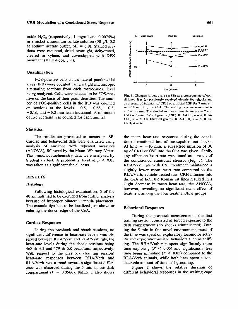
During the preshock and shock sessions, nosignificant difference in heart-rate levels was ob-served between RHA/Verh and RLA/Verh rats, theheart-rate levels during the shock sessions being468 ± 6.3 and 479 ± 5.0 beats/min, respectively.With respect to the preshock (training session)heart-rate responses between RHA/Verh andRLA/Verh rats, a trend toward a significant differ-ence was observed during the 5 min in the darkcompartment (P = 0.0566). Figure 1 also shows
Fig. 1. Changes in heart-rate (±SE) as a consequence of con-ditioned fear for previously received electric foot-shocks andas a result of infusion of CRH or artificial CSF for 7 min at t= —10 min into the CeA. The waiting cage measurement isat t = —1 min. The shock-box measurements are at t = 2 minand t = 5 min. Control groups (CSF): RLA-CSF, n = 8, RHA-CSF, n = 8. CRH-treated groups: RLA-CRH, n = 8; RHA-CRH, n = 6.
the mean heart-rate responses during the condi-tioned emotional test of inescapable foot-shocks.At time = —10 min, a stress-free infusion of 30ng of CRH or CSF into the CeA was given. Hardlyany effect on heart-rate was found as a result ofthe conditioned emotional stressor (Fig. 1). TheRHA/Verh rats with CSF treatment maintained aslightly lower mean heart rate compared to theRLA/Verh, vehicle-treated rats. CRH infusion intothe CeA of both the Roman rat lines resulted in aslight decrease in mean heart-rate, the ANOVA,however, revealing no significant main effect oftreatment among the four treatment/line groups.
Behavioral Responses
During the preshock measurements, the firsttraining session consisted of forced exposure to thedark compartment (no shocks administered). Dur-ing the 5 min in this novel environment, most ofthe time was spent on exploratory locomotor activ-ity and exploration-related behaviors such as sniff-ing. The RHA/Verh rats spent significantly moretime exploring (P < 0.05) and significantly lesstime being immobile (P < 0.05) compared to theRLA/Verh animals, while both lines spent a con-siderable amount of time self-grooming.
Figure 2 shows the relative duration ofdifferent behavioral responses in the waiting cage
552 Wiersma, Knollema, Konsman, Bohus, and Koolhaas
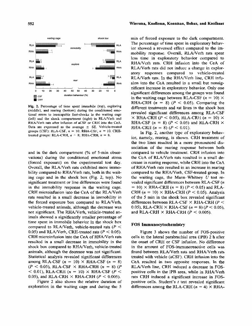
Fig. 2. Percentage of time spent immobile (top), exploring(middle), and rearing (bottom) during the conditioned emo-tional stress to inescapable foot-shocks in the waiting cage(left) and the shock compartment (right) in RLA/Verh andRHA/Verh rats after infusion of aCSF or CRH into the CeA.Data are expressed as the average ± SE. Vehicle-treatedgroups (CSF): RLA-CSF, n = 10; RHA-CSF, n = 10. CRH-treated groups: RLA-CRH, n = 8; RHA-CRH, n = 8.
and in the dark compartment (% of 5-min obser-vations) during the conditioned emotional stress(forced exposure) on the experimental test day.Overall, the RLA/Verh rats exhibited more immo-bility compared to RHA/Verh rats, both in the wait-ing cage and in the shock box (Fig. 2, top). Nosignificant treatment or line differences were foundin the immobility response in the waiting cage.CRH microinfusion into the CeA of the RLA/Verhrats resulted in a small decrease in immobility inthe forced exposure box compared to RLA/Verh,vehicle-treated animals, although the decrease wasnot significant. The RHA/Verh, vehicle-treated an-imals showed a significantly smaller percentage oftime spent in immobile behavior in the shock boxcompared to RLA/Verh, vehicle-treated rats (P <0.05) and RLA/Verh, CRH-treated rats (P < 0.05).CRH microinfusion into the CeA of RHA/Verh ratsresulted in a small decrease in immobility in theshock box compared to RHA/Verh, vehicle-treatedanimals, although the decrease was not significant.Statistical analysis revealed significant differencesamong RLA-CSF (n = 10) X RHA-CSF (n = 8)(P < 0.05), RLA-CSF X RHA-CRH (n = 8) (P< 0.01), RLA-CRH (n = 10) X RHA-CSF (P <0.05), and RLA-CRH X RHA-CRH (P < 0.005).
Figure 2 also shows the relative duration ofexploration in the waiting cage and during the 5
min of forced exposure to the dark compartment.The percentage of time spent in exploratory behav-ior showed a reversed effect compared to the im-mobility response. Overall, RLA/Verh rats spentless time in exploratory behavior compared toRHA/Verh rats. CRH infusion into the CeA ofRLA/Verh rats did not induce a change in explor-atory responses compared to vehicle-treatedRLA/Verh rats. In the RHA/Verh line, CRH infu-sion into the CeA resulted in a small but nonsig-nificant increase in exploratory behavior. Only onesignificant differences among the groups was foundin the waiting cage between RLA-CSF (n = 10) XRHA-CRH (n = 8) (P < 0.05). Comparing thedifferent treatments and rat lines in the shock boxrevealed significant differences among RLA-CSFX RHA-CRH (P < 0.05), RLA-CRH (n = 10) XRHA-CSF (n = 8) (P < 0.05) and RLA-CRH XRHA-CRH (n = 8) (P < 0.01).
In Fig. 2, another type of exploratory behav-ior, namely, rearing, is shown. CRH treatment ofthe two lines resulted in a more pronounced dis-sociation of the rearing response between bothcompared to vehicle treatment. CRH infusion intothe CeA of RLA/Verh rats resulted in a small de-crease in rearing response, while CRH into the CeAof RHA/Verh rats resulted in an increase in rearingcompared to the RHA/Verh, CSF-treated group. Inthe waiting cage, the Mann-Whitney U test re-vealed significant differences between RLA-CSF (n= 10) X RHA-CRH (n = 8) (P < 0.05) and RLA-CRH (n = 10) X RHA-CRH (P < 0.05). Analysisof the 5 min in the shock box revealed significantdifferences between RLA-CSF X RHA-CRH (P <0.05), RLA-CRH X RHA-CSF (n = 8) (P < 0.05),and RLA-CRH X RHA-CRH (P < 0.005).
FOS Immunocytochemistry
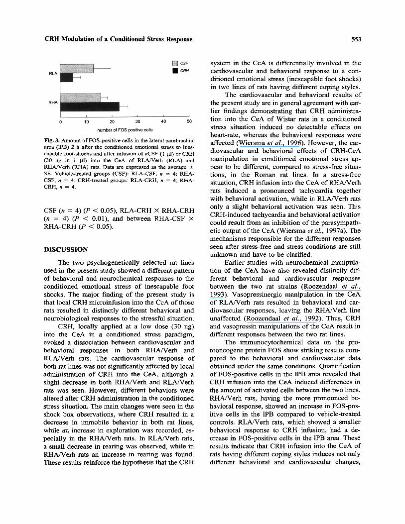
Figure 3 shows the number of FOS-positivecells in the lateral parabrachial area (1PB) 2 h afterthe onset of CRH or CSF infusion. No differencein the amount of FOS-immunoreactive cells wasfound between RLA/Verh rats and RHA/Verh ratstreated with vehicle (aCSF). CRH infusion into theCeA resulted in two opposite responses. In theRLA/Verh line, CRH induced a decrease in FOS-positive cells in the 1PB area, while in RHA/Verhrats CRH induced a significant increase in FOS-positive cells. Student's t test revealed significantdifferences among the RLA-CRH (n = 4) X RHA-
CRH Modulation of a Conditioned Stress Response 553
Fig. 3. Amount of FOS-positive cells in the lateral parabrachialarea (1PB) 2 h after the conditioned emotional stress to ines-capable foot-shocks and after infusion of aCSF (1 ul) or CRH(30 ng in 1 ul) into the CeA of RLA/Verh (RLA) andRHA/Verh (RHA) rats. Data are expressed as the average ±SE. Vehicle-treated groups (CSF): RLA-CSF, n = 4; RHA-CSF, n = 4. CRH-treated groups: RLA-CRH, n = 4; RHA-CRH, n = 4.
CSF (n = 4) (P < 0.05), RLA-CRH X RHA-CRH(n = 4) (P < 0.01), and between RHA-CSF XRHA-CRH (P < 0.05).
DISCUSSION
The two psychogenetically selected rat linesused in the present study showed a different patternof behavioral and neurochemical responses to theconditioned emotional stress of inescapable footshocks. The major finding of the present study isthat local CRH microinfusion into the CeA of thoserats resulted in distinctly different behavioral andneurobiological responses to the stressful situation.
CRH, locally applied at a low dose (30 ng)into the CeA in a conditioned stress paradigm,evoked a dissociation between cardiovascular andbehavioral responses in both RHA/Verh andRLA/Verh rats. The cardiovascular response ofboth rat lines was not significantly affected by localadministration of CRH into the CeA, although aslight decrease in both RHA/Verh and RLA/Verhrats was seen. However, different behaviors werealtered after CRH administration in the conditionedstress situation. The main changes were seen in theshock box observations, where CRH resulted in adecrease in immobile behavior in both rat lines,while an increase in exploration was recorded, es-pecially in the RHA/Verh rats. In RLA/Verh rats,a small decrease in rearing was observed, while inRHA/Verh rats an increase in rearing was found.These results reinforce the hypothesis that the CRH
system in the CeA is differentially involved in thecardiovascular and behavioral response to a con-ditioned emotional stress (inescapable foot shocks)in two lines of rats having different coping styles.
The cardiovascular and behavioral results ofthe present study are in general agreement with ear-lier findings demonstrating that CRH administra-tion into the CeA of Wistar rats in a conditionedstress situation induced no detectable effects onheart-rate, whereas the behavioral responses wereaffected (Wiersma et al., 1996). However, the car-diovascular and behavioral effects of CRH-CeAmanipulation in conditioned emotional stress ap-pear to be different, compared to stress-free situa-tions, in the Roman rat lines. In a stress-freesituation, CRH infusion into the CeA of RHA/Verhrats induced a pronounced tachycardia togetherwith behavioral activation, while in RLA/Verh ratsonly a slight behavioral activation was seen. ThisCRH-induced tachycardia and behavioral activationcould result from an inhibition of the parasympath-etic output of the CeA (Wiersma et al., 1997a). Themechanisms responsible for the different responsesseen after stress-free and stress conditions are stillunknown and have to be clarified.
Earlier studies with neurochemical manipula-tion of the CeA have also revealed distinctly dif-ferent behavioral and cardiovascular responsesbetween the two rat strains (Roozendaal et al.,1993). Vasopressinergic manipulation in the CeAof RLA/Verh rats resulted in behavioral and car-diovascular responses, leaving the RHA/Verh lineunaffected (Roozendaal et al., 1992). Thus, CRHand vasopressin manipulations of the CeA result indifferent responses between the two rat lines.
The immunocytochemical data on the pro-tooncogene protein FOS show striking results com-pared to the behavioral and cardiovascular dataobtained under the same conditions. Quantificationof FOS-positive cells in the 1PB area revealed thatCRH infusion into the CeA induced differences inthe amount of activated cells between the two lines.RHA/Verh rats, having the more pronounced be-havioral response, showed an increase in FOS-pos-itive cells in the 1PB compared to vehicle-treatedcontrols. RLA/Verh rats, which showed a smallerbehavioral response to CRH infusion, had a de-crease in FOS-positive cells in the 1PB area. Theseresults indicate that CRH infusion into the CeA ofrats having different coping styles induces not onlydifferent behavioral and cardiovascular changes,
554 Wiersma, Knollema, Konsman, Bohus, and Koolhaas
but also two distinct neurobiological responses.This suggests that the CeA output to the 1PB isdifferentially modulated between the Roman linesthrough administration of exogenous CRH into theCeA.
The amount of FOS-positive cells in the 1PBfound in this study after CRH infusion into the CeAin the forced exposure situation resulted in com-pletely opposite responses compared to the effectsof CRH treatment under stress-free conditions,where CRH infusion into the CeA induced an in-crease in FOS-positive cells in the 1PB area ofRLA/Verh rats and a decrease in FOS-positive cellsin RHA/Verh rats (Wiersma et al., 1997a). Thisdissociation of FOS responses in the stress-free andthe conditioned stress situation strengthens the ideathat these animals use their neurochemical sub-strates in different ways, depending on the situationwith which the animal has to deal. Thus, a finemodulation of the CRH-CeA system occurs in ratsusing different coping styles. The difference in sen-sitivity to CRH infusion between the Roman linescan be explained by different mechanisms. A dif-ference may occur at the receptor level either innumber or in affinity. However, sensitivity changesdue to postreceptor processes cannot be ruled out.The fact that the CRH-CeA output to the 1PB areasis differentially modulated between the Romanlines by CRH infusion into the CeA may reveal theunderlying mechanisms for the differential behav-ioral responses the Roman rat lines display in dif-ferent stress situations (Aubry et al., 1995;Castanon et al., 1994; Chauloff et al., 1994; Dris-coll and Battig, 1982; Ferre et al., 1995; Wiersmaet al., 1997a).
In conclusion, the present results reveal line-specific differences in behavioral and neurochemi-cal responses to exogenous CRH administrationinto the CeA, suggesting that the CRH-CeA systemin these two rat lines are differentially modulated.
ACKNOWLEDGMENTS
The authors wish to thank Dr. P. Driscoll forkindly providing the RHA/Verh and RLA/Verhrats. This study was financially supported by theCouncil of Geological and Biological Sciences ofthe Netherlands Organization for Scientific Re-search within the research program "Neuropep-tides and Behaviour," SLW-BION Grant805-16-206.
REFERENCESAder, R., Weijnen, J. A. W. N., and Moleman, P. (1972). Re-
tention of a passive avoidance response as a function ofthe intensity and duration of electric shock. Psychon. Sci.26:125-128.
Andreae, L. C., and Herbert, J . (1993). Expression of c-fos inrestricted areas of the basal forebrain and brainstem fol-lowing single or combined intraventricular infusions ofvasopressin and corticotropin-releasing factor. Neuro-science 53:735-748.
Aubry, J. M., Bartanusz, V., Driscoll, P., Schulz, P., Steimer,T., and Kiss, J. Z. (1995). Corticotropin-releasing factorand vasopressin mRNA levels in Roman high- and low-avoidance rats: Response to open-field exposure. Neu-roendocrinology 61:89—97.
Bakke, H. K., Bogsnes, A., and Murison, R. (1990). Studieson the interaction between icv effects of CRF and CNSnoradrenaline depletion. Physiol. Behav 47:1253-1260.
Bohus, B. (1974). Telemetred heart rate responses of the ratduring free and learned behavior. Biotelemetry 1:193-201.
Bohus, B., Benus, R. F., Fokkema, D. S., Koolhaas, J. M.,Nyakas, C., van Oortmerssen, G. A., Prins, A. J. A., deRuiter, A. J. H., Scheurink, A. J. W., and Steffens, A. B.(1987). Neuroendocrine states and behavioral and physi-ological stress responses. In de Kloet, E. R., Wiegant, V.M., and de Wied, D. (eds.), Progress in Brain Research:Neuropeptides and Brain Function, Elsevier, Amsterdam,New York, Oxford, pp. 57-70.
Britton, D. R., Koob, G. F., Rivier, J., and Vale, W. (1982).Intraventricular corticotropin-releasing factor enhancesbehavioral effects of novelty. Life Sci. 31:363-367.
Brown, M. R., and Fisher, L. A. (1985). Corticotropin-releas-ing factor: effects on the autonomic nervous system andvisceral systems. Fed. Proc. 44:243-248.
Castanon, N., and Mormede, P. (1994). Psychobiogenetics—Adapted tools for the study of the coupling between be-havioral and neuroendocrine traits of emotional reactivity.Psychoneuroendocrinology 19:257-282.
Castanon, N., Dulluc, J., LeMoal, M., and Mormede, P.(1994). Maturation of the behavioral and neuroendocrinedifferences between the Roman rat lines. Physiol. Behav.55:775-782.
Chaouloff, F., Castanon, N., and Mormede, P. (1994). Para-doxical differences in animal models of anxiety amongthe Roman rat lines. Neurosci. Lett. 182:217-221.
D'Angio, M., Serrano, A., Driscoll, P., and Scatton, B. (1988).Stressful environmental stimuli increase extracellular DO-PAC levels in the prefrontal cortex of hypoemotional (Ro-man high-avoidance) but not hyperemotional (Romanlow-avoidance) rats. An in vivo voltametric study. BrainRes. 451:237-247.
Diamant, M., and De Wied, D. (1991). Autonomic and behav-ioral effects of centrally administered corticotropin-re-leasing factor in rats. Endocrinology 129:446—454.
Driscoll, P., and Battig, K. (1982). Behavioral, emotional andneurochemical profiles in rats selected for extreme differ-ences in active, two-way avoidance performance. In Lie-blich, I. (ed.), Genetics of the Brain, Elsevier, Amsterdam,pp. 95-123.
Driscoll, P., Dedek, J., D'Angio, M., Claustre, Y., and Scatton,B. (1990). A genetically-based model for divergent stressresponses: behavioral, neurochemical and hormonal as-pects. In Pliska, V., and Stranzinger, G. (eds.), Farm An-imals in Biomedical Research, Verlag Paul Parey,Hamburg and Berlin, pp. 97-107.
CRH Modulation of a Conditioned Stress Response 555
Dunn, A. J., and Berridge, C. W. (1990). Physiological andbehavioral responses to corticotropin-releasing factor ad-ministration: Is CRF a mediator of anxiety or stress re-sponses? Brain Res. Rev. 15:71-100.
Ferre, P., Fernandez-Teruel, A., Escorihuela, R. M., Driscoll,P., Corda, M. G., Giorgi, O. and Tobena, A. (1995). Be-havior of the Roman/Verh High- and Low-Avoidance ratlines in anxiety tests: Relationship with defecation andself-grooming. Physiol. Behav. 58:1209-1213.
Fisher, L. A. (1988). Corticotropin-releasing factor: Centralnervous system effects on baroreflex control of heart rate.Life Sci. 42:2645-2649.
Fisher, L. A., and Brown, M. R. (1991). Central regulation ofstress responses: Regulation of the autonomic nervoussystem and visceral function by corticotropin releasingfactor-41. Bailliere's Clinical Endocrinology and Metab-olism, pp. 35—50.
Koolhaas, J. M. (1994). Individual coping strategies and vul-nerability to stress pathology. Homeostasis 35:24—27.
Koolhaas, J. M., and Bohus, B. (1991). Coping strategies andcardiovascular risk: A study of rats and mice. In Appels,A. (ed.), Behavioral Observations in Cardiovascular Re-search, Swets & Zeitlinger B.V., Amsterdam/Lisse, pp.45-58.
Korte, S. M., Bouws, G. A. H., and Bohus, B. (1993). Centralactions of corticotropin-releasing hormone (CRH) on be-havioral, neuroendocrine, and cardiovascular regula-tion—Brain corticoid receptor involvement. Horm.Behav. 27:167-183.
Korte, S. M., KorteBouws, G. A. H., Bohus, B., and Koob, G.F. (1994). Effect of corticotropin-releasing factor antag-onist on behavioural and neuroendocrine responses duringexposure to defensive burying paradigm in rats. Physiol.Behav. 56:115-120.
Kurosawa, M., Sato, A., Swenson, R. S., and Takahashi, Y.(1986). Sympatho-adrenal medullary functions in re-sponse to intracerebroventricular injected corticotropin-re-leasing factor in anesthetized rats. Brain Res. 367:250-257.
Meerlo, P., Overkamp, G. J. F., and Koolhaas, J. M. (1997).Behavioural and physiological consequences of a singlesocial defeat in Roman high and low avoidance rats. Psy-choneuroendocrinology 22:155—168.
Owens, M. J., and Nemeroff, C. B. (1991). Physiology andpharmacology of corticotropin-releasing factor. Pharma-col. Rev. 43(4):425-473.
Paxinos, G., and Watson, C. (1982). The Rat Brain in Ster-eotaxic Coordinates, Academic Press, New York.
Roozendaal, B., Koolhaas, J. M., and Bohus, B. (1993). Post-training norepinephrine infusion into the central amygdaladifferentially enhances later retention in Roman high-avoidance and low-avoidance rats. Behav. Neurosci.107:575-57???.
Roozendaal, B., Wiersma, A., Driscoll, P., Koolhaas, J. M.,and Bohus, B. (1992). Vasopressinergic modulation ofstress responses in the central amygdala of the Romanhigh-avoidance and low-avoidance rat. Brain Res. 596:35-40.
Sherman, J. E., and Kalin, N. H. (1988). Icv-CRH alters stress-induced freezing behavior without affecting pain sensitiv-ity. Pharmacol. Biochem. Behav. 30:801-807.
Spadaro, F., Berridge, C. W., Baldwin, H. A., and Dunn, A.J. (1990). Corticotropin-releasing factor acts via a third-ventricle site to reduce exploratory behavior in rats. Phar-macol. Biochem. Behav. 36:305-309.
Steimer, Th., Driscoll, P., and Schulz, P. (1997). Brain metab-olism of progesterone, coping behaviour and emotionalreactivity in male rats from two psychogenetically se-lected lines. J. Neuroendocrinol. 9:169—175.
Sutton, R. E., Koob, G. F., LeMoal, M., Rivier, J., and Vale,W. (1982). Corticotropin releasing factor produces behav-ioural activation in rats. Nature 297:331-333.
Veldhuis, H. D., and De Wied, D. (1984). Differential behav-ioral actions of corticotropin-releasing factor (CRF).Pharmacol. Biochem. Behav. 21:707-713.
Wiersma, A., Bohus, B., and Koolhaas, J. M. (1993). Corti-cotropin-releasing hormone microinfusion in the centralamygdala diminishes a cardiac parasympathetic outflowunder stress-free conditions. Brain Res. 625:219-227.
Wiersma, A., Baauw, A. D., Bohus, B., and Koolhaas, J. M.(1995). Behavioural activation produced by CRH but notA-helical CRH (CRH-receptor antagonist) when microin-fused into the central nucleus of the amygdala understress-free conditions. Psychoneuroendocrinology 20:423-432.
Wiersma, A., Bohus, B., and Koolhaas, J. M. (1996). Corti-cotropin-releasing hormone microinfusion in the centralamygdala enhances active behaviour responses in the con-ditioned defensive burying paradigm. Stress 1:113-122.
Wiersma, A., Konsman, J. P., Knollema, S., Bohus, B., andKoolhaas, J. M. (1997a). Differential cardiac, behavioural,and neuroanatomical modulation of central amygdaloidcorticotropin-releasing hormone mechanisms in the Ro-man high-avoidance and low-avoidance rats understress-free conditions. Psychoneuroendocrinology (inpress).
Wiersma, A., Tuinstra, T., and Koolhaas, J. M. (1997b). Cor-ticotropin-releasing hormone into the basolateral nucleusof the amygdala does not induce any change in cardio-vascular, neuroendocrine or behavioural output in astress-free condition. Brain Res. (in press).
Willig, F., M'Harzi, M., Bardelay, C., Viet, D., and Delacour,J. (1991). Roman strains as a psychogenetic model for thestudy of working memory: Behavioral and biochemicaldata. Pharmacol. Biochem. Behav. 40:7—16.