+ All Categories
Home > Documents > Diversity of endocervical microbiota associated with genital ...

Diversity of endocervical microbiota associated with genital ...

Date post: 20-Feb-2023
Category:
Upload: khangminh22
View: 0 times
Download: 0 times
Share this document with a friend
16
RESEARCH ARTICLE Diversity of endocervical microbiota associated with genital Chlamydia trachomatis infection and infertility among women visiting obstetrics and gynecology clinics in Malaysia Heng Choon Cheong 1 , Polly Soo Xi Yap 1 , Chun Wie Chong 2 , Yi Ying Cheok 1 , Chalystha Yie Qin Lee 1 , Grace Min Yi Tan 1 , Sofiah Sulaiman 3 , Jamiyah Hassan 3 , Negar Shafiei Sabet 4 , Chung Yeng Looi 5 *, Rishein Gupta 6 , Bernard Arulanandam 6 , Sazaly AbuBakar 1,7 , Cindy Shuan Ju Teh 1 , Li Yen Chang 1 , Won Fen Wong ID 1 * 1 Department of Medical Microbiology, Faculty of Medicine, University of Malaya, Kuala Lumpur, Malaysia, 2 School of Pharmacy, Monash University Malaysia, Jalan Lagoon Selatan, Bandar Sunway, Selangor, Malaysia, 3 Department of Obstetrics and Gynecology, Faculty of medicine, University of Malaya, Kuala Lumpur, Malaysia, 4 Faculty of Medicine, SEGi University, Kota Damansara, Selangor, Malaysia, 5 School of Bioscience, Taylor’s University, Subang Jaya, Selangor, Malaysia, 6 Center of Excellence in Infection Genomics, South Texas Center for Emerging Infectious Diseases, University of Texas at San Antonio, San Antonio, Texas, United States of America, 7 Tropical Infectious Disease Research and Education Center, University of Malaya, Kuala Lumpur, Malaysia * [email protected] (WFW); [email protected] (CYL) Abstract The cervical microbiota constitutes an important protective barrier against the invasion of pathogenic microorganisms. A disruption of microbiota within the cervical milieu has been suggested to be a driving factor of sexually transmitted infections. These include Chlamydia trachomatis which frequently causes serious reproductive sequelae such as infertility in women. In this study, we profiled the cervical microbial composition of a population of 70 reproductive-age Malaysian women; among which 40 (57.1%) were diagnosed with genital C. trachomatis infection, and 30 (42.8%) without C. trachomatis infection. Our findings showed a distinct compositional difference between the cervical microbiota of C. trachoma- tis-infected subjects and subjects without C. trachomatis infection. Specifically, significant elevations of mostly strict and facultative anaerobes such as Streptococcus, Megasphaera, Prevotella, and Veillonella in the cervical microbiota of C. trachomatis-positive women were detected. The results from the current study highlights an interaction of C. trachomatis with the environmental microbiome in the endocervical region. Introduction Alongside Trichomonas vaginalis, Neisseria gonorrhoeae, and Treponema pallidum, genital Chlamydia trachomatis infection is one of the four most common curable sexually transmitted infections (STIs) in the world, with approximately 131 million cases of new infections being reported in year 2012 [1, 2]. The seroprevalence for C. trachomatis infection was high (45.5%) in Malaysia with a large percentage (94.4%) of infection among prostitutes [3]. Chlamydial PLOS ONE | https://doi.org/10.1371/journal.pone.0224658 November 18, 2019 1 / 16 a1111111111 a1111111111 a1111111111 a1111111111 a1111111111 OPEN ACCESS Citation: Cheong HC, Yap PSX, Chong CW, Cheok YY, Lee CYQ, Tan GMY, et al. (2019) Diversity of endocervical microbiota associated with genital Chlamydia trachomatis infection and infertility among women visiting obstetrics and gynecology clinics in Malaysia. PLoS ONE 14(11): e0224658. https://doi.org/10.1371/journal.pone.0224658 Editor: S.M. Bruisten, GGD Amsterdam, NETHERLANDS Received: April 15, 2019 Accepted: October 18, 2019 Published: November 18, 2019 Copyright: © 2019 Cheong et al. This is an open access article distributed under the terms of the Creative Commons Attribution License, which permits unrestricted use, distribution, and reproduction in any medium, provided the original author and source are credited. Data Availability Statement: The 16S data are available from the NCBI SRA respiratory (BioProject accession no. PRJNA566293). Funding: This study was supported by the Fundamental Research Grant Scheme (FP014- 2017A) from the Malaysian Ministry of Education to WFW, and University of Malaya Specialist Center (UMSC) C.A.R.E. Fund (PV061-2018) to SS. The funders had no role in study design, data collection
Transcript

RESEARCH ARTICLE

Diversity of endocervical microbiota

associated with genital Chlamydia trachomatis

infection and infertility among women visiting

obstetrics and gynecology clinics in Malaysia

Heng Choon Cheong1, Polly Soo Xi Yap1, Chun Wie Chong2, Yi Ying Cheok1, Chalystha

Yie Qin Lee1, Grace Min Yi Tan1, Sofiah Sulaiman3, Jamiyah Hassan3, Negar

Shafiei Sabet4, Chung Yeng Looi5*, Rishein Gupta6, Bernard Arulanandam6,

Sazaly AbuBakar1,7, Cindy Shuan Ju Teh1, Li Yen Chang1, Won Fen WongID1*

1 Department of Medical Microbiology, Faculty of Medicine, University of Malaya, Kuala Lumpur, Malaysia,

2 School of Pharmacy, Monash University Malaysia, Jalan Lagoon Selatan, Bandar Sunway, Selangor,

Malaysia, 3 Department of Obstetrics and Gynecology, Faculty of medicine, University of Malaya, Kuala

Lumpur, Malaysia, 4 Faculty of Medicine, SEGi University, Kota Damansara, Selangor, Malaysia, 5 School

of Bioscience, Taylor’s University, Subang Jaya, Selangor, Malaysia, 6 Center of Excellence in Infection

Genomics, South Texas Center for Emerging Infectious Diseases, University of Texas at San Antonio, San

Antonio, Texas, United States of America, 7 Tropical Infectious Disease Research and Education Center,

University of Malaya, Kuala Lumpur, Malaysia

* [email protected] (WFW); [email protected] (CYL)

Abstract

The cervical microbiota constitutes an important protective barrier against the invasion of

pathogenic microorganisms. A disruption of microbiota within the cervical milieu has been

suggested to be a driving factor of sexually transmitted infections. These include Chlamydia

trachomatis which frequently causes serious reproductive sequelae such as infertility in

women. In this study, we profiled the cervical microbial composition of a population of 70

reproductive-age Malaysian women; among which 40 (57.1%) were diagnosed with genital

C. trachomatis infection, and 30 (42.8%) without C. trachomatis infection. Our findings

showed a distinct compositional difference between the cervical microbiota of C. trachoma-

tis-infected subjects and subjects without C. trachomatis infection. Specifically, significant

elevations of mostly strict and facultative anaerobes such as Streptococcus, Megasphaera,

Prevotella, and Veillonella in the cervical microbiota of C. trachomatis-positive women were

detected. The results from the current study highlights an interaction of C. trachomatis with

the environmental microbiome in the endocervical region.

Introduction

Alongside Trichomonas vaginalis, Neisseria gonorrhoeae, and Treponema pallidum, genital

Chlamydia trachomatis infection is one of the four most common curable sexually transmitted

infections (STIs) in the world, with approximately 131 million cases of new infections being

reported in year 2012 [1, 2]. The seroprevalence for C. trachomatis infection was high (45.5%)

in Malaysia with a large percentage (94.4%) of infection among prostitutes [3]. Chlamydial

PLOS ONE | https://doi.org/10.1371/journal.pone.0224658 November 18, 2019 1 / 16

a1111111111

a1111111111

a1111111111

a1111111111

a1111111111

OPEN ACCESS

Citation: Cheong HC, Yap PSX, Chong CW, Cheok

YY, Lee CYQ, Tan GMY, et al. (2019) Diversity of

endocervical microbiota associated with genital

Chlamydia trachomatis infection and infertility

among women visiting obstetrics and gynecology

clinics in Malaysia. PLoS ONE 14(11): e0224658.

https://doi.org/10.1371/journal.pone.0224658

Editor: S.M. Bruisten, GGD Amsterdam,

NETHERLANDS

Received: April 15, 2019

Accepted: October 18, 2019

Published: November 18, 2019

Copyright: © 2019 Cheong et al. This is an open

access article distributed under the terms of the

Creative Commons Attribution License, which

permits unrestricted use, distribution, and

reproduction in any medium, provided the original

author and source are credited.

Data Availability Statement: The 16S data are

available from the NCBI SRA respiratory

(BioProject accession no. PRJNA566293).

Funding: This study was supported by the

Fundamental Research Grant Scheme (FP014-

2017A) from the Malaysian Ministry of Education

to WFW, and University of Malaya Specialist Center

(UMSC) C.A.R.E. Fund (PV061-2018) to SS. The

funders had no role in study design, data collection

infection also occurs frequently among women with fertility disorders and gynecological prob-

lems in Malaysia (22.7%-51.1%), indicating widespread infection within the country [4, 5].

Although a cure for C. trachomatis infection is achievable using appropriate antibiotics for

most cases (� 97%) [6, 7], a large proportion of asymptomatic cases (50–70%) combined with

high rates of reinfection remain the significant challenges to ongoing efforts targeted at pre-

venting bacterial dissemination and reducing damages related to infection [8, 9]. C. trachoma-tis infections of the genital tract in females are characterized by a vast spectrum of genital tract

pathologies that include mucopurulent cervicitis, urethritis, and salpingitis, which could fur-

ther lead to pelvic inflammatory disease (PID), infertility, ectopic pregnancy and cervical can-

cer [10–12]. Further, genital chlamydial infection is also linked to preterm delivery and

spontaneous abortion, as well as neonatal conjunctivitis [8, 13–15].

An increasing number of studies have highlighted disruption of vaginal microbiome as a

predisposing factor for infection by urogenital pathogens. A healthy cervicovaginal microbiota

is typically dominated by bacteria of the genus Lactobacillus [16]. Lactobacillus spp. exert their

protective role in the female reproductive tract against the invasion of pathogenic microorgan-

isms by maintaining the acidity of the mucosal environment, inhibiting the adhesion of patho-

gens, and producing bactericidal compounds such as hydrogen peroxide (H2O2) [17]. Under

in vitro condition, Lactobacillus spp. are potent inhibitors of C. trachomatis largely due to their

lactic acid producing capacities [18–20]. In case of bacterial vaginosis, a state of microbial

imbalance within the vaginal environment where Lactobacillus spp. were replaced by other

anaerobic bacteria has been linked to the increase transmission of STIs, including infections

caused by C. trachomatis, Neisseria gonorrhoeae, Trichomonas vaginalis, human papillomavi-

rus (HPV), and human immunodeficiency virus (HIV) [21–25].

Microbiota studies in recent years have shown that women with genital C. trachomatis infec-

tion exhibit increased colonization of anaerobic bacteria in the cervicovaginal region, although a

causal relationship cannot be determined due to the use of a cross-sectional rather than a longitu-

dinal study design. This suggests the presence of a crosstalk between microbiome community

composition and the risk of chlamydial infection, which may dictate the subsequent course and

outcome of female reproductive tract disorders [26–29]. However, most of the studies were con-

ducted primarily among the European and South African populations, the data obtained are likely

different from the microbiota diversity in the Asian cohort. In this present study, we evaluated the

alteration of endocervical microbiome in association with C. trachomatis infection among a

cohort of women in Malaysia by performing a 16S rRNA metagenomic sequencing analysis.

Materials and methods

Study population

Our patient cohort comprised 77 female subjects attending the gynecology outpatient clinics at

the University of Malaya Medical Centre (a referral centre for subspecialties including early preg-

nancy, infertility, oncology and general gynaecology) from year 2010 to 2014. All patients were

thoroughly briefed about the purpose of the study and written informed consents were obtained

from each subject prior to participation in this study. The information pertinent to visits to gyne-

cology clinic including reasons for referral, menstruation, symptoms of genital and urinary tract

infection, obstetric and medical histories were documented prior to sample collection by physi-

cians. Patients attending the gynecology clinics for different purposes, especially those diagnosed

with infertility, were randomly sampled. From the cohort collection of 180 patients, those with

low DNA concentration or insufficient DNA quantity were excluded. A total of 77 samples com-

prising of approximately 42 C. trachomatis-infected and 35 non-infected samples were then ran-

domly selected from the total patient cohort. The criteria for inclusion were females at the

Endocervical microbiota in Chlamydia trachomatis infected females

PLOS ONE | https://doi.org/10.1371/journal.pone.0224658 November 18, 2019 2 / 16

and analysis, decision to publish, or preparation of

the manuscript.

Competing interests: The authors have declared

that no competing interests exist.

reproductive age (18 to 40 years old), while the criteria for exclusion were positive urine preg-

nancy test, recent antibiotic therapy, and genital tuberculosis. The definition of primary infertility

used in the present investigation is the inability to achieve conception after a minimum period of

one year of regular sexual intercourse without the use of contraception, whereas secondary infer-

tility refers to the failure to conceive after the last child birth. Ethical approval was granted by the

Ethics Committee of the University of Malaya Medical Centre Medical Research Ethics Commit-

tee (MREC) before commencement of this project (Reference number 908.109).

Specimen processing and diagnosis

Endocervical swabs were collected by the healthcare workers from the endocervix using

UTM-RT universal transport media tubes (Copan, Brescia, Italy) and processed as described

previously [5]. In brief, the transport tubes containing the endocervical swab was vortex mixed

and the homogenate was centrifuged at 10,000× g. The resultant pellet was lysed and separated

using phenol chloroform: isoamyl alcohol. DNA was precipitated with 1:10 volume of 3 M

sodium acetate and isopropanol. After overnight storage at -20˚C, the DNA pellet was washed

and then eluted in 10 mM Tris-HCl, pH 8.5. Detection of genital C. trachomatis infection was

performed using a combination technique of nested-PCR and RT-PCR as described previously

[5] using primer pairs (Supporting Information S1 Table) specifically targeting the C. tracho-matis MOMP and cryptic plasmid genes alongside a separate amplification of human β-globin

gene that served as a positive control for successful DNA extraction. All tests were run along

with a positive (bacterial DNA) and negative (non-template control) samples.

16S rRNA library preparation and HiSeq sequencing

16S rRNA library was prepared using the protocols outlined in the 16S metagenomic sequencing

library preparation part 15044223-B (Illumina, San Diego, CA). Amplification of 16S rRNA

V3-V4 hypervariable regions was conducted in a 25 μl PCR reaction containing 2.5 μl of template

DNA (5 ng/μl), 5 μl of 1 μM V3-V4 forward primer (50-CCTACGGGNGGCWGCAG-30) and

reverse primer (50-GACTACHVGGGTATCTA-30), and 12.5 μl of 2× HiFi hotstart readymix

(KapaBiosystems, Wilmington, MA). The PCR cycling conditions consisted of an initial denatur-

ation at 95˚C for 3 min, 25 cycles of 95˚C for 30 s, 55˚C for 30 s, and 72˚C for 30 s, followed by a

final extension at 72˚C for 5 min. PCR products were analyzed by 1.5% agarose gel electrophore-

sis and subsequently purified using Agencourt AMPure XP beads (Beckman Coulter, Brea, CA).

A second PCR was performed to introduce unique dual indices to the ends of the amplified

DNA. The PCR master mix comprised 5 μl of purified PCR products, 5 μl of Nextera XT index

primer 1 and 2 (Illumina), 25 μl of 2× HiFi hotstart readymix (KapaBiosystems), and 10 μl of

sterile water to a final reaction volume of 50 μl. Samples were amplified using the following

protocol: 95˚C for 3 min, 8 cycles of 95˚C for 30 s, 55˚C for 30 s, and 72˚C for 30 s, as well as a

final extension at 72˚C for 5 min. DNA libraries were re-purified with Agencourt AMPure XP

beads (Beckman Coulter) and DNA quantity was measured with Qubit dsDNA HS assay (Life

Technologies, Carlsbad, CA). Libraries were loaded on Agilent 2100 Bioanalyzer (Agilent

Technologies, Palo, Alto, CA) to evaluate average fragment size and yield using Agilent high

sensitivity DNA kit (Agilent Technologies) in accordance with the manufacturer’s protocol.

Library qualities were assessed with an Mx3000P qPCR system (Agilent Technologies) using a

library quantification kit (KapaBiosystems).

Following initial library quality control on the MiSeq system with the MiSeq V2 reagent kit

(Illumina), the libraries were pooled at equimolar concentration with 15% PhiX spike-in. The

final library was then subjected to paired-end 2× 250 bp sequencing on a HiSeq 2500 platform

using HiSeq rapid SBS kit V2 (Illumina).

Endocervical microbiota in Chlamydia trachomatis infected females

PLOS ONE | https://doi.org/10.1371/journal.pone.0224658 November 18, 2019 3 / 16

Sequencing data analysis

Raw reads generated from Illumina paired-end sequencing were processed and quality filtered

by using Mothur version 1.4.0 [30]. Briefly, 46,503,524 paired-end sequences were joined into

contigs using Make.contigs command. Sequences were filtered by using the following criteria:

minimum 20 bp overlapped, maximum 6 bp homopolymer and no ambiguous nucleotide.

Approximately 50% of the sequences was removed based on the filter and a total of 23,029,897

sequences were used for subsequent procedures.

Clustering and assignment of operational taxonomic unit (OTUs) was performed using

Recreated SILVA SEED Database Release 132 (https://mothur.org/wiki/Silva_reference_files).

Chimeric sequences were identified and removed using VSEARCH within the Mothur pipe-

line. The final dataset consisted of 669 OTUs from 10141568 sequences (min = 40476;

max = 211854), with mean length of 464 bp. Sequences for OTUs were subjected to a search in

NCBI’s database using the BLASTn algorithm to identify taxa at the species level (http://blast.

ncbi.nlm.nih.gov/Blast.cgi). For ease of comparison, the data was rarefied to equal depth of

40476 sequences per sample. Alpha diversity metrices including Shannon Diversity Index,

Simpson Diversity Index and Pielou’s Evenness were calculated and projected in bar graphs

using MicrobiomeSeq package. In addition, bar charts were constructed using phyloseq pack-

age [31] to display the proportional differences in genus and phylum across groups.

The beta-diversity (overall differences in bacterial composition across infection i.e. non-

infected versus C. trachomatis-infected; and fertility i.e. fertile versus infertile/miscarriage sta-

tus) was evaluated using Canonical analysis of principal coordinates (CAP) and Permutational

Multivariate Analysis of Variance (PERMANOVA). Further, taxa showing significant differ-

ences in abundance between infection status i.e. chlamydial-infected and non-infected patients

were identified using negative log binomial model implemented in DESeq2 R package [32].

Correction of multiple correction was conducted using Benjamini-Hochberg (BH) procedure

implemented in DESeq2. Differentially expressed phylum genus and OTUs were selected by

using BH adjusted P-value cut off of 0.01.

Results

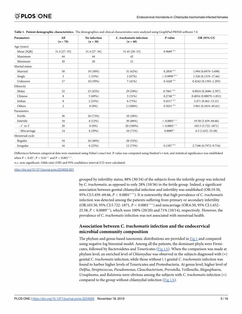
Patients’ demographics

The study cohort consisted of 77 voluntarily participating women of reproductive age (20–44)

presenting to the Obstetrics and Gynecology clinic at the University of Malaya Medical Center

from the year 2010 to 2014. After quality filtering and chimera removal, 7 samples which gener-

ated fewer than 40,000 16S rRNA gene amplicon reads were omitted from further analyses. The

final population comprised 70 subjects with ages ranging from 20 to 44 (mean age: 31.4; IQR:

27–35). Of the 70 subjects enrolled, 50 were married, 3 were divorced, and the remaining 17

women had not disclosed their marital status. The study cohort comprised 74.3% (52/70) sub-

jects from the Malay ethnic group, Chinese and Indians each contributing 11.4% (8/70), while

2.4% (2/70) were of other ethnic backgrounds, which describes the demographic diversity of the

multiethnic communities in Malaysia (Table 1). Among all, 51.4% (34/70) subjects were grouped

as infertile including 20 diagnosed with primary or secondary infertility and 14 patients with

miscarriage experience. Besides, 22.9% (16/70) of the subjects reported menstrual irregularity.

The relationship of C. trachomatis infection with infertility

A total of 40 out of 70 participants were infected with genital C. trachomatis based on the diag-

nostic test result. No significant correlation between chlamydial infection was detected with

demographics such as age, marital status, as well as ethnicity (Table 1). When the subjects were

Endocervical microbiota in Chlamydia trachomatis infected females

PLOS ONE | https://doi.org/10.1371/journal.pone.0224658 November 18, 2019 4 / 16

grouped by infertility status, 88% (30/34) of the subjects from the infertile group was infected

by C. trachomatis, as opposed to only 28% (10/36) in the fertile group. Indeed, a significant

association between genital chlamydial infection and infertility was established (OR:19.50,

95% CI:5.459–69.66, P< 0.0001���). It is noteworthy that high prevalence of C. trachomatisinfection was detected among the patients suffering from primary or secondary infertility

(OR:103.50, 95% CI:5.722–1871, P< 0.0001���) and miscarriage (OR:6.50, 95% CI:1.652–

25.58, P< 0.0089��), which were 100% (20/20) and 71% (10/14), respectively. However, the

prevalence of C. trachomatis infection was not associated with menstrual health.

Association between C. trachomatis infection and the endocervical

microbial community composition

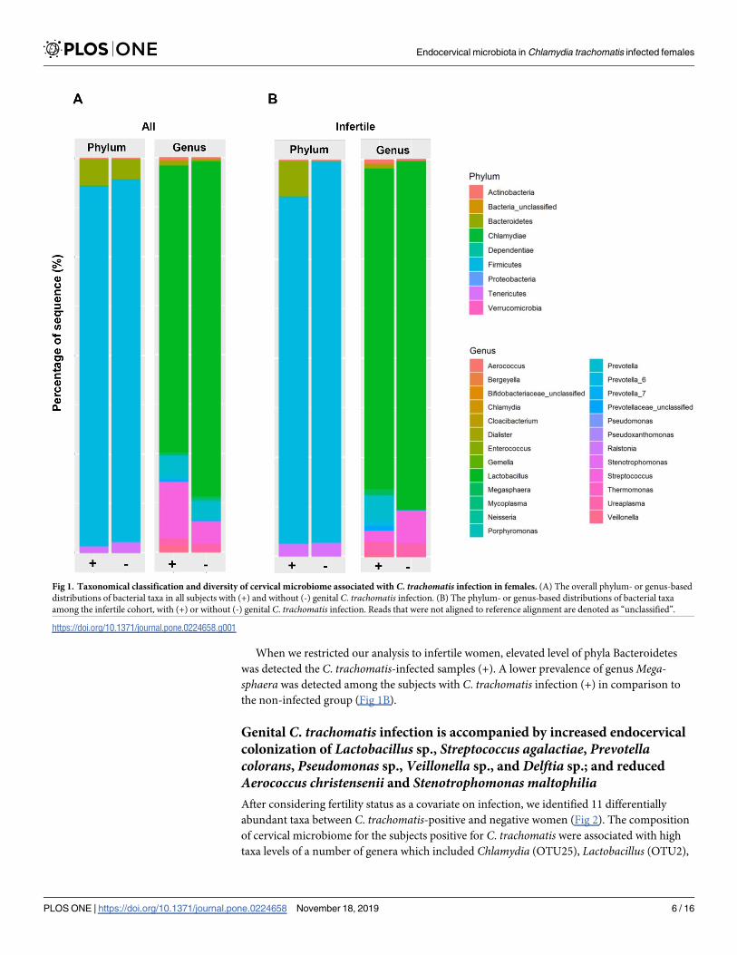
The phylum and genus based taxonomic distributions are provided in Fig 1 and compared

using negative log binomial model. Among all the patients, the dominant phyla were Firmi-

cutes, followed by Bacteroidetes and Tenericutes (Fig 1A). When the comparison was made at

phylum level, an enriched level of Chlamydiae was observed in the subjects diagnosed with (+)

genital C. trachomatis infection; while those without (-) genital C. trachomatis infection was

found to harbor higher levels of Tenericutes and Proteobacteria. At genus level, higher level of

Delftia, Streptococcus, Pseudomonas, Cloacibacterium, Prevotella, Veillonella, Megasphaera,

Ureaplasma, and Ralstonia were obvious among the subjects with C. trachomatis infection (+)

compared to the group without chlamydial infection (Fig 1A).

Table 1. Patient demographic characteristics. The demographics and clinical characteristics were analyzed using GraphPad PRISM software 7.0.

Parameters All

(n = 70)

No infection

(n = 30)

C. trachomatis infection

(n = 40)

P-value OR (95% CI)

Age (years)

Mean [IQR] 31.4 [27–35] 31.4 [27–36] 31.45 [28–35] 0.9689 n.s.

Maximum 44 44 42

Minimum 20 20 21

Marital status

Married 50 19 (38%) 31 (62%) 0.2850 n.s. 1.994 (0.6979–5.698)

Single 3 1 (33%) 2 (67%) > 0.9999 n.s.. 1.526 (0.1319–17.66)

Unknown 17 10 (59%) 7 (41%) 0.1628 n.s. 0.4242 (0.1392–1.293)

Ethnicity

Malay 52 23 (42%) 29 (58%) 0.7861 n.s.. 0.8024 (0.2686–2.397)

Chinese 8 5 (69%) 3 (31%) 0.2748 n.s. 0.4054 (0.08879–1.851)

Indian 8 2 (25%) 6 (75%) 0.4517 n.s. 2.471 (0.462–13.21)

Others 2 0 (0%) 2 (100%) 0.5031 n.s. 3.961 (0.1833–85.61)

Parameters

Fertile 36 26 (72%) 10 (28%)

Infertile 34 4 (12%) 30 (88%) < 0.0001��� 19.50 (5.459–69.66)

- 1˚ or 2˚ 20 0 (0%) 20 (100%) < 0.0001��� 103.5 (5.722–1871)

- Miscarriage 14 4 (29%) 10 (71%) 0.0089�� 6.5 (1.652–25.58)

Menstrual cycle

Regular 54 26 (48%) 28 (52%)

Irregular 16 4 (25%) 12 (75%) 0.1507 n.s. 2.7186 (0.7972–9.734)

Differences between categorical data were examined using Fisher’s exact test. P-value was computed using Student’s t-test, and statistical significance was established

when P < 0.05�, P < 0.01�� and P < 0.001���.

n.s.: non-significant. Odds ratio (OR) and 95% confidence interval (CI) were calculated.

https://doi.org/10.1371/journal.pone.0224658.t001

Endocervical microbiota in Chlamydia trachomatis infected females

PLOS ONE | https://doi.org/10.1371/journal.pone.0224658 November 18, 2019 5 / 16

When we restricted our analysis to infertile women, elevated level of phyla Bacteroidetes

was detected the C. trachomatis-infected samples (+). A lower prevalence of genus Mega-sphaera was detected among the subjects with C. trachomatis infection (+) in comparison to

the non-infected group (Fig 1B).

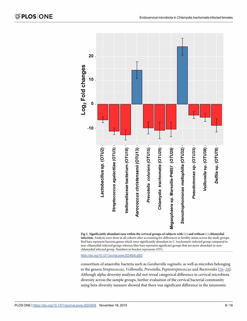
Genital C. trachomatis infection is accompanied by increased endocervical

colonization of Lactobacillus sp., Streptococcus agalactiae, Prevotellacolorans, Pseudomonas sp., Veillonella sp., and Delftia sp.; and reduced

Aerococcus christensenii and Stenotrophomonas maltophiliaAfter considering fertility status as a covariate on infection, we identified 11 differentially

abundant taxa between C. trachomatis-positive and negative women (Fig 2). The composition

of cervical microbiome for the subjects positive for C. trachomatis were associated with high

taxa levels of a number of genera which included Chlamydia (OTU25), Lactobacillus (OTU2),

Fig 1. Taxonomical classification and diversity of cervical microbiome associated with C. trachomatis infection in females. (A) The overall phylum- or genus-based

distributions of bacterial taxa in all subjects with (+) and without (-) genital C. trachomatis infection. (B) The phylum- or genus-based distributions of bacterial taxa

among the infertile cohort, with (+) or without (-) genital C. trachomatis infection. Reads that were not aligned to reference alignment are denoted as “unclassified”.

https://doi.org/10.1371/journal.pone.0224658.g001

Endocervical microbiota in Chlamydia trachomatis infected females

PLOS ONE | https://doi.org/10.1371/journal.pone.0224658 November 18, 2019 6 / 16

Streptococcus (OTU3), Megasphaera (OTU10/29), Prevotella_6 (OTU15), Pseudomonas(OTU33), Veillonella (OTU39), as well as Delftia (OTU70). Meanwhile, the genera Aerococcus(OTU13) and Stenotrophomonas (OTU32) were lower among subjects with C. trachomatisinfection compared to those without chlamydial infection.

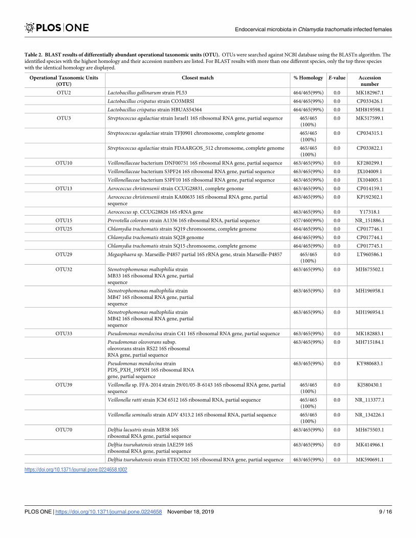
Sequence for each OTU was blasted using NCBI database to identify the microorganisms at

species level. The species which matched the highest sequence homology with the input are as

shown in Table 2. The abundant species in the C. trachomatis infected group were Lactobacil-lus gallinarum or Lactobacillus crispatus (OTU2), Streptococcus agalactiae (OTU3), Prevotellacolorans (OTU15), Pseudomonas mendocina or Psudomonas oleovorans (OTU33), Veillonellaratti or Veillonella seminalis (OTU39), and Delftia lacustris or Delftia tsuruhatensis (OTU70).

Whereas the abundant species in the non-chlamydial infected group were recognized as Aero-coccus christensenii (OTU13), Stenotrophomonas maltophilia (OTU32). Some OTUs, for exam-

ple OTU10 and OTU29, were unable to be identified at species level because of limited

information in the database.

Taxonomic diversity profiles of the endocervical microbiota in the presence

of C. trachomatis infection

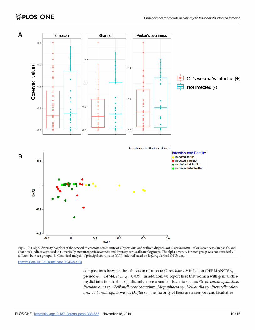
Alpha diversity measures including Pielou’s evenness alongside Simpson’s and Shannon’s

indices were examined (Fig 3A). There is a general trend of higher alpha diversity indices in

non-C. trachomatis infected than those in the C. trachomatis-infected subjects. However, the

difference did not achieve statistical significance at alpha = 0.05 (Fig 3A). Beta diversity

between the groups was further assessed by CAP (Fig 3B) and PERMANOVA analysis

(Table 3). In CAP, separation between non-infected and infected groups was observed along

axis CAP1. Indeed, a significance in bacterial compositional difference between groups with

different infection status was apparent (pseudo-F = 1.4744, P(perm) = 0.039). However, statisti-

cal significance was not achieved between groups with different fertility status (pseudo-

F = 1.384, P(perm) = 0.079).

Discussion

In this study, most of the patients diagnosed with C. trachomatis (88%) experienced primary

or secondary infertility (100%, P<0.0001) as well as miscarriage (71%, P = 0.0089). A prior epi-

demiological study has indicated that women with genital C. trachomatis infection are 30%

more likely to develop PID, ectopic pregnancy, as well as tubal factor infertility (TFI) than

those without, whereas infections with C. trachomatis elevate the risk of PID by an additional

20% [33]. Our results are consistent with this finding, whereby a significant positive correla-

tion between C. trachomatis and infertility was detected. Given the high prevalence of C. tra-chomatis infection and the severity of disease that ensues from the infection, a more effective

screening and treatment strategy is warranted to curb the dissemination of C. trachomatis in

the population. Notably, our study cohort comprised three major ethnic groups, namely the

Malays, Chinese, Indians, as well as ethnic minority participants of other nationalities. How-

ever, due to the small sample size, the differences between ethnicities were not compared.

The vaginal flora, which constitutes 9% of the human microbiome, have been shown to

play a critical role in the maintenance of overall health condition and disease state of the female

reproductive tract [34]. Many studies have demonstrated that women with bacterial vaginosis

are at higher risk of other sexually transmitted pathogens that includes C. trachomatis, signify-

ing the importance of microbiota homeostasis and its role as a determinant in disease outcome

[21, 23, 24, 35]. While the precise etiologic agents in bacterial vaginosis remain unclear, the

prevailing view is that the disease is polymicrobial in nature caused by overgrowth of a

Endocervical microbiota in Chlamydia trachomatis infected females

PLOS ONE | https://doi.org/10.1371/journal.pone.0224658 November 18, 2019 7 / 16

consortium of anaerobic bacteria such as Gardnerella vaginalis, as well as microbes belonging

to the genera Streptococcus, Veillonella, Prevotella, Peptostreptococcus and Bacteroides [36–38].

Although alpha-diversity analyses did not reveal categorical difference in cervical microbiota

diversity across the sample groups, further evaluation of the cervical bacterial community

using beta-diversity measure showed that there was significant difference in the taxonomic

Fig 2. Significantly abundant taxa within the cervical groups of subjects with (+) and without (-) chlamydial

infection. Analysis were done in all cohorts after accounting for differences in fertility status across the study groups.

Red bars represent bacteria genus which were significantly abundant in C. trachomatis-infected group compared to

non-chlamydial-infected group; whereas blue bars represent significant groups that are more abundant in non-

chlamydial infected group. Numbers in bracket represents OTU.

https://doi.org/10.1371/journal.pone.0224658.g002

Endocervical microbiota in Chlamydia trachomatis infected females

PLOS ONE | https://doi.org/10.1371/journal.pone.0224658 November 18, 2019 8 / 16

Table 2. BLAST results of differentially abundant operational taxonomic units (OTU). OTUs were searched against NCBI database using the BLASTn algorithm. The

identified species with the highest homology and their accession numbers are listed. For BLAST results with more than one different species, only the top three species

with the identical homology are displayed.

Operational Taxonomic Units

(OTU)

Closest match % Homology E-value Accession

number

OTU2 Lactobacillus gallinarum strain PL53 464/465(99%) 0.0 MK182967.1

Lactobacillus crispatus strain CO3MRSI 464/465(99%) 0.0 CP033426.1

Lactobacillus crispatus strain HBUAS54364 464/465(99%) 0.0 MH819598.1

OTU3 Streptococcus agalactiae strain Israel1 16S ribosomal RNA gene, partial sequence 465/465

(100%)

0.0 MK517599.1

Streptococcus agalactiae strain TFJ0901 chromosome, complete genome 465/465

(100%)

0.0 CP034315.1

Streptococcus agalactiae strain FDAARGOS_512 chromosome, complete genome 465/465

(100%)

0.0 CP033822.1

OTU10 Veillonellaceae bacterium DNF00751 16S ribosomal RNA gene, partial sequence 463/465(99%) 0.0 KF280299.1

Veillonellaceae bacterium S3PF24 16S ribosomal RNA gene, partial sequence 463/465(99%) 0.0 JX104009.1

Veillonellaceae bacterium S3PF10 16S ribosomal RNA gene, partial sequence 463/465(99%) 0.0 JX104005.1

OTU13 Aerococcus christensenii strain CCUG28831, complete genome 463/465(99%) 0.0 CP014159.1

Aerococcus christensenii strain KA00635 16S ribosomal RNA gene, partial

sequence

463/465(99%) 0.0 KP192302.1

Aerococcus sp. CCUG28826 16S rRNA gene 463/465(99%) 0.0 Y17318.1

OTU15 Prevotella colorans strain A1336 16S ribosomal RNA, partial sequence 457/460(99%) 0.0 NR_151886.1

OTU25 Chlamydia trachomatis strain SQ19 chromosome, complete genome 464/465(99%) 0.0 CP017746.1

Chlamydia trachomatis strain SQ28 genome 464/465(99%) 0.0 CP017744.1

Chlamydia trachomatis strain SQ15 chromosome, complete genome 464/465(99%) 0.0 CP017745.1

OTU29 Megasphaera sp. Marseille-P4857 partial 16S rRNA gene, strain Marseille-P4857 465/465

(100%)

0.0 LT960586.1

OTU32 Stenotrophomonas maltophilia strain

MB33 16S ribosomal RNA gene, partial

sequence

463/465(99%) 0.0 MH675502.1

Stenotrophomonas maltophilia strain

MB47 16S ribosomal RNA gene, partial

sequence

463/465(99%) 0.0 MH196958.1

Stenotrophomonas maltophilia strain

MB42 16S ribosomal RNA gene, partial

sequence

463/465(99%) 0.0 MH196954.1

OTU33 Pseudomonas mendocina strain C41 16S ribosomal RNA gene, partial sequence 463/465(99%) 0.0 MK182883.1

Pseudomonas oleovorans subsp.

oleovorans strain RS22 16S ribosomal

RNA gene, partial sequence

463/465(99%) 0.0 MH715184.1

Pseudomonas mendocina strain

PDS_PXH_19PXH 16S ribosomal RNA

gene, partial sequence

463/465(99%) 0.0 KY980683.1

OTU39 Veillonella sp. FFA-2014 strain 29/01/05-B-6143 16S ribosomal RNA gene, partial

sequence

465/465

(100%)

0.0 KJ580430.1

Veillonella ratti strain JCM 6512 16S ribosomal RNA, partial sequence 465/465

(100%)

0.0 NR_113377.1

Veillonella seminalis strain ADV 4313.2 16S ribosomal RNA, partial sequence 465/465

(100%)

0.0 NR_134226.1

OTU70 Delftia lacustris strain MB38 16S

ribosomal RNA gene, partial sequence

463/465(99%) 0.0 MH675503.1

Delftia tsuruhatensis strain IAE259 16S

ribosomal RNA gene, partial sequence

463/465(99%) 0.0 MK414966.1

Delftia tsuruhatensis strain ETEOC02 16S ribosomal RNA gene, partial sequence 463/465(99%) 0.0 MK590691.1

https://doi.org/10.1371/journal.pone.0224658.t002

Endocervical microbiota in Chlamydia trachomatis infected females

PLOS ONE | https://doi.org/10.1371/journal.pone.0224658 November 18, 2019 9 / 16

compositions between the subjects in relation to C. trachomatis infection (PERMANOVA,

pseudo-F = 1.4744, P(perm) = 0.039). In addition, we report here that women with genital chla-

mydial infection harbor significantly more abundant bacteria such as Streptococcus agalactiae,Pseudomonas sp., Veillonellaceae bacterium, Megasphaera sp., Veillonella sp., Prevotella color-ans, Veillonella sp., as well as Delftia sp., the majority of these are anaerobes and facultative

Fig 3. (A) Alpha diversity boxplots of the cervical microbiota community of subjects with and without diagnosis of C. trachomatis. Pielou’s evenness, Simpson’s, and

Shannon’s indices were used to numerically measure species evenness and diversity across all sample groups. The alpha diversity for each group was not statistically

different between groups. (B) Canonical analysis of principal coordinates (CAP) inferred based on log2 regularized OTUs data.

https://doi.org/10.1371/journal.pone.0224658.g003

Endocervical microbiota in Chlamydia trachomatis infected females

PLOS ONE | https://doi.org/10.1371/journal.pone.0224658 November 18, 2019 10 / 16

anaerobes which have been implicated in BV [36–39]. Other studies have also reported an

increase in the colonization of diverse anaerobic bacteria in the cervicovaginal environment of

women with C. trachomatis infection [26–28, 40, 41]. It is currently unknown whether Delftiacan modulate the host susceptibility to other pathogens. Delftia tsuruhatensis appears to con-

stitute the normal microbiota of the human vulva but there is evidence to suggest that its abun-

dance in amniotic fluid and blood is associated with preterm birth [42, 43]. The significant

presence of Delftia sp. in our C. trachomatis-positive group, therefore, merits further study to

better elucidate its role in the female endocervical microbiota.

Differential abundance analysis (DESeq2) further showed that the taxon Lactobacillus cris-patus and L. gallinarum was significantly elevated in the microbiota of women infected with C.

trachomatis relative to those without C. trachomatis infection. Evidence is emerging that prev-

alence of particular Lactobacillus provides fewer protection compared to other members of the

same genus. L. crispatus has been found to inhibit C. trachomatis infection, replication, as well

as attachment to host cells; these effects are likely related to its production of lactic acid that

lowers the surrounding pH and inhibits chlamydial growth [18–20]. However, it was reported

recently that L. crispatus promotes repair of injured vaginal epithelial cells by promoting the

secretion of vascular endothelial growth factor A (VEGF), which may help to reduce subse-

quent acquisition of urogenital tract infections [44]. L. crispatus was reported as one of the

major compositions of endocervix microbiota among the asymptomatic C. trachomatis-infected patients in South Africa [28]. Meanwhile, L. gallinarum has previously been found in

a high percentage of HPV-infected women in Beijing, China [45]. L. iners (OTU1) (data not

shown) was the most abundant taxon in the endocervical milieu of both infected and non-

infected group. Such dominance has been previously reported among normal healthy repro-

ductive Thai and Chinese women [46, 47]. L. iners has been shown to be negatively associated

with unfavorable reproductive health outcomes. Although commonly detected in the vagina of

healthy reproductive age women [48], a high prevalence of L. iners has been shown to be a pre-

dictive factor for bacterial vaginosis and pre-term delivery [49–52]. In fact, L. iners-dominated

microbiota has been linked to increased predisposition to acquisition of C. trachomatis, HIV,

as well as M. genitalium, indicating its double-edged sword role in female reproductive health

[26, 53–55]. However, we were unable to unequivocally determine the identity of the Lactoba-cillus in the present study as the sequences of L. gallinarum and L. crispatus had the identical

homology in the BLAST results.

Conversely, subjects negative for C. trachomatis displayed significantly greater abundance

of Aerococcus christensenii. Certain members of the genus Aerococcus including A. urinae, A.

sanguinocola, and A. viridans are rare pathogens involved in urinary tract infections particu-

larly among individuals with comorbidities [56–58]. In a similar study, Di Pietro et al. interro-

gated the microbial constituents of a population of Italian women infected with C. trachomatis

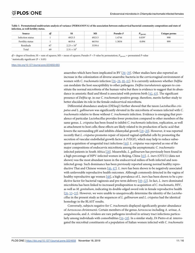
Table 3. Permutational multivariate analysis of variance (PERMANOVA) of the association between endocervical bacterial community composition and state of

infection, as well fertility status.

Source df SS MS Pseudo-F P(perm) Unique perms

Infection status 1 4923.5 4923.5 1.4744 0.039� 998

Fertility status 1 4619.6 4619.6 1.3834 0.094 999

Residuals 67 2.23 × 10⁵ 3339.4

Total 69 2.31 × 10⁵

df = degree of freedom; SS = sum of squares; MS = mean of squares; Pseudo-F = F value by permutation; P(perm) = permutated P-value

�statistically significant (P < 0.05)

https://doi.org/10.1371/journal.pone.0224658.t003

Endocervical microbiota in Chlamydia trachomatis infected females

PLOS ONE | https://doi.org/10.1371/journal.pone.0224658 November 18, 2019 11 / 16

and identified A. christensenii as the species closely associated with chlamydial infection [40].

Balle et al. showed that A. christensenii was only weakly related to C. trachomatis infection in a

cohort of South African adolescents [28]. A. christensenii has previously been implicated as a

cause of chorioamnionitis in a case report [59]. Beyond that, much remains unknown about

the role of A. christensenii in the female genital tract. Currently, there is a paucity of informa-

tion concerning the significance of Stenotrophomonas in the context of female reproductive

health. S. maltophilia is considered as a human commensal and is a frequently isolated micro-

organism in urine specimens. Although rare, S. maltophilia has been reported to be a cause of

nosocomial urinary tract infection [60, 61].

In conclusion, our study showed significant difference in endocervical microbiota between

C. trachomatis-infected and non-infected cohorts among patients visiting gynecology clinics

from Malaysia. However, we highlighted that genital C. trachomatis infection was not directly

connected to increased cervical bacterial diversity, as no significant differences were observed

in data richness and evenness. Further, we showed that infection with C. trachomatis was

related to elevated prevalence of mostly strict and facultative anaerobes, however disruption of

the sequencing data by PCR artefact should also be alerted [62]. Future studies involving a

larger cohort with more stringent criteria will allow us to delineate the endocervical microbial

landscape of women living in Malaysia. Nevertheless, the role of the anaerobes in disease pro-

gression is currently unknown, hence a further study to evaluate the involvement of taxa such

as A. christensenii and S. maltophilia in chlamydial infection is warranted.

Supporting information

S1 Table. Primers used in the present study for diagnosis of C. trachomatis.(DOCX)

Acknowledgments

We would like to extend our gratitude to all the study participants in this study and the dedi-

cated team of nurses at the Department of Obstetrics and Gynecology at the University Malaya

Medical Center for their assistance in collecting the clinical specimens.

Author Contributions

Conceptualization: Cindy Shuan Ju Teh, Li Yen Chang, Won Fen Wong.

Data curation: Heng Choon Cheong.

Formal analysis: Heng Choon Cheong, Chun Wie Chong, Yi Ying Cheok, Chalystha Yie Qin

Lee, Grace Min Yi Tan.

Investigation: Heng Choon Cheong, Rishein Gupta, Bernard Arulanandam.

Methodology: Polly Soo Xi Yap, Cindy Shuan Ju Teh.

Project administration: Sazaly AbuBakar, Won Fen Wong.

Resources: Sofiah Sulaiman, Jamiyah Hassan, Negar Shafiei Sabet.

Supervision: Li Yen Chang.

Validation: Heng Choon Cheong.

Writing – original draft: Heng Choon Cheong, Chun Wie Chong, Yi Ying Cheok, Chalystha

Yie Qin Lee, Grace Min Yi Tan.

Endocervical microbiota in Chlamydia trachomatis infected females

PLOS ONE | https://doi.org/10.1371/journal.pone.0224658 November 18, 2019 12 / 16

Writing – review & editing: Chung Yeng Looi, Won Fen Wong.

References1. Newman L, Rowley J, Vander Hoorn S, Wijesooriya NS, Unemo M, Low N, et al. Global Estimates of

the Prevalence and Incidence of Four Curable Sexually Transmitted Infections in 2012 Based on Sys-

tematic Review and Global Reporting. PLoS One. 2015; 10(12):e0143304. https://doi.org/10.1371/

journal.pone.0143304 PMID: 26646541; PubMed Central PMCID: PMC4672879.

2. Unemo M, Bradshaw CS, Hocking JS, de Vries HJC, Francis SC, Mabey D, et al. Sexually transmitted

infections: challenges ahead. Lancet Infect Dis. 2017; 17(8):e235–e79. Epub 2017/07/14. https://doi.

org/10.1016/S1473-3099(17)30310-9 PMID: 28701272.

3. Ngeow YF, Rachagan SP, Ramachandran S. Prevalence of chlamydial antibody in Malaysians. J Clin

Pathol. 1990; 43(5):400–2. Epub 1990/05/01. https://doi.org/10.1136/jcp.43.5.400 PMID: 2196283;

PubMed Central PMCID: PMC502444.

4. Ravindran J, Tan YI, Ngeow YF. The prevalence of Chlamydia trachomatis in patients with pelvic

inflammatory disease. Med J Malaysia. 1998; 53(1):16–21. Epub 2000/09/01. PMID: 10968132.

5. Yeow TC, Wong WF, Sabet NS, Sulaiman S, Shahhosseini F, Tan GM, et al. Prevalence of plasmid-

bearing and plasmid-free Chlamydia trachomatis infection among women who visited obstetrics and

gynecology clinics in Malaysia. BMC Microbiol. 2016; 16:45. Epub 2016/03/19. https://doi.org/10.1186/

s12866-016-0671-1 PMID: 26987367; PubMed Central PMCID: PMC4797335.

6. Lau CY, Qureshi AK. Azithromycin versus doxycycline for genital chlamydial infections: a meta-analysis

of randomized clinical trials. Sex Transm Dis. 2002; 29(9):497–502. Epub 2002/09/10. https://doi.org/

10.1097/00007435-200209000-00001 PMID: 12218839.

7. Miller KE. Diagnosis and treatment of Chlamydia trachomatis infection. Am Fam Physician. 2006; 73

(8):1411–6. PMID: 16669564.

8. Spiliopoulou A, Lakiotis V, Vittoraki A, Zavou D, Mauri D. Chlamydia trachomatis: time for screening?

Clin Microbiol Infect. 2005; 11(9):687–9. https://doi.org/10.1111/j.1469-0691.2005.01187.x PMID:

16104982.

9. Walker J, Tabrizi SN, Fairley CK, Chen MY, Bradshaw CS, Twin J, et al. Chlamydia trachomatis inci-

dence and re-infection among young women—behavioural and microbiological characteristics. PLoS

One. 2012; 7(5):e37778. https://doi.org/10.1371/journal.pone.0037778 PMID: 22662220; PubMed

Central PMCID: PMC3360595.

10. Brunham RC, Rey-Ladino J. Immunology of Chlamydia infection: implications for a Chlamydia tracho-

matis vaccine. Nat Rev Immunol. 2005; 5(2):149–61. https://doi.org/10.1038/nri1551 PMID: 15688042.

11. Moss NJ, Ahrens K, Kent CK, Klausner JD. The decline in clinical sequelae of genital Chlamydia tracho-

matis infection supports current control strategies. J Infect Dis. 2006; 193(9):1336–8; author reply 8–9.

https://doi.org/10.1086/503114 PMID: 16586376.

12. Mackern-Oberti JP, Motrich RD, Breser ML, Sanchez LR, Cuffini C, Rivero VE. Chlamydia trachomatis

infection of the male genital tract: an update. J Reprod Immunol. 2013; 100(1):37–53. https://doi.org/10.

1016/j.jri.2013.05.002 PMID: 23870458.

13. Manavi K. A review on infection with Chlamydia trachomatis. Best Pract Res Clin Obstet Gynaecol.

2006; 20(6):941–51. https://doi.org/10.1016/j.bpobgyn.2006.06.003 PMID: 16934531.

14. Wilkowska-Trojniel M, Zdrodowska-Stefanow B, Ostaszewska-Puchalska I, Redzko S, Przepiesc J,

Zdrodowski M. The influence of Chlamydia trachomatis infection on spontaneous abortions. Adv Med

Sci. 2009; 54(1):86–90. https://doi.org/10.2478/v10039-009-0008-5 PMID: 19403438.

15. Rours GI, Duijts L, Moll HA, Arends LR, de Groot R, Jaddoe VW, et al. Chlamydia trachomatis infection

during pregnancy associated with preterm delivery: a population-based prospective cohort study. Eur J

Epidemiol. 2011; 26(6):493–502. https://doi.org/10.1007/s10654-011-9586-1 PMID: 21538042;

PubMed Central PMCID: PMC3115062.

16. Kaminska D, Gajecka M. Is the role of human female reproductive tract microbiota underestimated?

Benef Microbes. 2017; 8(3):327–43. Epub 2017/05/16. https://doi.org/10.3920/BM2015.0174 PMID:

28504576.

17. Cribby S, Taylor M, Reid G. Vaginal microbiota and the use of probiotics. Interdiscip Perspect Infect

Dis. 2008; 2008:256490. Epub 2008/01/01. https://doi.org/10.1155/2008/256490 PMID: 19343185;

PubMed Central PMCID: PMC2662373.

18. Gong Z, Luna Y, Yu P, Fan H. Lactobacilli inactivate Chlamydia trachomatis through lactic acid but not

H2O2. PLoS One. 2014; 9(9):e107758. Epub 2014/09/13. https://doi.org/10.1371/journal.pone.

0107758 PMID: 25215504; PubMed Central PMCID: PMC4162611.

Endocervical microbiota in Chlamydia trachomatis infected females

PLOS ONE | https://doi.org/10.1371/journal.pone.0224658 November 18, 2019 13 / 16

19. Mastromarino P, Di Pietro M, Schiavoni G, Nardis C, Gentile M, Sessa R. Effects of vaginal lactobacilli

in Chlamydia trachomatis infection. Int J Med Microbiol. 2014; 304(5–6):654–61. Epub 2014/05/31.

https://doi.org/10.1016/j.ijmm.2014.04.006 PMID: 24875405.

20. Nardini P, Nahui Palomino RA, Parolin C, Laghi L, Foschi C, Cevenini R, et al. Lactobacillus crispatus

inhibits the infectivity of Chlamydia trachomatis elementary bodies, in vitro study. Sci Rep. 2016;

6:29024. Epub 2016/06/30. https://doi.org/10.1038/srep29024 PMID: 27354249; PubMed Central

PMCID: PMC4926251.

21. Gillet E, Meys JF, Verstraelen H, Bosire C, De Sutter P, Temmerman M, et al. Bacterial vaginosis is

associated with uterine cervical human papillomavirus infection: a meta-analysis. BMC Infect Dis. 2011;

11:10. Epub 2011/01/13. https://doi.org/10.1186/1471-2334-11-10 PMID: 21223574; PubMed Central

PMCID: PMC3023697.

22. Brotman RM, Bradford LL, Conrad M, Gajer P, Ault K, Peralta L, et al. Association between Trichomo-

nas vaginalis and vaginal bacterial community composition among reproductive-age women. Sex

Transm Dis. 2012; 39(10):807–12. Epub 2012/09/26. https://doi.org/10.1097/OLQ.0b013e3182631c79

PMID: 23007708; PubMed Central PMCID: PMC3458234.

23. Gallo MF, Macaluso M, Warner L, Fleenor ME, Hook EW 3rd, Brill I, et al. Bacterial vaginosis, gonor-

rhea, and chlamydial infection among women attending a sexually transmitted disease clinic: a longitu-

dinal analysis of possible causal links. Ann Epidemiol. 2012; 22(3):213–20. Epub 2011/12/24. https://

doi.org/10.1016/j.annepidem.2011.11.005 PMID: 22192490.

24. Bautista CT, Wurapa EK, Sateren WB, Morris SM, Hollingsworth BP, Sanchez JL. Association of Bacte-

rial Vaginosis With Chlamydia and Gonorrhea Among Women in the U.S. Army. Am J Prev Med. 2017;

52(5):632–9. Epub 2016/11/07. https://doi.org/10.1016/j.amepre.2016.09.016 PMID: 27816380.

25. Gosmann C, Anahtar MN, Handley SA, Farcasanu M, Abu-Ali G, Bowman BA, et al. Lactobacillus-Defi-

cient Cervicovaginal Bacterial Communities Are Associated with Increased HIV Acquisition in Young

South African Women. Immunity. 2017; 46(1):29–37. https://doi.org/10.1016/j.immuni.2016.12.013

PMID: 28087240; PubMed Central PMCID: PMC5270628.

26. van der Veer C, Bruisten SM, van der Helm JJ, de Vries HJ, van Houdt R. The Cervicovaginal Micro-

biota in Women Notified for Chlamydia trachomatis Infection: A Case-Control Study at the Sexually

Transmitted Infection Outpatient Clinic in Amsterdam, The Netherlands. Clin Infect Dis. 2017; 64(1):24–

31. Epub 2016/08/28. https://doi.org/10.1093/cid/ciw586 PMID: 27567124.

27. Filardo S, Di Pietro M, Porpora MG, Recine N, Farcomeni A, Latino MA, et al. Diversity of Cervical

Microbiota in Asymptomatic Chlamydia trachomatis Genital Infection: A Pilot Study. Front Cell Infect

Microbiol. 2017; 7:321. Epub 2017/08/05. https://doi.org/10.3389/fcimb.2017.00321 PMID: 28770172;

PubMed Central PMCID: PMC5509768.

28. Balle C, Lennard K, Dabee S, Barnabas SL, Jaumdally SZ, Gasper MA, et al. Endocervical and vaginal

microbiota in South African adolescents with asymptomatic Chlamydia trachomatis infection. Sci Rep.

2018; 8(1):11109. Epub 2018/07/25. https://doi.org/10.1038/s41598-018-29320-x PMID: 30038262;

PubMed Central PMCID: PMC6056523.

29. Tamarelle J, de Barbeyrac B, Le Hen I, Thiebaut A, Bebear C, Ravel J, et al. Vaginal microbiota compo-

sition and association with prevalent Chlamydia trachomatis infection: a cross-sectional study of young

women attending a STI clinic in France. Sex Transm Infect. 2018. Epub 2018/01/24. https://doi.org/10.

1136/sextrans-2017-053346 PMID: 29358524.

30. Schloss PD, Westcott SL, Ryabin T, Hall JR, Hartmann M, Hollister EB, et al. Introducing mothur: open-

source, platform-independent, community-supported software for describing and comparing microbial

communities. Appl Environ Microbiol. 2009; 75(23):7537–41. Epub 2009/10/06. https://doi.org/10.1128/

AEM.01541-09 PubMed Central PMCID: PMC2786419. PMID: 19801464

31. McMurdie PJ, Holmes S. phyloseq: an R package for reproducible interactive analysis and graphics of

microbiome census data. PLoS One. 2013; 8(4):e61217. Epub 2013/05/01. https://doi.org/10.1371/

journal.pone.0061217 PMID: 23630581; PubMed Central PMCID: PMC3632530.

32. Love MI, Huber W, Anders S. Moderated estimation of fold change and dispersion for RNA-seq data

with DESeq2. Genome Biol. 2014; 15(12):550. Epub 2014/12/18. https://doi.org/10.1186/s13059-014-

0550-8 PMID: 25516281; PubMed Central PMCID: PMC4302049.

33. Davies B, Turner KM, Frolund M, Ward H, May MT, Rasmussen S, et al. Risk of reproductive complica-

tions following chlamydia testing: a population-based retrospective cohort study in Denmark. Lancet

Infect Dis. 2016; 16(9):1057–64. https://doi.org/10.1016/S1473-3099(16)30092-5 PMID: 27289389.

34. Group NHW, Peterson J, Garges S, Giovanni M, McInnes P, Wang L, et al. The NIH Human Micro-

biome Project. Genome Res. 2009; 19(12):2317–23. Epub 2009/10/13. https://doi.org/10.1101/gr.

096651.109 PMID: 19819907; PubMed Central PMCID: PMC2792171.

Endocervical microbiota in Chlamydia trachomatis infected females

PLOS ONE | https://doi.org/10.1371/journal.pone.0224658 November 18, 2019 14 / 16

35. Wiesenfeld HC, Hillier SL, Krohn MA, Landers DV, Sweet RL. Bacterial vaginosis is a strong predictor

of Neisseria gonorrhoeae and Chlamydia trachomatis infection. Clin Infect Dis. 2003; 36(5):663–8.

Epub 2003/02/21. https://doi.org/10.1086/367658 PMID: 12594649.

36. Biagi E, Vitali B, Pugliese C, Candela M, Donders GG, Brigidi P. Quantitative variations in the vaginal

bacterial population associated with asymptomatic infections: a real-time polymerase chain reaction

study. Eur J Clin Microbiol Infect Dis. 2009; 28(3):281–5. Epub 2008/09/03. https://doi.org/10.1007/

s10096-008-0617-0 PMID: 18762999.

37. Turovskiy Y, Sutyak Noll K, Chikindas ML. The aetiology of bacterial vaginosis. J Appl Microbiol. 2011;

110(5):1105–28. Epub 2011/02/22. https://doi.org/10.1111/j.1365-2672.2011.04977.x PMID:

21332897; PubMed Central PMCID: PMC3072448.

38. Nelson TM, Borgogna JL, Brotman RM, Ravel J, Walk ST, Yeoman CJ. Vaginal biogenic amines: bio-

markers of bacterial vaginosis or precursors to vaginal dysbiosis? Front Physiol. 2015; 6:253. Epub

2015/10/21. https://doi.org/10.3389/fphys.2015.00253 PMID: 26483694; PubMed Central PMCID:

PMC4586437.

39. Ranjit E, Raghubanshi BR, Maskey S, Parajuli P. Prevalence of Bacterial Vaginosis and Its Association

with Risk Factors among Nonpregnant Women: A Hospital Based Study. Int J Microbiol. 2018;

2018:8349601. Epub 2018/04/26. https://doi.org/10.1155/2018/8349601 PMID: 29692813; PubMed

Central PMCID: PMC5859802.

40. Di Pietro M, Filardo S, Porpora MG, Recine N, Latino MA, Sessa R. HPV/Chlamydia trachomatis co-

infection: metagenomic analysis of cervical microbiota in asymptomatic women. New Microbiol. 2018;

41(1):34–41. Epub 2018/01/10. PMID: 29313867.

41. Witkin SS. Vaginal microbiome studies in pregnancy must also analyse host factors. BJOG. 2019; 126

(3):359. Epub 2018/05/24. https://doi.org/10.1111/1471-0528.15300 PMID: 29791773.

42. DiGiulio DB, Romero R, Amogan HP, Kusanovic JP, Bik EM, Gotsch F, et al. Microbial prevalence,

diversity and abundance in amniotic fluid during preterm labor: a molecular and culture-based investiga-

tion. PLoS One. 2008; 3(8):e3056. Epub 2008/08/30. https://doi.org/10.1371/journal.pone.0003056

PMID: 18725970; PubMed Central PMCID: PMC2516597.

43. You Y-A, Yoo JY, Kwon EJ, Kim YJ. Blood Microbial Communities During Pregnancy Are Associated

With Preterm Birth. Frontiers in Microbiology. 2019; 10(1122). https://doi.org/10.3389/fmicb.2019.

01122 PMID: 31214131

44. Takada K, Komine-Aizawa S, Kuramochi T, Ito S, Trinh QD, Pham NTK, et al. Lactobacillus crispatus

accelerates re-epithelialization in vaginal epithelial cell line MS74. Am J Reprod Immunol. 2018; 80(3):

e13027. Epub 2018/08/26. https://doi.org/10.1111/aji.13027 PMID: 30144195.

45. Gao W, Weng J, Gao Y, Chen X. Comparison of the vaginal microbiota diversity of women with and

without human papillomavirus infection: a cross-sectional study. BMC Infect Dis. 2013; 13:271. Epub

2013/06/14. https://doi.org/10.1186/1471-2334-13-271 PMID: 23758857; PubMed Central PMCID:

PMC3684509.

46. Shi Y, Chen L, Tong J, Xu C. Preliminary characterization of vaginal microbiota in healthy Chinese

women using cultivation-independent methods. J Obstet Gynaecol Res. 2009; 35(3):525–32. Epub

2009/06/17. https://doi.org/10.1111/j.1447-0756.2008.00971.x PMID: 19527394.

47. Sirichoat A, Buppasiri P, Engchanil C, Namwat W, Faksri K, Sankuntaw N, et al. Characterization of

vaginal microbiota in Thai women. PeerJ. 2018; 6:e5977. Epub 2018/12/01. https://doi.org/10.7717/

peerj.5977 PMID: 30498641; PubMed Central PMCID: PMC6252066.

48. Ravel J, Gajer P, Abdo Z, Schneider GM, Koenig SS, McCulle SL, et al. Vaginal microbiome of repro-

ductive-age women. Proc Natl Acad Sci U S A. 2011; 108 Suppl 1:4680–7. Epub 2010/06/11. https://

doi.org/10.1073/pnas.1002611107 PMID: 20534435; PubMed Central PMCID: PMC3063603.

49. Tamrakar R, Yamada T, Furuta I, Cho K, Morikawa M, Yamada H, et al. Association between Lactoba-

cillus species and bacterial vaginosis-related bacteria, and bacterial vaginosis scores in pregnant Japa-

nese women. BMC Infect Dis. 2007; 7:128. Epub 2007/11/08. https://doi.org/10.1186/1471-2334-7-128

PMID: 17986357; PubMed Central PMCID: PMC2212641.

50. Macklaim JM, Gloor GB, Anukam KC, Cribby S, Reid G. At the crossroads of vaginal health and dis-

ease, the genome sequence of Lactobacillus iners AB-1. Proc Natl Acad Sci U S A. 2011; 108 Suppl

1:4688–95. Epub 2010/11/10. https://doi.org/10.1073/pnas.1000086107 PMID: 21059957; PubMed

Central PMCID: PMC3063587.

51. Kindinger LM, Bennett PR, Lee YS, Marchesi JR, Smith A, Cacciatore S, et al. The interaction between

vaginal microbiota, cervical length, and vaginal progesterone treatment for preterm birth risk. Micro-

biome. 2017; 5(1):6. Epub 2017/01/21. https://doi.org/10.1186/s40168-016-0223-9 PMID: 28103952;

PubMed Central PMCID: PMC5244550.

52. Stafford GP, Parker JL, Amabebe E, Kistler J, Reynolds S, Stern V, et al. Spontaneous Preterm Birth Is

Associated with Differential Expression of Vaginal Metabolites by Lactobacilli-Dominated Microflora.

Endocervical microbiota in Chlamydia trachomatis infected females

PLOS ONE | https://doi.org/10.1371/journal.pone.0224658 November 18, 2019 15 / 16

Front Physiol. 2017; 8:615. Epub 2017/09/08. https://doi.org/10.3389/fphys.2017.00615 PMID:

28878691; PubMed Central PMCID: PMC5572350.

53. Borgdorff H, Tsivtsivadze E, Verhelst R, Marzorati M, Jurriaans S, Ndayisaba GF, et al. Lactobacillus-

dominated cervicovaginal microbiota associated with reduced HIV/STI prevalence and genital HIV viral

load in African women. ISME J. 2014; 8(9):1781–93. Epub 2014/03/07. https://doi.org/10.1038/ismej.

2014.26 PMID: 24599071; PubMed Central PMCID: PMC4139719.

54. Molenaar MC, Singer M, Ouburg S. The two-sided role of the vaginal microbiome in Chlamydia tracho-

matis and Mycoplasma genitalium pathogenesis. J Reprod Immunol. 2018; 130:11–7. Epub 2018/08/

28. https://doi.org/10.1016/j.jri.2018.08.006 PMID: 30149363.

55. van Houdt R, Ma B, Bruisten SM, Speksnijder A, Ravel J, de Vries HJC. Lactobacillus iners-dominated

vaginal microbiota is associated with increased susceptibility to Chlamydia trachomatis infection in

Dutch women: a case-control study. Sex Transm Infect. 2018; 94(2):117–23. Epub 2017/09/28. https://

doi.org/10.1136/sextrans-2017-053133 PMID: 28947665; PubMed Central PMCID: PMC6083440.

56. Mohan B, Zaman K, Anand N, Taneja N. Aerococcus Viridans: A Rare Pathogen Causing Urinary Tract

Infection. J Clin Diagn Res. 2017; 11(1):DR01–DR3. Epub 2017/03/10. https://doi.org/10.7860/JCDR/

2017/23997.9229 PMID: 28273968; PubMed Central PMCID: PMC5324413.

57. Higgins A, Garg T. Aerococcus urinae: An Emerging Cause of Urinary Tract Infection in Older Adults

with Multimorbidity and Urologic Cancer. Urol Case Rep. 2017; 13:24–5. Epub 2017/04/25. https://doi.

org/10.1016/j.eucr.2017.03.022 PMID: 28435789; PubMed Central PMCID: PMC5393163.

58. Zhang Q, Kwoh C, Attorri S, Clarridge JE 3rd. Aerococcus urinae in urinary tract infections. J Clin Micro-

biol. 2000; 38(4):1703–5. Epub 2000/04/04. PMID: 10747177; PubMed Central PMCID: PMC86536.

59. Carlstein C, Marie Soes L, Jorgen Christensen J. Aerococcus christensenii as Part of Severe Polymi-

crobial Chorioamnionitis in a Pregnant Woman. Open Microbiol J. 2016; 10:27–31. Epub 2016/03/26.

https://doi.org/10.2174/1874285801610010027 PMID: 27014376; PubMed Central PMCID:

PMC4787314.

60. Vartivarian SE, Papadakis KA, Anaissie EJ. Stenotrophomonas (Xanthomonas) maltophilia urinary

tract infection. A disease that is usually severe and complicated. Arch Intern Med. 1996; 156(4):433–5.

Epub 1996/02/26. PMID: 8607729.

61. Denton M, Kerr KG. Microbiological and clinical aspects of infection associated with Stenotrophomonas

maltophilia. Clin Microbiol Rev. 1998; 11(1):57–80. Epub 1998/02/11. PMID: 9457429; PubMed Central

PMCID: PMC121376.

62. Kebschull JM, Zador AM. Sources of PCR-induced distortions in high-throughput sequencing data sets.

Nucleic Acids Res. 2015; 43(21):e143. Epub 2015/07/19. https://doi.org/10.1093/nar/gkv717 PMID:

26187991; PubMed Central PMCID: PMC4666380.

Endocervical microbiota in Chlamydia trachomatis infected females

PLOS ONE | https://doi.org/10.1371/journal.pone.0224658 November 18, 2019 16 / 16


Recommended