Effects of triaryl phosphates on mouse and human nuclear receptors Paavo Honkakoski a,* , Jorma J. Palvimo b , Leena Penttila ¨ c , Jouko Vepsa ¨la ¨inen d , Seppo Auriola c a Department of Pharmaceutics, University of Kuopio, P.O. Box 1627, FIN-70211, Kuopio, Finland b Biomedicum Helsinki, Institute of Biomedicine, University of Helsinki, FIN-00014, Helsinki, Finland c Department of Pharmaceutical Chemistry, University of Kuopio, P.O. Box 1627, FIN-70211, Kuopio, Finland d Department of Chemistry, University of Kuopio, P.O. Box 1627, FIN-70211, Kuopio, Finland Received 13 June 2003; accepted 25 August 2003 Abstract The constitutively active receptor (CAR) is a crucial regulator of genes encoding for enzymes active in drug/steroid oxidation, conjugation, and transport. In our attempt to isolate the endogenous inhibitory ligand(s) for the mouse CAR, we found surprisingly that the inhibitory activity was associated with di- and tri-isopropylated phenyl phosphates that were present in livers of untreated mice. Trans- activation experiments in mammalian cells with synthetic compounds verified that mouse CAR was inhibited by various isopropylated phenyl phosphates (40–80%). Such triaryl phosphates are widely used as fire retardants, lubricants, and plasticizers, and some of them are known to disturb reproduction by currently unknown mechanisms. Equipped with the finding that these compounds could interact with mouse CAR, we proceeded to determine their functional effects on other nuclear receptors. Human CAR and pregnane X receptor (PXR) were variably activated (2–5-fold) by triaryl phosphates while mouse PXR, peroxisome proliferator-activated receptor-a, and vitamin D receptor were refractory. Among steroid hormone receptors, the human androgen receptor was inhibited by triphenyl phosphate and di-ortho-isopropylated phenyl phosphate (40–50%) and activated by di- and tri-para-substituted phenyl phosphates (2-fold). Our results add to the list of CAR and PXR activators and suggest steroid-dependent biological pathways that may contribute to the reproductive effects of triaryl phosphates. # 2003 Elsevier Inc. All rights reserved. Keywords: Organophosphates; Triaryl phosphates; Nuclear receptor; Activation; Mouse; Human 1. Introduction NRs are ligand-dependent DNA-binding transcription factors that are encoded by a superfamily of 48 genes [1]. The NRs transmit extra- or intracellular signals directly to gene transcription machinery, in response to steroid and thyroid hormones, fatty acid and cholesterol derivatives, and vitamins A and D. NRs control profound cellular processes such as growth and differentiation, carbohydrate and lipid metabolism, and endocrine physiology [2]. Therefore, dis- turbances in function of NRs may result in clinical mani- festations. Such disturbances include hormone resistance syndromes and cancers where interindividual differences in NR structure result in abnormal ligand binding or changes in responses to NR co-regulators [3,4]. On the other hand, environmental or diet-derived chemicals (xenobiotics) may disturb hormonal balance via binding to NRs [5–7]. Xenobiotics may also affect hormonal balance through activation of CYP gene expression. CYPs are essential not only in the metabolism of xenobiotics but also in the control of both formation and degradation of endogenous compounds (endobiotics) which include NR ligands [8]. Because CYP genes are regulated by NRs [9], exposure to xenobiotics may lead to increased expression of CYP enzymes and enhanced xeno- and endobiotic metabolism. While the ligand specificities of steroid hormone recep- tors have been well established, many of the so-called Biochemical Pharmacology 67 (2004) 97–106 0006-2952/$ – see front matter # 2003 Elsevier Inc. All rights reserved. doi:10.1016/j.bcp.2003.08.037 * Corresponding author. Tel.: þ358-17-162490; fax: þ358-17-162252. E-mail address: paavo.honkakoski@uku.fi (P. Honkakoski). Abbreviations: AR, androgen receptor; CAR, constitutively active receptor; CYP, cytochrome P450; ERa, estrogen receptor-a; GR, glucocorticoid receptor; NR, nuclear receptor; PPARa, peroxisome proliferator-activated receptor-a; PR, progesterone receptor; PXR, preg- nane X receptor; TCPOBOP, 1,4-bis[2-(3,5-dichloropyridyloxy)]benzene; TPP, triphenyl phosphate; TTP, tri-p-methyl phenyl phosphate. Isopropy- lated triaryl phosphates are abbreviated by the generic symbol o-, m-, p- iPrXN, where o-, m-, and p- denote the position of the isopropyl (iPr) substituent on the phenyl moiety, and XN denotes the number of isopropylated phenyl moieties (N ¼ 1, 2, or 3).
Transcript
Effects of triaryl phosphates on mouse and human nuclear receptors
Paavo Honkakoskia,*, Jorma J. Palvimob, Leena Penttilac,Jouko Vepsalainend, Seppo Auriolac
aDepartment of Pharmaceutics, University of Kuopio, P.O. Box 1627, FIN-70211, Kuopio, FinlandbBiomedicum Helsinki, Institute of Biomedicine, University of Helsinki, FIN-00014, Helsinki, Finland
cDepartment of Pharmaceutical Chemistry, University of Kuopio, P.O. Box 1627, FIN-70211, Kuopio, FinlanddDepartment of Chemistry, University of Kuopio, P.O. Box 1627, FIN-70211, Kuopio, Finland
Received 13 June 2003; accepted 25 August 2003
Abstract
The constitutively active receptor (CAR) is a crucial regulator of genes encoding for enzymes active in drug/steroid oxidation,
conjugation, and transport. In our attempt to isolate the endogenous inhibitory ligand(s) for the mouse CAR, we found surprisingly that the
inhibitory activity was associated with di- and tri-isopropylated phenyl phosphates that were present in livers of untreated mice. Trans-
activation experiments in mammalian cells with synthetic compounds verified that mouse CAR was inhibited by various isopropylated
phenyl phosphates (40–80%). Such triaryl phosphates are widely used as fire retardants, lubricants, and plasticizers, and some of them are
known to disturb reproduction by currently unknown mechanisms. Equipped with the finding that these compounds could interact with
mouse CAR, we proceeded to determine their functional effects on other nuclear receptors. Human CAR and pregnane X receptor (PXR)
were variably activated (2–5-fold) by triaryl phosphates while mouse PXR, peroxisome proliferator-activated receptor-a, and vitamin D
receptor were refractory. Among steroid hormone receptors, the human androgen receptor was inhibited by triphenyl phosphate and
di-ortho-isopropylated phenyl phosphate (40–50%) and activated by di- and tri-para-substituted phenyl phosphates (2-fold). Our results
add to the list of CAR and PXR activators and suggest steroid-dependent biological pathways that may contribute to the reproductive
effects of triaryl phosphates.
# 2003 Elsevier Inc. All rights reserved.
Keywords: Organophosphates; Triaryl phosphates; Nuclear receptor; Activation; Mouse; Human
1. Introduction
NRs are ligand-dependent DNA-binding transcription
factors that are encoded by a superfamily of 48 genes [1].
The NRs transmit extra- or intracellular signals directly to
gene transcription machinery, in response to steroid and
thyroid hormones, fatty acid and cholesterol derivatives, and
vitamins A and D. NRs control profound cellular processes
such as growth and differentiation, carbohydrate and lipid
metabolism, and endocrine physiology [2]. Therefore, dis-
turbances in function of NRs may result in clinical mani-
festations. Such disturbances include hormone resistance
syndromes and cancers where interindividual differences in
NR structure result in abnormal ligand binding or changes
in responses to NR co-regulators [3,4]. On the other hand,
environmental or diet-derived chemicals (xenobiotics)
may disturb hormonal balance via binding to NRs [5–7].
Xenobiotics may also affect hormonal balance through
activation of CYP gene expression. CYPs are essential
not only in the metabolism of xenobiotics but also in the
control of both formation and degradation of endogenous
compounds (endobiotics) which include NR ligands [8].
Because CYP genes are regulated by NRs [9], exposure to
xenobiotics may lead to increased expression of CYP
enzymes and enhanced xeno- and endobiotic metabolism.
While the ligand specificities of steroid hormone recep-
tors have been well established, many of the so-called
Biochemical Pharmacology 67 (2004) 97–106
0006-2952/$ – see front matter # 2003 Elsevier Inc. All rights reserved.
12 m � 0:2 mm, film thickness 0.33 mm) with a temperature
gradient (2 min at 1208, followed by 180–3208 over
20 min). The molecules were ionized by 36 eV electrons
and the mass range m/z 50–700 was scanned in 0.9 s. The
compounds were identified by comparisons with the data in
Wiley mass spectral library. The identification of the com-
pounds was verified by running a commercial tri-(isopro-
pylphenyl) phosphate standard (Chem Service Inc.) with the
GC–MS instrument. The elemental composition of selected
compounds was then determined with a high resolution
GC–MS instrument (VG250, 70-SE) with above GC con-
ditions. The accurate mass measurement was carried out
automatically with the data system. Perfluorokerosene was
used as the reference compound.
2.5. Reporter plasmids
pCMVb was purchased from Clontech Inc. pARE2-
TATA-LUC reporter contains two AR response elements
in front of minimal TATA sequence [23]. Hormone
response elements in the ARE2TATA promoter mediate
also GR- and PR-dependent signaling [24]. pERE2TATA-
LUC was constructed in the same way except that the
inserted 45-base pair oligomer contained two ER response
elements [25]. The reporter plasmids pPBREM-tk-luc
[26], pXREM-3A4-luc [27] and pDR1x4-tk-luc [21] which
respond to CAR, PXR, and PPARa, respectively, have been
described.
2.6. Expression plasmids
The sources of expression vectors for mouse and human
PPARa, PXR, and CAR, and for human vitamin D receptor
have been described [21]. Human AR, GR and ERaexpression vectors have been reported earlier [23,25,28].
pSG5-hPR1 was gift from Dr. Pierre Chambon (INSERM,
Illkirch, France). All plasmids were purified with Qiagen
columns.
2.7. Cell culture and transfection with mouse and
human CAR, PXR, and PPARa
HEK293 cells (ATCC CRL-1573) were grown in phe-
nol-free Dulbecco’s modified Eagle medium supplemented
with 10% fetal bovine serum (FBS) and antibiotics. One day
prior to transfection, 70 � 103 cells per well were seeded
on 48-well plates in medium containing 5% delipidated
FBS (Sigma). After an overnight incubation, the medium
was changed and the cells were transfected using calcium
phosphate method. In the mouse CAR deactivation assay,
each well received pCMVb (100 ng), UAS4-tk-luc (25 ng)
[29] and CMV-GAL4-mouse CAR (12.5 ng) [21]. In
experiments with purified test chemicals, each well
received pCMVb (100 ng), expression vectors (12.5 ng)
for full-length mouse or human CAR, PXR, or PPARareceptors, and the respective reporter gene (25 ng). After a
4-hr transfection period, the medium was changed. The
fresh medium additionally contained either samples from
HPLC fractions that were first evaporated and then dis-
solved in isopropanol (mouse CAR assay), or purified test
chemicals. Controls for mouse CAR included 10 mM
androstenol (suppressor) and 0.5 mM TCPOBOP (activa-
tor) [30]. To account for the known species differences in
activation of xenobiotic-activated NRs [17], positive con-
trols for human CAR, mouse PXR, human PXR and both
PPARa species were 2 mM clotrimazole, 10 mM RU486,
10 mM rifampicin and 10 mM Wy-14,643, respectively
[21]. Cells were then cultured for 40 hr prior to washing
with PBS and lysis of the cells. Luciferase and b-galacto-
sidase activities [26] were determined from 20 mL of
lysates in 96-well plates using the Victor2TM multiplate
reader (Perkin-Elmer Wallac).
2.8. Cell culture and transfections with human
GR, PR, AR, and ERa
COS-1 cells (ATCC CRL-1650) were maintained in
Dulbecco’s minimal essential medium containing antibio-
tics and 10% FBS. Cells were seeded onto 12-well plates
and transfected 24 hr later by FuGene transfection method
(Roche Molecular Biochemicals). In brief, 30 � 103 cells
in each well received 290 ng of the luciferase reporter
plasmid, 20 ng of pCMVb internal control plasmid and
20 ng of different steroid receptor expression vectors. Four
hours before transfection, the medium was changed to one
containing 10% charcoal-stripped FBS. Twenty hours after
transfection, the cells received fresh medium containing
2% charcoal-stripped FBS with or without indicated ster-
oid agonist (10 nM) or test compound. Forty-eight hours
after transfection, the cells were harvested, lysed in Repor-
ter Lysis Buffer (Promega), and the cleared supernatants
were used for luciferase measurements with reagents
from Promega using a Luminoskan Ascent luminometer
(Labsystems) and for b-galactosidase assays as described
[31,32].
2.9. Statistical methods
After normalization for transfection efficiency using
b-galactosidase activity, reporter gene activities are expres-
sed relative to those of vehicle (set at unity). Analysis of
variance was employed. P-values <0.05 as compared to
vehicle were considered statistically significant.
P. Honkakoski et al. / Biochemical Pharmacology 67 (2004) 97–106 99
3. Results
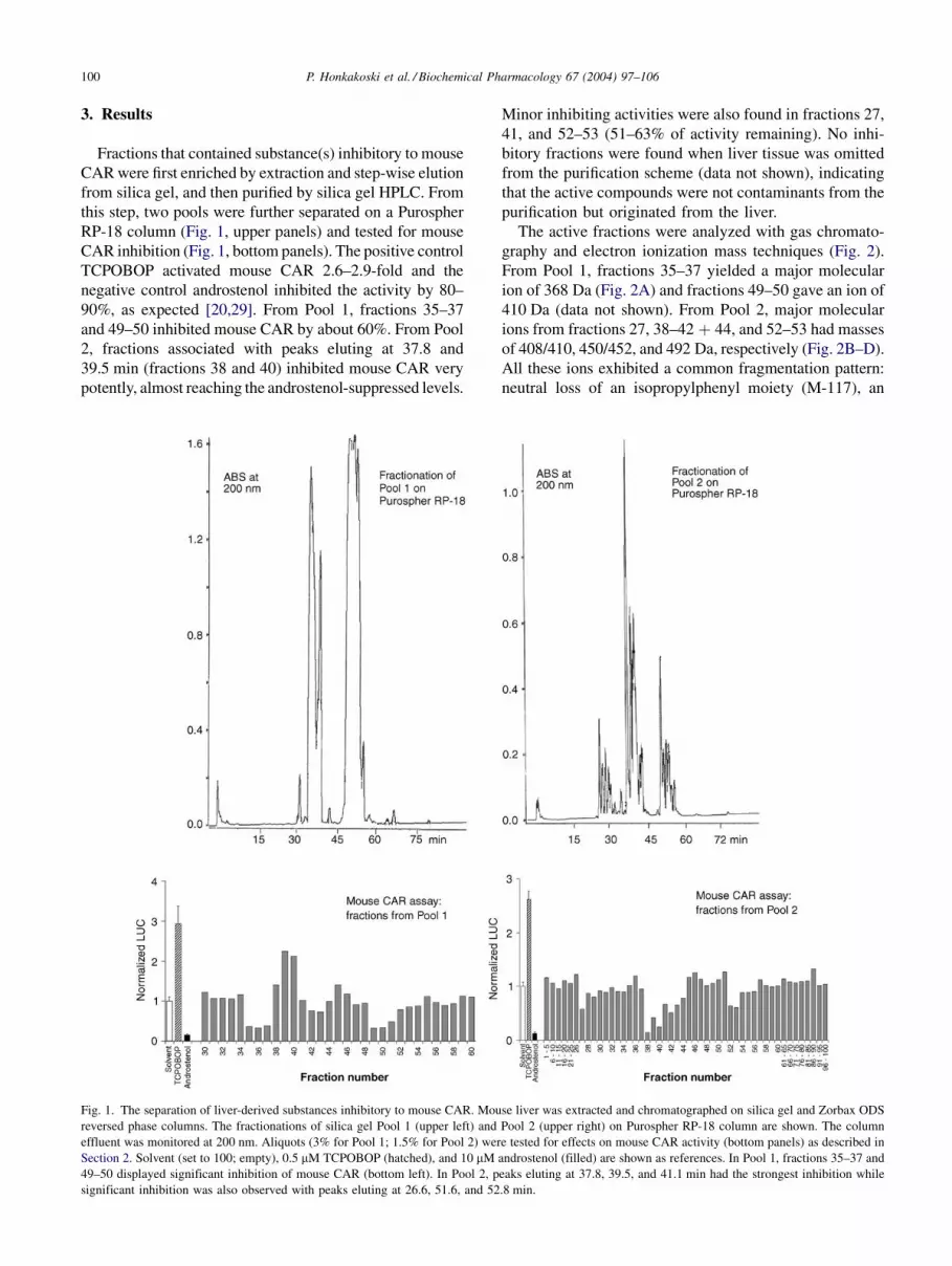
Fractions that contained substance(s) inhibitory to mouse
CAR were first enriched by extraction and step-wise elution
from silica gel, and then purified by silica gel HPLC. From
this step, two pools were further separated on a Purospher
RP-18 column (Fig. 1, upper panels) and tested for mouse
CAR inhibition (Fig. 1, bottom panels). The positive control
TCPOBOP activated mouse CAR 2.6–2.9-fold and the
negative control androstenol inhibited the activity by 80–
90%, as expected [20,29]. From Pool 1, fractions 35–37
and 49–50 inhibited mouse CAR by about 60%. From Pool
2, fractions associated with peaks eluting at 37.8 and
39.5 min (fractions 38 and 40) inhibited mouse CAR very
potently, almost reaching the androstenol-suppressed levels.
Minor inhibiting activities were also found in fractions 27,
41, and 52–53 (51–63% of activity remaining). No inhi-
bitory fractions were found when liver tissue was omitted
from the purification scheme (data not shown), indicating
that the active compounds were not contaminants from the
purification but originated from the liver.
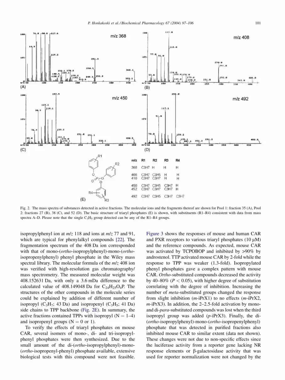
The active fractions were analyzed with gas chromato-
graphy and electron ionization mass techniques (Fig. 2).
From Pool 1, fractions 35–37 yielded a major molecular
ion of 368 Da (Fig. 2A) and fractions 49–50 gave an ion of
410 Da (data not shown). From Pool 2, major molecular
ions from fractions 27, 38–42 þ 44, and 52–53 had masses
of 408/410, 450/452, and 492 Da, respectively (Fig. 2B–D).
All these ions exhibited a common fragmentation pattern:
neutral loss of an isopropylphenyl moiety (M-117), an
Fig. 1. The separation of liver-derived substances inhibitory to mouse CAR. Mouse liver was extracted and chromatographed on silica gel and Zorbax ODS
reversed phase columns. The fractionations of silica gel Pool 1 (upper left) and Pool 2 (upper right) on Purospher RP-18 column are shown. The column
effluent was monitored at 200 nm. Aliquots (3% for Pool 1; 1.5% for Pool 2) were tested for effects on mouse CAR activity (bottom panels) as described in
Section 2. Solvent (set to 100; empty), 0.5 mM TCPOBOP (hatched), and 10 mM androstenol (filled) are shown as references. In Pool 1, fractions 35–37 and
49–50 displayed significant inhibition of mouse CAR (bottom left). In Pool 2, peaks eluting at 37.8, 39.5, and 41.1 min had the strongest inhibition while
significant inhibition was also observed with peaks eluting at 26.6, 51.6, and 52.8 min.
100 P. Honkakoski et al. / Biochemical Pharmacology 67 (2004) 97–106
isopropylphenyl ion at m/z 118 and ions at m/z 77 and 91,
which are typical for phenylalkyl compounds [22]. The
fragmentation spectrum of the 408 Da ion corresponded
with that of mono-(ortho-isopropylphenyl)-mono-(ortho-
isopropenylphenyl) phenyl phosphate in the Wiley mass
spectral library. The molecular formula of the m/z 408 ion
was verified with high-resolution gas chromatography/
mass spectrometry. The measured molecular weight was
408.152631 Da, with only a 3.6 mDa difference to the
calculated value of 408.149048 Da for C24H25O4P. The
structures of the other compounds in the molecule series
could be explained by addition of different number of
isopropyl (C3H7; 43 Da) and isopropenyl (C3H5; 41 Da)
side chains to TPP backbone (Fig. 2E). In summary, the
active fractions contained TPPs with isopropyl (N ¼ 1–4)
and isopropenyl groups (N ¼ 0 or 1).
To verify the effects of triaryl phosphates on mouse
CAR, several isomers of mono-, di- and tri-isopropyl-
phenyl phosphates were then synthesized. Due to the
small amount of the di-(ortho-isopropylphenyl)-mono-
phosphate that was detected in purified fractions also
inhibited mouse CAR to similar extent (data not shown).
These changes were not due to non-specific effects since
the luciferase activity from a reporter gene lacking NR
response elements or b-galactosidase activity that was
used for reporter normalization were not changed by the
Fig. 2. The mass spectra of substances detected in active fractions. The molecular ions and the fragments thereof are shown for Pool 1: fraction 35 (A), Pool
2: fractions 27 (B), 38 (C), and 52 (D). The basic structure of triaryl phosphates (E) is shown, with substituents (R1–R4) consistent with data from mass
spectra A–D. Please note that the single C3H5 group detected can be any of the R1–R4 groups.
P. Honkakoski et al. / Biochemical Pharmacology 67 (2004) 97–106 101
compounds (data not shown). No overt toxicity was evident
after the exposure of the cells to 10 mM triaryl phosphates.
To summarize, these data confirmed that isopropylated
TPPs that were identified in the liver are indeed capable
of inhibiting mouse CAR.
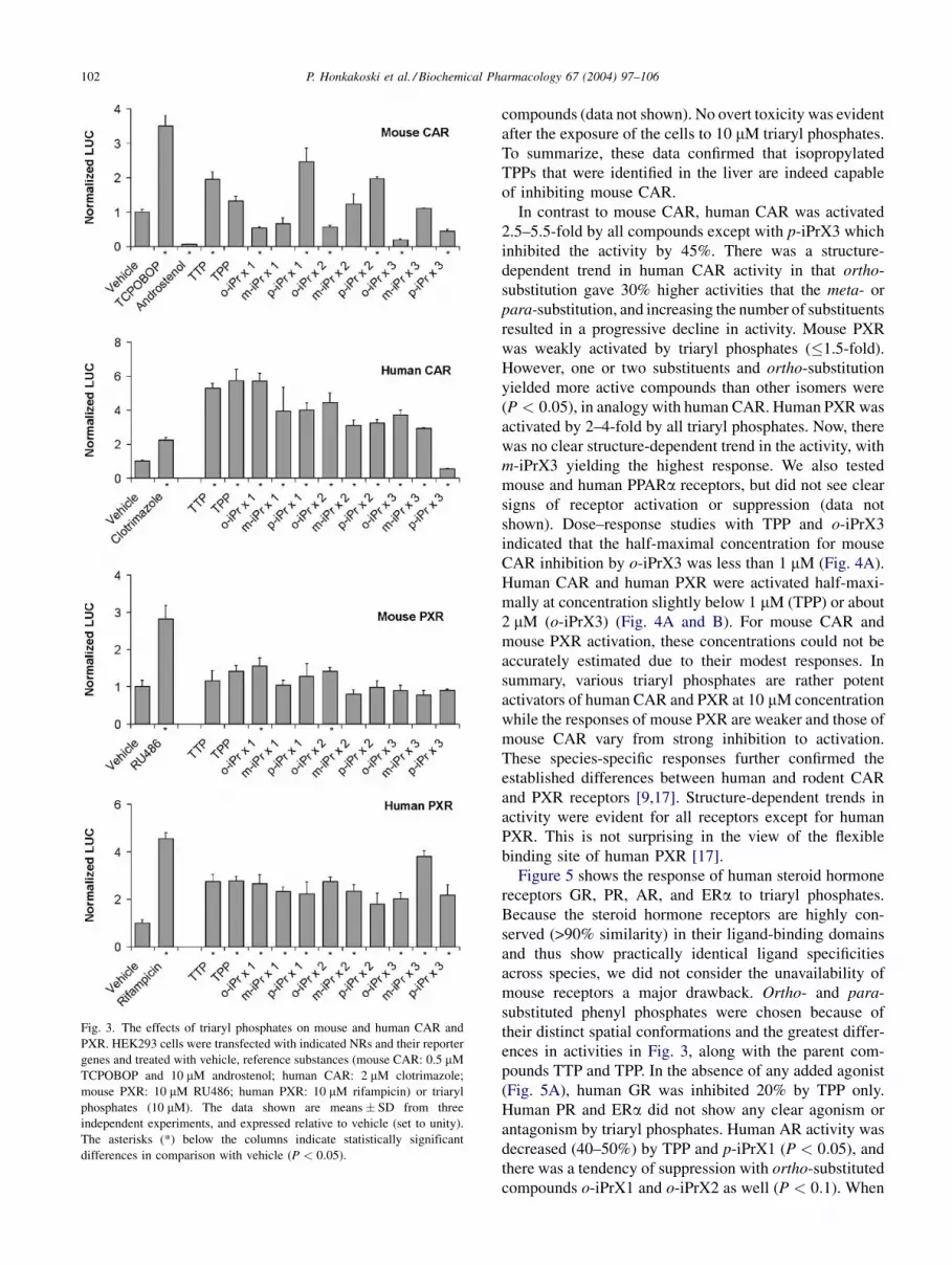
In contrast to mouse CAR, human CAR was activated
2.5–5.5-fold by all compounds except with p-iPrX3 which
inhibited the activity by 45%. There was a structure-
dependent trend in human CAR activity in that ortho-
substitution gave 30% higher activities that the meta- or
para-substitution, and increasing the number of substituents
resulted in a progressive decline in activity. Mouse PXR
was weakly activated by triaryl phosphates (�1.5-fold).
However, one or two substituents and ortho-substitution
yielded more active compounds than other isomers were
(P < 0:05), in analogy with human CAR. Human PXR was
activated by 2–4-fold by all triaryl phosphates. Now, there
was no clear structure-dependent trend in the activity, with
m-iPrX3 yielding the highest response. We also tested
mouse and human PPARa receptors, but did not see clear
signs of receptor activation or suppression (data not
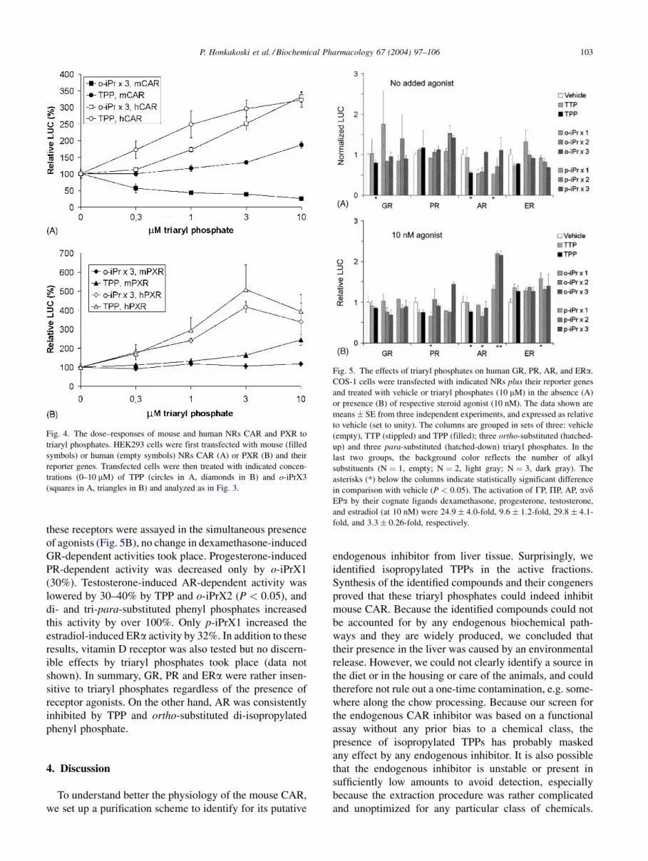
shown). Dose–response studies with TPP and o-iPrX3
indicated that the half-maximal concentration for mouse
CAR inhibition by o-iPrX3 was less than 1 mM (Fig. 4A).
Human CAR and human PXR were activated half-maxi-
mally at concentration slightly below 1 mM (TPP) or about
2 mM (o-iPrX3) (Fig. 4A and B). For mouse CAR and
mouse PXR activation, these concentrations could not be
accurately estimated due to their modest responses. In
summary, various triaryl phosphates are rather potent
activators of human CAR and PXR at 10 mM concentration
while the responses of mouse PXR are weaker and those of
mouse CAR vary from strong inhibition to activation.
These species-specific responses further confirmed the
established differences between human and rodent CAR
and PXR receptors [9,17]. Structure-dependent trends in
activity were evident for all receptors except for human
PXR. This is not surprising in the view of the flexible
binding site of human PXR [17].
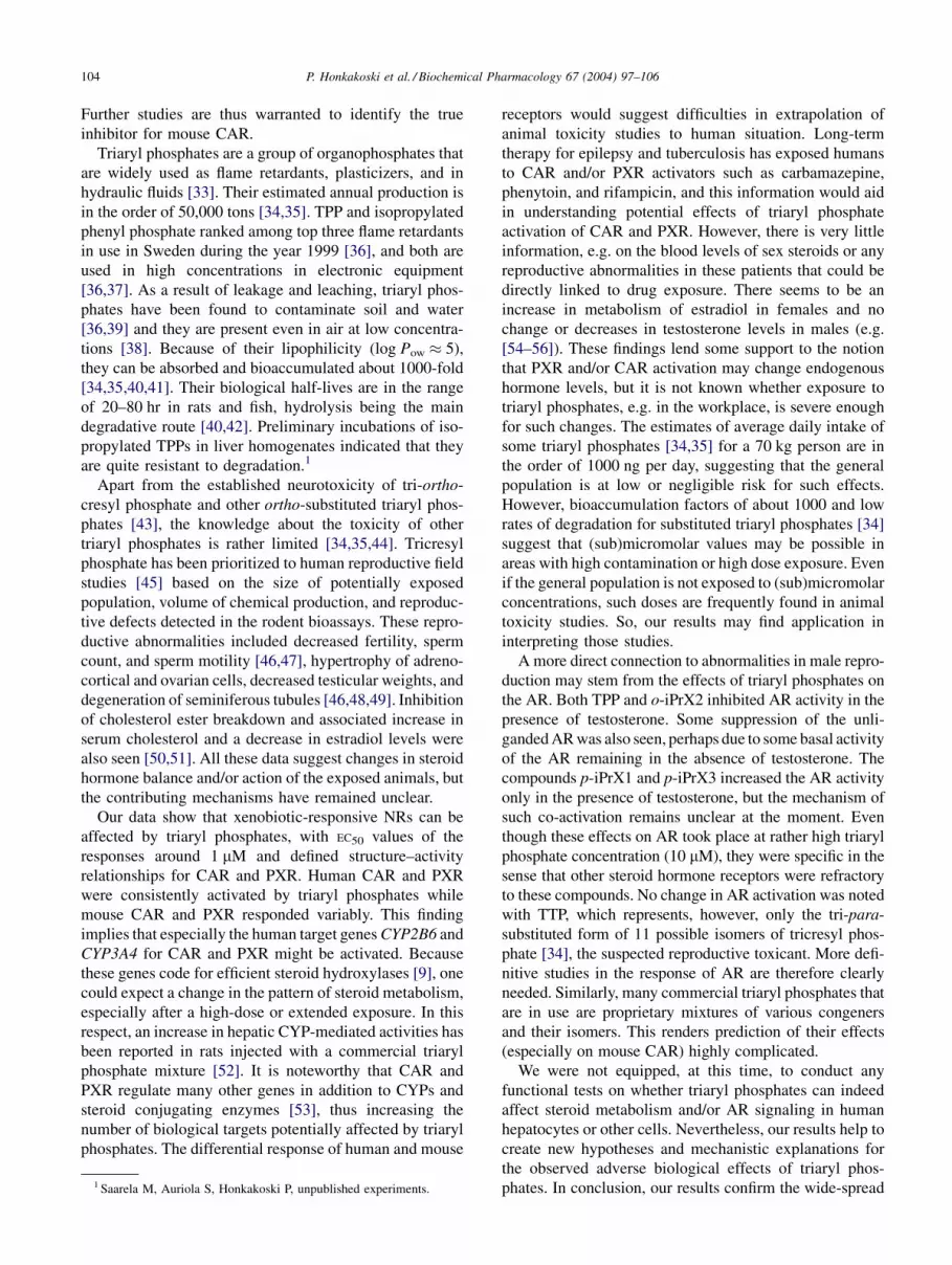
Figure 5 shows the response of human steroid hormone
receptors GR, PR, AR, and ERa to triaryl phosphates.
Because the steroid hormone receptors are highly con-
served (>90% similarity) in their ligand-binding domains
and thus show practically identical ligand specificities
across species, we did not consider the unavailability of
mouse receptors a major drawback. Ortho- and para-
substituted phenyl phosphates were chosen because of
their distinct spatial conformations and the greatest differ-
ences in activities in Fig. 3, along with the parent com-
pounds TTP and TPP. In the absence of any added agonist
(Fig. 5A), human GR was inhibited 20% by TPP only.
Human PR and ERa did not show any clear agonism or
antagonism by triaryl phosphates. Human AR activity was
decreased (40–50%) by TPP and p-iPrX1 (P < 0:05), and
there was a tendency of suppression with ortho-substituted
compounds o-iPrX1 and o-iPrX2 as well (P < 0:1). When
Fig. 3. The effects of triaryl phosphates on mouse and human CAR and
PXR. HEK293 cells were transfected with indicated NRs and their reporter
genes and treated with vehicle, reference substances (mouse CAR: 0.5 mM
TCPOBOP and 10 mM androstenol; human CAR: 2 mM clotrimazole;
mouse PXR: 10 mM RU486; human PXR: 10 mM rifampicin) or triaryl
phosphates (10 mM). The data shown are means � SD from three
independent experiments, and expressed relative to vehicle (set to unity).
The asterisks (*) below the columns indicate statistically significant
differences in comparison with vehicle (P < 0:05).
102 P. Honkakoski et al. / Biochemical Pharmacology 67 (2004) 97–106
these receptors were assayed in the simultaneous presence
of agonists (Fig. 5B), no change in dexamethasone-induced
GR-dependent activities took place. Progesterone-induced
PR-dependent activity was decreased only by o-iPrX1
(30%). Testosterone-induced AR-dependent activity was
lowered by 30–40% by TPP and o-iPrX2 (P < 0:05), and
di- and tri-para-substituted phenyl phosphates increased
this activity by over 100%. Only p-iPrX1 increased the
estradiol-induced ERa activity by 32%. In addition to these
results, vitamin D receptor was also tested but no discern-
ible effects by triaryl phosphates took place (data not
shown). In summary, GR, PR and ERa were rather insen-
sitive to triaryl phosphates regardless of the presence of
receptor agonists. On the other hand, AR was consistently
inhibited by TPP and ortho-substituted di-isopropylated
phenyl phosphate.
4. Discussion
To understand better the physiology of the mouse CAR,
we set up a purification scheme to identify for its putative
endogenous inhibitor from liver tissue. Surprisingly, we
identified isopropylated TPPs in the active fractions.
Synthesis of the identified compounds and their congeners
proved that these triaryl phosphates could indeed inhibit
mouse CAR. Because the identified compounds could not
be accounted for by any endogenous biochemical path-
ways and they are widely produced, we concluded that
their presence in the liver was caused by an environmental
release. However, we could not clearly identify a source in
the diet or in the housing or care of the animals, and could
therefore not rule out a one-time contamination, e.g. some-
where along the chow processing. Because our screen for
the endogenous CAR inhibitor was based on a functional
assay without any prior bias to a chemical class, the
presence of isopropylated TPPs has probably masked
any effect by any endogenous inhibitor. It is also possible
that the endogenous inhibitor is unstable or present in
sufficiently low amounts to avoid detection, especially
because the extraction procedure was rather complicated
and unoptimized for any particular class of chemicals.
Fig. 4. The dose–responses of mouse and human NRs CAR and PXR to
triaryl phosphates. HEK293 cells were first transfected with mouse (filled
symbols) or human (empty symbols) NRs CAR (A) or PXR (B) and their
reporter genes. Transfected cells were then treated with indicated concen-
trations (0–10 mM) of TPP (circles in A, diamonds in B) and o-iPrX3
(squares in A, triangles in B) and analyzed as in Fig. 3.
Fig. 5. The effects of triaryl phosphates on human GR, PR, AR, and ERa.
COS-1 cells were transfected with indicated NRs plus their reporter genes
and treated with vehicle or triaryl phosphates (10 mM) in the absence (A)
or presence (B) of respective steroid agonist (10 nM). The data shown are
means � SE from three independent experiments, and expressed as relative
to vehicle (set to unity). The columns are grouped in sets of three: vehicle
(empty), TTP (stippled) and TPP (filled); three ortho-substituted (hatched-
up) and three para-substituted (hatched-down) triaryl phosphates. In the
last two groups, the background color reflects the number of alkyl
substituents (N ¼ 1, empty; N ¼ 2, light gray; N ¼ 3, dark gray). The
asterisks (*) below the columns indicate statistically significant difference
in comparison with vehicle (P < 0:05). The activation of GP, PP, AP, andEPa by their cognate ligands dexamethasone, progesterone, testosterone,
and estradiol (at 10 nM) were 24:9 � 4:0-fold, 9:6 � 1:2-fold, 29:8 � 4:1-
fold, and 3:3 � 0:26-fold, respectively.
P. Honkakoski et al. / Biochemical Pharmacology 67 (2004) 97–106 103
Further studies are thus warranted to identify the true
inhibitor for mouse CAR.
Triaryl phosphates are a group of organophosphates that
are widely used as flame retardants, plasticizers, and in
hydraulic fluids [33]. Their estimated annual production is
in the order of 50,000 tons [34,35]. TPP and isopropylated
phenyl phosphate ranked among top three flame retardants
in use in Sweden during the year 1999 [36], and both are
used in high concentrations in electronic equipment
[36,37]. As a result of leakage and leaching, triaryl phos-
phates have been found to contaminate soil and water
[36,39] and they are present even in air at low concentra-
tions [38]. Because of their lipophilicity (log Pow � 5),
they can be absorbed and bioaccumulated about 1000-fold
[34,35,40,41]. Their biological half-lives are in the range
of 20–80 hr in rats and fish, hydrolysis being the main
degradative route [40,42]. Preliminary incubations of iso-
propylated TPPs in liver homogenates indicated that they
are quite resistant to degradation.1
Apart from the established neurotoxicity of tri-ortho-
cresyl phosphate and other ortho-substituted triaryl phos-
phates [43], the knowledge about the toxicity of other
triaryl phosphates is rather limited [34,35,44]. Tricresyl
phosphate has been prioritized to human reproductive field
studies [45] based on the size of potentially exposed
population, volume of chemical production, and reproduc-
tive defects detected in the rodent bioassays. These repro-
ductive abnormalities included decreased fertility, sperm
count, and sperm motility [46,47], hypertrophy of adreno-
cortical and ovarian cells, decreased testicular weights, and
degeneration of seminiferous tubules [46,48,49]. Inhibition
of cholesterol ester breakdown and associated increase in
serum cholesterol and a decrease in estradiol levels were
also seen [50,51]. All these data suggest changes in steroid
hormone balance and/or action of the exposed animals, but
the contributing mechanisms have remained unclear.
Our data show that xenobiotic-responsive NRs can be
affected by triaryl phosphates, with EC50 values of the
responses around 1 mM and defined structure–activity
relationships for CAR and PXR. Human CAR and PXR
were consistently activated by triaryl phosphates while
mouse CAR and PXR responded variably. This finding
implies that especially the human target genes CYP2B6 and
CYP3A4 for CAR and PXR might be activated. Because
these genes code for efficient steroid hydroxylases [9], one
could expect a change in the pattern of steroid metabolism,
especially after a high-dose or extended exposure. In this
respect, an increase in hepatic CYP-mediated activities has
been reported in rats injected with a commercial triaryl
phosphate mixture [52]. It is noteworthy that CAR and
PXR regulate many other genes in addition to CYPs and
steroid conjugating enzymes [53], thus increasing the
number of biological targets potentially affected by triaryl
phosphates. The differential response of human and mouse
receptors would suggest difficulties in extrapolation of
animal toxicity studies to human situation. Long-term
therapy for epilepsy and tuberculosis has exposed humans
to CAR and/or PXR activators such as carbamazepine,
phenytoin, and rifampicin, and this information would aid
in understanding potential effects of triaryl phosphate
activation of CAR and PXR. However, there is very little
information, e.g. on the blood levels of sex steroids or any
reproductive abnormalities in these patients that could be
directly linked to drug exposure. There seems to be an
increase in metabolism of estradiol in females and no
change or decreases in testosterone levels in males (e.g.
[54–56]). These findings lend some support to the notion
that PXR and/or CAR activation may change endogenous
hormone levels, but it is not known whether exposure to
triaryl phosphates, e.g. in the workplace, is severe enough
for such changes. The estimates of average daily intake of
some triaryl phosphates [34,35] for a 70 kg person are in
the order of 1000 ng per day, suggesting that the general
population is at low or negligible risk for such effects.
However, bioaccumulation factors of about 1000 and low
rates of degradation for substituted triaryl phosphates [34]
suggest that (sub)micromolar values may be possible in
areas with high contamination or high dose exposure. Even
if the general population is not exposed to (sub)micromolar
concentrations, such doses are frequently found in animal
toxicity studies. So, our results may find application in
interpreting those studies.
A more direct connection to abnormalities in male repro-
duction may stem from the effects of triaryl phosphates on
the AR. Both TPP and o-iPrX2 inhibited AR activity in the
presence of testosterone. Some suppression of the unli-
ganded AR was also seen, perhaps due to some basal activity
of the AR remaining in the absence of testosterone. The
compounds p-iPrX1 and p-iPrX3 increased the AR activity
only in the presence of testosterone, but the mechanism of
such co-activation remains unclear at the moment. Even
though these effects on AR took place at rather high triaryl
phosphate concentration (10 mM), they were specific in the
sense that other steroid hormone receptors were refractory
to these compounds. No change in AR activation was noted
with TTP, which represents, however, only the tri-para-
substituted form of 11 possible isomers of tricresyl phos-
phate [34], the suspected reproductive toxicant. More defi-
nitive studies in the response of AR are therefore clearly
needed. Similarly, many commercial triaryl phosphates that
are in use are proprietary mixtures of various congeners
and their isomers. This renders prediction of their effects
(especially on mouse CAR) highly complicated.
We were not equipped, at this time, to conduct any
functional tests on whether triaryl phosphates can indeed
affect steroid metabolism and/or AR signaling in human
hepatocytes or other cells. Nevertheless, our results help to
create new hypotheses and mechanistic explanations for
the observed adverse biological effects of triaryl phos-
phates. In conclusion, our results confirm the wide-spread1 Saarela M, Auriola S, Honkakoski P, unpublished experiments.
104 P. Honkakoski et al. / Biochemical Pharmacology 67 (2004) 97–106
contamination by triaryl phosphates, and indicate for the
first time that triaryl phosphates are efficient activators of
human CAR and PXR, the main regulators of steroid
hormone-metabolizing CYP and conjugating enzymes,
and suggest biological pathways that might be affected
by triaryl phosphates.
Acknowledgments
Excellent technical assistance by Saija Kotola, Hanna
Eskelinen, and Kaarina Pitkanen is gratefully acknowl-
edged. We wish to thank Dr. Pekka Savolainen for his
expertise in synthesis, and Dr. Veli-Pekka Ranta, and
Dr. Hannu Taipale for their advice on HPLC purifications.
Dr. Thorvald Staaf, Dr. Hakan Carlsson, and Prof. Conny
Ostman are acknowledged for their gifts of triaryl phos-
phate preparations and kind advice during the study.
This work was supported by grants from the Academy
of Finland, the Finnish Foundation for Cancer Research
and Biocentrum, Helsinki.
References
[1] Maglich JM, Sluder A, Guan X, Shi Y, McKee DD, Carrick K, Kamdar
K, Willson TM, Moore JT, Comparison of complete nuclear receptor
sets from the human, Caenorhabditis elegans and Drosophila gen-