4 Chapter Enrichment of Perforate Septal Pore Caps from the Basidiomycetous Fungus Rhizoctonia solani by Combined Use of French Press, Isopycnic Centrifugation, and Triton X-100 Kenneth G.A. van Driel, Arend F. van Peer, Han A.B. Wösten, Arie J. Verkleij, Teun Boekhout & Wally H. Müller Published in Journal of Microbiological Methods doi:10.1016/j.mimet.2007.09.013
Transcript
4Chapter
Enrichment of Perforate Septal Pore Caps from the Basidiomycetous Fungus Rhizoctonia solani by Combined Use of French Press, Isopycnic Centrifugation, and Triton X-100
Kenneth G.A. van Driel, Arend F. van Peer, Han A.B. Wösten, Arie J. Verkleij, Teun Boekhout & Wally H. Müller
Published in Journal of Microbiological Methods doi:10.1016/j.mimet.2007.09.013
Chapter 4
80
ABSTRACT
Septal pore caps occur in many filamentous Basidiomycota fungi. They are located at both sides of the dolipore septum and are at their base connected to the endoplasmic reticulum. The septal pore cap ultrastructure has been described extensively, but its composition and function are not yet known. To enable biochemical and functional analyses, we here describe an enrichment method for perforate septal pore caps from Rhizoctonia solani. Our method is based on the combined use of French press, isopycnic centrifugation using a discontinuous sucrose gradient, and a treatment with Triton X-100. Enrichment was monitored by the use of scanning electron microscopy and transmission electron microscopy. Using the same isolation method, smaller septal pore caps were isolated from two other basidiomycetous fungi as well. Furthermore, we showed that pore-occluding material co-purified with the septal pore caps. This observation supports the hypothesis that septal pore caps play a key role in the plugging process of the septal pores in filamentous Basidiomycota fungi.
INTRODUCTION
The septal pore cap (SPC) or parenthesome is a membranous structure located at both sides of the dolipore septum in many filamentous Basidiomycota fungi (Moore & McAlear, 1962; Bracker & Butler, 1963). In 1958, Girbardt described for the first time the SPC ultrastructure at the septum of Trametes versicolor by the use of transmission electron microscopy (cited as Polystictus versicolor; Girbardt, 1958). Since then, many studies on the SPC ultrastructure have followed (e.g. Bracker & Butler, 1963; Setliff et al., 1972; Khan & Kimbrough, 1982; Patrignani & Pellegrini, 1986; Wells, 1994; Moore, 1996; Keller, 1997; Müller et al., 1998b, 2000b). These studies have shown that the ultrastructure of SPCs is diverse and can be divided into the following types: the vesicular (saccular, cupulate or tubular) type, the imperforate (or continuous) type, and the perforate type (McLaughlin et al., 1995). The absence or presence of a SPC, the SPC-type, and septal morphology are important markers that reflect the main phylogenetic groups in the Basidiomycota (Khan & Kimbrough, 1982; Wells, 1994; McLaughlin et al., 1995; Moore, 1996; Müller et al., 2000b, Weiss et al., 2004; Chapter 2).
Although the ultrastructure of SPCs has been studied extensively, its precise function is still a matter of speculation. As the base of SPCs is continuous with the endoplasmic reticulum (ER), the SPC has been proposed to be a subdomain of the ER (Girbardt, 1961; Bracker & Butler, 1963; Müller et al., 1995a). However, differences in calcium-affinity sites between the ER and SPCs have been observed by the use of zinc-iodin-osmium tetroxide staining (Müller et al., 1995a, 1998a). Possibly, SPCs act as a repository for
Enrichment of septal pore caps from Rhizoctonia solani
81
certain proteins that are produced and processed in the ER, and that are translocated from the SPC to the septal pore when pore sealing is needed in cases of stress or hyphal damage (Müller et al., 1998a). Several other functions of SPCs have been suggested: they may act as a sieve to discriminate between organelles that pass the pore (Wilsenach & Kessel, 1965), they may guide cytoplasmic streams to the pore (Orlovich & Ashford, 1994), or they may function in protoplasmic streaming, by which SPCs protect the dolipore for accidental closing or diminishing the pore by organelles that could hit the sides of the swelling in case SPCs are absent (Bracker & Butler, 1964). Furthermore, in Schizophyllum commune, Rhizoctonia solani, and Pisolithus tinctorius filamentous structures were observed that connect the inside of the SPC with the pore-occluding material (Orlovich & Ashford, 1994; Müller et al., 1999, 2000a). These observations suggest that SPCs play a key role in plug formation after hyphal damage or stress and consequently, are of importance in intercellular communication within the hyphae (Thielke, 1972; Aylmore et al., 1984; Markham, 1994; Müller et al., 1999, 2000a).
The many ultrastructural studies of SPCs have led to a number of hypotheses on SPC functioning. Biochemical analysis of SPCs may help to understand its function in the hyphal cells of the Basidiomycota. However, an isolation procedure of SPCs necessary to biochemically study these organelles has never been published. We here describe for the first time a method to enrich SPCs from the basidiomycetous fungus R. solani. Using the same procedure, we could also isolate SPCs from two other species belonging to the Rhizoctonia sensu lato (s.l.) complex (Müller et al., 1998b), namely Thanatephorus cucumeris and Ceratobasidium cornigerum. Furthermore, we showed that a structural complex consisting of SPCs attached by filaments to pore-occluding material could be isolated. The presented method will also allow the future isolation and biochemical analysis of proteins of perforate SPCs from different basidiomycetous fungi.
MATERIALS & METHODS
Organisms, Media and Culture ConditionsRhizoctonia solani (CBS 346.84), Thanatephorus cucumeris (CBS 700.82), and Ceratobasidium cornigerum (CBS 132.82) were grown on malt extract agar (Oxoid, Hampshire, UK) at 25oC for 4 days. A Sorvall omni-mixer (Kendro Laboratory Products GmbH, Langenselbold, Germany) was used for 10 sec at speed 4 to homogenize the culture in 100 ml complete medium (20.0 g glucose, 2.0 g peptone L37 (Oxoid), 2.0 g yeast extract (Difco, Detroit, MI, USA), 0.5 g MgSO4.7H2O, 0.46 g KH2PO4, 1.0 g K2HPO4 per liter) containing 100 mg/l Penicillin-G (Yamanouchi Pharma, Leiderdorp, The Netherlands) and 100,000 units/l Streptomycin (Radiumfarma-Fissiopharma, Naples, Italy). After growing for 48 hr at 175 rpm and 25oC, the culture was again homogenized in an omni-mixer for 10 sec at
Chapter 4
82
speed 4. Thereafter, 10 ml of this homogenate was used to inoculate 100 ml complete medium and growth was allowed for 3 days at 175 rpm and 25oC.
Subcellular Fractionation by Isopycnic CentrifugationThe mycelium from three-days-old submerged cultures was harvested by centrifugation at 2000 rpm and 4oC for 3 min in a Mistral 400 centrifuge (MSE Scientific Instruments, West Sussex, UK). Subsequently, the mycelium was washed twice in HEPES/KAc buffer (20 mM HEPES, pH 6.8, 50 mM potassium acetate (KAc), 200 mM D-sorbitol, 1 mM EDTA) (Rieder & Emr, 2000) and resuspended in half a volume of HEPES/KAc buffer supplemented with 1/200 volume of protease inhibitor cocktail (Sigma-Aldrich, St. Louis, MO, USA). The mycelium was disrupted by two passages of 500 PSI (equivalent to 3447 kPa) through a French press (American Instrument Company, Silver Spring, MD, USA), resulting into homogeneous fungal cell extracts.
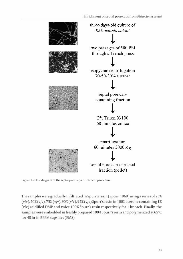
For isopycnic centrifugation, 12 ml of fungal cell extracts was layered on top of a discontinuous gradient consisting of 8 ml 70% (w/v), 8 ml 50% (w/v), and 8 ml 30% (w/v) sucrose in HEPES/KAc buffer in a 38.5 ml polyallomer thinwall ultracentrifuge tube (Herolab GmbH, Wiesloch, Germany). After centrifugation at 85,000 × g (21,500 rpm) and 4oC for 90 min in a Centrikon T-2180 ultracentrifuge (Kontron Instruments, Watford, UK), fractions were collected from underneath using a pasteur pipette with a 180 degrees bent tip. The fractions were diluted with HEPES/KAc buffer containing 2% (w/v) Triton X-100 (GE Healthcare, Uppsala, Sweden) to a final sucrose concentration of 10% (w/v). After one hour on ice, fractions were centrifuged at 5,000 × g (5,300 rpm) and 4oC for 1 hr. The resulting pellet was resuspended in 1 ml HEPES/KAc buffer and further processed for electron microscopy as described below. To analyze the supernatant (remaining after the 5,000 × g centrifugation step) for the presence of SPCs, the supernatant was centrifuged at 100,000 × g (23,500 rpm) and 4oC for another 45 min, and subsequently the resulting pellet was resuspended in 1 ml HEPES/KAc buffer and processed for electron microscopy. A flow chart of the SPC-enrichment procedure is presented in Figure 1.
Transmission Electron MicroscopyThe obtained fraction samples were fixed chemically or by high-pressure freezing. For the chemical fixation, samples were fixed in 1.5% glutaraldehyde (Agar Scientific Ltd, Essex, UK) buffered with 20 mM HEPES, pH 6.8, overnight at 4oC. Samples were washed twice in PBS and post-fixed in 1% (w/v) aqueous osmium tetroxide (EMS, Hatfield, PA, USA) for one hour at room temperature. Subsequently, the samples were washed twice in distilled water, followed by gradual dehydration in a series of ascending concentrations of acetone, namely 60% (v/v), 70% (v/v), 80% (v/v), 90% (v/v), and three times of 100% acetone containing 1% (v/v) acidified 2,2-dimethoxypropane (DMP) for 30 min each.
Enrichment of septal pore caps from Rhizoctonia solani
83
Figure 1 – Flow diagram of the septal pore cap-enrichment procedure.
The samples were gradually infiltrated in Spurr’s resin (Spurr, 1969) using a series of 25% (v/v), 50% (v/v), 75% (v/v), 90% (v/v), 95% (v/v) Spurr’s resin in 100% acetone containing 1% (v/v) acidified DMP and twice 100% Spurr’s resin respectively for 1 hr each. Finally, the samples were embedded in freshly prepared 100% Spurr’s resin and polymerized at 65oC for 48 hr in BEEM capsules (EMS).
Chapter 4
84
Mycelium of R. solani was subjected to high-pressure freezing (HPF) and freeze-substitution as described by Müller et al. (1998a). To high-pressure freeze the SPC-enriched fraction samples, an aluminum lecithin-coated planchette (100 µm-deep well) used for HPF (Engineering Office M. Wohlwend GMbH, Sennwald, Switzerland) was dipped into the SPC-enriched pellet and we drew material from the bottom of the tube. A second lecithin-coated planchette (300 µm-deep well) was placed with the flat side as a lid on top of the first planchette, thus subjecting 100 µm thick SPC-enriched pellet material to HPF by the use of a Leica EM HPF (Leica Microsystems, Vienna, Switzerland). After separating the planchettes in liquid nitrogen, the samples were freeze-substituted in a mixture of 1% (w/v) osmium tetroxide, 3% (v/v) glutaraldehyde, and 0.3% (w/v) uranyl acetate in anhydrous methanol at -85oC for 2 days. After rinsing with methanol, the samples were gradually infiltrated using a series of 25% (v/v), 50% (v/v), 75% (v/v) lowicryl HM20 (EMS) for 2 hr each, and three times 100% lowicryl HM20 for 1 day each. Finally, the samples were low-temperature embedded in lowicryl HM20, and were polymerized after 48 hours at -35oC and 24 hr at room temperature under UV light.
Sections of about 90 nm and 350 nm were cut with a diamond knife (Diatome, Hatfield, PA, USA) using an ULTRACUT E ultramicrotome (Leica Microsystems, Vienna, Austria). The sections were picked up with formvar film-coated, carbon-stabilized copper grids (hexagonal 150 mesh Veco grids, EMS). Sections were contrasted with 4% (w/v) aqueous uranyl acetate (Merck) for 10 min and 0.4% (w/v) aqueous lead citrate (Merck) for 2 min (Venable & Coggeshall, 1965). The sections were viewed with a TECNAI 10 (FEI Company, Eindhoven, The Netherlands) transmission electron microscope at an acceleration voltage of 100 kV. Scanning Electron MicroscopyA small aliquot of the R. solani SPC-enriched fraction was placed on a formvar film-coated, carbon-stabilized grid and incubated for 10 min at room temperature. Thereafter, excess of fluid was removed carefully by touching the edge of the grid with a piece of Whatman paper, and subsequently the grid was air-dried overnight. Then, it was incubated 2 min with 4% (w/v) aqueous uranyl acetate, and subsequently washed thoroughly in distilled water. The grid was dried carefully with a piece of Whatman paper and further air-dried. Finally, the grids were mounted on a stub, coated with 5 nm Pt/Pd by using a Cressington sputter coater 208HR (Cressington Scientific Instruments, Watford, UK), and viewed in a XL30 scanning electron microscope (FEI Company) at an acceleration voltage of 15 kV and a working distance of about 8 mm.
Enrichment of septal pore caps from Rhizoctonia solani
85
RESULTS
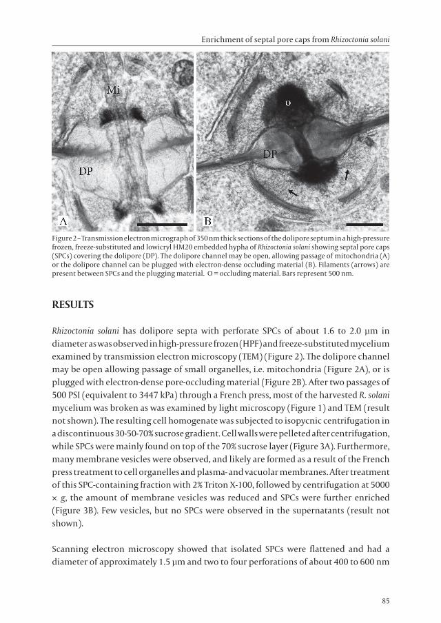
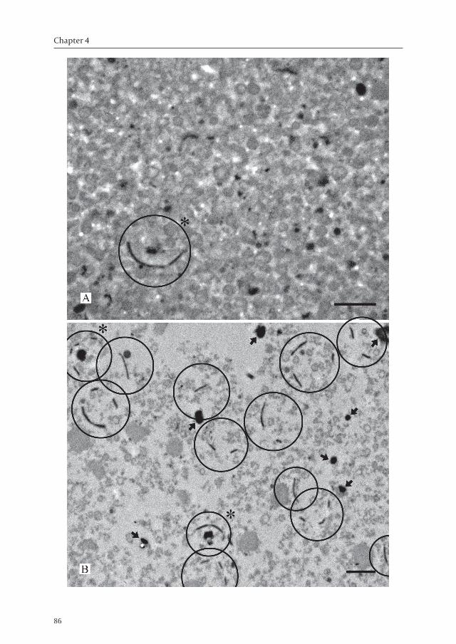
Rhizoctonia solani has dolipore septa with perforate SPCs of about 1.6 to 2.0 µm in diameter as was observed in high-pressure frozen (HPF) and freeze-substituted mycelium examined by transmission electron microscopy (TEM) (Figure 2). The dolipore channel may be open allowing passage of small organelles, i.e. mitochondria (Figure 2A), or is plugged with electron-dense pore-occluding material (Figure 2B). After two passages of 500 PSI (equivalent to 3447 kPa) through a French press, most of the harvested R. solani mycelium was broken as was examined by light microscopy (Figure 1) and TEM (result not shown). The resulting cell homogenate was subjected to isopycnic centrifugation in a discontinuous 30-50-70% sucrose gradient. Cell walls were pelleted after centrifugation, while SPCs were mainly found on top of the 70% sucrose layer (Figure 3A). Furthermore, many membrane vesicles were observed, and likely are formed as a result of the French press treatment to cell organelles and plasma- and vacuolar membranes. After treatment of this SPC-containing fraction with 2% Triton X-100, followed by centrifugation at 5000 × g, the amount of membrane vesicles was reduced and SPCs were further enriched (Figure 3B). Few vesicles, but no SPCs were observed in the supernatants (result not shown).
Scanning electron microscopy showed that isolated SPCs were flattened and had a diameter of approximately 1.5 µm and two to four perforations of about 400 to 600 nm
Figure 2 – Transmission electron micrograph of 350 nm thick sections of the dolipore septum in a high-pressure frozen, freeze-substituted and lowicryl HM20 embedded hypha of Rhizoctonia solani showing septal pore caps (SPCs) covering the dolipore (DP). The dolipore channel may be open, allowing passage of mitochondria (A) or the dolipore channel can be plugged with electron-dense occluding material (B). Filaments (arrows) are present between SPCs and the plugging material. O = occluding material. Bars represent 500 nm.
Chapter 4
86
Enrichment of septal pore caps from Rhizoctonia solani
87
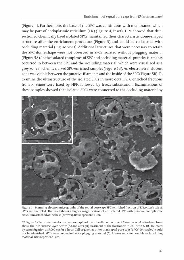
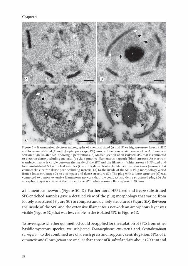
(Figure 4). Furthermore, the base of the SPC was continuous with membranes, which may be part of endoplasmic reticulum (ER) (Figure 4, inset). TEM showed that thin-sectioned chemically fixed isolated SPCs maintained their characteristic dome-shaped structure after the enrichment procedure (Figure 5) and could be co-isolated with occluding material (Figure 5B-D). Additional structures that were necessary to retain the SPC dome-shape were not observed in SPCs isolated without plugging material (Figure 5A). In the isolated complexes of SPC and occluding material, putative filaments occurred in between the SPC and the occluding material, which were visualized as a grey zone in chemical fixed SPC-enriched samples (Figure 5B). An electron-translucent zone was visible between the putative filaments and the inside of the SPC (Figure 5B). To examine the ultrastructure of the isolated SPCs in more detail, SPC-enriched fractions from R. solani were fixed by HPF, followed by freeze-substitution. Examinations of these samples showed that isolated SPCs were connected to the occluding material by
Figure 4 – Scanning electron micrographs of the septal pore cap (SPC) enriched fraction of Rhizoctonia solani. SPCs are encircled. The inset shows a higher magnification of an isolated SPC with putative endoplasmic reticulum attached at the base (arrows). Bars represent 1 µm.
Ô Figure 3 – Transmission electron micrographs of the subcellular fraction of Rhizoctonia solani isolated from above the 70% sucrose layer before (A) and after (B) treatment of the fraction with 2% Triton X-100 followed by centrifugation at 5,000 × g for 1 hour. Cell organelles other than septal pore caps (SPCs) (encircled) could not be identified. SPCs were co-purified with plugging material (*). Arrows indicate possible isolated plug material. Bars represent 1µm.
Chapter 4
88
a filamentous network (Figure 5C, D). Furthermore, HPF-fixed and freeze-substituted SPC-enriched samples gave a detailed view of the plug morphology that varied from loosely structured (Figure 5C) to compact and densely structured (Figure 5D). Between the inside of the SPC and the extensive filamentous network an amorphous layer was visible (Figure 5C) that was less visible in the isolated SPC in Figure 5D.
To investigate whether our method could be applied for the isolation of SPCs from other basidiomycetous species, we subjected Thanatephorus cucumeris and Ceratobasidium cornigerum to the combined use of French press and isopycnic centrifugation. SPCs of T. cucumeris and C. cornigerum are smaller than those of R. solani and are about 1200 nm and
Figure 5 – Transmission electron micrographs of chemical fixed (A and B) or high-pressure frozen (HPF) and freeze-substituted (C and D) septal pore cap (SPC) enriched fractions of Rhizoctonia solani. A) Transverse section of an isolated SPC showing 3 perforations. B) Median section of an isolated SPC that is connected to electron-dense occluding material (o) via a putative filamentous network (black arrows). An electron-translucent zone is visible between the inside of the SPC and the filaments (white arrows). HPF-fixed and freeze-substituted SPC-enriched samples (C and D) show clearly the filamentous structures (arrows) that connect the electron-dense pore-occluding material (o) to the inside of the SPCs. Plug morphology varied from a loose structure (C), to a compact and dense structure (D). The plug with a loose structure (C) was connected to a more extensive filamentous network than the compact and dense structured plug (D). An amorphous layer is visible at the inside of the SPC (white arrows). Bars represent 200 nm.
Enrichment of septal pore caps from Rhizoctonia solani
89
850 nm in diameter respectively. Cell homogenates were successfully generated and for both, SPCs could be isolated after isopycnic centrifugation on top of the 70% sucrose layer (result not shown). However, the SPC-enriched fraction of C. cornigerum contained also mycelial fragments and remnants of broken cell walls, whereas T. cucumeris cell walls and mycelial fragments passed the 70% sucrose layer and were pelleted at the bottom of the ultracentrifuge tube, like in R. solani (result not shown). Moreover, C. cornigerum SPCs were also found in the 50% sucrose layer (result not shown). SPC fractions from C. cornigerum and T. cucumeris contained complexes of SPCs co-isolated with electron-dense occluding material as observed in R. solani SPC-enriched fractions (result not shown).
DISCUSSION
A wealth of information is available on the ultrastructure of SPCs in Basidiomycota, but about the function one can only speculate at present. The ultrastructure of the SPC in Rhizoctonia solani has been extensively studied by transmission electron microscopy (TEM) (Bracker & Butler, 1963, 1964; Setliff et al., 1972), scanning electron microscopy (SEM) (Lisker et al., 1975; Müller et al., 1998b) and automated electron tomography (Müller et al., 2000a). However, SPCs have never been subjected to biochemical analysis of the proteins residing in the SPCs. Prior to analysis of these SPC-related proteins, a method is needed to enrich SPCs. Ideally, this method must also be applicable to other basidiomycetous fungi.
Fungal organelles can be isolated using several approaches. For example, Penicillium chrysogenum microbodies were isolated by protoplasting followed by isopycnic centrifugation (Müller et al., 1995b). Protoplasting does not affect the structural integrity of SPCs (Müller et al., 1998a), however, the yield of SPC-containing protoplasts is low and release of SPCs from protoplasts was unsuccessful (E. Boon & W.H Müller, unpublished results). Woronin bodies from Neurospora crassa were enriched after the cells were frozen in liquid nitrogen and ground to a fine powder that was separated on a sucrose cushion (Jedd & Chua, 2000). This study led to the findings that Woronin bodies are pre-formed peroxisomes that consist of HEX-1 protein and are necessary for septal pore sealing in filamentous Ascomycota (Jedd & Chua, 2000). More recently, laser microdissection by the use of a PALM laserbeam system resulted into isolated septal regions of R. solani (Van Driel et al., 2007). Though fungal septa including SPCs were successfully isolated, laser microdissection is a very laborious technique that is not available in every laboratory. Furthermore, the isolation of proteins from sectioned fungal septa has not been optimized yet. Compared to described isolation methods, the success of our SPC isolation method is based on the combination of French press, isopycnic centrifugation, and a treatment with Triton X-100 that resulted into a high
Chapter 4
90
yield of isolated SPCs (Figure 3B).
Cell homogenates of R. solani, T. cucumeris, and C. cornigerum were prepared by passage of the mycelium through a French press. We experienced that the smaller hyphae of C. cornigerum were more resistant to the pressure of the French press than the broader hyphae of R. solani and T. cucumeris, resulting in more mycelial fragments present in the fractions. Therefore, to generate cell homogenates from other fungi one may need additional passages or higher PSI values to break all the cells. By combining the French press with isopycnic centrifugation we could isolate the different sized SPCs as visualized by TEM. However, we cannot rule out that by the inclusion of the French press step in our method, parts of the SPCs will be ripped into smaller components that are not recognized in the SPC-enriched fraction by TEM. Still, when isolating these SPC-components together with the enriched intact SPCs, this will result into a high yield of SPC components enhancing future biochemical studies.
To enrich the SPCs, we included a treatment with Triton X-100 detergent to solubilize the many membrane structures and vesicles that developed after French press treatment and isopycnic centrifugation. Triton X-100 is a non-ionic detergent often used to solubilize lipid membranes to isolate membrane proteins or detergent-resistant membrane domains like lipid rafts (reviewed by London & Brown, 2000). Interestingly, this Triton-treatment did not solubilize the SPC membranes, the filamentous network or the pore-occluding material as was only seen in the HPF-fixed and freeze-substituted SPC-enriched samples. Therefore, the membranes of the perforate SPCs of R. solani may not exclusively build-up by lipids that are solubilized by detergent, but they also may consist other components like sphingolipids or proteins that keep the SPC detergent-resistant. Wheat germ agglutinin labeling showed that N-acetyl glucosamine residues are present in SPCs (Benhamou et al., 1993; Van Driel et al., 2007) that may indicate the presence of glycoproteins in the SPC matrix, in the SPC-membranes or in both. Alternatively, a fibrous layer on top of the SPC (Figures 5C and 5D) that was also observed in other studies (C.E. Bracker, pers. comm.; Müller et al., 2000a) and the amorphous layer at the inside of the SPC (Figure 5C) that was also observed in S. commune (Müller et al., 1998a) may prevent SPC membrane solubilization by Triton X-100.
Next to analysis of the fractions by TEM, SEM was used as a fast and easy-to-use method to analyze the quality of the SPCs and the degree of enrichment. SEM analysis showed that SPCs in the R. solani SPC-enriched fraction were slightly reduced in size and the characteristic dome-shaped morphology was flattened compared to earlier observations (Müller et al., 1998b). Both observations may be a result of the air-drying procedure in the preparation for SEM analysis. In contrast, TEM analysis showed isolated SPCs with the characteristic dome-shaped morphology. In addition, isolated SPCs that were not
Enrichment of septal pore caps from Rhizoctonia solani
91
associated with plugging material did not show any additional structures that were necessary to retain the dome-shaped structure of SPCs as was proposed by Orlovich and Ashford (1994). This was also demonstrated in free-lying SPCs in Schizophyllum commune protoplasts (Müller et al., 1998a), which showed the same morphology as in intact hyphal cells. These observations, together with the fact that the SPC structure was not broken down or solubilized after the enrichment procedure that includes passages through a French press and a Triton X-100 treatment, we conclude that SPCs are resistant to high pressures and non-ionic surfactants.
The isolation of structural complexes consisting of SPCs connected to pore-occluding material by a filamentous network shows the close interaction of these two structures. Chemically fixed fractions showed an electron-translucent zone between the inside of the SPC and the filaments. We assume this zone is due to the preparation, because in HPF-fixed and freeze-substituted samples, an amorphous layer was visible instead, which agrees with previous observations (Müller et al., 1998a). This may be material from the SPC that contributes to the build-up of filaments or pore-occluding material. We, however, cannot rule out that during the isolation method proteins may be trapped into this zone due to the many filaments that are present (Figure 5C). When few filaments are present, these proteins then can be easily washed off (Figure 5D). The filamentous network between SPCs and occluding material agrees with previously reported observations of SPCs in intact hyphae after automated electron tomography (Müller et al., 2000a). The filamentous connections between SPCs and pore-occluding material and the isolation of these three structures as a structural complex suggest that SPCs may take part in the plugging process of dolipores and thereby fulfill a crucial role in maintaining cell homeostasis. Furthermore, the filamentous network showed dynamics in density and was found to be more extensive when the plug was loosely structured (Figure 5C) instead of compact (Figure 5D). In addition, those parts of the SPC that are connected with the filaments show a less electron dense SPC matrix than the remainder of the SPC matrix (Müller et al., 2000a). This indicates that the filaments between SPC and occluding material may be involved in the formation of pore-occluding material.
Chapter 4
92
REFERENCES
Aylmore, R.C., Wakley, G.E. & Todd, N.K. (1984) Septal sealing in the basidiomycete Coriolus versicolor. J. Gen. Microbiol. 130, 2975 – 2982.
Benhamou, N., Broglie, K., Broglie, R. & Chet, I. (1993) Antifungal effect of bean chitinase on Rhizoctonia solani: ultrastructural changes and cytochemical aspects of chitin breakdown. Can. J. Microbiol. 39, 318 – 328.
Bracker, C.E. & Butler, E.E. (1963) The ultrastructure and development of septa in hyphae of Rhizoctonia solani. Mycologia 55, 35 – 58.
Bracker, C.E. & Butler, E.E. (1964) Function of the septal pore apparatus in Rhizoctonia solani during protoplasmic streaming. J. Cell Biol. 21, 152 – 157.
Girbardt, M. (1958) Über die Substruktur von Polystictus versicolor. Arch. Mikrobiol. 28, 255 – 269.Girbardt, M. (1961) Licht- und Elektronenmikroskopische Untersuchungen an Polystictus versicolor.
II. Die Feinstruktur von Grundplasma und Mitochondrien. Arch. Mikrobiol. 39, 351 – 359.Jedd, G. & Chua, N-H. (2000) A new self-assembled peroxisomal vesicle required for efficient
resealing of the plasma membrane. Nat. Cell Biol. 2, 226 – 231.Keller, J. (1997) Atlas des Basidiomycetes vus aux microscopes electroniques. Union des Societes
Suisses de Mycologie, Neuchâtel, Suisse.Khan, S.R. & Kimbrough, J.W. (1982) A reevaluation of the Basidiomycetes based upon septal and
basidial structures. Mycotaxon 15, 103 – 120.Lisker, N., Katan, J. & Henis, Y. (1975) Scanning electron microscopy of the septal pore apparatus
of Rhizoctonia solani. Can. J. Bot. 53, 1801 – 1804.London, E. & Brown, D.A. (2000) Insolubility of lipids in Triton X-100: physical origin and
Markham, P. (1994) Occlusions of septal pores in filamentous fungi. Mycol. Res. 98, 1089 – 1106.McLaughlin, D.J., Frieders, E.M. & Lü, H. (1995) A microscopist’s view of heterobasidiomycete
phylogeny. Stud. Mycol. 38, 91 – 109.Moore, R.T. & McAlear, J.H. (1962) Fine structure of Mycota. 7. Observations on septa of Ascomycetes
and Basidiomycetes. Am. J. Bot. 49, 86 – 94.Moore, R.T. (1996) The dolipore/parenthesome septum in modern taxonomy. Pp. 13 – 35. In
Rhizoctonia species: Taxonomy, molecular biology, ecology, pathology and disease control. Sneh, B., Jabaji-Hare, S., Neate, S. & Dijst, G. (eds.), Kluwer Academic Publishers, Dordrecht, The Netherlands.
Müller, W.H., Van Aelst, A.C., Van der Krift, T.P. & Boekhout, T. (1995a) Novel approaches to visualize the septal pore cap. Stud. Mycol. 38, 111 – 117.
Müller, W.H., Essers, J., Humbel, B.M. & Verkleij, A.J. (1995b) Enrichment of Penicillium chrysogenum microbodies by isopycnic centrifugation in nycodenz as visualized with immuno-electron microscopy. Biochim. Biophys. Acta 1245, 215 – 220.
Müller, W.H., Montijn, R.C., Humbel, B.M., Van Aelst, A.C., Boon, E.J.M.C., Van der Krift, T.P. & Boekhout, T. (1998a) Structural differences between two types of basidiomycete septal pore caps. Microbiology 144, 1721 – 1730.
Müller, W.H., Stalpers, J.A., Van Aelst, A.C., Van der Krift, T.P. & Boekhout, T. (1998b) Field emission gun-scanning electron microscopy of septal pore caps of selected species in the Rhizoctonia s.l. complex. Mycologia 90, 170 – 179.
Müller, W.H., Humbel, B.M., Van Aelst, A.C., Van der Krift, T.P. & Boekhout, T. (1999) The perforate septal pore cap of Basidiomycetes. Pp. 120 – 129. In Plasmodesmata. Structure, function, role in cell communication. Van Bel, A.J.E. & Van Kesteren, W.J.P. (eds.), Springer-Verlag, Berlin, Germany.
Müller, W.H., Koster, A.J., Humbel, B.M., Ziese, U., Verkleij, A.J., Van Aelst, A.C., Van der Krift, T.P., Montijn, R. & Boekhout, T. (2000a) Automated electron tomography of the septal pore cap in Rhizoctonia solani. J. Struct. Biol. 131, 10 – 18.
Enrichment of septal pore caps from Rhizoctonia solani
93
Müller, W.H., Stalpers, J.A., Van Aelst, A.C., De Jong, M.D.M., Van der Krift, T.P. & Boekhout, T. (2000b) The taxonomic position of Asterodon, Asterostroma and Coltricia inferred from the septal pore cap ultrastructure. Mycol. Res. 104, 1485 – 1491.
Orlovich, D.A. & Ashford, A.E. (1994) Structure and development of the dolipore septum in Pisolithus tinctorius. Protoplasma 178, 66 – 80.
Patrignani, G. & Pellegrini, S. (1986) Fine structures of the fungal septa on varieties of basidiomycetes. Caryologia 39, 239 – 250.
Rieder, S.E. & Emr, S.D. (2000) Isolation of subcellular fractions from the yeast Saccharomyces cerevisiae. Pp. 3.8.45. In Current protocols in cell biology. Bonifacino, J.S., Dasso, M., Lippincott-Schwartz, J., Harford, J.B. & K.M. Yamada, K.M. (eds.), John Wiley and Sons, New York, USA.
Setliff, E.C., MacDonald, W.L. & Patton, R.F. (1972) Fine structure of the septal pore apparatus in Polyporus tomentosus, Poria latemarginata, and Rhizoctonia solani. Can. J. Bot. 50, 2559 – 2563.
Spurr, A.R. (1969) A low viscosity resin embedding medium for electron microscopy. J. Ultrastruct. Res. 26, 31 – 43.
Thielke, C. (1972) Die Dolipore der Basidiomyceten. Arch. Mikrobiol. 82, 31 – 37.Van Driel, K.G.A., Boekhout, T, Wösten, H.A.B., Verkleij, A.J. & Müller, W.H. (2007) Laser
microdissection of fungal septa as visualized by scanning electron microscopy. Fungal Genet. Biol. 44, 466 – 473.
Venable, J.H. & Coggeshall, R. (1965) A simplified lead citrate stain for use in electron microscopy. J. Cell Biol. 25, 407 – 408.
Weiss, M., Bauer, R. & Begerow, D. (2004) Spotlights on heterobasidiomycetes. Pp. 7 – 48. In Frontiers in Basidiomycete Mycology. Agerer, M., Piepenbring & M., Blanz, P. (eds.), IHW-Verlag, Berlin, Germany.
Wells, K. (1994) Jelly fungi, then and now. Mycologia 86, 18 – 48.Wilsenach, R. & Kessel, M. (1965) On the function and the structure of the septal pore of Polyporus