Evolutionary Biology Redux John S Torday Perspectives in Biology and Medicine, Volume 56, Number 4, Autumn 2013, pp. 455-484 (Article) Published by The Johns Hopkins University Press DOI: 10.1353/pbm.2013.0038 For additional information about this article Access provided by UCLA Library (12 Jun 2014 14:14 GMT) http://muse.jhu.edu/journals/pbm/summary/v056/56.4.torday.html
Transcript
Evolutionary Biology Redux
John S Torday
Perspectives in Biology and Medicine, Volume 56, Number 4, Autumn2013, pp. 455-484 (Article)
Published by The Johns Hopkins University PressDOI: 10.1353/pbm.2013.0038
For additional information about this article
Access provided by UCLA Library (12 Jun 2014 14:14 GMT)
ABSTRACT This article offers a novel, enlightened concept for determining the mechanism of evolution. It is based on homeostasis, which distinguishes life from non-life and as such is the universal mechanism for the evolution of all living organisms. This view of evolution is logical, mechanistic, non-scalar, predictive, testable, and falsi-fiable, and it illuminates the epistemological relationships between physics and biology, ontogeny and phylogeny, development and aging, ultimate and proximate causation, health and disease. In addition to validating Haeckel’s biogenetic law and Lamarckian epigenetics, reflecting the enabling value of the cellular approach, this perspective also expresses the evolutionary process at the cell-molecular level, since the mechanism of cell communication itself is universal in biology, in keeping with a Kuhnian paradigm shift. This approach may even elucidate the nature and evolution of consciousness as a manifestation of the cellular continuum from unicellular to multicellular life. We need such a functional genomic mechanism for the process of evolution if we are to make progress in biology and medicine. Like Copernican heliocentrism, a cellular approach to evolution may fundamentally change humankind’s perceptions about our place in the universe.
Life is that which can mix oil and water. —Robert Frost
This article proposes a mechanistic approach for understanding the first principles of physiology based on evolutionary precepts, an approach that
challenges the prevailing descriptive paradigm. It was motivated by recently pub-lished, novel insights to the cell-molecular mechanisms of lung evolution as a common denominator for the cell-cell signaling mechanisms of embryogene-
UCLA Evolutionary Medicine Program, Harbor–UCLA, Department of Pediatrics, 1124 West Carson Street, Torrance, CA 90502-2006.
E-mail: [email protected] author wishes to thank Clark Barrett, Neil Blackstone, Aaron Blaisdell, Juanita Jellyman, Ellen
Larsen, Bill Mahon, Virender Rehan, and Andrew Reynolds for their comments and suggestions. This work was supported in part by NIH grant HL55268.
Evolutionary Biology Redux
John S. Torday
John S. Torday
456 Perspectives in Biology and Medicine
sis, homeostasis, and regeneration (Torday and Rehan 2012). As used here, the concept of homeostasis transcends the maintenance of stability, referring to the underlying cellular-molecular mechanisms involved in the developmental estab-lishment, physiologic modification, and ultimate inheritance of biologic traits (Torday and Rehan 2007).
Evolutionary literature is replete with metaphors that have sustained interest in this hermeneutic topic. But metaphoric thinking has bogged down evolutionary biology in description ever since Darwin first coined the term “natural selection” to provide an ultimate mechanism for evolution. Such metaphors as natural selection and random mutation are difficult to subject to rigorous experimental testing. By contrast, the cellular/molecular approach is imminently testable, because it is predicated on gain and loss of function experiments conducted routinely in contemporary developmental biology. By viewing development as the truncated form of phylogeny, the entire history of the organism as evolution can be tested empirically.
There have been many attempts to systematize the formation of complex physiology. For example, Walter Cannon (1932) formulated the concept that biological systems were designed to “trigger physiological responses to maintain the constancy of the internal environment in face of disturbances of external surroundings,” which he termed homeostasis. He emphasized the need for reassembling the data being amassed for the components of biological systems into the context of whole organism function. Hence, Weibel, Taylor, and Bolis (1991) tested their theory of “symmorphosis,” the idea that physiology has evolved to optimize biologic function. Harold Morowitz (1968) is a proponent of the concept that the energy that flows through a system organizes that system. West, Brown, and Enquist (1999) have derived a general model for allometry, including a mathematical model demonstrating that metabolism complies with the M3/4 rule. Horowitz (1945) has suggested that all of biochemistry can be reduced to hierarchical networks, or “shells.” The significance of all of these observations is that the investigators acknowledge that there are fundamental rules of physiology, but they do not address how and why these rules have evolved. This review applies the mechanism of cell-cell signaling in sustaining and perpetuating homeostasis, starting with the reduction in intracellular entropy as the organizing principle for metazoan evolution.
Even to the naïve observer, it is intuitively obvious that there are patterns of size and shape in biology. Darwin was a master at tracing these patterns and defining a process by which they might have evolved through descent with modification, as well as a descriptive mechanism, natural selection. However, such metaphors are grossly inadequate in the age of genomics, since they do not allow for testable, refutable hypotheses. Without an understanding of how and why evolution has occurred, we cannot take advantage of the underlying principles, particularly as they might apply to human physiology and medicine. This problem
Evolutionary Biology Redux
457autumn 2013 • volume 56, number 4
recurs in various ways that are euphemistically referred to as “counterintuitive”—an expedient way of dismissing observations that cannot be explained by the prevailing descriptive paradigm. For example, why is it that organ systems have coevolved to link such disparate functions as lipid metabolism and respiration (lung alveolar surfactant and gas exchange), photoreception and circadian rhythms (the pineal as the “third eye”), and blood volume control and erythropoiesis (renal)? Why did ear ossicles evolve from fish jaws?
Alternatively, with the aid of genomics as the basis for biologic analyses, I reconsider the process of evolution from a unique cellular-molecular signaling perspective, because that is where the process emanated from and has evolved to. Such a Kuhnian paradigm shift would allow us to distinguish forest and trees, and to understand how the evolution of structure and function lends itself to the application of genomics to medicine. It seems intuitively obvious that there are fundamental commonalities between ontogeny and phylogeny, given that both start from single cells and form progressively more complex structures through cell-cell interactions mediated by growth factors and their cognate receptors. By systematically focusing on such cell-molecular developmental mechanisms as serial homologies across vertebrate classes, as is inferred from cladograms, it may ultimately be possible to determine the mechanisms of evolution.
The networks of genes that derive from the proposed algorithm can be used to generate a self-organizing map, offering dynamic new ways of thinking about how the genomic elements of physiologic systems are recombined and permuted through evolution to generate novelty based on cellular principles of phylogeny and development, rather than on static descriptions of structure and function. This is analogous to the periodic table being constructed based on atomic number as an independent “self-organizing principle” for the physical elements. And like the periodic table of elements, which predicts new elements (Scerri and Worrall 2001), the biologic algorithm would predict novel gene regulatory networks (GRNs), or DNA segments in cells that interact with each other through their RNA and protein products. Ultimately, this biological space-time hologram could reveal the underlying rules for the first principles of physiology. Our laboratory has devised several models with which to test this evolutionary cell-molecular concept: the developing rat and mouse, the embryonic chick, the Xenopus tadpole, and zebra fish. These models offer a concerted developmental and phylogenetic approach for determining specific functional GRNs across phyla.
Since there is currently no unifying theory for biology (Brenner 2010), we continue blindly collecting information in the name of knowledge, just as Linnaeus did when he founded biology as a discipline in the 18th century. The physicist Ernest Rutherford was of the opinion that “All science is either physics or stamp collecting” (Birks 1963). Nowadays, the practice of amassing more and more information is validated by the theory of informatics, which posits that, given enough data, one can solve even the most complex problems, including
John S. Torday
458 Perspectives in Biology and Medicine
that of evolution (Goryanin 2010; Weiss, Buchanan, and Lambert 2011). As a result, contemporary genomic, proteomic, and interactomic analyses are merely collections of data that are hypothesis-generating, not predictive algorithms. DNA is merely a further reduction of the problem, not its solution. Solving for evolution as “all of biology,” as famously stated by Ted Dobzhansky (1973), would provide us with such a tool, but its discovery has eluded us for centuries. The prevailing theory of evolution is based on top-down natural selection, survival of the fittest, and descent with modification. These metaphoric properties of evolution are attractive because they appeal to our common sense, yet they merely lull us into complacency, convincing us that we have figured out the process, while the actual mechanism of evolution lies elsewhere. The psychologist Gerhart Wiebe calls this the “well-informed futility syndrome”: the more informed you are, the more you think of knowledge as power (Steingraber 2011).
Metaphors are heuristically useful, but as Denis Noble (2013) counsels, such “ladders” should be removed from under us so that knowledge can advance (or the ladder becomes a “crutch”). For example, Darwin didn’t know about genes, DNA, or soluble growth factors and their cell-surface receptors, which weren’t discovered until 1978 (Todaro and de Larco 1978). As a result, instead of enabling the discovery of the basis for physiology, evolution theory has continued to obfuscate and misdirect our understanding of its true nature (Bard 2011; Noble 2013). Witness the popularity of intelligent design, the religiously based belief that challenges and often supersedes evolution theory. By rights, it should have no credibility whatsoever in an age of reason and evidence-based science and medicine. Yet it is viewed by many as a—or the—way of thinking about the origins of life (Behe 1996). In a recent essay entitled “Moon Man: What Galileo Saw,” Adam Gopnik (2013) refers to “smart accommodationists in favor of evolution,” for whom evolution is not an alternative to intelligent design, it is intelligent design, seen from the viewpoint of a “truly intelligent designer.” In my opinion, this is a failure of commitment to science as the means of human progress.
In the midst of the sea change we are now experiencing in the post-genomic era, it is essential that we step back and reevaluate our perspective on biology. For example, the take-home message of the Human Genome Project was that humans have fewer genes than a carrot (25,000 vs. 40,000, respectively), whereas it had been predicted that we would have at least 100,000 genes, based on the number of genes found in worms, flies, and other model organisms. So much for a predictive paradigm, and yet another example of the absence of a logic for biology. The fact that we humans have fewer than the predicted number of genes obviously doesn’t mean we are “simpler.” It is more likely that we have used fewer genes more effectively to adapt to our environment as a result of heretofore unidentified evolutionary processes (Torday and Rehan 2012).
For evolution to be scientifically testable, we need specific mechanisms, not just a deus ex machina, like natural selection or survival of the fittest. Since
Evolutionary Biology Redux
459autumn 2013 • volume 56, number 4
embryology generates form and function through well-known cell-molecular mechanisms, that is a logical place to look. Evolutionary developmental biology, or evo-devo (Hall 1999), is an attempt to identify such a mechanism, but for historic reasons evolution theory has rejected cell biology (Smocovitis 1996). I have proposed a cellular-molecular approach to evolution that effectively integrates biology by focusing on homeostasis as the underlying selection pressure, since it was the initial reduction in entropic energy, or “negentropy,” within primitive cells (Torday and Rehan 2012). Homeostasis is a fundamental principle of biology, without which there could be no Linnaean hierarchy of species, or the ability to recognize discrete species, despite development under a variety of environmental conditions. To my knowledge, this is the first such proposed mechanism for evolution that utilizes contemporary biologic principles of development.
Since homeostasis has been universally accepted as the fundament of physiology for more than 50 years, it is remarkable that it has not previously been adopted as a paradigm for understanding evolution. Soft tissues in general, and visceral organs in particular, have not been studied evolutionarily for two reasons: there is no fossil record, and internal selection, or adaptation due to modification of visceral organs, has been out of favor for centuries.
Semantically, it is illogical to think of stasis and change simultaneously giving rise to novel structures and functions. As R. G. B. Reid (1985) phrased the paradox: “If homeostasis is characterized as constancy, and evolution as change, how could the homeostatic condition possibly evolve?” For another case in point, homeostatic evolution is dependent on internal selection, which is rejected by evolutionists (Smuts 1926), though in the past several notables have extolled its merits, including Aristotle (entelechy), Whyte (coordinative conditions), Riedl (burden), and the orthogonalists Remane, Rensch, and Osch. L. L. Whyte (1968), in particular, was a major proponent of internal selection. He intuited that cells were hierarchically ordered systems, and in his Unitary Principle in Physics and Biology (1949), he surmised that “all mutations to new stable patterns may necessarily possess favourable or unfavourable properties in relation to the self-stabilizing organization of the system.” But without knowing of contemporary internal physiologic regulation at the cellular-molecular level, there was no scientific basis for such speculation.
In a similar vein, Horowitz (1945) rationalized the evolution of biochemical pathways based on metabolic change through interactions between the external and internal environments. He imagined an organism that could not synthesize a biochemical substance essential for life, forcing it to obtain the substance from the environment or become extinct. When that substance in the environment was exhausted as a result of reproductive success, those organisms that possessed the last enzyme in the biosynthetic pathway could make use of its immediate precursor, converting it to the end product, until the supply of the immediate precursor was also exhausted, and so on. Only then could those organisms that had the
John S. Torday
460 Perspectives in Biology and Medicine
next-to-last enzyme survive, iteratively, until the complete biosynthetic pathway was ultimately in place. This clever, descriptive analysis relies on natural selection but fails to provide a biologic mechanism for the serial homologies. It essentially describes how selection through cellular interactions for homeostasis works, but without actually determining the nature of the molecular intermediaries selected for by the effects of external and internal stresses.
Armed with knowledge of the cell-molecular basis for embryologic development and homeostasis, we have provided empiric evidence for such interactions between internal and external selection pressures to understand the ontogeny and phylogeny of the physiologic basis for lung evolution (Torday and Rehan 2011). That perspective was enabled by the discovery of soluble growth factors as the mediators of organogenesis during embryologic development (de Larco and Todaro 1978), a principle that has been overarched by the evolutionists, who favor mutation and selection. This is a historic problem, since the evolutionists parted company with embryologists (as the forerunners of cell biologists) at the end of the 19th century and have never looked back (Smocovitis 1996). Without a working knowledge of cell biology, such a cellular premise for evolution is moot.
Cell Communication: The Essence of Evolution
The cooperation that underlies endosymbiosis in the emergence of eukaryotes has evolved from metabolic processes to cellular forms that have been recapitulated throughout the evolution of multicellular organisms as phylogeny and ontogeny. Take, for example, the epithelial-mesenchymal interactions that form tissues and organs during embryogenesis. Such interactions are necessary for both the formation of the liver, as well as its homeostatic control of lipids, which shuttle back and forth between stellate cells and hepatocytes. The epithelial-mesenchymal cell-cell interactions that control development and regulation of endocrine tissues such as the adrenals, gonads, prostate and mammary glands can be viewed similarly.
In the cell-cell communication model of lung development and homeostasis I have formulated, lipids maintain the structural integrity of the alveoli. Epithelial type II cells in the corners of the alveoli produce surfactant, a lipid-protein complex. As lung air volume increases and decreases with breathing, physical force (or stretch) on the alveoli regulates surfactant production and secretion. The specialized connective tissue cells of the alveolar wall, or lipofibroblasts (McGowan and Torday 1997), actively recruit lipids from the circulation and transfer them to the epithelial type II cells for surfactant phospholipid synthesis.
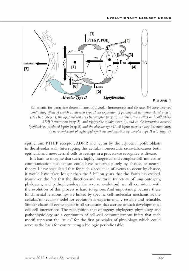
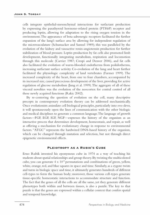
Lipofibroblast lipid uptake and storage, or neutral lipid trafficking, is mediated by adipocyte differentiation-related protein (ADRP), which is under the control of the parathyroid hormone-related protein (PTHrP) signaling pathway (Torday and Rehan 2007b; Figure 1). This series of functionally integrated proteins is expressed compartmentally: PTHrP, surfactant, and the leptin receptor in the
Evolutionary Biology Redux
461autumn 2013 • volume 56, number 4
epithelium; PTHrP receptor, ADRP, and leptin by the adjacent lipofibroblasts in the alveolar wall. Interrupting this cellular homeostatic cross-talk causes both epithelial and mesodermal cells to readapt in a process we recognize as disease.
It is hard to imagine that such a highly integrated and complex cell-molecular communication mechanism could have occurred purely by chance, or neutral theory. I have speculated that for such a sequence of events to occur by chance, it would have taken longer than the 5 billion years that the Earth has existed. Moreover, the fact that the direction and vectorial trajectory of lung ontogeny, phylogeny, and pathophysiology (as reverse evolution) are all consistent with the evolution of this process is hard to ignore. And importantly, because these fundamental relationships are linked by specific cell-molecular mechanisms, the cellular/molecular model for evolution is experimentally testable and refutable. Similar chains of events occur in all structures that ascribe to such developmental cell-cell interactions. The recognition that ontogeny, phylogeny, physiology, and pathophysiology are a continuum of cell-cell communications infers that such motifs represent the “rules” for the first principles of physiology, which could serve as the basis for constructing a biologic periodic table.
Figure 1
Schematic for paracrine determinants of alveolar homeostasis and disease. We have observed coordinating effects of stretch on alveolar type II cell expression of parathyroid hormone-related protein (PTHrP) (step 1), the lipofibroblast PTHrP receptor (step 2), its downstream effect on lipofibroblast
ADRP expression (step 3), and triglyceride uptake (step 4), and on the interaction between lipofibroblast-produced leptin (step 5) and the alveolar type II cell leptin receptor (step 6), stimulating
de novo surfactant phospholipid synthesis and secretion by alveolar type II cells (step 7).
John S. Torday
462 Perspectives in Biology and Medicine
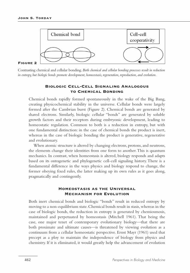
Figure 2
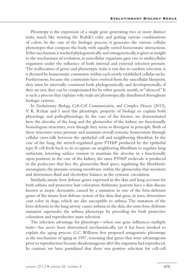
Contrasting chemical and cellular bonding. Both chemical and cellular bonding processes result in reduction in entropy, but biologic bonds promote development, homeostasis, regeneration, reproduction, and evolution.
Biologic Cell-Cell Signaling Analogous to Chemical Bonding
Chemical bonds rapidly formed spontaneously in the wake of the Big Bang, creating physicochemical stability in the universe. Cellular bonds were largely formed after the Cambrian burst (Figure 2). Chemical bonds are generated by shared electrons. Similarly, biologic cellular “bonds” are generated by soluble growth factors and their receptors during embryonic development, leading to homeostatic regulation. Common to both is a reduction in entropy, but with one fundamental distinction: in the case of chemical bonds the product is inert, whereas in the case of biologic bonding the product is generative, regenerative and evolutionary.
When atomic structure is altered by changing electrons, protons, and neutrons, the elements change their identities from one form to another. This is quantum mechanics. In contrast, when homeostasis is altered, biology responds and adapts based on its ontogenetic and phylogenetic cell-cell signaling history. There is a fundamental difference in the ways physics and biology respond to change, the former obeying fixed rules, the latter making up its own rules as it goes along, pragmatically and contingently.
Homeostasis as the Universal Mechanism for Evolution
Both inert chemical bonds and biologic “bonds” result in reduced entropy by moving to a non-equilibrium state. Chemical bonds result in stasis, whereas in the case of biologic bonds, the reduction in entropy is generated by chemiosmosis, maintained and perpetuated by homeostasis (Mitchell 1961). That being the case, one major tenet of contemporary evolutionary biology—that there are both proximate and ultimate causes—is threatened by viewing evolution as a continuum from a cellular homeostatic perspective. Ernst Mayr (1961) used that precept as a ploy to maintain the independence of biology from physics and chemistry. If it is eliminated, it would greatly help the advancement of evolution
Evolutionary Biology Redux
463autumn 2013 • volume 56, number 4
theory, moving it away from dichotomous mutation and selection, towards a unified theory of biology. Since homeostasis is the basis for all of life, and therefore the site for evolutionary selection pressure, this concept applies universally to all living organisms.
Transcending Time and Space
We have previously shown that viewing lung ontogeny and phylogeny from the common denominator of cell-cell interactive paracrine signaling reveals that they are one and the same mechanism of morphogenesis, suggesting that time is superfluous to understanding vertebrate evolution (Torday and Rehan 2009a, 2009b, 2009c). The time variable is an artifact of descriptive biology. As a result, when the processes of lung ontogeny and phylogeny are merged together, lung evolution is comprehensively reducible to the facilitation of gas exchange, beginning with the introduction of cholesterol into the cell membranes of unicellular organisms (Miao et al. 2002). This is exclusive to eukaryotes, since prokaryotes are devoid of cholesterol; this fundamental structural difference in the cell membranes of eukaryotes facilitated the interactions between the external and internal environments of the cell. One can visualize the unicellular origins of vertebrate evolution by focusing on cholesterol, which Conrad Bloch (1979) considered to be a “molecular fossil”: the synthesis of cholesterol and its insertion into the cell membrane rendered the membrane deformable, allowing increased gas exchange due to the thinning of the cell membrane (respiration), facilitating both endocytosis and exocytosis (metabolism), and enhanced cell movement (locomotion) through cytoplasmic streaming (Torday and Rehan 2012).
Notably, metabolism, respiration, and locomotion are the three driving forces behind vertebrate evolution (Carrier 1987). As a result, the unicellular plasmalemma is a functional homolog for the skin, lung, gut, kidney, and brain of metazoans. Functional homologies for all of these complex physiologic structures are linked together molecularly through cholesterol utility, either directly (lung~surfactant, brain~myelinization, skin~stratum corneum), or derivatively, through barrier function for all of these traits. Therefore, the unicellular state is the biologic life form that metazoans are derived from. And the morpho-space that is filled by the biota (Gould and Lewontin 1979), like time, is also an epiphenomenon that distracts us from understanding the Ur mechanisms of vertebrate evolution.
Seeking a Universal Language for Biology, Medicine, and Evolution
In the current modality, biologic phenomena are anecdotal, giving rise to descriptive medicine and evolution. As a result, the languages of all three have created a Tower of Babel. Finding a common language seems daunting, yet Thomas Kuhn (1962) characterized a paradigm shift as a change in the language. The cellular approach to
John S. Torday
464 Perspectives in Biology and Medicine
evolution simplifies the problem, leveling the differences between ontogeny and phylogeny as the histories (short- and long-term, respectively) of the organism and providing a deeper understanding of how and why metazoan structures and functions have evolved from their unicellular origins. When physiologic traits are reduced to their cellular and molecular components—independent of species, age, gender—the differences between them dissipate, allowing for a new perspective on molecular homologies.
The language of cell communication is universal. For example, epidermal growth factor signaling between cell-types as it applies to development, homeostasis, regeneration, and repair—how and why it is used during the history of the organism—can be mapped out, annotated, and integrated with other up- and downstream signaling pathways, such as notch, fox, transforming growth factor beta, wingless/int, beta catenin, and bone morphogenetic proteins, using the functional phenotypes as templates for ultimately determining the rules of organization.
Genetic Assimilation as a Case in Point
A classic example of how a change in our perspective on the process of evolution would impact on the language of evolutionary biology is genetic assimilation. Genetic assimilation states that an organism’s phenotype can change across environments (phenotypic plasticity), and that selection can operate both on the expression of traits within particular environments and on the shape of the reaction norm itself. Such terms as canalization, genetic landscape (cell-cell signaling), adaptive peaks and valleys (cell-cell signaling for homeostatic set-points), plasticity, adaptive inactivation of the canalizing system under environmental stress, reaction norm, directed preadaptation, non-directed preadaptation, atavism, and the cost of plasticity (homeostasis as energetics) all describe environmental effects on phenotypes that assume that genes are the underlying determinants of these manifestations. However, the specific genes involved in genetic assimilation are rarely directly examined, either observationally or in experimental manipulations (gain and loss of function) to test hypotheses (Abzhanov et al. 2006). I assume that is because it is assumed that the genetic changes are the result of spontaneous mutations and the selection for such mutations. Consequently, all of this terminology is non-mechanistic, derivative, descriptive, a posteriori thinking.
In contrast, the cell-molecular homeostatic model for evolution and stability addresses how the external environment generates homeostasis developmentally at the cellular level and determines homeostatic set-points in adaptation to the environment (the reaction norm) through specific environmental effectors (growth factors and their receptors, second messengers, inflammatory mediators, shear stress, biochemistry, mechanotransduction, apoptosis, stem cells, DNA repair, cross-over mutation, gene duplication, other) that may or may not alter the homeostatic set-point (reaction norm). This is a highly mechanistic, heritable, plastic process that
Evolutionary Biology Redux
465autumn 2013 • volume 56, number 4
lends itself to understanding evolution at the cellular, tissue, organ, system, and population levels, mediated by physiologically linked mechanisms throughout, without having to invoke random chance mechanisms to bridge different scales of evolutionary change. In other words, it is an integrated mechanism that can often be traced all the way back to its unicellular origins.
Utility of the Approach
By adopting the proposed cell communication approach for problem solving in biology or medicine, there would be no intellectual boundaries: one would no longer be restricted to one discipline, cell-type, tissue, organ, species, or to development, homeostasis, pathophysiology, or regeneration. All data related to the question at hand would be useful in the broadest context of biology. This approach would also allow understanding causality based on first principles, rather than on relativistic phenomena—health as the absence of disease, pathology as signs and symptoms, biologic traits as monogenetic. In essence, biology and medicine would be based on hard science instead of approximations. How a particular gene forms structure-function relationships in all of these contexts would now be accessible and useful to understanding the gene’s ultimate role(s) in homeostasis, physiology, and disease processes. This a priori approach to biology and medicine is predictive, ubiquitous, testable, and refutable, and the results generated by such a comprehensive analysis are universal, durable, and falsifiable. Many functional components could be tested by this approach in a wide variety of conditions, both homeostatic and pathologic, to determine if they comply with the predicted parallelisms within and between traits.
The model allows for the consideration of knockouts that don’t produce altered phenotypes resulting from adaptive compensatory mechanisms useful in treating disease; for understanding the phenomenon of cryptic genes that emerge during disease processes as the evolutionary recapitulation of ontogeny and phylogeny in service to retrograde control of homeostasis; for regarding chronic disease as retrograde evolution; and for viewing integrated physiology as the aggregate history of the organism. Ultimately, this approach would generate a predictive algorithm for functional genomics, proteomics, interactomics, and more.
Such descriptive concepts as plasticity, evolvability, systems biology, and homology, when looked at from a cellular perspective, are far more comprehendible than when they are seen from the vantage point of superficial description and metaphoric thinking. For example, plasticity is likely delimited by the range of reaction norms for any given biologic trait, while evolvability reflects the nature of the cell-molecular linkage between the external environmental stressor or mutation and the internal deep homology being challenged.
Importantly, this evolutionary model of physiology also lends itself to thinking about health and disease as a continuum, rather than as a mutually exclusive dichotomy, revealing the true nature of aging, for example, as an integral part of
John S. Torday
466 Perspectives in Biology and Medicine
the life cycle, not merely as a consequence of cumulative pathology. Ultimately, this epistemological change in our view of biology and medicine would form the basis for a bioethics based on logic, rather than on anecdote and subjective a posteriori reasoning. This approach thus provides a platform for rational, ethical health-care policies, and for effective societal resource allocation.
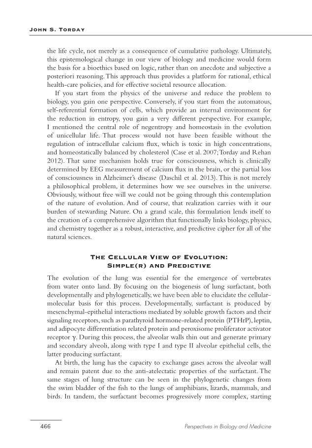
If you start from the physics of the universe and reduce the problem to biology, you gain one perspective. Conversely, if you start from the automatous, self-referential formation of cells, which provide an internal environment for the reduction in entropy, you gain a very different perspective. For example, I mentioned the central role of negentropy and homeostasis in the evolution of unicellular life. That process would not have been feasible without the regulation of intracellular calcium flux, which is toxic in high concentrations, and homeostatically balanced by cholesterol (Case et al. 2007; Torday and Rehan 2012). That same mechanism holds true for consciousness, which is clinically determined by EEG measurement of calcium flux in the brain, or the partial loss of consciousness in Alzheimer’s disease (Daschil et al. 2013). This is not merely a philosophical problem, it determines how we see ourselves in the universe. Obviously, without free will we could not be going through this contemplation of the nature of evolution. And of course, that realization carries with it our burden of stewarding Nature. On a grand scale, this formulation lends itself to the creation of a comprehensive algorithm that functionally links biology, physics, and chemistry together as a robust, interactive, and predictive cipher for all of the natural sciences.
The Cellular View of Evolution: Simple(r) and Predictive
The evolution of the lung was essential for the emergence of vertebrates from water onto land. By focusing on the biogenesis of lung surfactant, both developmentally and phylogenetically, we have been able to elucidate the cellular-molecular basis for this process. Developmentally, surfactant is produced by mesenchymal-epithelial interactions mediated by soluble growth factors and their signaling receptors, such as parathyroid hormone-related protein (PTHrP), leptin, and adipocyte differentiation related protein and peroxisome proliferator activator receptor γ. During this process, the alveolar walls thin out and generate primary and secondary alveoli, along with type I and type II alveolar epithelial cells, the latter producing surfactant.
At birth, the lung has the capacity to exchange gases across the alveolar wall and remain patent due to the anti-atelectatic properties of the surfactant. The same stages of lung structure can be seen in the phylogenetic changes from the swim bladder of the fish to the lungs of amphibians, lizards, mammals, and birds. In tandem, the surfactant becomes progressively more complex, starting
Evolutionary Biology Redux
467autumn 2013 • volume 56, number 4
with cholesterol in the swim bladder, followed sequentially by phospholipid mixtures and surfactant apoproteins. Thus, the cellular-molecular reduction of the physiology of the alveolus, in combination with the molecular mechanism for surfactant production, reveals the congruence of lung ontogeny and phylogeny in adaptation to atmospheric oxygen for metabolic drive.
The N+1th Generation Zygote and Evolutionary Selection
Recent scientific evidence indicates that, contrary to popular belief, the epigenetic “marks” in both the gonads and soma accumulated during the life cycle are not expunged during meiosis. This suggests that evolution is actually a mechanism for gleaning information from the environment to inform future generations, or for the success of the zygote-as-future adult.
Barring philosophical questions and quandaries about fetal rights, it is interesting to ponder what the evolutionary process actually constitutes. The hypothesis that the zygotic or unicellular state is the phenotype being selected for is attractive, since it is at that phase of the vertebrate life cycle that the skin, lung, kidney, and brain phenotypes (re)coalesce in the unicellular plasma membrane, both phylogenetically and ontogenetically. The recrudescence of these functions at the first stage of development may represent an evolutionary fail-safe mechanism for any epigenetic or mutational modifications acquired during the prior life cycle, ensuring the effective, overall vertical integration of all of the homeostatic mechanisms that have evolved from that mutation, by putting it into the ontogenetic/phylogenetic/homeostatic “context” of the developing embryo. This may actually be an atavistic trait, harkening back to the tried-and-true binary fission method of reproduction manifested by our unicellular ancestors for the first 4.5 billion years of life on Earth.
And perhaps this is why Haeckel was right after all: ontogeny does recapitulate phylogeny. The fact that even the asexually reproducing slime mold acquires epigenetic marks from the environment encourages me to think that this Lamarckian evolutionary strategy may be universal among biota.
This novel perspective on evolution, in which the primary selection pressure is for the zygote rather than the adult stage of the life cycle, is analogous with the shift in perspective from an Earth-centered to a heliocentric solar system. The recognition and recalibration of this and other anthropocentrisms, such as the Rights of Man and the Great Chain of Being, have proven important for the advancement of the human species, though of late our intellectual growth in the wake of the Enlightenment has been challenged by social, nutritional, cultural, and, most recently, climatic forces—all of which are extensions of human evolution as sociobiology. Perhaps the realization that we humans have evolved big brains as our answer to survival of the fittest is no different from other species
John S. Torday
468 Perspectives in Biology and Medicine
evolving eyesight, smell, or running ability, flying, or swimming. That is to say, all species are the products of their respective environments, which are ever-changing, and we are all equals in the eyes of Nature.
The Darwinian Biologic Space-Time Continuum
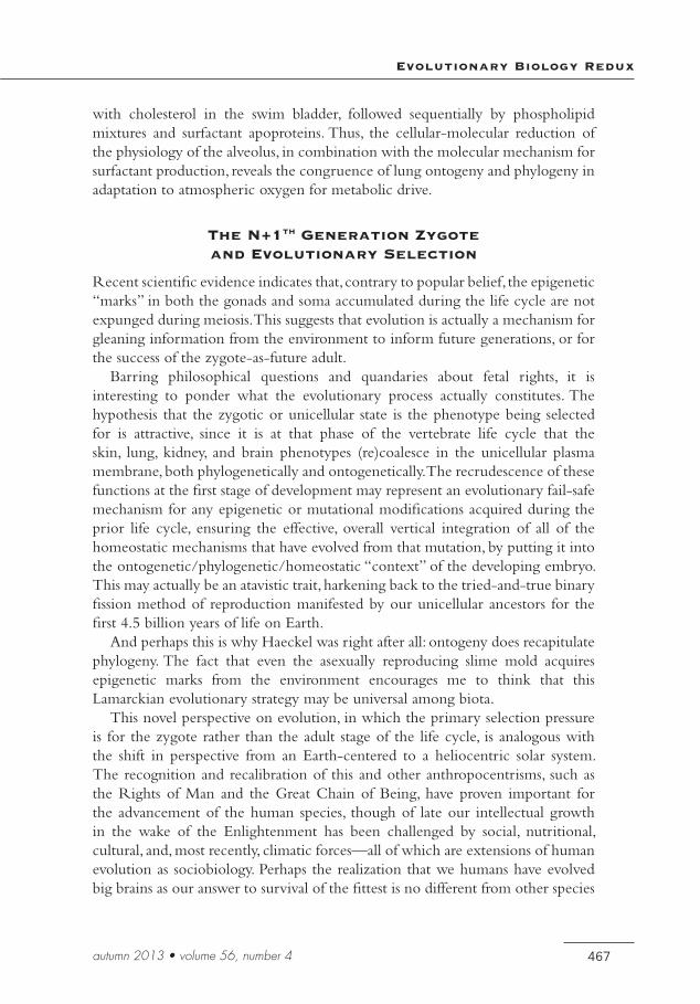
Darwin saw a continuum of speciation based on principles of natural selection, not the anthropocentric Great Chain of Being. However, Darwin’s explanation for the biologic patterns he observed was survival of the fittest, which is a metaphor for the evolutionary process but does not provide a paradigm for drilling down to the cell/molecular origins of life. Such a mechanistic model is essential if we are going to take full advantage of the knowledge gleaned from the genomes of humans and other model organisms. For example, the cell-molecular mechanism of lung evolution based on the evolution of pulmonary surfactant infers that there is a cellular continuum from development to homeostasis and regeneration/repair (Torday and Rehan 2007a). This concept of the process of lung evolution, like a cladogram, also infers a vectorial direction and magnitude of change.
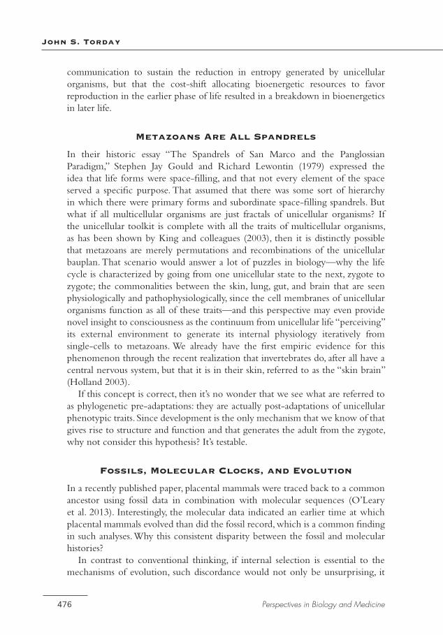
That perspective is not unlike Einstein’s vision of traveling in tandem with a light beam through space, which gave him the insight to the physical continuum from Brownian movement to the photoelectric effect and relativity theory in his annus mirabilis of 1905. Einstein was severely criticized for being intuitive (Isaacson 2007), yet his thought process has been borne out by Popperian experimentation. The space-time continuum that emerged from that epiphany has similarities to the space-time continuum of lung biology (see Figure 3). Seen from a cell-cell signaling perspective, lung ontogeny, phylogeny, homeostasis, and regeneration (as evolution in reverse) are a series of simultaneous equations that form the continuum of lung evolution as the solution for all of the equations. And since this model is largely based on universal developmental principles, the evolution of all other tissues and organs is also amenable to the same analytic approach. This analysis assumes that there are enough “molecular fossil” data to solve the evolutionary equation, since mathematically there must be as many variables as equations—hence the utilization of all of the available, relevant phenotypic functional genomic data sets.
Reverse-Engineering of Physiologic Traits as a Portal for Viewing Evolution
The premise of the approach I have taken to evolution is that by tracing the ligand-receptor cell communications that determine the pulmonary surfactant phenotype backwards in time and space, both within and between species, ontogenetically and phylogenetically, we would be able to understand the cell-molecular mechanisms that have fashioned the lung through external and internal environmental selection pressures. I have referred to this as a “middle-
Evolutionary Biology Redux
469autumn 2013 • volume 56, number 4
out” approach, in contrast to the traditional top-down or bottom-up strategies. Horowitz (1945) formulated a similar approach to the evolution of biochemical pathways by assuming a retrograde mode of evolution. He envisioned an organism that could not synthesize an essential biochemical substance, so it acquired it from the environment. When the supply of that substance in the environment was exhausted, those organisms that possessed the last enzyme in the biosynthetic pathway could make use of the immediate precursor and convert it to the end product, until the supply of the immediate precursor was also exhausted. Then only those organisms that possessed the second to last enzyme could survive, and so on, until the biosynthetic pathway was completely established.
This approach describes the functional phenotype for the evolution of a biosynthetic pathway, whereas the cell-molecular, middle-out approach would provide the mechanistic series of ligand-receptor homeostatic mechanisms that determined those biosynthetic pathways from phenotypes to genes, providing a way of understanding physiology from its origins. Selection pressure on such ligand-receptor GRNs has generated both evolutionary stability and novelty through gene duplication, gene mutation, redundancy, alternative pathways, compensatory mechanisms, positive and balancing selection pressures, and so forth. Such genetic modifications have been manifested in the structural and functional changes in the blood-gas barrier, primarily its thinning out as a direct result of adaptive phylogenetic changes in the composition and physiologic regulation
Figure 3
Lung evolution expressed as a series of simultaneous equations. Lung evolution (middle line), as the aggregate of cell-cell signaling mechanisms for ontogeny, phylogeny, homeostasis, and repair (evolution in reverse), mediated by PTHrP signaling. Phenotypic data sets are expressed as simultaneous equations,
forming the continuum of lung evolution as the mathematical “solution.” Boxes represent strings of incomplete “molecular fossil” data.
John S. Torday
470 Perspectives in Biology and Medicine
of lung surfactant, as described eloquently in a series of studies by Orgeig and Daniels from 1994 on (see, for example, Orgeig et al. 2007). The reverse-engineering of these phenotypic changes in the blood-gas barrier form the basis for our functional genomic approach to lung evolution.
Cell-Cell Communication as the Basis for Metazoan Evolution
Based on the middle-out, cell communication model, how might biologic evolution have begun? One school of thought is that cellular organisms emerged as a result of the wetting and drying of lipids due to the diurnal rhythms of the sun, abetted by the waxing and waning of the moon, which gave rise to micelles, semi-permeable lipid membranes that would have provided a protected environment for the reduction of entropy through enzymatic catalysis. Over the ensuing 4.5 billion years, such unicellular organisms have evolved in adaptation to their physical surrounds, eukaryotes evolving from prokaryotes, the former being distinguished from the latter by the presence of a nuclear envelope. And the eukaryotic acquisition of mitochondria from prokaryotes, described by the Endosymbiosis Theory, similarly emerged as a result of the on-going competition between pro- and eukaryotes. Then, about 500 million years ago, unicellular organisms began cooperating with one another metabolically. In the case of the prokaryotes, this took the form of such phenomena as biofilm and quorum sensing. Those “inventions,” in turn, may have been the positive selection pressure for eukaryotes to also cooperate (or become extinct) by evolving the cell-cell signaling mechanisms that we recognize today as the soluble growth factors and cell-surface cognate receptors that universally mediate morphogenesis, homeostasis, regeneration, and reproduction. However, it should be borne in mind that this process began with a decrease in entropy, arguably defying the Second Law of Thermodynamics. You cannot defy the laws of physics forever: there has got to be payback, since matter and energy cannot be created or destroyed.
Unicellular organisms are “immortalized” by reproducing through binary fission, whereas eukaryotes evolved sexual reproduction as a means of communicating their genetic information from one generation to the next. Since the overall evolutionary selection pressure for vertebrate evolution is for reproductive success, the distribution of bioenergetics is asymmetrically distributed throughout the life cycle, being biased in favor of the reproductive phase. The trade-off is that the cellular machinery must ultimately fail due to the omnipresence of bacteria, oxidative stress and all the other environmental forces that initiated the evolutionary strategy. The result is a decrease in bioenergetic resources after the reproductive phase, which we recognize descriptively as aging, resulting in such phenomena as increased oxidative stress, lipid peroxidation, protein misfolding, endoplasmic reticulum stress, and failure of other such metabolic mechanisms,
Evolutionary Biology Redux
471autumn 2013 • volume 56, number 4
which are commonly thought to be the causes of aging. However, the cell-cell communication model predicts that the decline in bioenergetics is what causes decreased cell communication as an energy-requiring process, ultimately culminating in the catastrophic failure of signaling, or death. But because the gene pool is immortalized by the communication of DNA from one generation to the next, in the final analysis, each phase of this perspective on the how and why of evolution is one of cell communication, initially between unicellular organisms and their physical environment, followed by cell communication as the basis for metazoan structure and function, and ultimately reproduction as communication of the environmental knowledge gleaned from generation to generation for adaptation and survival.
How Genes Determine Cell Cross-Talk
The greatest challenge we face in the post-genomic era is to effectively integrate functionally relevant genomic data to determine physiologic first principles, and how they can be used to decode complex biologic traits. This problem is usually addressed stochastically as systems biology, by analyzing large genomic data sets to identify genes that are associated with structural and functional phenotypes; whether they are causal is more often than not seemingly ignored. This approach is merely an extrapolation from descriptive systematic biology, beginning with Linnaeus’s invention of binomial nomenclature.
Systems biology can be viewed at several different levels—the gene, the transcript, the protein, the cell, the organ, the organ system, or the population—and clearly, evolution could have impacted the underlying process at any one of these levels. There are many such analyses in the literature, but they don’t provide (vertically) integrated, functional genomic, evolutionary mechanisms that lead to novel insights to the process, let alone further experimentation and ultimately prediction. Selection pressure—intrinsic, extrinsic, or both—must be applied at a level where it can have the necessary homeostatic effect for survival, the level where the genetic expression is functionally integrated with the phenotype. The middle-out approach offers the advantage of minimizing a posteriori assumptions by focusing on GRNs. GRNs govern the expression levels of the mRNAs and proteins that generate form and function, particularly those that have evolved using the same conserved ontogenetic/phylogenetic, homeostatic, and regenerative cell-molecular motifs.
Those vertically integrated, cell-to-functional phenotype mechanisms that best represent physiology across species and development, particularly that of the lung, are archetypes for the analytic approach being advocated. The rise in oxygen in the Phanerozoic era gave rise to alveolar lipofibroblasts (Falkowski et al. 2005). This concept is corroborated by the observation by Barbara Wold’s laboratory that muscle stem cells spontaneously differentiate into adipocytes in 21% oxygen, but
John S. Torday
472 Perspectives in Biology and Medicine
not in 6% oxygen, likely due to the effects of oxygen on fibroblast differentiation (Csete et al. 2001; Higuchi et al. 2013;Qintero et al. 2012; Saitoh et al 2012). And perhaps this phenomenon explains the positive selection for the cytoprotective effect of the neutral lipids stored in lipofibroblasts (Torday et al. 2001). This adaptation was followed by the stretch-regulated production of leptin by these cells, perhaps in response to positive selection for endothermy by somatic fat cells (Mezentseva, Kumaratilake and Newman 2008; Torday and Rehan 2002). Leptin is a molecular homolog of interleukin-6, an inflammatory cytokine thought to have fostered endothermy. The increase in body temperature from 25oC (ambient temperature) to 37oC (body temperature) would have rendered lung surfactant 300% more surface-active, leading to selection pressure for a PTHrP stretch-regulated mechanism for the integration of surfactant production and alveolar capillary perfusion, since PTHrP is a potent vasodilator (Gao and Raj 2005; Torday, Sanchez-Esteban, and Rubin 1998). This cell-molecular series of evolved homologies, coupled together by alternating external and internal selection pressures, is well recognized in conventional descriptive physiology as ventilation-perfusion matching (Burrowes et al. 2008). Furthermore, it is known that the stretch-regulated mechanism for parathyroid hormone-related protein (PTHrP) expression is intrinsic to alveolar epithelial type II cells, because in a microgravity environment these cells will contract, resulting in decreased PTHrP expression (Torday 2003). This trait may have originated as selection pressure for the expression of PTHrP in the fish swim bladder in adaptation to gravity (buoyancy) for efficient feeding (Zheng et al. 2011); the functional homology between gas exchange for buoyancy and respiration uses the same genes expressed within the epithelium and mesenchyme, derived from the esophagus for both structures (Korzh et al. 2011).
Tiktaalik: An Object Lesson in Cell-Molecular Evolution
PTHrP signaling provides the mechanistic basis for the morphing of fish into tetrapods, like Neil Shubin’s Tiktaalik, discovered in 2004 (Shubin, Daeschler, and Jenkins 2006). All of the essential water-to-land adaptations—lung, skin, kidney, gut, and brain—would have been facilitated by a timely gene duplication of the PTHrP receptor that seemingly occurred just as fish evolved into amphibians (Aya et al. 1999; Gu et al. 2012; Karparien et al. 1994; Pinheiro et al 2012; Rubin et al. 2004;Yan et al. 2012). This event seems fortuitous for the sake of vertebrate evolution from water to land, but seen in the context of cellular mechanisms, it may have been a direct consequence of the generation of excess oxygen radicals and lipid peroxides, since such substances can be generated locally by vascular wall shear stress in microcirculations like those of the alveolus and glomerulus, causing context-specific adaptive gene mutations (Galhardo, Hastings and Rosenberg 2007; Stevens 2011;Takahashi, Huynh-Do and Daniel 1998). Such extrinsic
Evolutionary Biology Redux
473autumn 2013 • volume 56, number 4
factors as oxygen and gravisensing implicate population genetic mechanisms for evolutionary selection, underpinned by the cell-cell signaling model for lung evolution.
In fact, if adaptation is thought of in the context of internal selection caused by vascular shear stress, the concept of plasticity becomes much more relevant, not to mention being testable; constitutive genes are the ones that were most vulnerable to mutation, since they were the genes being targeted by such selection mechanisms. And perhaps such unconventional internal selection was followed by classic Darwinian population selection for those members of the species that were best fit to regulate those constitutive genes to survive, rendering the newly evolved homeostatic mechanism regulatable. Theoretically, this may have been due to the fact that regulated mechanisms would be more resilient, and therefore less likely to generate mutagens than non-regulated constitutive genes. And this may also explain why humans have fewer than predicted genes.
There have been numerous attempts to reconstruct biology from its component parts. Darwinian thought fostered the works of Haeckel, Waddington, Riel, Seilacher, and Gould, to name only a few of those who have attempted to further our insights to evolution. And more recently, Morowitz (1968) and West and colleagues (1997) have gained much notoriety by formulating comprehensive analyses of physiology, but the problem with their approaches is that they reason backwards from existing structures and functions. They do not predict the changes that have occurred over the course of evolution, even given all the moving parts, and they thus leave biology as a loosely linked series of anecdotes (Raff 1996), and medicine as virtually non-predictive and ultimately incomplete in its philosophic and functional scope.
Systems biology attempts to provide a mechanistic, causal, and explanatory linkage between genotypes and their associated phenotypes by studying the structure and dynamics of the convoluted molecular interactive networks that regulate cells and tissues in development, homeostasis, and aging. It aims at developing and integrating experimental and mathematical techniques in pursuit of principles that would make the nature of cellular phenotypes more intelligible, and their control more deliberate. While this effort is motivated by the practical need and desire to cure disease, or at least make the symptoms go away, it also reflects a desire for a theoretical framework by which to deconvolute the complexities of the cell and the organism.
Exploiting the Evolution of the Lung to Understand Other Physiologic Traits
By focusing on the progressive cell-molecular changes in the lung that have occurred ontogenetically and phylogenetically, we have been able to trace other cell-molecular milestones in vertebrate evolution. Lipofibroblasts spontaneously form from muscle cells in a 21% oxygen environment (Csete et al. 2001). These
John S. Torday
474 Perspectives in Biology and Medicine
cells integrate epithelial-mesenchymal interactions for surfactant production by expressing the parathyroid hormone-related protein (PTHrP) receptor and producing leptin, allowing for adaptation to the rising oxygen tension in the environment. The appearance of beta-adrenergic receptors facilitated the further expansion of the lung’s surface area by allowing for independent regulation of the microvasculature (Schumacker and Samsel 1989); this was paralleled by the evolution of the kidney and vasoactive renin-angiotensin production for further stabilization of blood pressure. Leptin production by fat cells also promoted limb development, functionally integrating metabolism, respiration and locomotion through this molecule (Carrier 1987; Crespi and Denver 2006), and fat cells also facilitated the evolution of warm-blooded endotherms from poikilotherms, increasing surfactant surface activity. Co-evolution of the lung and heart further facilitated the physiologic complexity of land vertebrates (Farmer 1999). The increased complexity of the heart, from one to four chambers, accompanied by its increased size, caused precocious development of the liver, providing for active regulation of glucose metabolism (Jung et al. 1999). The aggregate of all of these visceral novelties was the evolution of the neocortex for central control of all these newly acquired functions (Rakic 2002).
By re-centering the question of evolution on the cell, many descriptive precepts in contemporary evolution theory can be addressed mechanistically. Once evolutionists assimilate cell biological principles, particularly into evo-devo, it will spontaneously open the lines of communication with the other biologic and medical disciplines to generate a common language. The language of growth factors—FGF, EGF, IGF, NGF—expresses the history of the organism as an interactive process that determines development, homeostasis, and repair, as well as offering a mechanism for evolutionary change in response to environmental factors. “ATGC” represents the hardwired DNA-based history of the organism, which can be changed through mutation and selection, but not through direct epigenetic environmental effects.
Pleiotropy as a Rubik’s Cube
Erno Rubik invented his eponymous cube in 1974 as a way of teaching his students about spatial relationships and group theory. By twisting the multicolored cube, you can generate 4 x 10¹⁹ permutations and combinations of green, yellow, white, orange, red, and blue squares in space and time. Similarly, as a zygote twists and turns in biologic space and time, it ultimately generates hundreds of different cell-types to form the human body; moreover, those various cell-types generate tissue-specific homeostatic interactions to accommodate structure and function. The fact that the genes of all the cells are all the same, yet they generate different phenotypes both within and between tissues, is also a puzzle. The key to that puzzle is that the genes are expressed within a cellular context that confers spatial and temporal knowledge.
Evolutionary Biology Redux
475autumn 2013 • volume 56, number 4
Pleiotropy is the expression of a single gene generating two or more distinct traits, much like twisting the Rubik’s cube and getting various combinations of colors. In the case of the biologic process, it generates the various cellular phenotypes that compose the body, with equally varied homeostatic interactions. If this mechanism is tracked phylogenetically and ontogenetically, it gives us insight to the mechanisms of evolution, as unicellular organisms gave rise to multicellular organisms under the influence of both internal and external selection pressure. The reallocation of genes and phenotypic traits is not due to random selection, it is dictated by homeostatic constraints within each newly established cellular niche. Furthermore, because the constraints have evolved from the unicellular blueprint, they must be internally consistent both phylogenetically and developmentally; if they are not, they can be compensated for by other genetic motifs, or “silenced.” It is such a process that explains why traits are pleiotropically distributed throughout biologic systems.
In Evolutionary Biology, Cell-Cell Communication, and Complex Disease (2012), V. K. Rehan and I used this pleiotropic property of biology to explain both physiology and pathophysiology. In the case of the former, we demonstrated how the alveolus of the lung and the glomerulus of the kidney are functionally homologous structures, even though they seem so divergent in principle. Both of these structures sense pressure and maintain overall systemic homeostasis through cellular cross-talk between the epithelial cell and neighboring fibroblast. In the case of the lung, the stretch-regulated gene PTHrP produced by the epithelial type II cell feeds back to its receptors on neighboring fibroblasts to regulate lung surfactant, lowering surface tension to maintain the alveolus in a functionally open position; in the case of the kidney, the same PTHrP molecule is produced in the podocytes that line the glomerular fluid space, regulating the fibroblastic mesangium, the pressure-sensing membrane within the glomerulus that monitors and determines fluid and electrolyte balance in the systemic circulation.
Similarly, innate host defense genes expressed in the skin and lung account for both asthma and protective hair coloration. Asthmatic patients have a skin disease known as atopic dermatitis, caused by a mutation in one of the beta-defensin genes of the innate host defense system of the skin; that gene, in turn, determines coat color in dogs, which are also susceptible to asthma. The mutation of the beta-defensin in the lung airway causes asthma; in the skin, the same beta-defensin mutation supersedes the asthma phenotype by providing for both protective coloration and reproductive mate selection.
The selection advantage for pleiotropy—when one gene influences multiple traits—has never been determined mechanistically, yet it has been invoked to explain the aging process. G.C. Williams first proposed antagonistic pleiotropy as the mechanism of aging in 1957, reasoning that genes that were advantageous prior to reproduction became disadvantageous after the organism had reproduced. In contrast, we have postulated that there was positive selection for cell-cell
John S. Torday
476 Perspectives in Biology and Medicine
communication to sustain the reduction in entropy generated by unicellular organisms, but that the cost-shift allocating bioenergetic resources to favor reproduction in the earlier phase of life resulted in a breakdown in bioenergetics in later life.
Metazoans Are All Spandrels
In their historic essay “The Spandrels of San Marco and the Panglossian Paradigm,” Stephen Jay Gould and Richard Lewontin (1979) expressed the idea that life forms were space-filling, and that not every element of the space served a specific purpose. That assumed that there was some sort of hierarchy in which there were primary forms and subordinate space-filling spandrels. But what if all multicellular organisms are just fractals of unicellular organisms? If the unicellular toolkit is complete with all the traits of multicellular organisms, as has been shown by King and colleagues (2003), then it is distinctly possible that metazoans are merely permutations and recombinations of the unicellular bauplan. That scenario would answer a lot of puzzles in biology—why the life cycle is characterized by going from one unicellular state to the next, zygote to zygote; the commonalities between the skin, lung, gut, and brain that are seen physiologically and pathophysiologically, since the cell membranes of unicellular organisms function as all of these traits—and this perspective may even provide novel insight to consciousness as the continuum from unicellular life “perceiving” its external environment to generate its internal physiology iteratively from single-cells to metazoans. We already have the first empiric evidence for this phenomenon through the recent realization that invertebrates do, after all have a central nervous system, but that it is in their skin, referred to as the “skin brain” (Holland 2003).
If this concept is correct, then it’s no wonder that we see what are referred to as phylogenetic pre-adaptations: they are actually post-adaptations of unicellular phenotypic traits. Since development is the only mechanism that we know of that gives rise to structure and function and that generates the adult from the zygote, why not consider this hypothesis? It’s testable.
Fossils, Molecular Clocks, and Evolution
In a recently published paper, placental mammals were traced back to a common ancestor using fossil data in combination with molecular sequences (O’Leary et al. 2013). Interestingly, the molecular data indicated an earlier time at which placental mammals evolved than did the fossil record, which is a common finding in such analyses. Why this consistent disparity between the fossil and molecular histories?
In contrast to conventional thinking, if internal selection is essential to the mechanisms of evolution, such discordance would not only be unsurprising, it
Evolutionary Biology Redux
477autumn 2013 • volume 56, number 4
should be expected. There would have been extended periods of internal cell-molecular functional adaptations in response to physiologic stress, microvascular shear stress, and remodeling, both genetic and epigenetic, followed by the formation of fossilizable structures. Yet because of the disdain evolutionists hold for internal selection, such a scenario is not even mentioned in that study or in the accompanying commentary (Yoder 2013). However, if one thinks of the practical and conceptual implications of internal selection, such disparities between fossil and molecular data would also explain punctuated equilibrium. Although the “missing” intermediate phenotypes may be invisible to the fossil record, recognizing that there may be molecular intermediates would encourage looking for molecular homologies in co-evolved species, or during embryogenesis, or homeostasis, or regeneration and repair (Torday and Rehan 2004).
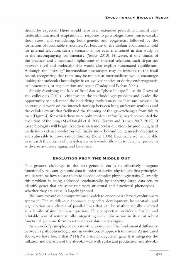
Simply dismissing the lack of fossil data as “ghost lineages”—as do Goswami and colleagues (2011)—circumvents the methodologic problem and evades the opportunity to understand the underlying evolutionary mechanisms involved. In contrast, our work on the interrelationship between lung surfactant synthesis and the cellular events that facilitated the thinning of the gas-exchanger from fish to man (Figure 4), for which there were only “molecular fossils,” has deconvoluted the evolution of the lung (MacDonald et al. 2006; Torday and Rehan 2007, 2012). If more biologists will begin to address such molecular questions by producing hard predictive evidence, evolution will finally move beyond being merely descriptive and vulnerable to unwarranted dismissal (Behe 1996). Eventually we may be able to unearth the origins of physiology, which would allow us to decipher problems as diverse as disease, aging, and bioethics.
Evolution from the Middle Out
The greatest challenge in the post-genomic era is to effectively integrate functionally relevant genomic data in order to derive physiologic first principles, and determine how to use them to decode complex physiologic traits. Currently, this problem is being addressed stochastically by analyzing large data sets to identify genes that are associated with structural and functional phenotypes—whether they are causal is largely ignored.
We must expand our computational models to encompass a broad, evolutionary approach. The middle-out approach engenders development, homeostasis, and regeneration as a cluster of parallel lines that can be mathematically analyzed as a family of simultaneous equations. This perspective provides a feasible and refutable way of systematically integrating such information in its most robust functional genomic form to retrace its evolutionary origins.
As a proof of principle, we can cite other examples of the fundamental difference between a pathophysiologic and an evolutionary approach to disease. As indicated above, we have found that PTHrP is a stretch-regulated gene that integrates the inflation and deflation of the alveolar wall with surfactant production and alveolar
John S. Torday
478 Perspectives in Biology and Medicine
capillary perfusion. PTHrP is classically thought of as a bone-related gene that regulates calcium flux. With this and the stretch effect in mind, we recalled that astronauts develop osteoporosis due to weightlessness. Others have pursued a more conventional pathophysiologic tack to this phenomenon, reasoning that postmenopausal women develop osteoporosis and are estrogen deficient, therefore estrogen replacement would be the appropriate treatment for osteoporosis. We, on the other hand, have tested the hypothesis that microgravity would inhibit PTHrP expression in bone and lung cells. I observed a decrease in PTHrP expression by osteoblasts and lung epithelial cells in free-fall attached to dextran beads, mimicking 0 x g. When these cells were put back in unit gravity, the expression of PTHrP returned to normal levels in both cell-types. We subsequently examined the PTHrP expression by the weight-bearing bones of rats flown in deep space for two weeks. Here too, we observed a significant decrease in PTHrP expression compared to ground-based littermate controls. The weightlessness effect was not seen in the parietal bone, which is non-weight bearing, consistent with the effect of unloading of the weight bearing bones due to 0 x g.
We have also taken an unconventional evo-devo biologic approach to chronic lung disease. We have pursued the concept that there is an evolutionary continuum from development to homeostasis and regeneration mediated by soluble growth factors. Based on that approach, we have discovered that the cell communication between the epithelium and mesoderm is critically important for
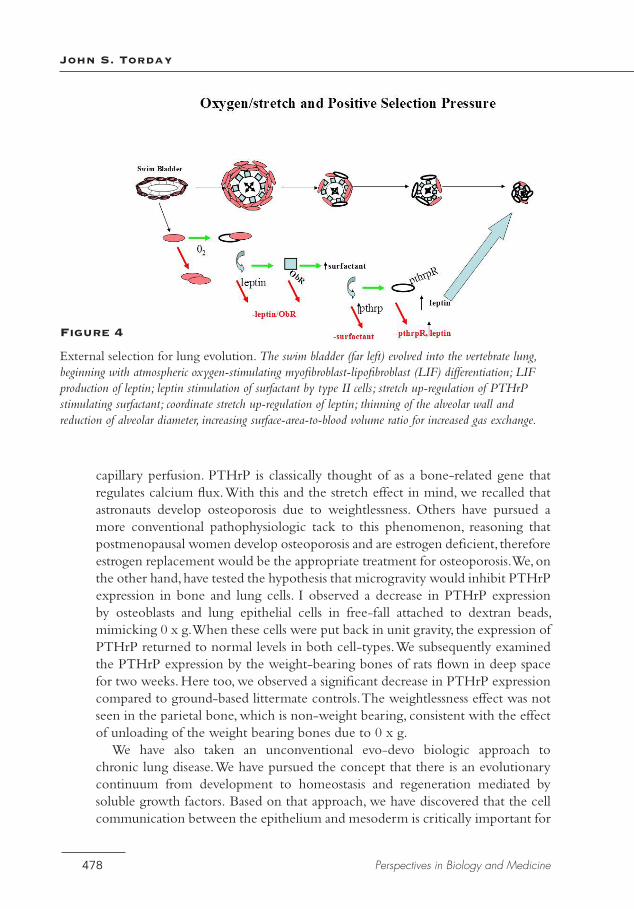
Figure 4
External selection for lung evolution. The swim bladder (far left) evolved into the vertebrate lung, beginning with atmospheric oxygen-stimulating myofibroblast-lipofibroblast (LIF) differentiation; LIF production of leptin; leptin stimulation of surfactant by type II cells; stretch up-regulation of PTHrP stimulating surfactant; coordinate stretch up-regulation of leptin; thinning of the alveolar wall and reduction of alveolar diameter, increasing surface-area-to-blood volume ratio for increased gas exchange.
Evolutionary Biology Redux
479autumn 2013 • volume 56, number 4
the development and maintenance of the alveolar lipofibroblast, and that when that signaling mechanism fails, the lipofibroblast “defaults” to its cellular origin as a muscle cell, or myofibroblast, the signature cell-type for fibrosis. Not only has this approach given us insight to the multifactorial causes of bronchopulmonary dysplasia (pressure, oxygen, infection, maternal smoking), but also to a novel treatment for this condition based on the use of peroxisome proliferator activated receptor gamma (PPARγ) as the nuclear transcription factor that determines the lipofibroblast phenotype. Thiazolidinediones are potent PPARγ agonists, and we have found that they can prevent or reverse the effects of all of the BPD-inducing agents we have studied, ranging from pressure to oxygen, infection and nicotine. This evo-devo approach may be far more successful in the treatment of this condition than more traditional, generic anti-inflammatory agents, such as antenatal or postnatal steroids, or prophylactic surfactant therapy.
As indicated earlier, evolutionary selection pressure generated metazoa through cell communication, leading to reproduction, aging, and death. There is accumulating evidence for the loss of cell communication in aging rats to support this perspective on the life cycle. By inference, the selection pressure for reproductive success may optimize for cell communication, and there is accumulating evidence in this regard as well. Therefore, one could devise strategies for “healthy aging” based on this premise, rather than accepting the inevitability of aging as a slow, degradative pathologic process. It has recently been shown, for example, that there is a subset of aging humans who experience a precipitous death rather than experiencing the slow loss of biologic function over years. These data suggest that what we conventionally think of as aging and death is pathology, not evolved biology.
Conclusion
With the aid of the human genome, we must address the evolutionary origins of human physiology based on both phylogenetic and ontogenetic mechanisms. Our approach may not directly identify such first principles, because we are missing intermediates from the molecular fossil record that failed to facilitate survival under stress, resulting in extinction. But some aspects of those failures were likely incorporated into other existing functional phenotypes, or into other molecularly related functional homologies, like those of the lung and kidney, photoreceptors and circadian rhythms, the crystallins of the lens of the eye, and liver enzymes (Torday and Rehan 2012; Vatine et al. 2011; Wistow et al. 1988). What this approach does provide is a robust means of formulating refutable hypotheses to determine the ultimate origins and first principles of physiology, by providing candidate genes for phenotypes hypothesized to have mediated evolutionary changes in structure or function. It also forms the basis for predictive medicine and even for bioethics. For if you start from the chemistry of the universe and reduce the
John S. Torday
480 Perspectives in Biology and Medicine
problem to biology, you get one perspective; if you start from the spontaneous formation of cells, which then provide an environment for the reduction in entropy, you gain a very different perspective. This is not merely a philosophical problem, it determines how we see ourselves in the universe, either as having free will or as being determined by inanimate physics and chemistry.
This paper proposes a paradigm shift in evolution theory towards a cellular perspective founded on homeostasis as the mechanism for evolution. We have suggested that such a shift would revolutionize biology and medicine, extricating us from descriptive biology based on DNA as its mechanism, towards cellular homeostasis mediated by cell-cell interactions. Such an approach would be in keeping with contemporary mechanisms of biology and medicine, leading to a predictive model for each, rather than the current post-dictive model, replete with paradoxes and internal inconsistencies (Torday and Rehan 2009). The scale-free simplifying characteristics of a cellular approach to evolution alone would justify its consideration, not to mention its Popperian refutability.
Kuhn (1962) stated that a scientific paradigm shift is marked by a change in the language. In the course of this paper, we have shown how the current language of evolutionary biology might change as a result of re-centering it on cellular homeostasis, starting at the beginnings of life. Noam Chomsky (1965) referred to such simplification of language theory as “explanatory adequacy.” And Maynard-Smith and Szathmáry (1995) consider language to be the epitome of evolutionary biology, so we have come full circle.
The cell-cell signaling model of physiological evolution is not a tautological “just so story.” It is based on mechanisms of cell-molecular embryogenesis, linked to phylogenesis through homeobox genes, for example. It is predictive for chronic disease. And, as proof of principle, it can also be used to effectively prevent chronic disease. Furthermore, this model of physiological evolution provides mechanistic, evolutionary links between different organs. By using common threads in the evolutionary fabric of biology, it enables us to solve complex problems.
Three thousand years of descriptive biology and medicine has brought us to the threshold of predictive molecular medicine. Now, aided by our knowledge of the human genome, we must address the evolutionary origins of human physiology based on the fundamental commonalities between phylogenetic and developmental mechanisms. In this new age of genomics, our reach must exceed our grasp. My hope is to engage you in this new approach to understanding physiology, by tracing the regulatory pathways affecting the basic operating unit for all of biology, the cell.
References
Abzhanov, A., et al. 2006. The calmodulin pathway and evolution of elongated beak morphology in Darwin’s finches. Nature 442:563–67.
Evolutionary Biology Redux
481autumn 2013 • volume 56, number 4
Aya, K., et al. 1999. Expression of parathyroid hormone-related peptide messenger ribonucleic acid in developing kidney. Kidney Int 55:1696–703.
Bard, J. B. 2011. The next evolutionary synthesis: From Lamarck and Darwin to genomic variation and systems biology. Cell Commun Signal 9:30–36.
Behe, M. J. 1996. Darwin’s black box: The biochemical challenge to evolution. New York: Touchstone.
Birks, J. B. 1963. Rutherford at Manchester. New York: W. A. Benjamin.Bloch, K. E. 1979. Speculations on the evolution of sterol structure and function. CRC
Crit Rev Biochem 7:1–5.Brenner, S. 2010. Sequences and consequences. Philos Trans R Soc Lond B Biol Sci 365:207–
12.Burrowes, K. S., et al. 2008. Towards a virtual lung: Multi-scale, multi-physics modelling of
the pulmonary system. Philos Trans A Math Phys Eng Sci 366:3247–63.Cannon, W. B. 1932. The wisdom of the body. New York: Norton.Carrier, D. R. 1987. The evolution of locomotor stamina in tetrapods: Circumventing a
mechanical constraint. Paleobiology 13:326–41.Case, R. M., et al. 2007 Evolution of calcium homeostasis: From birth of the first cell to
an omnipresent signalling system. Cell Calcium 42:345–50.Chomsky, N. 1965. Aspects of the theory of syntax. Cambridge: MIT Press.Crespi, E. J., and R. J. Denver. 2006. Leptin (ob gene) of the South African clawed frog
Xenopus laevis. Proc Natl Acad Sci USA 103:10092–97.Csete, M., et al. 2001. Oxygen-mediated regulation of skeletal muscle satellite cell
proliferation and adipogenesis in culture. J Cell Physiol 189:189–96.Daschil, N., et al. 2013. CaV1.2 calcium channel expression in reactive astrocytes is
associated with the formation of amyloid-plaques in an Alzheimer’s disease mouse model. J Alzheimers Dis 37:439–51.
de Larco, J. E., and G. J. Todaro. 1978. Growth factors from murine sarcoma virus-transformed cells. Proc Natl Acad Sci USA 75:4001–5.
Dobzhansky, T. 1973. Nothing in biology makes sense except in the light of evolution. Am Biol Teacher 35:125–29.
Falkowski, P. G., et al. 2005. The rise of oxygen over the past 205 million years and the evolution of large placental mammals. Science 309:2202–4.
Farmer, C. G. 1999. Evolution of the vertebrate cardio-pulmonary system. Annu Rev Physiol 61:573–92.
Galhardo, R. S., P. J. Hastings, and S. M. Rosenberg. 2007. Mutation as a stress response and the regulation of evolvability. Crit Rev Biochem Mol Biol 42:399–435.
Gao, Y., and J. U. Raj. 2005. Parathyroid hormone-related protein-mediated responses in pulmonary arteries and veins of newborn lambs. Am J Physiol Lung Cell Mol Physiol 289:L60–L66.
Gopnik, A. 2013. Moon man: What Galileo saw. New Yorker.Goryanin, I. 2010 Computational optimization and biological evolution. Biochem Soc Trans
38:1206–9.Goswami, A., et al. 2011. A radiation of arboreal basal eutherian mammals beginning in
the Late Cretaceous of India. Proc Natl Acad Sci USA 108:16333–38.
John S. Torday
482 Perspectives in Biology and Medicine
Gould, S. J, and R. C. Lewontin. 1979. The Spandrels of San Marco and the Panglossian paradigm: A critique of the adaptationist programme. Proc R Soc Lond B Biol Sci 205:581–98.
Gu, Z., et al. 2012. Absence of PTHrP nuclear localization and carboxyl terminus sequences leads to abnormal brain development and function. PLoS One 7:e41542–53.
Hall, B. K. 1999. Evolutionary developmental biology. Rueil-Malmaison: Springer.Higuchi, M., et al. 2013. Differentiation of human adipose-derived stem cells into fat
involves reactive oxygen species and Forkhead box O1 mediated upregulation of antioxidant enzymes. Stem Cells Dev 22:878–88.
Holland, N. D. 2003. Early central nervous system evolution: An era of skin brains? Nat Rev Neurosci 4:617–27.
Horowitz, N. H. 1945. On the evolution of biochemical syntheses. Proc Natl Acad Sci USA 31:153–57.
Isaacson W. 2007. Einstein: His life and universe. New York: Simon and Schuster.Jung, J., et al. 1999. Initiation of mammalian liver development from endoderm by
fibroblasts growth factors, Science 284:1998–2003.Karperien, M., et al. 1994. Expression pattern of parathyroid hormone/parathyroid
hormone related peptide receptor mRNA in mouse postimplantation embryos indicates involvement in multiple developmental processes. Mech Dev 47:29–42.
King, N., C. T. Hittinger, and S. B. Carroll. 2003. Evolution of key cell signaling and adhesion protein families predates animal origins. Science 301:361–63.
Korzh, S., et al. 2011. The interaction of epithelial Ihha and mesenchymal Fgf10 in zebrafish esophageal and swimbladder development. Dev Biol 359:262–76.
Kuhn, T. S. 1962. The structure of scientific revolutions. Chicago: Univ. of Chicago Press.MacDonald, B. A., et al. 2006. Zebrafish to humans: Evolution of the alpha3-chain of type
IV collagen and emergence of the autoimmune epitopes associated with Goodpasture syndrome. Blood 107:1908–15.
Maynard Smith, J., and E. Szathmáry. 1995. The major transitions in evolution. Oxford: Oxford Univ. Press.
Mayr, E. 1961. Cause and effect in biology. Science 134:1501–6.McGowan, S. E., and J. S. Torday. 1997. The pulmonary lipofibroblast (lipid interstitial cell)
and its contributions to alveolar development. Annu Rev Physiol 59:43–62.Mezentseva, N. V., J. S. Kumaratilake, and S. A. Newman. 2008. The brown adipocyte
differentiation pathway in birds: an evolutionary road not taken. BMC Biol 6:17–30.Miao, L., et al. 2002. From lanosterol to cholesterol: Structural evolution and differential
effects on lipid bilayers. Biophys J 82:1429–44.Mitchell, P. 1961. Coupling of phosphorylation to electron and hydrogen transfer by a
chemi-osmotic type of mechanism. Nature 191:144–48.Morowitz, H. J. 1968. Energy flow in biology. Amsterdam: Academic Press.Noble, D. 2013. Physiology is rocking the foundations of evolutionary biology. Exp Physiol
98:1235–43.O’Leary, M. A., et al. 2013. The placental mammal ancestor and the post-K-Pg radiation
of placentals. Science 339:662–67.Orgeig, S., et al. 2007. The anatomy, physics, and physiology of gas exchange surfaces: Is
there a universal function for pulmonary surfactant in animal respiratory structures? Integr Comp Biol 47:610–27.
Evolutionary Biology Redux
483autumn 2013 • volume 56, number 4
Pinheiro, P. L., et al. 2012. Functional characterization and evolution of PTH/PTHrP receptors: Insights from the chicken. BMC Evol Biol 12:110–25.
Quintero, P., et al. 2012. Effects of hyperoxia exposure on metabolic markers and gene expression in 3T3-L1 adipocytes. J Physiol Biochem 68:663–69.
Raff, R. 1996. The shape of life: Genes, development, and the evolution of animal form. Chicago: Univ. of Chicago Press.
Rakic, P. 2009. Evolution of the neocortex: A perspective from developmental biology. Nat Rev Neurosci 10:724–35.
Rehan, V. K., and J. S. Torday. 2012. PPARγ signaling mediates the evolution, development, homeostasis, and repair of the lung. PPAR Res 2012:289867–75.
Reid, R. G. B. 1985. Evolution theory: The unfinished synthesis. Ithaca: Cornell Univ. Press.Rubin, L. P, et al. 2004. Arrested pulmonary alveolar cytodifferentiation and defective
surfactant synthesis in mice missing the gene for parathyroid hormone-related protein. Dev Dyn 230:278–89.
Saitoh, Y., et al. 2012. Polyhydroxylated fullerene C₆₀(OH)₄₄ suppresses intracellular lipid accumulation together with repression of intracellular superoxide anion radicals and subsequent PPARγ2 expression during spontaneous differentiation of OP9 preadipocytes into adipocytes. Mol Cell Biochem 366:191–200.
Scerri, E. R., and J. Worrall. 2001. Prediction and the periodic table. Stud Hist Philos Sci A 32:407–52.
Schumacker, P. T., and R. W. Samsel. 1989. Oxygen delivery and uptake by peripheral tissues: Physiology and pathophysiology. Crit Care Clin 5:255–69.
Shubin, N. H., E. B. Daeschler, and F. A. Jenkins, Jr . 2006. The pectoral fin of Tiktaalik roseae and the origin of the tetrapod limb. Nature 440:764–71.
Smocovitis, V. 1996. Unifying biology. Princeton: Princeton Univ. Press.Smuts, J. C. 1926. Holism and evolution. London: Macmillan.Steingraber, S. 2011. Raising Elijah. Boston: Da Capo Press.Stevens, T. 2011. Functional and molecular heterogeneity of pulmonary endothelial cells.
Proc Am Thorac Soc 8:453–57.Takahashi, T., U. Huynh-Do, and T. O. Daniel. 1998. Renal microvascular assembly and
repair: Power and promise of molecular definition. Kidney Int 53:826–35.Todaro, G. J., and J. E. De Larco. 1978 Growth factors produced by sarcoma virus-
transformed cells. Cancer Res 38:4147–54.Torday, J. S. 2003. Parathyroid hormone-related protein is a gravisensor in lung and bone
cell biology. Adv Space Res 32:1569–76.Torday, J. S., and V. K. Rehan. 2002. Stretch-stimulated surfactant synthesis is coordinated
by the paracrine actions of PTHrP and leptin. Am J Physiol Lung Cell Mol Physiol 283:L130–35.
Torday, J. S., and V. K. Rehan. 2004. Deconvoluting lung evolution using functional/comparative genomics. Am J Respir Cell Mol Biol 31:8–12.
Torday, J. S., and V. K. Rehan. 2007a. The evolutionary continuum from lung development to homeostasis and repair. Am J Physiol Lung Cell Mol Physiol 292:L608–11.
Torday, J. S., and V. K. Rehan. 2007b. Developmental cell/molecular biologic approach to the etiology and treatment of bronchopulmonary dysplasia. Pediatr Res 62(1):2–7.
Torday, J. S., and V. K. Rehan. 2009a. Exploiting cellular-developmental evolution as the scientific basis for preventive medicine. Med Hypotheses 72:596–602.
John S. Torday
484 Perspectives in Biology and Medicine
Torday, J. S., and V. K. Rehan. 2009b. The evolution of cell communication: The road not taken. Cell Commun Insights 2:17–25.
Torday, J. S., and V. K. Rehan. 2009c. Lung evolution as a cipher for physiology. Physiol Genomics 38:1–6.
Torday, J. S., and V. K. Rehan. 2011. A cell-molecular approach predicts vertebrate evolution. Mol Biol Evol 28:2973–81.
Torday, J. S., and V. K. Rehan. 2012. Evolutionary biology, cell-cell communication and complex disease. Hoboken: Wiley-Blackwell.
Torday, J. S., J. Sanchez-Esteban, and L. P. Rubin. 1998. Paracrine mediators of mechanotransduction in lung development. Am J Med Sci 316:205–8.
Torday, J. S., et al. 2001. Biologic role of fetal lung fibroblast triglycerides as antioxidants. Pediatr Res 49:843–49.
Vatine, G., et al. 2011. It’s time to swim! Zebrafish and the circadian clock. FEBS Lett 585:1485–94.
Weibel, E. R., C. R. Taylor, and H. Hoppeler. 1991. The concept of symmorphosis: A testable hypothesis of structure-function relationship. Proc Natl Acad Sci USA 88:10357–61.
Weiss, K. M., A. V. Buchanan, and B. W. Lambert. 2011. The Red Queen and her king: Cooperation at all levels of life. Am J Phys Anthropol 146:3–18.
West, G. B., J. H. Brown, and B. J. Enquist. 1997. A general model for the origin of allometric scaling laws in biology. Science 276:122–26.
West, G. B., J. H. Brown, and B. J. Enquist. 1999. The fourth dimension of life: Fractal geometry and allometric scaling of organisms. Science 284:1677–79.
Whyte, L. L. 1949. The unitary principle in physics and biology. London: Cresset Press.Whyte, L. L. 1968. Internal factors in evolution. London: Tavistock.Williams, G. C. 1957. Pleiotropy, natural election, and the evolution of senescence.
Evolution 11:398–411.Wistow, G. J., et al. 1988. Tau-crystallin/alpha-enolase: One gene encodes both an enzyme
and a lens structural protein. J Cell Biol 107:2729-36.Yan,Y. L., et al. 2012. Duplicated zebrafish co-orthologs of parathyroid hormone-related
peptide (PTHrP, Pthlh) play different roles in craniofacial skeletogenesis. Endocrinology 214:421–35.
Yoder, A. D. 2013. Evolution: Fossils versus clocks. Science 339:656–58.Zheng, W., et al. 2011. Comparative transcriptome analyses indicate molecular homology
of zebrafish swimbladder and mammalian lung. PLoS One 6:e24019–32.