Phylogenetic Analysis of Phenotypically Characterized Cryptococcus laurentii Isolates Reveals High Frequency of Cryptic Species Kennio Ferreira-Paim 1 *, Thatiana Bragine Ferreira 1 , Leonardo Andrade-Silva 1 , Delio Jose Mora 1 , Deborah J. Springer 2 , Joseph Heitman 2,3,4 , Fernanda Machado Fonseca 1 , Dulcilena Matos 5 , Ma ´ rcia Souza Carvalho Melhem 5 , Mario Leo ´ n Silva-Vergara 1 1 Department of Infectious and Parasitic Diseases, Triangulo Mineiro Federal University, Uberaba, Minas Gerais, Brazil, 2 Department of Molecular Genetics and Microbiology, Duke University Medical Center, Durham, North Carolina, United States of America, 3 Department of Medicine, Duke University Medical Center, Durham, North Carolina, United States of America, 4 Department of Pharmacology and Cancer Biology, Duke University Medical Center, Durham, North Carolina, United States of America, 5 Public Health Reference Laboratory, Adolfo Lutz Institute, Sa ˜o Paulo, Sa ˜o Paulo, Brazil Abstract Background: Although Cryptococcus laurentii has been considered saprophytic and its taxonomy is still being described, several cases of human infections have already reported. This study aimed to evaluate molecular aspects of C. laurentii isolates from Brazil, Botswana, Canada, and the United States. Methods: In this study, 100 phenotypically identified C. laurentii isolates were evaluated by sequencing the 18S nuclear ribosomal small subunit rRNA gene (18S-SSU), D1/D2 region of 28S nuclear ribosomal large subunit rRNA gene (28S-LSU), and the internal transcribed spacer (ITS) of the ribosomal region. Results: BLAST searches using 550-bp, 650-bp, and 550-bp sequenced amplicons obtained from the 18S-SSU, 28S-LSU, and the ITS region led to the identification of 75 C. laurentii strains that shared 99–100% identity with C. laurentii CBS 139. A total of nine isolates shared 99% identity with both Bullera sp. VY-68 and C. laurentii RY1. One isolate shared 99% identity with Cryptococcus rajasthanensis CBS 10406, and eight isolates shared 100% identity with Cryptococcus sp. APSS 862 according to the 28S-LSU and ITS regions and designated as Cryptococcus aspenensis sp. nov. (CBS 13867). While 16 isolates shared 99% identity with Cryptococcus flavescens CBS 942 according to the 18S-SSU sequence, only six were confirmed using the 28S- LSU and ITS region sequences. The remaining 10 shared 99% identity with Cryptococcus terrestris CBS 10810, which was recently described in Brazil. Through concatenated sequence analyses, seven sequence types in C. laurentii, three in C. flavescens, one in C. terrestris, and one in the C. aspenensis sp. nov. were identified. Conclusions: Sequencing permitted the characterization of 75% of the environmental C. laurentii isolates from different geographical areas and the identification of seven haplotypes of this species. Among sequenced regions, the increased variability of the ITS region in comparison to the 18S-SSU and 28S-LSU regions reinforces its applicability as a DNA barcode. Citation: Ferreira-Paim K, Ferreira TB, Andrade-Silva L, Mora DJ, Springer DJ, et al. (2014) Phylogenetic Analysis of Phenotypically Characterized Cryptococcus laurentii Isolates Reveals High Frequency of Cryptic Species. PLoS ONE 9(9): e108633. doi:10.1371/journal.pone.0108633 Editor: Anuradha Chowdhary, V.P.Chest Institute, India Received May 13, 2014; Accepted August 22, 2014; Published September 24, 2014 Copyright: ß 2014 Ferreira-Paim et al. This is an open-access article distributed under the terms of the Creative Commons Attribution License, which permits unrestricted use, distribution, and reproduction in any medium, provided the original author and source are credited. Data Availability: The authors confirm that all data underlying the findings are fully available without restriction. All relevant data are within the paper. Funding: This work was supported by Fundac ¸a ˜o de Amparo a Pesquisa de Minas Gerais-FAPEMIG APQ-01735-10 [to M.L.S.V.]. K.F.P. is a research fellow of CAPES: Process number 9313/13-3. The funders had no role in study design, data collection and analysis, decision to publish, or preparation of the manuscript. Competing Interests: The authors have declared that no competing interests exist. * Email: [email protected]Introduction The Cryptococcus genus includes more than 100 species of which most are considered non-pathogenic, with the exceptions of Cryptococcus neoformans and Cryptococcus gattii. During the last decade Cryptococcus laurentii has occasionally been described to infect severely immunocompromised hosts [1–3]. In most of these reports from which isolates were obtained, the blood and the cerebrospinal fluid (CSF) were the predominant sources [2–5]. C. laurentii was first identified from palm wine in the Congo by Kufferath in 1920 as Torula laurentii [6]. This isolate was then reclassified as Torulopsis laurentii [7] and renamed in 1950 as Cryptococcus laurentii (CBS 139) [8]. Later in Japan, an isolate with identical phenotypic characteristics was described as Torula flavescens [9], reclassified in 1922 as Torulopsis flavescens [7], and then renamed as Cryptococcus flavescens (CBS 942) [8]. Cryptococcus aureus, Cryptococcus carnescens, and Cryptococcus peneaus, in addition to C. flavescens, were also considered to be synonymous of C. laurentii until phylogenetic analysis of the internal transcribed spacer (ITS) and D1/D2 region of 28S nuclear ribosomal large subunit rRNA gene (28S-LSU) demon- strated that they are different species [10,11]. PLOS ONE | www.plosone.org 1 September 2014 | Volume 9 | Issue 9 | e108633
Transcript
Phylogenetic Analysis of Phenotypically CharacterizedCryptococcus laurentii Isolates Reveals High Frequencyof Cryptic Species
Kennio Ferreira-Paim1*, Thatiana Bragine Ferreira1, Leonardo Andrade-Silva1, Delio Jose Mora1,
Deborah J. Springer2, Joseph Heitman2,3,4, Fernanda Machado Fonseca1, Dulcilena Matos5, Marcia
Souza Carvalho Melhem5, Mario Leon Silva-Vergara1
1Department of Infectious and Parasitic Diseases, Triangulo Mineiro Federal University, Uberaba, Minas Gerais, Brazil, 2Department of Molecular Genetics and
Microbiology, Duke University Medical Center, Durham, North Carolina, United States of America, 3Department of Medicine, Duke University Medical Center, Durham,
North Carolina, United States of America, 4Department of Pharmacology and Cancer Biology, Duke University Medical Center, Durham, North Carolina, United States of
America, 5 Public Health Reference Laboratory, Adolfo Lutz Institute, Sao Paulo, Sao Paulo, Brazil
Abstract
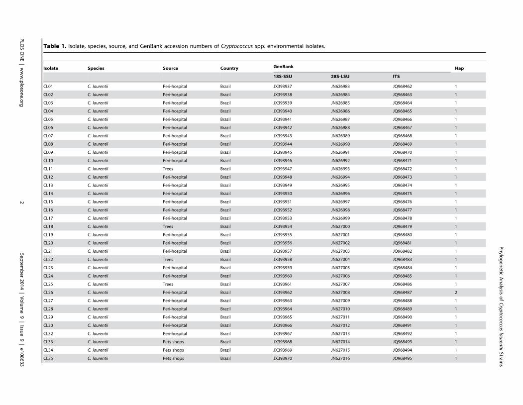
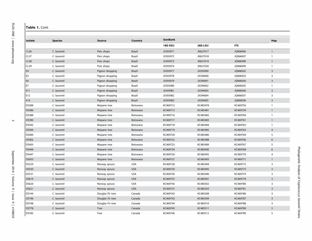
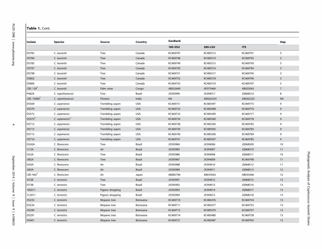
Background: Although Cryptococcus laurentii has been considered saprophytic and its taxonomy is still being described,several cases of human infections have already reported. This study aimed to evaluate molecular aspects of C. laurentiiisolates from Brazil, Botswana, Canada, and the United States.
Methods: In this study, 100 phenotypically identified C. laurentii isolates were evaluated by sequencing the 18S nuclearribosomal small subunit rRNA gene (18S-SSU), D1/D2 region of 28S nuclear ribosomal large subunit rRNA gene (28S-LSU),and the internal transcribed spacer (ITS) of the ribosomal region.
Results: BLAST searches using 550-bp, 650-bp, and 550-bp sequenced amplicons obtained from the 18S-SSU, 28S-LSU, andthe ITS region led to the identification of 75 C. laurentii strains that shared 99–100% identity with C. laurentii CBS 139. A totalof nine isolates shared 99% identity with both Bullera sp. VY-68 and C. laurentii RY1. One isolate shared 99% identity withCryptococcus rajasthanensis CBS 10406, and eight isolates shared 100% identity with Cryptococcus sp. APSS 862 according tothe 28S-LSU and ITS regions and designated as Cryptococcus aspenensis sp. nov. (CBS 13867). While 16 isolates shared 99%identity with Cryptococcus flavescens CBS 942 according to the 18S-SSU sequence, only six were confirmed using the 28S-LSU and ITS region sequences. The remaining 10 shared 99% identity with Cryptococcus terrestris CBS 10810, which wasrecently described in Brazil. Through concatenated sequence analyses, seven sequence types in C. laurentii, three in C.flavescens, one in C. terrestris, and one in the C. aspenensis sp. nov. were identified.
Conclusions: Sequencing permitted the characterization of 75% of the environmental C. laurentii isolates from differentgeographical areas and the identification of seven haplotypes of this species. Among sequenced regions, the increasedvariability of the ITS region in comparison to the 18S-SSU and 28S-LSU regions reinforces its applicability as a DNA barcode.
Citation: Ferreira-Paim K, Ferreira TB, Andrade-Silva L, Mora DJ, Springer DJ, et al. (2014) Phylogenetic Analysis of Phenotypically Characterized Cryptococcuslaurentii Isolates Reveals High Frequency of Cryptic Species. PLoS ONE 9(9): e108633. doi:10.1371/journal.pone.0108633
Editor: Anuradha Chowdhary, V.P.Chest Institute, India
Received May 13, 2014; Accepted August 22, 2014; Published September 24, 2014
Copyright: � 2014 Ferreira-Paim et al. This is an open-access article distributed under the terms of the Creative Commons Attribution License, which permitsunrestricted use, distribution, and reproduction in any medium, provided the original author and source are credited.
Data Availability: The authors confirm that all data underlying the findings are fully available without restriction. All relevant data are within the paper.
Funding: This work was supported by Fundacao de Amparo a Pesquisa de Minas Gerais-FAPEMIG APQ-01735-10 [to M.L.S.V.]. K.F.P. is a research fellow of CAPES:Process number 9313/13-3. The funders had no role in study design, data collection and analysis, decision to publish, or preparation of the manuscript.
Competing Interests: The authors have declared that no competing interests exist.
The Cryptococcus genus includes more than 100 species of
which most are considered non-pathogenic, with the exceptions of
Cryptococcus neoformans and Cryptococcus gattii. During the last
decade Cryptococcus laurentii has occasionally been described to
infect severely immunocompromised hosts [1–3]. In most of these
reports from which isolates were obtained, the blood and the
cerebrospinal fluid (CSF) were the predominant sources [2–5].
C. laurentii was first identified from palm wine in the Congo by
Kufferath in 1920 as Torula laurentii [6]. This isolate was then
reclassified as Torulopsis laurentii [7] and renamed in 1950 as
Cryptococcus laurentii (CBS 139) [8]. Later in Japan, an isolate
with identical phenotypic characteristics was described as Torulaflavescens [9], reclassified in 1922 as Torulopsis flavescens [7], andthen renamed as Cryptococcus flavescens (CBS 942) [8].
Cryptococcus aureus, Cryptococcus carnescens, and Cryptococcuspeneaus, in addition to C. flavescens, were also considered to be
synonymous of C. laurentii until phylogenetic analysis of the
internal transcribed spacer (ITS) and D1/D2 region of 28S
nuclear ribosomal large subunit rRNA gene (28S-LSU) demon-
strated that they are different species [10,11].
PLOS ONE | www.plosone.org 1 September 2014 | Volume 9 | Issue 9 | e108633
To confirm the haplotypes obtained by median-joining
networks the analyses were replicated in MLSTest software
(available at http://mlstest.codeplex.com) and graphed by goe-
BURST algorithm in PHILOVIZ software [33,34]. The mini-
mum spanning tree representing the comparison between the
isolates sources and their haplotype was also calculated by
goeBURST.
Coalescent species analysesIn order to estimate the time divergence between species and
haplotypes, the interspecific and intraspecific net nucleotides
substitutions (d) and standard error of the concatenated sequences
were calculated in accordance to Kimura [27] with a bootstrap
(500 replicates) as a variance method in the MEGA 6.0 software
[35,36]. The distance and standard error between closest species e.
g. (C. laurentii x C. rajasthanensis 0.01660.003; C. aspenensis xC. flavescens 0.07160.007; C. terrestris x C. flavescens0.00960.002) were obtained and applied in the equation
d=2lt, where d is the number of nucleotide substitutions per
site between a pair of sequences, t is the divergence time, and l the
rate of nucleotide substitution. Here, we applied the constant (l)
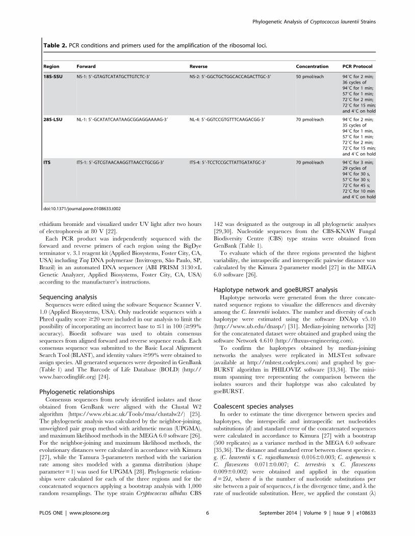
Table 2. PCR conditions and primers used for the amplification of the ribosomal loci.
Region Forward Reverse Concentration PCR Protocol
18S-SSU NS-1: 59-GTAGTCATATGCTTGTCTC-39 NS-2: 59-GGCTGCTGGCACCAGACTTGC-39 50 pmol/each 94uC for 2 min;36 cycles of94uC for 1 min;57uC for 1 min;72uC for 2 min;72uC for 15 min;and 4uC on hold
28S-LSU NL-1: 59-GCATATCAATAAGCGGAGGAAAAG-39 NL-4: 59-GGTCCGTGTTTCAAGACGG-39 70 pmol/each 94uC for 2 min;35 cycles of94uC for 1 min,57uC for 1 min;72uC for 2 min;72uC for 15 min;and 4uC on hold
ITS ITS-1: 59-GTCGTAACAAGGTTAACCTGCGG-39 ITS-4: 59-TCCTCCGCTTATTGATATGC-39 70 pmol/each 94uC for 3 min;29 cycles of94uC for 30 s,57uC for 30 s;72uC for 45 s;72uC for 10 minand 4uC on hold
doi:10.1371/journal.pone.0108633.t002
Phylogenetic Analysis of Cryptococcus laurentii Strains
PLOS ONE | www.plosone.org 6 September 2014 | Volume 9 | Issue 9 | e108633
1029/bp/year previously obtained for the Eurotiomycetes lineagedue to the absence of a known fossil for C. laurentii species [36].The resulting time of divergence were used as prior parameters to
calibrate the tree in the coalescent analyses.
The optimal molecular evolutionary model for the concatenated
sequences was determined using the corrected Akaike Information
Criterion (AICc) as executed in the software jModelTest 2 [37,38].
The optimal molecular evolutionary model General Time
Reversible (GTR+I+G) with the respective parameters: AC:
raffinose, sacarose, sorbose, xylose, and 2-keto-glutarate. However,
the isolates were negative for fermentation of dextrose and
assimilation of inulin and potassium nitrate. FACS analysis
indicated that most of the isolates are haploid (Figure S1).
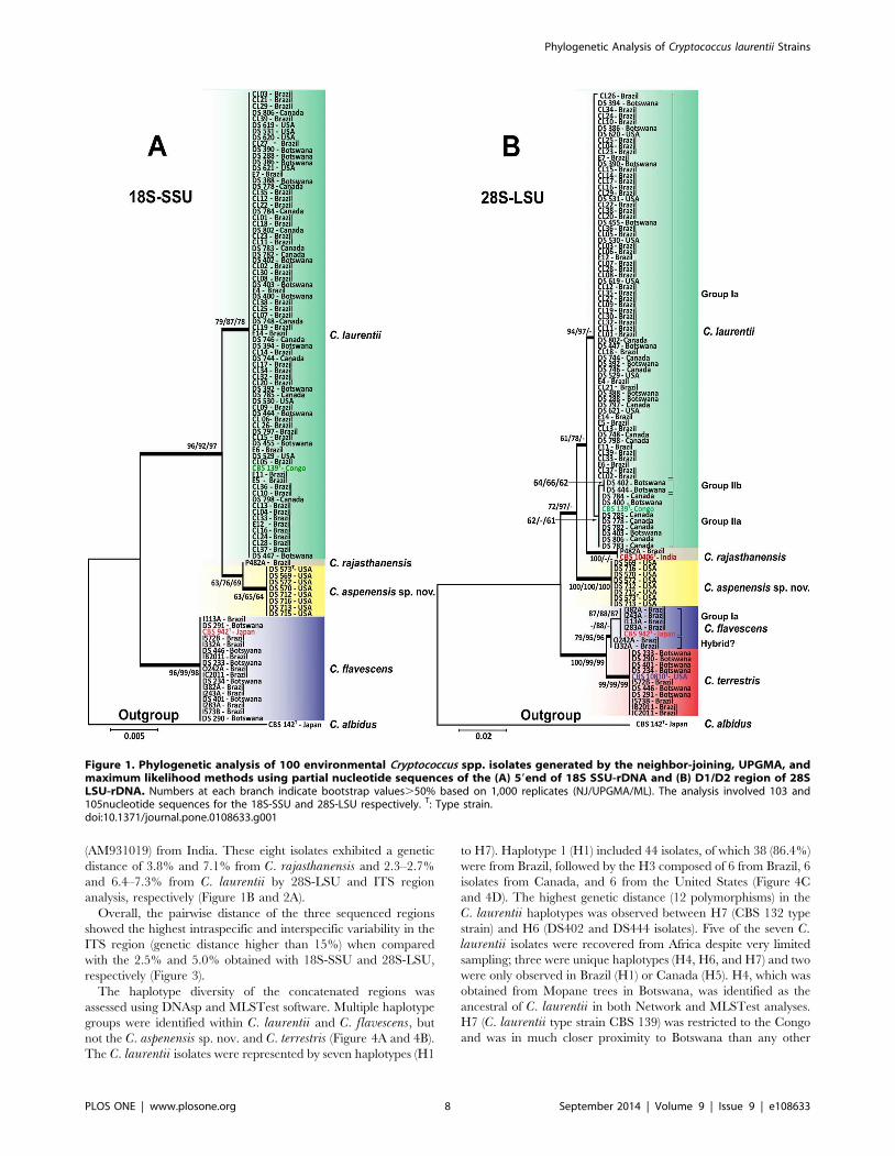
A 550-bp product was amplified from the 59 end of 18S-SSU
and sequenced with the primers NS-1 and NS-2, from which a
339-bp alignment was obtained. In this analysis, 75 isolates shared
99–100% identity with the C. laurentii CBS 139 (AB032640) type
strain. Another 16 isolates shared 99% identity with C. flavescensCBS 942 (AB085796). The remaining 9 shared 99% identity with
both Bullera sp. VY-68 (AB110694) from Japan and with C.laurentii RY1 from India (EF063147). High bootstrap values
generated by neighbor-joining, UPGMA, and maximum likeli-
hood analyses supported the differentiation of the following clades:
C. laurentii (bootstrap values of 79, 87, and 78, respectively), C.flavescens (bootstrap values of 96, 99, and 98, respectively) and
Bullera sp./C. laurentii (bootstrap values of 63, 65, and 64,
respectively) (Figure 1A).
Due to the low genetic variability of the C. laurentii clade
obtained at the 59 end of the 18S-SSU gene, we sequenced two
additional ribosomal loci: D1/D2 of the 28S-LSU and the ITS
gene regions. The alignment and analysis of the 530-bp long
amplicon of the 28S-LSU region confirmed the identification of 75
isolates as C. laurentii and showed more intraspecific variability
differentiating three major groups (group Ia, IIa, and IIb) within
C. laurentii isolates. Of the 16 C. flavescens isolates identified by
the 18S-SSU sequencing, only 6 were confirmed in the C.flavescens clade by the 28S-LSU region with high bootstrap values
of 79 (neighbor-joining), 95 (UPGMA), and 96 (maximum
likelihood). The C. flavescens clade was split into two groups
(Group Ia and a possible hybrid) by 28S-LSU and three groups
(Groups Ia, Ib, and a possible hybrid) by ITS and analyses of the
1,328-bp amplicon of the concatenated regions. The two possible
hybrid isolates I332A and O242A from Brazil were more related
to C. terrestris in the ITS and concatenated sequences analyses
(Figure 1B and 2). The remaining 10 isolates shared 99% identity
with C. terrestris (CBS 10810), which has been recently described
in Brazil and the United States (Figure 1B).
Among the nine Bullera sp./C. laurentii isolates identified by
the 18S-SSU, isolate P482A shared 99% identity of the 28S-LSU
and ITS regions with Cryptococcus rajasthanensis CBS 10406
(AM262324) from India. The eight remaining isolates (DS569,
DS570, DS572, DS573, DS712, DS713, DS715, and DS716)
recovered from a trembling aspen tree (Populus tremuloides) weredesignated as Cryptococcus aspenensis sp. nov. because they shared100% identity with two undescribed isolates of Cryptococcus sp.
APSS-862 (FM178286) and Cryptococcus sp. APSS-823
Phylogenetic Analysis of Cryptococcus laurentii Strains
PLOS ONE | www.plosone.org 7 September 2014 | Volume 9 | Issue 9 | e108633
(AM931019) from India. These eight isolates exhibited a genetic
distance of 3.8% and 7.1% from C. rajasthanensis and 2.3–2.7%
and 6.4–7.3% from C. laurentii by 28S-LSU and ITS region
analysis, respectively (Figure 1B and 2A).
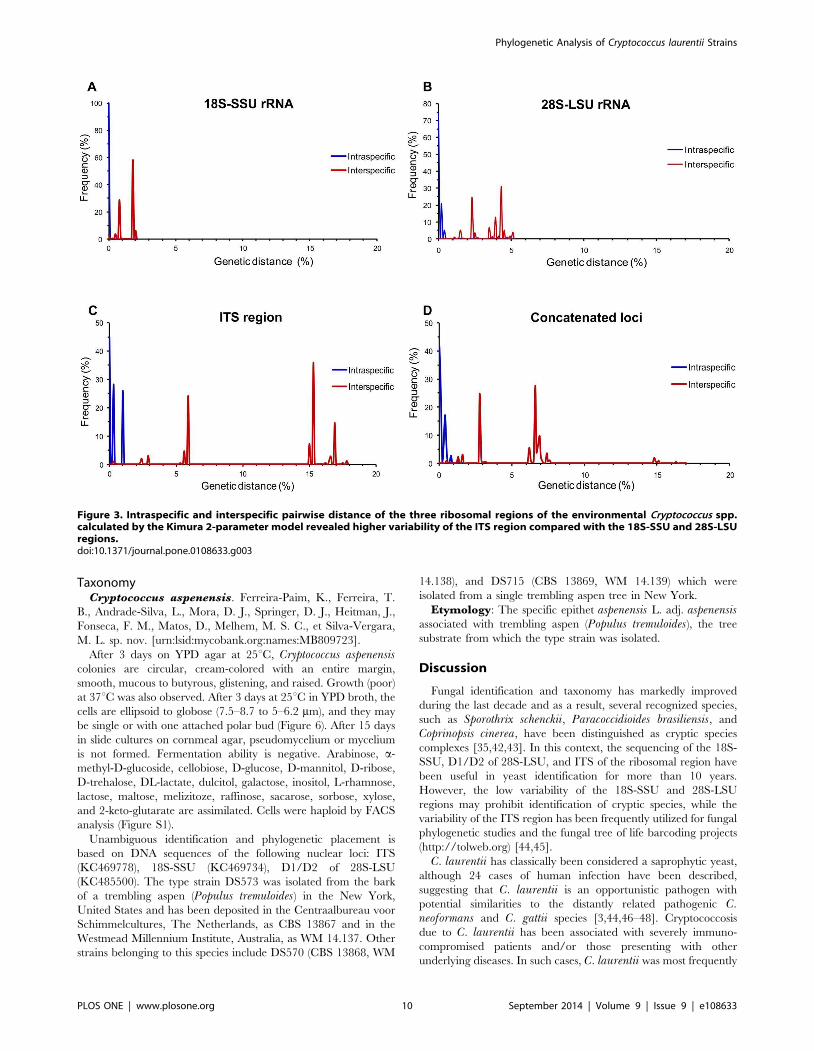
Overall, the pairwise distance of the three sequenced regions
showed the highest intraspecific and interspecific variability in the
ITS region (genetic distance higher than 15%) when compared
with the 2.5% and 5.0% obtained with 18S-SSU and 28S-LSU,
respectively (Figure 3).
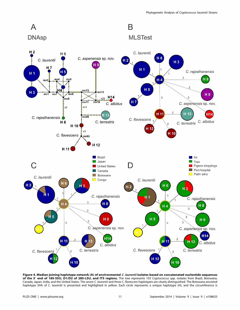
The haplotype diversity of the concatenated regions was
assessed using DNAsp and MLSTest software. Multiple haplotype
groups were identified within C. laurentii and C. flavescens, butnot the C. aspenensis sp. nov. and C. terrestris (Figure 4A and 4B).
The C. laurentii isolates were represented by seven haplotypes (H1
to H7). Haplotype 1 (H1) included 44 isolates, of which 38 (86.4%)
were from Brazil, followed by the H3 composed of 6 from Brazil, 6
isolates from Canada, and 6 from the United States (Figure 4C
and 4D). The highest genetic distance (12 polymorphisms) in the
C. laurentii haplotypes was observed between H7 (CBS 132 type
strain) and H6 (DS402 and DS444 isolates). Five of the seven C.laurentii isolates were recovered from Africa despite very limited
sampling; three were unique haplotypes (H4, H6, and H7) and two
were only observed in Brazil (H1) or Canada (H5). H4, which was
obtained from Mopane trees in Botswana, was identified as the
ancestral of C. laurentii in both Network and MLSTest analyses.
H7 (C. laurentii type strain CBS 139) was restricted to the Congo
and was in much closer proximity to Botswana than any other
Figure 1. Phylogenetic analysis of 100 environmental Cryptococcus spp. isolates generated by the neighbor-joining, UPGMA, andmaximum likelihood methods using partial nucleotide sequences of the (A) 59end of 18S SSU-rDNA and (B) D1/D2 region of 28SLSU-rDNA. Numbers at each branch indicate bootstrap values.50% based on 1,000 replicates (NJ/UPGMA/ML). The analysis involved 103 and105nucleotide sequences for the 18S-SSU and 28S-LSU respectively. T: Type strain.doi:10.1371/journal.pone.0108633.g001
Phylogenetic Analysis of Cryptococcus laurentii Strains
PLOS ONE | www.plosone.org 8 September 2014 | Volume 9 | Issue 9 | e108633
sample region. H3 was distinct from Botswana and was comprised
of isolates from North and South America (Figure 4).
Three haplotypes were identified in C. flavescens isolates (H10,
H11, and H12), with the ancestral haplotype H11 restricted to
Brazil. H10 presented the highest genetic distance (9 polymor-
phisms) when compared with H11 and H12 (2 polymorphisms).
H10 was also positioned closer to the C. terrestris haplotype H13
and could be a unique species, or ancestral genotype, or
recombinant hybrid isolate between C. flavescens and C. terrestris.The C. aspenensis sp. nov. H9 was a completely unique genotype
from New York, USA (Figure 4).
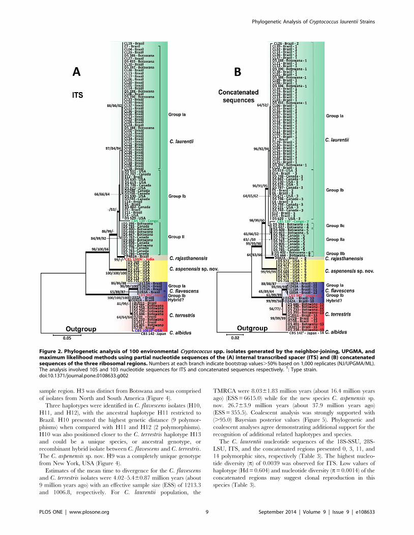
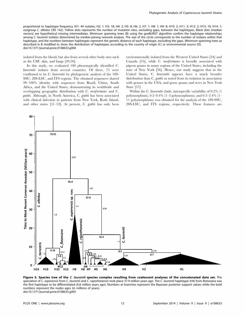
Estimates of the mean time to divergence for the C. flavescensand C. terrestris isolates were 4.02–5.460.87 million years (about
9 million years ago) with an effective sample size (ESS) of 1213.3
and 1006.8, respectively. For C. laurentii population, the
TMRCA were 8.0361.83 million years (about 16.4 million years
ago) (ESS=6615.0) while for the new species C. aspenensis sp.
nov. 26.763.9 million years (about 37.9 million years ago)
(ESS=355.5). Coalescent analysis was strongly supported with
(.95.0) Bayesian posterior values (Figure 5). Phylogenetic and
coalescent analyses agree demonstrating additional support for the
recognition of additional related haplotypes and species.
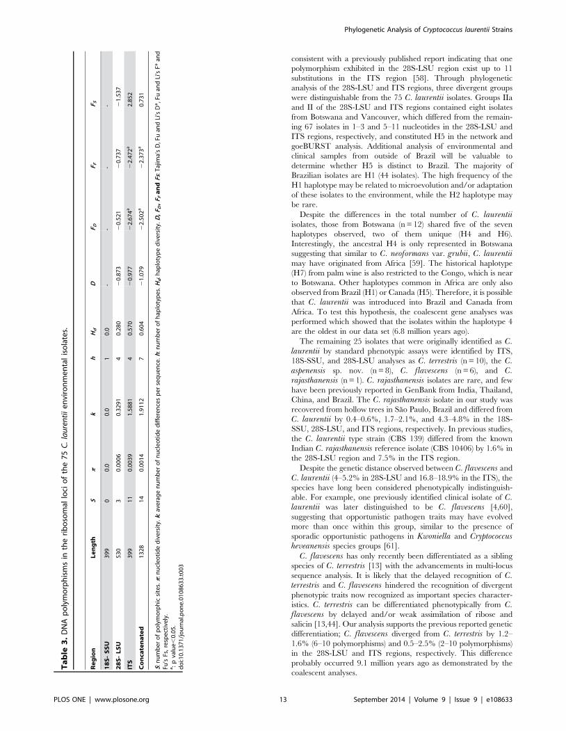
The C. laurentii nucleotide sequences of the 18S-SSU, 28S-
LSU, ITS, and the concatenated regions presented 0, 3, 11, and
14 polymorphic sites, respectively (Table 3). The highest nucleo-
tide diversity (p) of 0.0039 was observed for ITS. Low values of
haplotype (Hd= 0.604) and nucleotide diversity (p=0.0014) of the
concatenated regions may suggest clonal reproduction in this
species (Table 3).
Figure 2. Phylogenetic analysis of 100 environmental Cryptococcus spp. isolates generated by the neighbor-joining, UPGMA, andmaximum likelihood methods using partial nucleotide sequences of the (A) internal transcribed spacer (ITS) and (B) concatenatedsequences of the three ribosomal regions. Numbers at each branch indicate bootstrap values.50% based on 1,000 replicates (NJ/UPGMA/ML).The analysis involved 105 and 103 nucleotide sequences for ITS and concatenated sequences respectively. T: Type strain.doi:10.1371/journal.pone.0108633.g002
Phylogenetic Analysis of Cryptococcus laurentii Strains
PLOS ONE | www.plosone.org 9 September 2014 | Volume 9 | Issue 9 | e108633
TaxonomyCryptococcus aspenensis. Ferreira-Paim, K., Ferreira, T.
B., Andrade-Silva, L., Mora, D. J., Springer, D. J., Heitman, J.,
Fonseca, F. M., Matos, D., Melhem, M. S. C., et Silva-Vergara,
M. L. sp. nov. [urn:lsid:mycobank.org:names:MB809723].
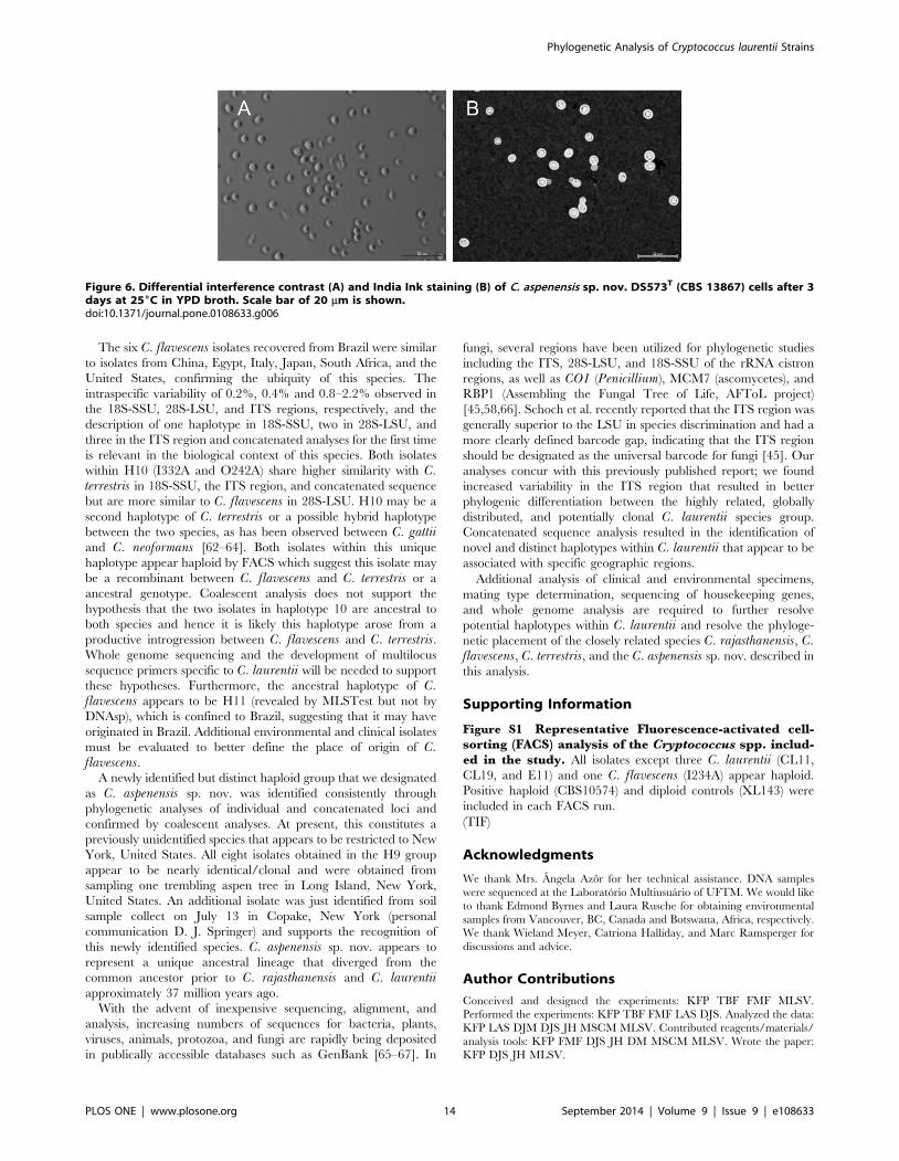
After 3 days on YPD agar at 25uC, Cryptococcus aspenensiscolonies are circular, cream-colored with an entire margin,
smooth, mucous to butyrous, glistening, and raised. Growth (poor)
at 37uC was also observed. After 3 days at 25uC in YPD broth, the
cells are ellipsoid to globose (7.5–8.7 to 5–6.2 mm), and they may
be single or with one attached polar bud (Figure 6). After 15 days
in slide cultures on cornmeal agar, pseudomycelium or mycelium
is not formed. Fermentation ability is negative. Arabinose, a-
and 2-keto-glutarate are assimilated. Cells were haploid by FACS
analysis (Figure S1).
Unambiguous identification and phylogenetic placement is
based on DNA sequences of the following nuclear loci: ITS
(KC469778), 18S-SSU (KC469734), D1/D2 of 28S-LSU
(KC485500). The type strain DS573 was isolated from the bark
of a trembling aspen (Populus tremuloides) in the New York,
United States and has been deposited in the Centraalbureau voor
Schimmelcultures, The Netherlands, as CBS 13867 and in the
Westmead Millennium Institute, Australia, as WM 14.137. Other
strains belonging to this species include DS570 (CBS 13868, WM
14.138), and DS715 (CBS 13869, WM 14.139) which were
isolated from a single trembling aspen tree in New York.
Etymology: The specific epithet aspenensis L. adj. aspenensisassociated with trembling aspen (Populus tremuloides), the tree
substrate from which the type strain was isolated.
Discussion
Fungal identification and taxonomy has markedly improved
during the last decade and as a result, several recognized species,
such as Sporothrix schenckii, Paracoccidioides brasiliensis, and
Coprinopsis cinerea, have been distinguished as cryptic species
complexes [35,42,43]. In this context, the sequencing of the 18S-
SSU, D1/D2 of 28S-LSU, and ITS of the ribosomal region have
been useful in yeast identification for more than 10 years.
However, the low variability of the 18S-SSU and 28S-LSU
regions may prohibit identification of cryptic species, while the
variability of the ITS region has been frequently utilized for fungal
phylogenetic studies and the fungal tree of life barcoding projects
(http://tolweb.org) [44,45].
C. laurentii has classically been considered a saprophytic yeast,
although 24 cases of human infection have been described,
suggesting that C. laurentii is an opportunistic pathogen with
potential similarities to the distantly related pathogenic C.neoformans and C. gattii species [3,44,46–48]. Cryptococcosis
due to C. laurentii has been associated with severely immuno-
compromised patients and/or those presenting with other
underlying diseases. In such cases, C. laurentii was most frequently
Figure 3. Intraspecific and interspecific pairwise distance of the three ribosomal regions of the environmental Cryptococcus spp.calculated by the Kimura 2-parameter model revealed higher variability of the ITS region compared with the 18S-SSU and 28S-LSUregions.doi:10.1371/journal.pone.0108633.g003
Phylogenetic Analysis of Cryptococcus laurentii Strains
PLOS ONE | www.plosone.org 10 September 2014 | Volume 9 | Issue 9 | e108633
Figure 4. Median-joining haplotype network (A) of environmental C. laurentii isolates based on concatenated nucleotide sequencesof the 59 end of 18S-SSU, D1/D2 of 28S-LSU, and ITS regions. The tree represents 103 Cryptococcus spp. isolates from Brazil, Botswana,Canada, Japan, India, and the United States. The seven C. laurentii and three C. flavescens haplotypes are clearly distinguished. The Botswana ancestralhaplotype (H4) of C. laurentii is presented and highlighted in yellow. Each circle represents a unique haplotype (H), and the circumference is
Phylogenetic Analysis of Cryptococcus laurentii Strains
PLOS ONE | www.plosone.org 11 September 2014 | Volume 9 | Issue 9 | e108633
isolated from the blood, but also from several other body sites such
as the CSF, skin, and lungs [49,50].
In this study, we evaluated 100 phenotypically identified C.laurentii isolates from several countries. Of these, 75 were
confirmed to be C. laurentii by phylogenetic analysis of the 18S-
SSU, 28S-LSU, and ITS regions. The obtained sequences shared
99–100% identity with sequences from Brazil, China, South
Africa, and the United States, demonstrating its worldwide and
overlapping geographic distribution with C. neoformans and C.gattii. Although, in North America, C. gattii has been associated
with clinical infection in patients from New York, Rode Island,
and other states [51–53]. At present, C. gattii has only been
environmentally isolated from the Western United States [54] and
Canada [55], while C. neoformans is broadly associated with
pigeon guano in many regions of the United States, including the
state of New York [56]. Hence, our study suggests that in the
United States, C. laurentii appears have a much broader
distribution than C. gattii as noted from its isolation in association
with grasses in the USA, and goose guano and trees in New York
State [57].
Within the C. laurentii clade, intraspecific variability of 0.2% (1
polymorphism), 0.2–0.4% (1–3 polymorphisms), and 0.3–2.4% (1–
11 polymorphisms) was obtained for the analysis of the 18S-SSU,
28S-LSU, and ITS regions, respectively. These features are
proportional to haplotype frequency (H1: 44 isolates; H2: 1; H3: 18; H4: 2; H5: 8; H6: 2; H7: 1; H8: 1; H9: 8; H10: 2; H11: 3; H12: 2; H13: 10; H14: 1;outgroup C. albidus CBS 142). Yellow dots represents the number of mutation sites, excluding gaps, between the haplotypes. Black dots (medianvectors) are hypothetical missing intermediates. Minimum spanning trees (B) using the goeBURST algorithm confirm the haplotype relationshipsamong C. laurentii isolates determined by median-joining network analysis. The size of the circle corresponds to the number of isolates within thathaplotype, and the numbers between haplotypes represent the genetic distance of each haplotype, excluding the gaps. Minimum spanning trees asdescribed in B modified to show the distribution of haplotypes according to the country of origin (C) or environmental source (D).doi:10.1371/journal.pone.0108633.g004
Figure 5. Species tree of the C. laurentii species complex resulting from coalescent analyses of the concatenated data set. Thespeciation of C. aspenensis from C. laurentii and C. rajasthanensis took place 37.9 million years ago. The C. laurentii haplotype (H4) from Botswana wasthe first haplotype to be differentiated (6.8 million years ago). Numbers at branches represent the Bayesian posterior support values while the boldnumbers represent the nodes ages (in millions of years).doi:10.1371/journal.pone.0108633.g005
Phylogenetic Analysis of Cryptococcus laurentii Strains
PLOS ONE | www.plosone.org 12 September 2014 | Volume 9 | Issue 9 | e108633
consistent with a previously published report indicating that one
polymorphism exhibited in the 28S-LSU region exist up to 11
substitutions in the ITS region [58]. Through phylogenetic
analysis of the 28S-LSU and ITS regions, three divergent groups
were distinguishable from the 75 C. laurentii isolates. Groups IIa
and II of the 28S-LSU and ITS regions contained eight isolates
from Botswana and Vancouver, which differed from the remain-
ing 67 isolates in 1–3 and 5–11 nucleotides in the 28S-LSU and
ITS regions, respectively, and constituted H5 in the network and
goeBURST analysis. Additional analysis of environmental and
clinical samples from outside of Brazil will be valuable to
determine whether H5 is distinct to Brazil. The majority of
Brazilian isolates are H1 (44 isolates). The high frequency of the
H1 haplotype may be related to microevolution and/or adaptation
of these isolates to the environment, while the H2 haplotype may
be rare.
Despite the differences in the total number of C. laurentiiisolates, those from Botswana (n= 12) shared five of the seven
haplotypes observed, two of them unique (H4 and H6).
Interestingly, the ancestral H4 is only represented in Botswana
suggesting that similar to C. neoformans var. grubii, C. laurentiimay have originated from Africa [59]. The historical haplotype
(H7) from palm wine is also restricted to the Congo, which is near
to Botswana. Other haplotypes common in Africa are only also
observed from Brazil (H1) or Canada (H5). Therefore, it is possible
that C. laurentii was introduced into Brazil and Canada from
Africa. To test this hypothesis, the coalescent gene analyses was
performed which showed that the isolates within the haplotype 4
are the oldest in our data set (6.8 million years ago).
The remaining 25 isolates that were originally identified as C.laurentii by standard phenotypic assays were identified by ITS,
18S-SSU, and 28S-LSU analyses as C. terrestris (n = 10), the C.aspenensis sp. nov. (n = 8), C. flavescens (n = 6), and C.rajasthanensis (n = 1). C. rajasthanensis isolates are rare, and few
have been previously reported in GenBank from India, Thailand,
China, and Brazil. The C. rajasthanensis isolate in our study was
recovered from hollow trees in Sao Paulo, Brazil and differed from
C. laurentii by 0.4–0.6%, 1.7–2.1%, and 4.3–4.8% in the 18S-
SSU, 28S-LSU, and ITS regions, respectively. In previous studies,
the C. laurentii type strain (CBS 139) differed from the known
Indian C. rajasthanensis reference isolate (CBS 10406) by 1.6% in
the 28S-LSU region and 7.5% in the ITS region.
Despite the genetic distance observed between C. flavescens andC. laurentii (4–5.2% in 28S-LSU and 16.8–18.9% in the ITS), the
species have long been considered phenotypically indistinguish-
able. For example, one previously identified clinical isolate of C.laurentii was later distinguished to be C. flavescens [4,60],
suggesting that opportunistic pathogen traits may have evolved
more than once within this group, similar to the presence of
sporadic opportunistic pathogens in Kwoniella and Cryptococcusheveanensis species groups [61].C. flavescens has only recently been differentiated as a sibling
species of C. terrestris [13] with the advancements in multi-locus
sequence analysis. It is likely that the delayed recognition of C.terrestris and C. flavescens hindered the recognition of divergent
phenotypic traits now recognized as important species character-
istics. C. terrestris can be differentiated phenotypically from C.flavescens by delayed and/or weak assimilation of ribose and
salicin [13,44]. Our analysis supports the previous reported genetic
differentiation; C. flavescens diverged from C. terrestris by 1.2–
1.6% (6–10 polymorphisms) and 0.5–2.5% (2–10 polymorphisms)
in the 28S-LSU and ITS regions, respectively. This difference
probably occurred 9.1 million years ago as demonstrated by the
sorting (FACS) analysis of the Cryptococcus spp. includ-
ed in the study. All isolates except three C. laurentii (CL11,CL19, and E11) and one C. flavescens (I234A) appear haploid.
Positive haploid (CBS10574) and diploid controls (XL143) were
included in each FACS run.
(TIF)
Acknowledgments
We thank Mrs. Angela Azor for her technical assistance. DNA samples
were sequenced at the Laboratorio Multiusuario of UFTM. We would like
to thank Edmond Byrnes and Laura Rusche for obtaining environmental
samples from Vancouver, BC, Canada and Botswana, Africa, respectively.
We thank Wieland Meyer, Catriona Halliday, and Marc Ramsperger for
discussions and advice.
Author Contributions
Conceived and designed the experiments: KFP TBF FMF MLSV.
Performed the experiments: KFP TBF FMF LAS DJS. Analyzed the data:
KFP LAS DJM DJS JH MSCM MLSV. Contributed reagents/materials/
analysis tools: KFP FMF DJS JH DM MSCM MLSV. Wrote the paper:
KFP DJS JH MLSV.
Figure 6. Differential interference contrast (A) and India Ink staining (B) of C. aspenensis sp. nov. DS573T (CBS 13867) cells after 3days at 256C in YPD broth. Scale bar of 20 mm is shown.doi:10.1371/journal.pone.0108633.g006
Phylogenetic Analysis of Cryptococcus laurentii Strains
PLOS ONE | www.plosone.org 14 September 2014 | Volume 9 | Issue 9 | e108633
References
1. Averbuch D, Boekhoutt T, Falk R, Engelhard D, Shapiro M, et al. (2002)Fungemia in a cancer patient caused by fluconazole-resistant Cryptococcuslaurentii. Med Mycol 40: 479–484.
2. Bauters TG, Swinne D, Boekhout T, Noens L, Nelis HJ (2002) Repeatedisolation of Cryptococcus laurentii from the oropharynx of an immunocompro-mized patient. Mycopathologia 153: 133–135.
3. Manfredi R, Fulgaro C, Sabbatani S, Legnani G, Fasulo G (2006) Emergence ofamphotericin B-resistant Cryptococcus laurentii meningoencephalitis shortlyafter treatment for Cryptococcus neoformans meningitis in a patient with AIDS.AIDS Patient Care STDS 20: 227–232.
4. Kordossis T, Avlami A, Velegraki A, Stefanou I, Georgakopoulos G, et al. (1998)First report of Cryptococcus laurentii meningitis and a fatal case of Cryptococcusalbidus cryptococcaemia in AIDS patients. Med Mycol 36: 335–339.
5. Banerjee P, Haider M, Trehan V, Mishra B, Thakur A, et al. (2013)Cryptococcus laurentii fungemia. Indian J Med Microbiol 31: 75–77.
6. Kufferath H (1920) Peut-on obtenir du mout de biere alcalin? Annales de laSociete royale des sciences medicales et naturelles de Bruxelles 74: 16–46.
7. Lodder J (1934) Die anaskosporegenen Hefen, I. Halfte. Verh K Ned Akad WetAfd Natuurkd v. 32: 1–256.
8. Skinner CE (1950) Generic name for imperfect yeasts, Cryptococcus orTorulopsis? The American Midland Naturalist Journal 43: 242–250.
9. Saito K (1922) Untersuchungen uber die atmospharischen Pilzkeime. MittJpn J Bot 1: 1–54.
10. Sugita T, Takashima M, Ikeda R, Nakase T, Shinoda T (2000) Intraspeciesdiversity of Cryptococcus laurentii as revealed by sequences of internaltranscribed spacer regions and 28S rRNA gene and taxonomic position of C.laurentii clinical isolates. J Clin Microbiol 38: 1468–1471.
11. Takashima M, Sugita T, Shinoda T, Nakase T (2003) Three new combinationsfrom the Cryptococcus laurentii complex: Cryptococcus aureus, Cryptococcuscarnescens and Cryptococcus peneaus. Int J Syst Evol Microbiol 53: 1187–1194.
12. Saluja P, Prasad GS (2007) Cryptococcus rajasthanensis sp. nov., an anamorphicyeast species related to Cryptococcus laurentii, isolated from Rajasthan, India.Int J Syst Evol Microbiol 57: 414–418.
13. Crestani J, Fontes Landell M, Faganello J, Henning Vainstein M, SimpsonVishniac H, et al. (2009) Cryptococcus terrestris sp. nov., a tremellaceous,anamorphic yeast phylogenetically related to Cryptococcus flavescens. Int J SystEvol Microbiol 59: 631–636.
14. Simon G, Simon G, Erdos M, Marodi L (2005) Invasive Cryptococcus laurentiidisease in a nine-year-old boy with X-linked hyper-immunoglobulin Msyndrome. Pediatr Infect Dis J 24: 935–937.
15. Rosario I, Soro G, Deniz S, Ferrer O, Acosta F, et al. (2009) Presence of C.albidus, C. laurentii and C. uniguttulatus in crop and droppings of pigeon lofts(Columba livia). Mycopathologia 169: 315–319.
16. Ferreira-Paim K, Andrade-Silva L, Mora DJ, Lages-Silva E, Pedrosa AL, et al.(2012) Antifungal susceptibility, enzymatic activity, PCR-fingerprinting and ITSsequencing of environmental Cryptococcus laurentii isolates from Uberaba,Minas Gerais, Brazil. Mycopathologia 174: 41–52.
17. Tay ST, Na SL, Tajuddin TH (2008) Natural occurrence and growth reactionon canavanine-glycine-bromothymol blue agar of non-neoformans Cryptococcusspp. in Malaysia. Mycoses 51: 515–519.
18. Granados DP, Castaneda E (2005) Isolation and characterization of Cryptococ-cus neoformans varieties recovered from natural sources in Bogota, Colombia,and study of ecological conditions in the area. Microb Ecol 49: 282–290.
19. Staib F (1963) Membrane filtration and guizotia abyssinica culture media for thedemonstration of Cryptococcus neoformans (Brown Color Effect). Z HygInfektionskr 149: 329–336.
20. Randhawa HS, Kowshik T, Khan ZU (2005) Efficacy of swabbing versus aconventional technique for isolation of Cryptococcus neoformans from decayedwood in tree trunk hollows. Med Mycol 43: 67–71.
21. Ferreira-Paim K, Andrade-Silva L, Mora DJ, Pedrosa AL, Rodrigues V, et al.(2011) Genotyping of Cryptococcus neoformans isolated from captive birds inUberaba, Minas Gerais, Brazil. Mycoses 54: e294–300.
22. Sugita T, Nishikawa A, Ikeda R, Shinoda T (1999) Identification of medicallyrelevant Trichosporon species based on sequences of internal transcribed spacerregions and construction of a database for Trichosporon identification. J ClinMicrobiol 37: 1985–1993.
23. Kurtzman CP, Robnett CJ (1997) Identification of clinically importantascomycetous yeasts based on nucleotide divergence in the 59 end of thelarge-subunit (26S) ribosomal DNA gene. J Clin Microbiol 35: 1216–1223.
24. Ratnasingham S, Hebert PD (2007) bold: The Barcode of Life Data System(http://www.barcodinglife.org). Mol Ecol Notes 7: 355–364.
25. Thompson JD, Higgins DG, Gibson TJ (1994) CLUSTAL W: improving thesensitivity of progressive multiple sequence alignment through sequenceweighting, position-specific gap penalties and weight matrix choice. NucleicAcids Res 22: 4673–4680.
26. Tamura K, Stecher G, Peterson D, Filipski A, Kumar S (2013) MEGA6:Molecular Evolutionary Genetics Analysis version 6.0. Mol Biol Evol 30: 2725–2729.
27. Kimura M (1980) A simple method for estimating evolutionary rates of basesubstitutions through comparative studies of nucleotide sequences. J Mol Evol16: 111–120.
28. Tamura K (1992) Estimation of the number of nucleotide substitutions whenthere are strong transition-transversion and G+C-content biases. Mol Biol Evol9: 678–687.
29. Fell JW, Roeijmans H, Boekhout T (1999) Cystofilobasidiales, a new order ofbasidiomycetous yeasts. Int J Syst Bacteriol 49 Pt 2: 907–913.
30. Scorzetti G, Petrescu I, Yarrow D, Fell JW (2000) Cryptococcus adeliensis sp.nov., a xylanase producing basidiomycetous yeast from Antarctica. Antonie VanLeeuwenhoek 77: 153–157.
31. Librado P, Rozas J (2009) DnaSP v5: a software for comprehensive analysis ofDNA polymorphism data. Bioinformatics 25: 1451–1452.
32. Bandelt HJ, Forster P, Rohl A (1999) Median-joining networks for inferringintraspecific phylogenies. Mol Biol Evol 16: 37–48.
33. Francisco AP, Bugalho M, Ramirez M, Carrico JA (2009) Global optimaleBURST analysis of multilocus typing data using a graphic matroid approach.BMC Bioinformatics 10: 152.
34. Francisco AP, Vaz C, Monteiro PT, Melo-Cristino J, Ramirez M, et al. (2012)PHYLOViZ: phylogenetic inference and data visualization for sequence basedtyping methods. BMC Bioinformatics 13: 87.
35. Teixeira MM, Theodoro RC, de Carvalho MJ, Fernandes L, Paes HC, et al.(2009) Phylogenetic analysis reveals a high level of speciation in theParacoccidioides genus. Mol Phylogenet Evol 52: 273–283.
36. Kasuga T, White TJ, Taylor JW (2002) Estimation of nucleotide substitutionrates in Eurotiomycete fungi. Mol Biol Evol 19: 2318–2324.
37. Darriba D, Taboada GL, Doallo R, Posada D (2012) jModelTest 2: moremodels, new heuristics and parallel computing. Nat Methods 9: 772.
38. Guindon S, Gascuel O (2003) A simple, fast, and accurate algorithm to estimatelarge phylogenies by maximum likelihood. Syst Biol 52: 696–704.
41. Tanaka R, Taguchi H, Takeo K, Miyaji M, Nishimura K (1996) Determinationof ploidy in Cryptococcus neoformans by flow cytometry. J Med Vet Mycol 34:299–301.
42. Marimon R, Cano J, Gene J, Sutton DA, Kawasaki M, et al. (2007) Sporothrixbrasiliensis, S. globosa, and S. mexicana, three new Sporothrix species of clinicalinterest. J Clin Microbiol 45: 3198–3206.
43. Nagy LG, Desjardin DE, Vagvolgyi C, Kemp R, Papp T (2013) Phylogeneticanalyses of Coprinopsis sections Lanatuli and Atramentarii identify multiplespecies within morphologically defined taxa. Mycologia 105: 112–124.
44. Fell JW, Boekhout T, Fonseca A, Scorzetti G, Statzell-Tallman A (2000)Biodiversity and systematics of basidiomycetous yeasts as determined by large-subunit rDNA D1/D2 domain sequence analysis. Int J Syst Evol Microbiol 50Pt 3: 1351–1371.
45. Schoch CL, Seifert KA, Huhndorf S, Robert V, Spouge JL, et al. (2012) Nuclearribosomal internal transcribed spacer (ITS) region as a universal DNA barcodemarker for fungi. Proc Natl Acad Sci U S A 109: 6241–6246.
46. Shankar EM, Kumarasamy N, Bella D, Renuka S, Kownhar H, et al. (2006)Pneumonia and pleural effusion due to Cryptococcus laurentii in a clinicallyproven case of AIDS. Can Respir J 13: 275–278.
47. Andrade-Silva L, Ferreira-Paim K, Silva-Vergara ML, Pedrosa AL (2010)Molecular characterization and evaluation of virulence factors of Cryptococcuslaurentii and Cryptococcus neoformans strains isolated from external hospitalareas. Fungal Biol 114: 438–445.
48. Danesi P, Firacative C, Cogliati M, Otranto D, Capelli G, et al. (2014)Multilocus sequence typing (MLST) and M13 PCR fingerprinting revealedheterogeneity amongst Cryptococcus species obtained from Italian veterinaryisolates. FEMS Yeast Res. doi: 10.1111/1567-1364.12178.
49. Johnson LB, Bradley SF, Kauffman CA (1998) Fungaemia due to Cryptococcuslaurentii and a review of non-neoformans cryptococcaemia. Mycoses 41: 277–280.
50. Kiertiburanakul S, Sungkanuparph S, Pracharktam R (2001) Cryptococcuslaurentii fungemia: A case report. J Infect Dis Antimicrob Agents 18: 112–114.
51. Lockhart SR, Iqbal N, Harris JR, Grossman NT, DeBess E, et al. (2013)Cryptococcus gattii in the United States: genotypic diversity of human andveterinary isolates. PLoS One 8: e74737.
52. McCulloh RJ, Phillips R, Perfect JR, Byrnes EJ 3rd, Heitman J, et al. (2011)Cryptococcus gattii genotype VGI infection in New England. Pediatr Infect Dis J30: 1111–1114.
53. Warren K, Amory C, Tobin E (2014) Meningitis Due to Cryptococcus gattii:First Reported Case of an Emerging Infectious Disease in an ImmunocompetentPatient Residing in the Northeast United States. Neurology 82: SupplementP2.323.
54. Byrnes EJ 3rd, Li W, Lewit Y, Ma H, Voelz K, et al. (2010) Emergence andpathogenicity of highly virulent Cryptococcus gattii genotypes in the northwestUnited States. PLoS Pathog 6: e1000850.
55. Kidd SE, Chow Y, Mak S, Bach PJ, Chen H, et al. (2007) Characterization ofenvironmental sources of the human and animal pathogen Cryptococcus gattii in
Phylogenetic Analysis of Cryptococcus laurentii Strains
PLOS ONE | www.plosone.org 15 September 2014 | Volume 9 | Issue 9 | e108633
British Columbia, Canada, and the Pacific Northwest of the United States. Appl
Environ Microbiol 73: 1433–1443.
56. Steenbergen JN, Casadevall A (2000) Prevalence of Cryptococcus neoformansvar. neoformans (Serotype D) and Cryptococcus neoformans var. grubii (SerotypeA) isolates in New York City. J Clin Microbiol 38: 1974–1976.
57. Filion T, Kidd S, Aguirre K (2006) Isolation of Cryptococcus laurentii fromCanada Goose guano in rural upstate New York. Mycopathologia 162: 363–
368.
58. Yurkov AM, Golubev WI (2013) Phylogenetic study of Cryptococcus laurentiimycocinogenic strains. Mycological Progress 12: 777–782.
59. Litvintseva AP, Carbone I, Rossouw J, Thakur R, Govender NP, et al. (2011)
Evidence that the human pathogenic fungus Cryptococcus neoformans var. grubiimay have evolved in Africa. PLoS One 6: e19688.
60. Brown JK, Hovmoller MS (2002) Aerial dispersal of pathogens on the global and
continental scales and its impact on plant disease. Science 297: 537–541.
Molecular and genetic evidence for a tetrapolar mating system in the
basidiomycetous yeast Kwoniella mangrovensis and two novel sibling species.Eukaryot Cell 12: 746–760.
62. Bovers M, Hagen F, Kuramae EE, Diaz MR, Spanjaard L, et al. (2006) Uniquehybrids between the fungal pathogens Cryptococcus neoformans and Cryptococ-cus gattii. FEMS Yeast Res 6: 599–607.
63. Bovers M, Hagen F, Kuramae EE, Hoogveld HL, Dromer F, et al. (2008) AIDSpatient death caused by novel Cryptococcus neoformans x C. gattii hybrid. EmergInfect Dis 14: 1105–1108.
64. Aminnejad M, Diaz M, Arabatzis M, Castaneda E, Lazera M, et al. (2012)Identification of novel hybrids between Cryptococcus neoformans var. grubii VNIand Cryptococcus gattii VGII. Mycopathologia 173: 337–346.
65. Hollingsworth PM, Graham SW, Little DP (2011) Choosing and using a plantDNA barcode. PLoS One 6: e19254.
66. Pino-Bodas R, Martin MP, Burgaz AR, Lumbsch HT (2013) Speciesdelimitation in Cladonia (Ascomycota): a challenge to the DNA barcodingphilosophy. Mol Ecol Resour 13: 1058–1068.
67. Scicluna SM, Tawari B, Clark CG (2006) DNA barcoding of blastocystis. Protist157: 77–85.
Phylogenetic Analysis of Cryptococcus laurentii Strains
PLOS ONE | www.plosone.org 16 September 2014 | Volume 9 | Issue 9 | e108633