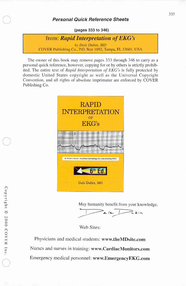
Date post: | 02-Feb-2023 |
Category: |
Documents |
Upload: | khangminh22 |
View: | 0 times |
Download: | 0 times |
no
—
era
—
®
to
ooo
n
O<
—
-
Personal Quick Reference Sheets
(pages 333 to 346)
from: Rapid Interpretation ofEKG'sby Dale Ditbin, MD
COVER Publishing Co., P.O. Box 1092, Tampa, FL 33601, USA
The owner of this book may remove pages 333 through 346 to carry as apersonal quick reference, however, copying for or by others is strictly prohibited. The entire text of Rapid Interpretation of EKG's is fully protected bydomestic United States copyright as well as the Universal CopyrightConvention, and all rights of absolute imprimatur are enforced by COVERPublishing Co.
RAPID
INTERPRETATIONOF
EKG's
Dr.Dubln's classic, simplified methodology for understanding EKG's
Dale Dubin, MD
May humanity benefit from your knowledge,
Web Sites:
Physicians and medical students: www.theMDsite.com
Nurses and nurses in training: www.CardiacMonitors.com
Emergency medical personnel: www.EmergencyEKG.com
333
534
Personal Quick Reference Sheets
Dubin's Method
for
Reading EKG's
from: Rapid Interpretation ofEKG'sbv Dale Dubin, MD
COVER Publishing Co., P.O. Box 1092, Tampa, FL 33601, USA
1. RATE (pages 65-96)Say "300, 150, 100" ..."75, 60, 50"
• but for bradycardia:rate = cycles/6 sec. strip X 10
2. RHYTHM (pages 97-202)Identify the basic rhythm, then scan tracing for prematurity,pauses, irregularity, and abnormal waves.
• Check for: P before each QRS.
QRS after each P.
• Check: PR intervals (for AV Blocks).QRS interval (for BBB).
• IfAxis Deviation, rule out Hemiblock.
3. AXIS (pages 203-242)• QRS above or below baseline for Axis Quadrant
(for Normal vs. R. or L. Axis Deviation).For Axis in degrees, find isoelectric QRS in a limb leadof Axis Quadrant using the "Axis in Degrees" chart.
• Axis rotation in the horizontal plane: (chest leads)find "transitional" (isoelectric) QRS.
4. HYPERTROPHY (pages 243-258) |IP wave for atrial hypertrophy. ^
R wave for Right Ventricular Hypertrophy. WS wave depth in V,.., q
+ R wave height in V5 for Left Ventricular Hypertrophy. rjo
o
5. INFARCTION (pages 259-308) gScan all leads for: q
• Q waves «
• Inverted T waves ao• ST segment elevation or depression 'Z
Find the location of the pathology (in the Left ventricle), o.and then identify the occluded coronary artery. °
no
13
—
©
o
oo
no<
-
Personal Quick Reference Sheets
Kclte (pages 65 to 96)
from: Rapid Interpretation ofEKG'sby Dale Dubin, MD
COVER Publishing Co., P.O. Box 1092, Tampa, FL 33601, USA
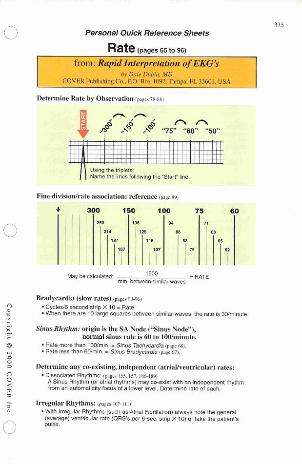
Determine Rate by Observation (pages 78-88)
l- f # jf "75" "60" "50"
11
fflJIJ 1 Using the triplets:j | Name the lines following the "Start" line.
Fine division/rate association: reference (page 89)
+ 300 150 100 75
250 136 94 71
60
214 125 88 68
187 115 83 65
167 107 79 62
May be calculated:1500
mm. between similar waves= RATE
Bradycardia (slow rates) (pages 90-96)• Cycles/6 second strip X 10 = Rate• When there are 10 large squares between similar waves, the rate is 30/minute.
Sinus Rhythm: origin is the SA Node ("Sinus Node"),normal sinus rate is 60 to 100/minute.
• Rate more than 100/min. = Sinus Tachycardia (page 68).• Rate less than 60/min. = Sinus Bradycardia (page 67).
Determine any co-existing, independent (atrial/ventricular) rates:• Dissociated Rhythms: (pages 155, 157.186-189)
ASinus Rhythm (or atrial rhythms) may co-exist with an independent rhythmfrom an automaticity focus of a lower level. Determine rate of each.
Irregular Rhythms: (pages 107-111)• With Irregular Rhythms (such as Atrial Fibrillation) always note the general
(average) ventricular rate (QRS's per 6-sec. strip X 10) or take the patient'spulse.
335
336
Personal Quick Reference Sheets
RhythlTl (pages 97 to 111)from: Rapid Interpretation ofEKG's
bv Dale Dubin, MDCOVER Publishing Co., P.O. Box 1092, Tampa, FL 33601. USA
* Identify basic rhythm......then scan entire tracing for pauses, premature beats,
irregularity, and abnormal waves.
•k Always:• Check for: P before each QRS.
QRS after each P.
• Check: PR intervals (for AV Blocks).QRS interval (for BBB).
• Has QRS vector shifted outside normal range? (to rule out Hemiblock).
Irregular Rhythms ipages 107-11n
Sinus Arrhythmia <page to©)Irregular rhythm that varieswith respiration.
All P waves are identical.
Considered normal.
Wandering Pacemaker (page 108)Irregular rhythm. P waveschange shape aspacemaker location varies.
Rate under 100/minute...
...but if the rate exceeds
100/minute, then it is called
Multifocal Atrial Tachycardia(page 109)
Atrial Fibrillation (pages no, 164-166)Irregular ventricular rhythm.Erratic atrial spikes(no P waves) frommultiple atrial automaticityfoci. Atrial dischargesmay be difficult to see.
L
.Aw ^sj \r\A^ /-/"W |/"\J |f»s
c1—<
<Xpq
>
o
u
—
o
c
Q
60
•—
a,Q
u
oo
@
to
oo
o
no
<
toPC
Personal Quick Reference Sheets
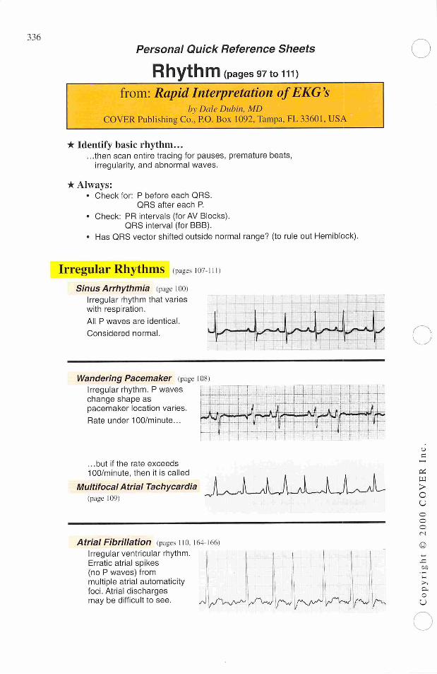
Rhythm continued (pages 112 to 145)from: Rapid Interpretation ofEKG's
by Dale Dubin, MDCOVER Publishing Co., P.O. Box 1092, Tampa, FL 33601, USA
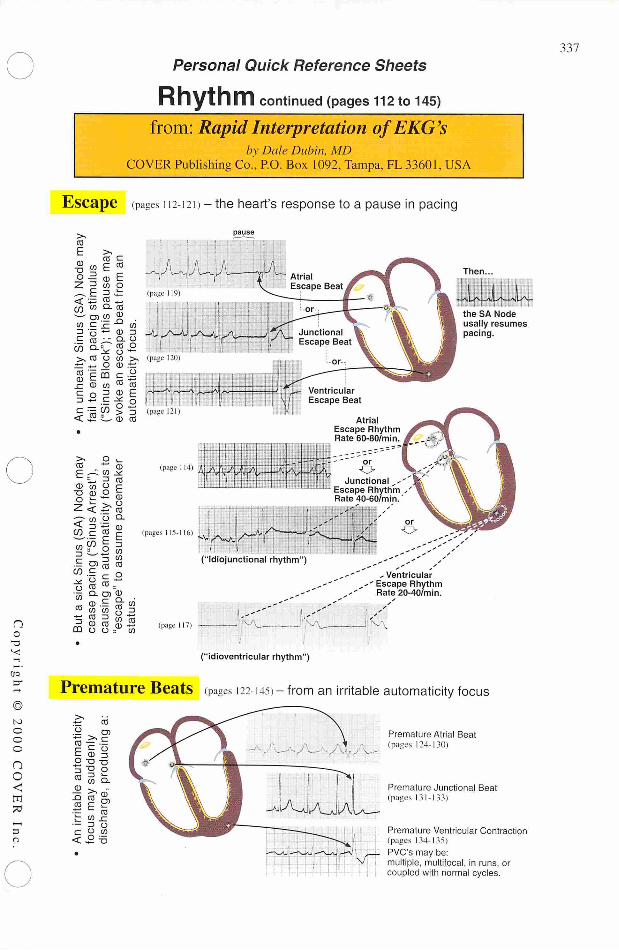
Escape (pages 112-121 >- the heart's response to a pause in pacing
CO
E >,m a
E CO
<B Em 0
Premature Beats
pause
AtrialEscape RhythmRate 60-80/min.
wpC-22£p\, oJunctional ,-
Escape RhythmRate 40-60/min.
, Ventricular
-' Escape RhythmRate 20-40/min.
1Then.
I II j 11111
4J±iJU^-—«v(LyN—
the SA Node
usally resumespacing.
("idioventricular rhythm")
(pages 122-14?)- from an irritable automaticity focus
Premature Atrial Beat(pages 124-130)
Premature Junctional Beat(pages 131-133)
Premature Ventricular Contraction(pages 134-135)
PVCs may be:multiple, multifocal, in runs, orcoupled with normal cycles.
337
338
Personal Quick Reference Sheets
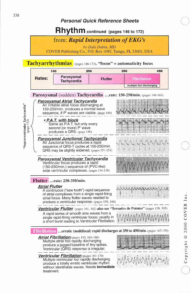
Rhythm continued (pages 146 to 172)from: Rapid Interpretation ofEKG's
by Dale Dubin, MDCOVER Publishing Co., P.O. Box 1092, Tampa, FL 33601, USA
Tachyarrhythmias (pages 146-172) "focus" = automaticity focus
150 250 350 450
Rates: ParoxysmalTachycardia
Flutter ^^^li illation
multiple foci discharging
e2^Z 2
a,9
X
Paroxysmal (sudden) Tachycardia ...rate: 150-250/min. (pages t46-i63)f Paroxysmal Atrial Tachycardia
An irritable atrial focus discharging at150-250/min. produces a normal wavesequence, if P' waves are visible, (page 149)
ajp;'"'ft:
iMmmMmMBi• P.A. T. with block 1 & :j^T'K ' k*- !l ^JU-M'f A'i;«
w Sp \m r V
second (or more) P' wave_U-rl —1_
-
—
att ._ _
I
Paroxysmal Junctional TachycardiaAV Junctional focus produces a rapidsequence of QRS-T cycles at 150-250/min.QRS may be slightly widened, (pages 151-153)
Paroxysmal Ventricular TachycardiaVentricular focus produces a rapid(150-250/min.) sequence of (PVC-like)wide ventricular complexes, (pages I54-158)
Flutter ...rate:250-350/min.
f\ ^ mj
n /•» /\ rt ^ r\Si 4, i\ S
1
\ ; v.
i
^ V
wmmww
Atrial FlutterA continuous ("saw 1of atrial complexes from a single rapid-firingatrial focus. Many flutter waves needed to
kjvtt!-tooth") rapid sequence JS^VJUMUVJifrom a single rapid-firinq I !
produce a ventricular response, (pages 159.160)
Ventricular Flutter (pages 161, 162) also see "Torsades de Pointes" (pages 158, 345)A rapid series of smooth sine waves from asingle rapid-firing ventricular focus; usually ina short burst leading to Ventricular Fibrillation.
Fibrillation .erratic (multifocal) rapid discharges at 350 to 450/min. (pages 167-170)
Atrial Fibrillation mages no. 164-166)Multiple atrial foci rapidly dischargingproduce a jagged baseline of tiny spikes.Ventricular (QRS) response is irregular.
Ventricular Fibrillation (pages i67-t70)Multiple ventricular foci rapidly dischargingproduce a totally erratic ventricular rhythmwithout identifiable waves. Needs immediatetreatment.
L:ii
^V^^MHWt"
iiiPi
pi
>
o
y
o
oc;
©
i_
>->c
c
U
@
to
©©Q
nc
<
-
Personal Quick Reference Sheets
Rhythm: ("heart") blocks (Pages 17310202)from: Rapid Interpretation ofEKG's
by Dale Dubin, MDCOVER Publishing Co., P.O. Box 1092, Tampa, FL 33601, USA
Sinus (SA) Block (page i74>An unhealthy Sinus (SA) Node misses one or more cycles (sinus pause)...
„,,.. the Sinus Node usually resumes pacing, buti i i pause i . , i .. i m IT
—A - -fi - A . A_ . A - 1\_ . the pause may evoke an escape responsefrom an automaticity focus, (pages 119-121)
AV BlOCk (pages 176-189)Blocks that delay or prevent atrial impulses from reaching the ventricles.
S 1° AV BlOCk ...prolonged PR interval (pages 176-178).3 PR interval is prolonged to greater« than .2 sec (one large square).
n 2 AV BlOCk ... some Pwaves without QRS response (page 179-185)| Wenckebach ...PR gradually lengthens with each
(pages 180-182, cycle until the last P wave in the
(/) o-> CD
n^ to_fc CD
183)
Mobitz ..(pages 181
series does not produce a QRS.
some P waves don't produce a QRS183) response. If "intermittent," an
occasional QRS is droped.
More advanced Mobitz block mayproduce a 3:1 (AV) pattern or evenhigher AV ratio (page 181).
2:1 AV Block .. .may be Mobitz or Wenckebach,(pages 182, 183) PR length and QRS width or
vagal maneuvers help differentiate. UA
3° ("complete") AV Block3° Block:
(page 188)
no P wave produces a QRS response (pages 186
P waves—SA Node origin.QRS's—if narrow, and if the '—ventricular rate is 40 to 60 per min.,then origin is a Junctional focus.
P waves—SA Node origin.QRS's—if PVC-like, and if the _a__a![ - - ..._•ventricular rate is 20 to 40 per min.,then origin is a Ventricular focus.
190)
a.
3° Block:
(page 189)
Bundle Branch BlOCk ...find R,R' in right or left chest leads (pages 191-202)Right BBB (pages 194-196) Left BBB (pages 194-197)
* Always Check:• is QRS within
3 tiny squares?
With Bundle BranchBlock the criteria for
ventricular hypertrophyare unreliable.
Caution:With Left BBB
infarction is difficultto determine on EKG.
QRS in V, or V2 QRS in V5 or VB
Hemiblock ...block of Anterior or Posterior fascicle of the Left Bundle Branch.(pages 295-305)
?1SZ shifted Anterior Hemiblock Posterior HemiblockoutsideNormal Axis shifts Leftward -» L.A.D. Axis shifts Rightward -» R.A.D.range? lookfor QS, lookfor S,Q,
1 J 13
(pages297-299) (pages300-302)
339
340
Personal Quick Reference Sheets
AXIS (pages 203 to 242)from: Rapid Interpretation ofEKG's
by Dale Dubin, MDCOVER Publishing Co., P.O. Box 1092, Tampa, FL 33601, USA
General Determination of Electrical Axis (pages 203-231)Is QRS positive (- -) or negative (-w-) in leads I and AVF?
Is Axis Normal? (page 227)
QRS in lead I (pages 215-222)...if the QRS is Positive (mainly abovebaseline), then the Vector points topositive (patient's left) side.
Normal-1 ^RS uPrish'in I;mtl AVF"two thumbs-up" sign
QRS in lead AVF (pages 223-226)...if the QRS is mainly Positive, thenthe Vector must point downward topositive half of the sphere.
Axis in Degrees (pages 233,2345 (Frontal Plane)After locating Axis Quadrant, find limb lead where QRS is most isoelectric:
Extreme Right Axis Deviationlead Axis
I -s. -90°
AVL —> -120°
III -» -150°
AVF -» -180°
Right Axis Deviationlead Axis
AVF -* +180°
II -» +150°
AVR -» +120°
I -^ +90°
First Determine Axis Quadrant(pages 214-231)
Left Axis Deviation
lead Axis
I -^ -90°
AVR -> -60°
II -+ -30°
AVF -» 0°
Normal Range
lead Axis
AVF —» 0°
III -* +30°
AVL -*• +60°
I -* +90°
Axis Rotation (left/right) in the Horizontal Plane (pages 236-242)Find transitional (isoelectric) QRS in a chest lead.
Patient's
Right
transitional QRSis "isoelectric"
! Patient':Left
o
a
W
>Ou
o
o
o
©
si60
c
0
a
©
ts)
Oo
o
o
c
<
7C
Personal Quick Reference Sheets
Hypertrophy (pages 243 to 258)from: Rapid Interpretation ofEKG's
by Dale Dubin, MDCOVER PublishingCo., P.O. Box 1092,Tampa, FL 33601, USA
Atrial Hypertrophy (pages 245-249)
Right Atrial Hypertrophy (page 248)
• large, diphasic P wave with tall initial component.Initial
component
i i ! 1 ! •!
Left Atrial Hypertrophy (page 249)
• large, diphasic P wave with wide terminal component.
terminalcomponent
Ventricular Hypertrophy (pages 250-258)
Right Ventricular Hypertrophy (pages 250-2521
• R wave greater than S in V . but R wave getsprogressively smaller from V - V,.
• S wave persists in V. and V„• R.A.D. with slightly widened QRS.• Rightward rotation in the horizontal plane.
Left Ventricular Hypertrophy (pages 253 257)
S wave in V (in mm.)
+ R wave in V. (in mm.)
Sum in mm. is more than 35 mm. with L.V.H.
• L.A.D. with slightly widened QRS.• Leftward rotation in the horizontal plane.
Inverted T wave:
slants downward
gradually, mbut up rapidly.
341
342
Personal Quick Reference Sheets
Infarction (pages259to308)from: RapidInterpretation ofEKG's
by Dale Dubin, MDCOVER Publishing Co., P.O. Box 1092, Tampa, FL33601, USA
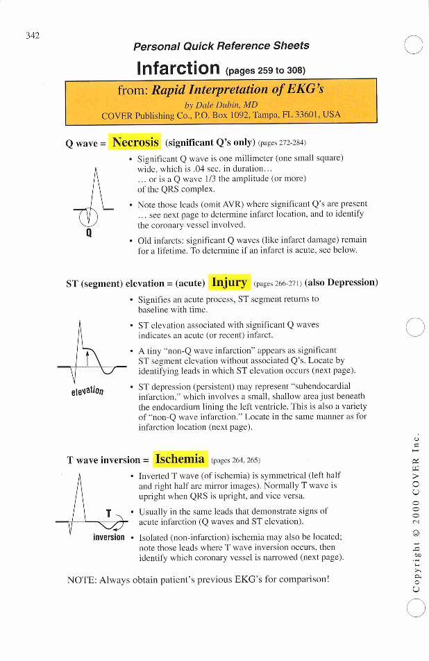
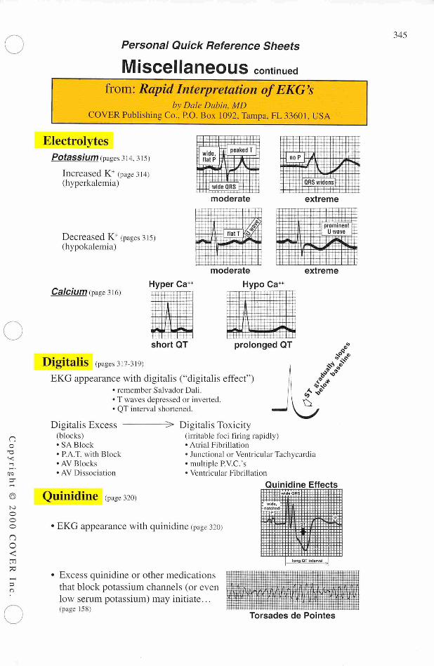
Q wave = Necrosis (significant Q's only) (pages 272 284)• Significant Q wave is one millimeter (one small square)
wide, which is .04 sec. in duration...... or is a Q wave 1/3 the amplitude (or more)of the QRS complex.
• Note those leads (omit AVR) where significant Q's are present... see next page to determine infarct location, and to identifythe coronary vessel involved.
• Old infarcts: significant Q waves (like infarct damage) remainfor a lifetime. To determine if an infarct is acute, see below.
ST (segment) elevation =(acute) Injury (pages 266-271) (also Depression)
• Signifies an acute process, ST segment returns tobaseline with time.
ST elevation associated with significant Q wavesindicates an acute (or recent) infarct.
A tiny "non-Q wave infarction" appears as significantST segment elevation without associated Q's. Locate byidentifying leads in which ST elevation occurs (next page).
ST depression (persistent) may represent "subendocardialinfarction," which involves a small, shallow area just beneaththe endocardium lining the left ventricle. This is also a varietyof "non-Q wave infarction." Locate in the same manner as forinfarction location (next page).
jleMat/oo
T wave inversion = Ischemia (pages 264. 265)
inversion
Inverted T wave (of ischemia) is symmetrical (left halfand right half are mirror images). Normally T wave isupright when QRS is upright, and vice versa.
Usually in the same leads that demonstrate signs ofacute infarction (Q waves and ST elevation).
Isolated (non-infarction) ischemia may also be located;note those leads where T wave inversion occurs, thenidentify which coronary vessel is narrowed (next page).
NOTE: Always obtain patient's previous EKG's for comparison!
o
a
>o
u
©c
c
©
JO
• —
i>sao
U
no
13
©
to
e3
o
nc<
rr
-
o
Personal Quick Reference Sheets
Infarction Location
— and —
Coronary Vessel Involvement(pages 259 to 308)
from: Rapid Interpretation ofEKG'sby Dale Dubin, MD
COVER Publishing Co., P.O. Box 1092,Tampa, FL 33601, USA
Coronary Artery Anatomy (Page 291)
Right CoronaryArtery
Left CoronaryArtery
circumflex
anterior
descending
Infarction Location/Coronary Vessel Involvement (pages 278 294)
Posterior• large R with
ST depression in V, &V2• mirror test or reversed
transillumination test
(Right Coronary Artery)(pages 282-286)
Inferior(diaphragmatic)Q's in inferior leads
II. III. and AVF
(R. or L. Coronary Artery)(pages 281, 294)
LateralQ's in lateral leads I and AVL
(Circumflex Coronary Artery)(pages 280, 292)
AnteriorQ's in V,, V2, V3, and V„(Anterior Descending
Coronary Artery)(pages 278, 292)
343
344
Personal Quick Reference Sheets
Miscellaneous (pages309to328)
from: RapidInterpretation ofEKG'sby Dale Dubin, MD
COVER PublishingCo., P.O. Box 1092,Tampa, FL 33601, USA
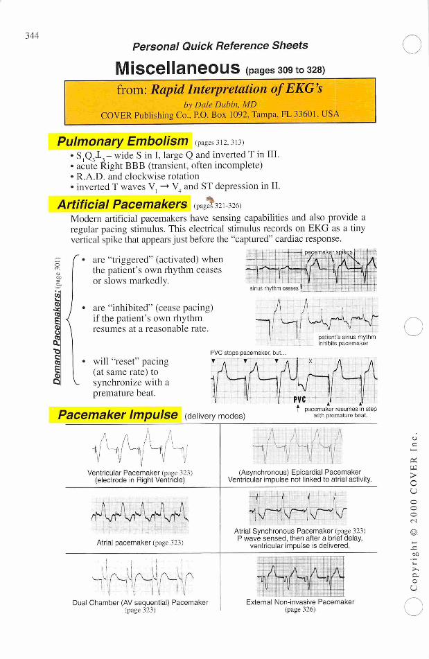
Pulmonary Embolism Pages 312313)• SjQjij- wide S in I, large Q and inverted T inIII.• acute Right BBB (transient, often incomplete)• R.A.D. and clockwise rotation• inverted T waves V —• V4 and ST depression in II.
Artificial Pacemakers ,^321326)Modern artificial pacemakers have sensing capabilities and also provide aregular pacing stimulus. This electrical stimulus records on EKG as a tinyvertical spike that appearsjust before the "captured" cardiac response.
= r>
V.
are "triggered" (activated) whenthe patient's own rhythm ceasesor slows markedly.
sinus rhythm ceases
are "inhibited" (cease pacing)if the patient's own rhythmresumes at a reasonable rate.
will "reset" pacing(at same rate) tosynchronize with apremature beat.
PVC stops pacemaker, but
• A • a •
,^\/wUsrV'T
patient's sinus rhythminhibits pacemaker
PVCT pacemaker resumes in step
with premature beat.Pacemaker Impulse (delivery modes)
AHli Hi I •—•/
Ventricular Pacemaker (page 323)(electrode in Right Ventricle)
tJ\ „sM aa r^\WtHW
Atrial pacemaker (page 323)
uaJJa4ii1
Dual Chamber (AV sequential) Pacemaker(page 323)
A Vv_
V f
m-V
A
(Asynchronous) Epicardial PacemakerVentricular impulse not linked to atrial activity.
SpwTlAtrial Synchronous Pacemaker (page 323)
P wave sensed, then after a brief delay,ventricular impulse is delivered.
•1 ••!• -
r -
-
~ A.m
- J -
"V 1
External Non-invasive Pacemaker(page 326)
•j
C—i
IKj
>c
u
o
3
3
—
O
O
no
—
era
—
©
3
3
nc
<
-
70
Personal Quick Reference Sheets
Miscellaneous continuedfrom: Rapid Interpretation ofEKG's
by Dale Dubin, MDCOVER Publishing Co., P.O. Box 1092, Tampa, FL 33601, USA
ElectrolytesPotassium (pages 314. 315)
Increased K+ (page 314)(hyperkalemia)
Decreased K+ (pages 315)(hypokalemia)
H+4 11111,1144-TTTT !l ! 1 —
wide,flat P i
peakedT••
k A --1•» #y. M
=1^F
5;- wide QRS ElI l l . l i . i •'! ' j
moderate
HSi^a
noP
QRS widensjy-LllLLUJilTextreme
extreme
Ca/C/Um (page 316)Hyper Ca++
moderate
Hypo Ca+
short QT prolonged QT
Digitalis (pages 317-319)EKG appearance with digitalis ("digitalis effect"
• remember Salvador Dali.
• T waves depressed or inverted.• QT interval shortened.
Digitalis Excess -(blocks)•SA Block
• RAT. with Block
• AV Blocks
• AV Dissociation
Quinidine (Page320)
^> Digitalis Toxicity(irritable foci firing rapidly)• Atrial Fibrillation
• Junctional or Ventricular Tachycardia• multiple P.V.C.'s• Ventricular Fibrillation
Quinidine Effects
EKG appearance with quinidine (page 320)
-notched ~P
:t:u::
3T 3j±"
long QT In erva 1
Excess quinidine or other medicationsthat block potassium channels (or evenlow serum potassium) may initiate...(page 158)
y|i^|p|||^|PTorsades de Pointes
345
346
Personal Quick Reference Sheets
Practical Tipsfrom: Rapid Interpretation ofEKG's
by Dale Dubin, MDCOVER Publishing Co.', P.O. Box 1092, Tampa, FL 33601, USA
Dubin's Quickie Conversion—for—
Patient's Weight from Pounds to Kilograms
Patient wt. in kg. = Half of patient's wt. (in lb.) minus 1/10 of that value.
Examples: 180lb. patient 160 lb. patient 140lb. patient(becomes 90 minus 9) (becomes 80 minus 8) (becomes 70 minus 7)
is 81 kg is 72 kg is 63 kg.
Modified Leads—for—
Cardiac MonitoringLocations are approximate. Some minor adjustment of electrode posi
tions may be necessary to obtain the best tracing. Identify the specificlead on each strip placed in the patient's record.
Sensor Electrode
+
* Ground, Neutral or Reference
Modified Lead I
Conventional Lead
Identification
Letter
R(orRA)L (or LA)G (or RL)
Color (inconsistent)
red
white
variable
Modified Lead II
MCI,To make litisMCltmove + electrode
to same
(mirror)
position onthepatient'sleft chest.
o
—
>c
u
3
3
3
<N
©
Si60
-
o