Alzheimer’s disease (AD), a central nervous system (CNS) neu-rodegenerative disorder, is the predominant cause of dementia inelderly people and is seen in Down’s syndrome individuals [1,2].Early-onset familial AD (FAD) patients carrying mutations inAPP, PS-1 or PS-2 encoding genes show increased b-amyloid(Ab)[1–42/43] and [x-42] peptide levels by altered amyloid pre-cursor protein (APP) processing, suggesting a major role for Abin AD pathogenesis [3]. Aggregated and polymerized Ab depositsextracellularly in AD brain as highly insoluble neuritic plaques of b-pleated amyloid fibrils (predominantly Ab[1–42/43] with less Ab[1–40]) and in cerebrovasculature as Ab[1–40] with lessAb[1–42], [45]. Toxicity of insoluble fibril Ab aggregates whichform at high concentrations [6] and of soluble oligomers atnanomolar concentrations [7] has been proposed. PeptidesAb[1–42/43] display higher fibrillogenicity compared to Ab[1–40]and exhibit earlier and selective deposition in AD, which is implicated in neurotoxicity and Ab deposition in FAD plaques[8,9].
Decreased total Ab or Ab[1–42] levels in AD CSF relative tocontrol patients, correlating with increased dementia severity,have been reported [10] but measuring Ab levels in biologicalfluids to diagnose AD is controversial. Diagnostic sensitivity andspecificity remain dependent on social, genetic, medical, psychi-atric and neurological examinations with confirmation by postmortem immunohistochemistry [11,12].
Using highly specific antibodies reactive with AD biologicalmarkers present in accessible bodily fluids, immunodiagnosticsoffer the opportunity for specific and sensitive detection of ADduring life. Recombinant techniques provide tools for the gen-eration of antibodies of enhanced specificity and sensitivity forAD markers, such as Ab in CSF/plasma, by incorporating tags,affinity enhancement, enzyme linkage, bispecificity and increasedavidity [13]. Although Ab[1–42] : Ab[1–40] plasma levels havebeen reported to be increased in Down syndrome and FAD cases[14,15], increased assay sensitivity may minimize the existing con-troversy in sporadic AD [16]. Recombinant antibodies also haveimportance in AD therapy. Immunization of mice with human Abhas been shown recently to inhibit amyloid plaque formation [17].‘Specific anti-Ab antibody-directed mopping-up’ of excess Abmay be therapeutic in AD. With these considerations in mind, the novel Ab-specific recombinant 1E8–4b Fab fragment was generated from Escherichia coli and compared with the parentantibody.
453
Generation of a recombinant Fab antibody reactive with the Alzheimer’sdisease-related Ab peptide
A. H. TAMMER, G. COIA*, R. CAPPAI, S. FULLER, C. L. MASTERS, P. HUDSON* & J. R. UNDERWOOD Department ofPathology, Melbourne University, Parkville, Victoria, and *CRC for Diagnostics, CSIRO Health Sciences and Nutrition, Parkville,
Victoria, Australia
(Accepted for publication 10 April 2002)
SUMMARY
A recombinant Fab antibody, designated 1E8–4b, which reacts with the Alzheimer’s disease (AD)-related Ab peptides, Ab[1–40], Ab[1–42] and Ab[1–43] has been developed. The 1E8–4b Fab was con-structed by cloning the VHCH1 and VLCL domains from the parent hybridoma 1E8 antibody, reportedpreviously to recognize these Ab peptides. Briefly, a C-terminal Flag tag sequence was incorporatedinto this construct, which was ligated into the vector pHFA2 and expressed in Escherichia coli. Fol-lowing purification on an M2 anti-Flag affinity column, the 1E8–4b recombinant Fab antibody wasshown to bind plaques within sections of brain tissue from CERAD-defined AD patients by immuno-histochemistry. ELISA, epitope mapping and immunoblotting confirmed the recognition of theAb[1–40/42/43] peptides by the 1E8–4b Fab. The 1E8–4b Fab did not recognize APP695 or APP770which contain the Ab sequence. The Ab specificity of the recombinant 1E8–4b Fab antibody was iden-tical to the parent 1E8 monoclonal antibody.
Keywords recombinant Fab antibody monoclonal antibody Alzheimer’s disease Ab peptide
Clin Exp Immunol 2002; 129:453–463
Correspondence: J. R. Underwood, Department of Pathology, Uni-versity of Melbourne, Parkville, Victoria 3010, Australia.
Generation of the immunoglobulin heavy and light chain constructsThe mRNA was extracted from 4·0 ¥ 106 hybridoma cells by Dyn-abeads mRNA Direct Kit (Dynal, Oslo, Norway). The hybrid-oma used was 1E8 [17–22] (SmithKlein Beecham Laboratories,Harlow, UK) [18]. The cDNA was generated by the Promega(Madison, WI, USA) reverse transcription system kit in a 20-mlreaction containing 1 ¥ RT buffer, 5 mM MgCl2, 5 mM DTT, 1 U/mlRNase inhibitor, 1 mM dNTP mix, 0·5 mg oligo (dT)15 primer/mgmRNA, 15 U AMV reverse transcriptase/mg mRNA, 1 mg mRNAand RNase-free ddH2O. The reaction was incubated at 42°C for1 h and inhibited at 99°C for 5 min. PCR was performed in a 100-ml reaction containing 0·50 pM each forward and reverseprimer (section 1·2), 0·20 mM dNTP (Amresco), 1 ¥ ThermoPolreaction buffer (New England BioLabs, Beverly, MA, USA), 100mg/ml BSA, 10 ml of cDNA and ddH2O. Samples were ‘hot started’by heating to 94°C for 1 min and adding 2 Units of Vent DNApolymerase (New England BioLabs, Beverly, MA, USA) per 100 ml reaction. Reactions were overlaid with 50 ml of mineral oil(Sigma, St Louis, MO, USA) and incubated at 94°C 1 min, 55°C1 min and 72°C 1 min for three cycles, followed by 94°C 1 min,55°C 1 min and 72°C 2 min, for 30 cycles on a FTS-1 thermalsequencer (Corbett Research, Mortlake, NSW, Australia). DNAwas purified by the BIO101 GeneClean (GC) Spin Kit (Inte-grated Sciences, NSW, Australia).
Primers used to generate heavy and light chain constructs
The VHCH1 construct was generated using the forward primer [CH1
degen(212–223)] (5¢ at taa gtc gac T/G/AAT T/CTT T/CTT GTCCAC C/TG/TC GGT G/CC/TT GCT GGC C/TGG GTG 3¢)modified from Kettleborough et al. [19] with data from Kabatet al. [20], containing a SalI site and the reverse primer [VH back3350] containing a SfiI site (5¢ tta tta ctc gcg gcc cag ccg gcc atggcc GAG GTC CAG CTG CAG CAG TC 3¢). The VLCL con-struct was generated using [CL forward 3964] containing a NotIsite (5¢ at gag ttt ttg ttc tgc ggc cgc ggc ACA CTC ATT CCT GTTGAA GCT CTT 3¢) and the reverse primer [VL back 3959] con-taining a NcoI site (5¢ c cgg gtg tct gcc atg gcc GAC ATT GTGATG ACC CAG TCT 3¢). All restriction sites are underlined.
Cloning the immunoglobulin heavy and light chain constructsPurified PCR fragments were cloned into pCR-Script Amp SK(+)vector and transformed into E. coli XL1-Blue MRF¢ Kan cellsusing pCR-Script Amp SK(+) an electroporation-competent cell cloning kit (Stratagene, CA, USA). Positive colonies werescreened by restriction enzyme digests of Miniprep DNA orcolony PCR. Following sequencing using the ABI PRISM dye terminator cycle sequencing ready reaction kit (Perkin Elmer,Norwalk, CT, USA), positive clones were digested with appro-priate enzymes, purified from 1% (w/v) low melt agarose gels andligated into a similarly digested and purified pHFA2 expressionvector [21]. The ligation mixture was transformed into E. coliHB2151 cells and plated onto 1·5% (w/v) agar plates containing2 ¥ YT, 100 mg/ml ampicillin and 1% (v/v) glucose, overnight at37°C. Positive colonies were screened as above and resequencedfor affirmation using pUC/M13 22-mer reverse sequencing primer(Promega, Madison, WI, USA) (5¢TCACACAGGAAACAGCTATGAC 3¢) and Fd gene III primer (Beckman, NSW, Australia)(5¢ACTTTCAACAGTCTATGCCGCG 3¢).
Expression and purification of the antibody Fab fragmentsE. coli HB2151 cells [22] containing the recombinant plasmidswere grown overnight at 37°C in 2 ¥ YT containing 100 mg/mlAmpicillin and 1% (v/v) glucose (2 ¥ YT/AMP/Glu). The fol-lowing day, cells were subcultured to an O.D. 600 nm value of 0·1 Unit into 2 ¥ YT/AMP. The cells were grown at 30°C,125 r.p.m. in an orbital incubator to an O.D. 600 nm value of 0·8–1·0 Units (about 3 h). IPTG was added to a final concen-tration of 1 mM and the cultures allowed to shake at 125 r.p.m.,25°C overnight.
Overnight culture medium was clarified by two successivecentrifugations at 10 000 gmax for 30 min, 4°C. The supernatant was treated with 2 mM EDTA, 0·1 mg/ml leupeptin, 0·1 mg/ml pepstatin, 1 mM PMSF and 0·02% (w/v) NaN3. The supernatantwas treated with 100 mg DNAaseI/l supernatant (SIGMA, StLouis, MO, USA) for 30 min at RT with stirring and then filteredthrough a 0·45-mm filter (Nalgene). The supernatant was concen-trated by a 60% (v/v) saturated ammonium sulphate (SAS) pre-cipitation as described in Coppola et al. [23]. The pellet wasresuspended in 1/40th of the original volume in TBS/NaN3 anddialysed against 100 volumes of TBS/NaN3 over 48 h with threechanges of buffer at 4°C. Dialysed samples were centrifuged at 10 000 gmax for 30 min at 4°C and the supernatant collected. The M2 anti-Flag antibody affinity column (Sigma, St Louis, MO, USA) was equilibrated with four column volumes ofTBS/NaN3/I (TBS/NaN3 containing 2 mM EDTA, 0·1 mg/ml leupeptin, 0·1 mg/ml Pepstatin, 2·5 mg/ml Aprotinin) at 1 ml/min at 4°C. The concentrated supernatant was treated with inhibitors(2 mM EDTA, 0·1 mg/ml leupeptin, 0·1 mg/ml Pepstatin, 5 mg/mlAprotinin), ultracentrifuged at 100 000 gmax 4°C for 1 h, filteredthrough a 0·22-mm filter (Millipore, Bedford, MA, USA) andapplied at 1 ml/min 4°C to the affinity column. The sample was recirculated over the column three times or overnight. Theaffinity column was then washed with four column volumes of TBS/NaN3/I at 1 ml/min 4°C. The bound antibody was elutedat 4°C with 0·1 M glycine-HCl/0·02% (w/v) NaN3 pH 3·5 at 1 ml/min. One-millilitre fractions were collected and immediatelyneutralized with 20–40 ml of 1 M Tris-HCl, pH 8·0. Fractions wereexamined by immunoblot for the presence of antibody and byELISA for antibody reactivity. Eluates containing active antibodywere pooled, concentrated in 2 ml Centricon concentrators (Millipore, Bedford, MA, USA) and buffer exchanged intoPBS/NaN3.
SDS-PAGE and immunoblotting analysisSDS-PAGE and immunoblotting were carried out under standardconditions [24]. Samples were boiled for 5 min in sample bufferand electrophoresed on 10% SDS-polyacrylamide gels. For reducing conditions, the sample buffer contained 20% (v/v) 2-mercaptoethanol (Sigma, St. Louis, MO, USA). The proteinswere transferred onto nitrocellulose (Biorad, Hercules, CA,USA) in a Biorad Mini Trans-Blot apparatus at 300 mA for 1 h.The recombinant antibody, containing C-terminal flag and tubulintags, was detected with mouse M2 anti-Flag (Sigma, St Louis, MO, USA) or rat antitubulin antibodies (Serotec, Oxford, UK),respectively, followed by goat anti-mouse immunoglobulincoupled to alkaline phosphatase (Promega, Madison, WI, USA)or goat anti-rat immunoglobulin coupled to alkaline phosphatase(Southern Biotechnology Associates), respectively, and devel-oped with a substrate solution containing fast red and naphtholphosphate AS-MX reagents (Sigma, St Louis, MO, USA).
Fig. 1. DNA and protein sequences of 1E8-4b Fab in the pHFA2 cloning vector. Both the heavy (VHCH1) and light (VLCL) chains of the1E8-4b Fab are preceeded by N-terminal pelB signal sequences (italicised). Non-coding regions located 5¢ to the pelB signal sequences areshown in lower case. C-terminal peptide tag sequences (tubulin (Glu Glu Phe) and Flag) are shown in bold. The framework, CDR andconstant regions are outlined above the sequence, according to Kabat et al. [20]. Important restriction sites are underlined.
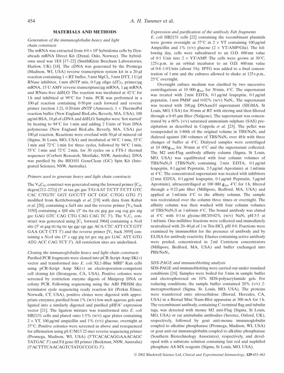
Fig. 2. Purification of recombinant 1E8-4b Fab-EEF/FLAG conjugatefrom E. coli culture supernatant as analysed by SDS-PAGE under non-reducing conditions and immunoblotting (lanes 1–5) or Coomassie Bril-liant Blue staining (lane 6). Immunoreactive protein bands were detectedusing M2 anti-Flag antibody followed by goat anti-mouse alkaline phos-phatase conjugated antibody and developed with Fast Red/AS-MX Naph-thol reagents. Lane 1: Culture supernatant; lane 2: Supernatant following60% (v/v) SAS precipitation; lane 3: Low-molecular-weight-proteinmarkers (with numbers to the left in kDa); lane 4: precipitated 1E8-4b Fabprotein following 60% (v/v) SAS precipitation; lane 5: pooled and con-centrated M2 anti-Flag affinity gel purified 1E8-4b Fab eluates; lane6: Coomassie stain of lane 5.
ELISA analysis of the 1E8–4b Fab fragmentIn order to analyse the specificity of the 1E8–4b recombinant Fab,96-well microtitre Cliniplates EB (Labsystems, Helsinki, Finland)were coated with 50 ng/well of the peptides Ab[1–40], Ab[1–42],Ab[1–43], APP 695 or APP 770 with or without serial dilutions incarbonate-bicarbonate buffer pH 9·6 for 4 h. After two washeswith PBS 100 ml/well of 1% (w/v) BSA was added for 2 h at 37°C.After two PBS washes, antibodies were incubated overnight at4°C at 10 mg/ml with serial dilutions or 5 mg/ml with titratedpeptide plates. Plates had three PBST washes and three PBSwashes. To detect the Fab and isotype control, mouse M2 anti-Flag antibody (Sigma, St Louis, MO, USA) was applied at 100 ng/well for 1 h, followed by three PBST washes and three PBSwashes, then rabbit anti-mouse immunoglobulins coupled tohorseradish peroxidase (Dako, Carpinteria, CA, USA) at 1/500dilution was added at 50 ml/well for 1 h. To detect the parent, anti-mouse immunoglobulins couple to horseradish peroxidase (Dako,Carpinteria, CA, USA) at 1/500 dilution was added at 50 ml/wellfor 1 h. ELISA plates were washed three times with PBST andthree times with PBS followed by 100 ml/well ‘o’-phenylenedi-amine (SIGMA, St Louis, MO, USA) developing solution for 1 h.The development reaction was stopped with 50 ml/well 8 M H2SO4
and the colour reaction determined at O.D. 490 nm on a Model450 microplate reader (Biorad, Hercules, CA, USA).
Detection of Ab and APP using immunoblottingThe following procedure was derived from Ida et al. [16]. Samplescontaining 100 ng of human Ab or recombinant APP were dena-tured, electrophoresed in 14% Tris-tricine gels and transferredonto nitrocellulose (Biorad, Hercules, CA, USA). Blots wereboiled in PBS for 5 min and blocked for 1·5 h with 1% (w/v) BSA.The Fab, isotype and parent antibodies were incubated overnightat 4°C (5 mg/ml of Fab or isotype and 50 mg/ml of parent). Theblots were washed once for 5 min then twice for 20 min withTBST. To detect the Fab and isotype antibodies, mouse M2 anti-Flag (Sigma, St Louis, MO, USA) was applied at 2 mg/ml, followedby one 5-min then two 20-min washes with TBST, followed byanti-mouse immunoglobulins coupled to horseradish peroxidase(Dako, Carpinteria, CA, USA) at 1/2000 for 1 h. To detect theparent, anti-mouse immunoglobulins coupled to horseradish per-oxidase (Dako, Carpinteria, CA, USA) was applied at 1/2000 for1 h. All blots were washed with TBST once for 5 min and twicefor 20 min Blots were rinsed with PBST and developed with the ECL development reagent system (Amersham PharmaciaBiotech, Buckinghamshire, UK).
Epitope mapping analysisTo determine the specificity of the Ab parent and Fab antibodies,Pepset analysis was undertaken comprising a set of 15 mer overlapping biotinylated peptides incrementing by three aminoacids per peptide and encompassing the human APP [695–652]sequence (Chiron Mimotopes Pty Ltd, Melbourne, Australia)[18]. The 96-well microtitre Cliniplates EB (Labsystems, Helsinki,Finland) were incubated with 500 ng/well streptavidin at 37°Covernight and evaporated to dryness. Then 50 ng/well of biotiny-lated peptides were incubated for 30 min followed by threewashes in PBS. BSA [1% (w/v)] (Sigma, St Louis, MO, USA) at100 ml/well was incubated at 37°C for 2 h and plates washed threetimes with PBS. The parent and Fab antibodies at 500 ng/wellwere incubated for 30 min and the plates washed three times with PBST and three times with PBS. To detect the parent
antibody, 50 ml/well anti-mouse immunoglobulins coupled tohorseradish peroxidase (Dako, Carpinteria, CA, USA) at 1/500was incubated for 30 min. To detect the Fab antibody 100 ng/wellM2 anti-Flag monoclonal antibody (Sigma, St Louis, MO, USA)was applied for 30 min, plates were washed three times with PBSTand three times with PBS, and 50 ml/well anti-mouse immunoglob-ulins coupled to horseradish peroxidase (Dako, Carpinteria, CA,USA) at 1/500 was incubated for 30 min. Plates were washed threetimes with PBST, three times with PBS and bound mouseimmunoglobulin or Fab was detected by the addition of 100 ml/well of ‘o’-phenylenediamine (Sigma, St Louis, MO, USA)development solution for 1 h. The development reaction wasstopped by adding 25 ml/well of 8 M H2SO4. The O.D. 490 nm was determined using a Model 450 microplate reader (Biorad,Hercules, CA, USA).
Immunohistochemistry of human AD (certified by CERADcriteria) and normal, non-AD control brainParaffin-embedded, formalin fixed, 4 mm tissue sections wereincubated over three changes of Shellex for 2 min each, followedby 2 min in 90% (v/v) ethanol and 70% (v/v) ethanol. Sectionswere washed with ddH2O and treated for 5 min in 80% (v/v)formic acid. After a ddH2O wash, sections were treated with 3% (v/v) H2O2 (BDH Chemicals, Poole, Dorset, UK) for 5 min,washed in ddH2O and TBS pH 7·4. Sections were blocked with 20% (v/v) rabbit serum in TBS pH 7·4 for 30 min and 100 ml/section of the parent, Fab and isotype control antibodieswere applied at either 20 mg/ml or 10 mg/ml for 1 h. Slides werewashed twice for 5 min with TBS pH 7·4. To detect the parentantibody 100 ml/section of anti-mouse immunoglobulins coupled to horseradish peroxidase (Dako, Carpinteria, CA, USA) wasapplied at 6·5 mg/ml for 1 h. To detect the Fab and isotype control,
Generation of Ab-specific recombinant Fab antibody 457
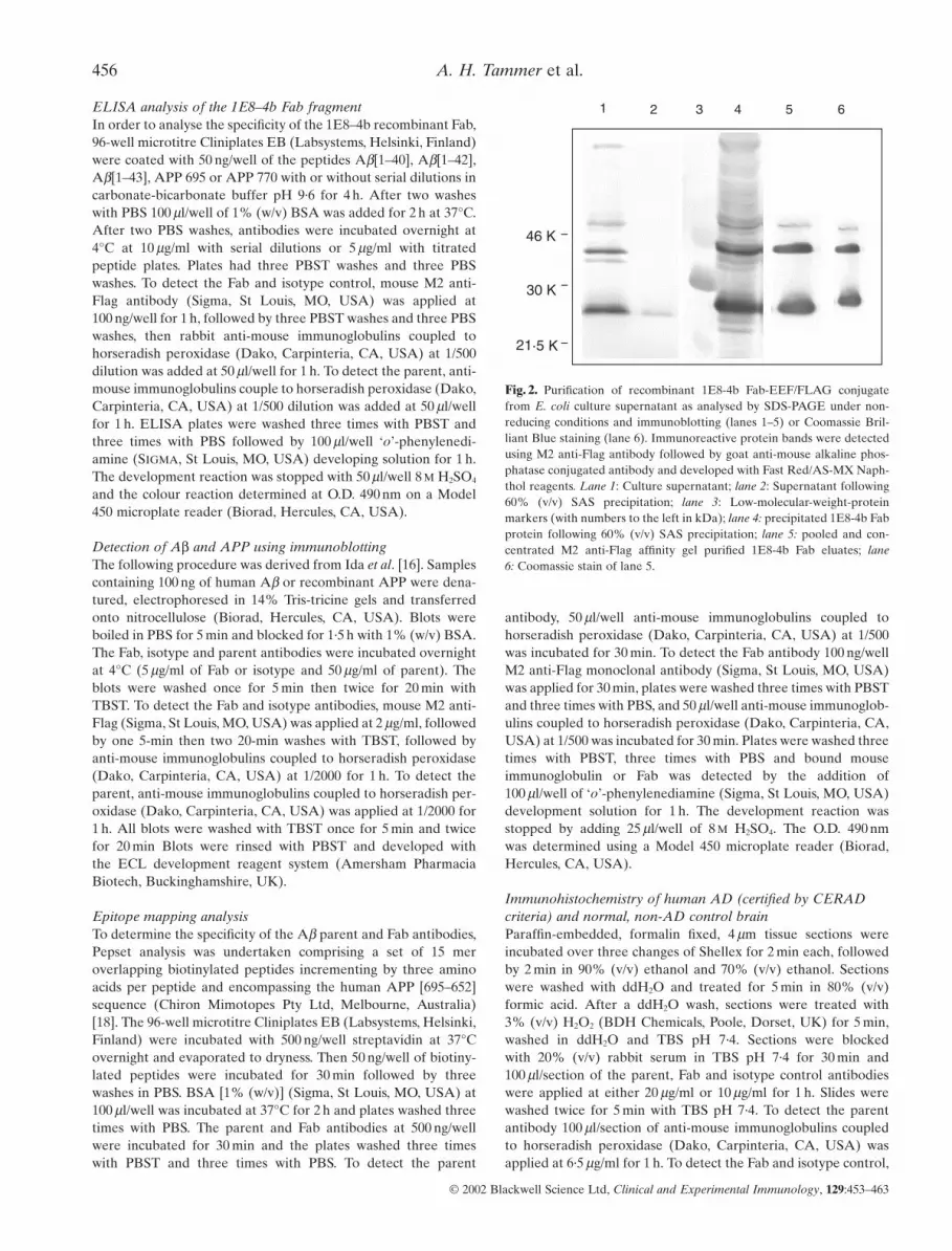
Fig. 3. ELISA depicting the reactivity of the parent 1E8 antibody (a) and the recombinant 1E8-4b Fab fragment (b) to Ab[1–40], Ab[1–42], Ab[1–43], APP695 and APP770 peptides. The antibody is titrated from 10 mg/ml with serial doubling dilutions, while the peptide isat a constant concentration of 50 ng/well. 1E8 was probed with antimouse-HRP while 1E8-4b was probed with M2 anti-Flag antibody andthen with antimouse-HRP. Detection of antibody reactivity to the peptides was via an ‘o’-phenylenediamine development mixture. Theisotype control used was M2 anti-Flag antibody which is a mouse IgG1.
100 ml/section of M2 anti-Flag monoclonal antibody (Sigma, StLouis, MO, USA) was applied at 10 mg/ml for 1 h followed by two5-min washes with TBS pH 7·4, then 100 ml/section of anti-mouseHRP (Dako, Carpinteria, CA, USA) was applied at 6·5 mg/ml for1 h. Sections were washed twice for 5 min with TBS pH 7·4 and developed with diaminobenzidine solution (BDH Chemicals,Poole, Dorset, UK) for 5 min. Sections were washed in TBSpH 7·4 and ddH2O, then counterstained in Mayer’s haematoxylinfor 1 min, washed in ddH2O, immersed in Scott’s tap water for30 s, washed in ddH2O, dehydrated in two changes of absolutealcohol, cleared in three changes of shellex (Shell Chemicals, Victoria, Australia) and mounted in DPX (BDH Chemicals,Poole, Dorset, UK).
Purification of monoclonal antibodiesThe mouse monoclonal antibody 1E8 was purified fromhybridoma culture supernatant by affinity chromatography on aProtein-G Hi-trap affinity column (Pharmacia Biotech, Sweden).Briefly, a 100-ml aliquot of hybridoma supernatant was recircu-lated overnight at 4°C using a peristaltic pump. The Protein G column containing bound antibody was washed with 30 ml of 0·1 M sodium phosphate buffer pH 7·4. Bound antibody waseluted with 0·1 M glycine-HCl pH 2·7 into 1 ml fractions contain-ing 42 ml/ml of 1·0 M Tris-HCl pH 9·0. Eluted fractions wereanalysed spectrophotometrically at A280 nm to determine theantibody-containing fractions. Peak fractions were pooled anddialysed against 100 volumes of PBS at 4°C. Following dialysis,
protein concentrations were measured spectrophotometricallyand the antibody stored at -20°C.
Preparation of the FAD PS-1 mutation brain sampleThe FAD brain sample containing the Australian PS-1 mutation(Leu219Pro) [25] was prepared from total brain tissue extract,homogenized in 1 ml TRIZOL (Life Technologies), incubated for 5 min at RT and 2 ml of chloroform was added. The samplewas shaken for 15 s, incubated for 2 min at RT and centrifuged at 12 000 gmax for 15 min, 4°C. Isopropyl alcohol (1·5 ml) was added
to the lower interphase, incubated 10 min at RT and centrifugedat 12 000 gmax for 10 min at 4°C. The pellet was washed three timeswith 2 ml of 0·3 M guanidine-HCl in 95% (v/v) ethanol for 20 minat RT and centrifuged at 7500 gmax for 5 min at 4°C after eachwash. The pellet was vortexed in absolute ethanol (2 ml), incu-bated 20 min at RT, centrifuged at 7500 gmax for 5 min at 4°C, air-dried and redissolved in 0·5 ml of 2% (w/v) SDS, by heatingand sonification. Insoluble material was centrifuged at 10 000 gmax
for 10 min at 4°C. The supernatant was aliquoted and frozen at -80°C.
2·0
1·5
1·0
0·5
0·0
2·0
1·5
1·0
0·5
0·0
2·0
1·5
1·0
0·5
0·0
2·0
1·5
1·0
0·5
0·0
2·0
1·5
1·0
0·5
0·0
2·0
1·5
1·0
0·5
0·0
10–2 10–1 100 101 102 10–2 10–1 100 101 102
A49
0 nm
(Antigen) ng
1E8 and Ab [1-40] 1E8-4b and Ab [1-40]
1E8 and Ab [1-42]
1E8 and Ab [1-43]1E8-4b and Ab [1-43]
1E8-4b and Ab [1-42]
Isotype control
(a) (b)
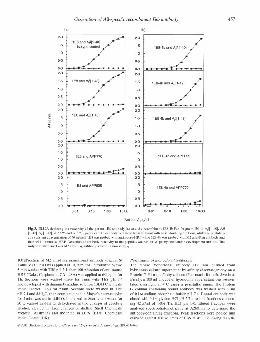
Fig. 4. Determination of the sensitivity of the parent antibody 1E8 (a) and the recombinant Fab antibody 1E8-4b (b) to the amyloid pep-tides Ab[1–40], Ab[1–42] and Ab[1–43] by ELISA. The peptides were serially diluted with doubling dilutions starting from 50 ng/ml whilethe concentrations of 1E8 and 1E8-4b were kept constant at 5 mg/ml. 1E8 was detected with antimouse-HRP conjugated antibody while1E8-4b was probed with M2 anti-Flag antibody and then detected with antimouse-HRP conjugated antibody. Detection was via an ‘o’-phenylenediamine development mixture. The isotype control used was M2 anti-Flag antibody which is a mouse IgG1.
Generation of Ab-specific recombinant Fab antibody 459
Generation, expression and purification of 1E8–4bFab antibody gene fragments from the mouse monoclonalhybridoma cell line 1E8 [17–22] were generated by PCR usingprimers generated from immunological sequences in Kabat et al.[20]. The VHCH1 construct was created with primers flankingamino acids 1–6/7 of the mouse variable heavy chain (VH) frame-work 1 region and amino acids 212–223 of the mouse first con-stant heavy chain region (CH1). The VLCL construct was createdwith primers flanking amino acids 1–7 of the variable light chain(VL) framework 1 region and amino acids 207–214 of the constantlight chain region (CL).
Constructs were cloned into pCR-Script Amp SK(+) vectors(Stratagene, CA, USA), derived from pBluescript® II SK(+)vectors (Stratagene, CA, USA). Six independent clones for both VHCH1 and VLCL were sequenced, generating a consensussequence. Sequence analysis in the Kabat and Wu database [20]
suggested the heavy and light chain constructs had not previouslybeen published. The entire VHCH1 chain spanned 213 amino acidsand the VH region belonged to the immunoglobulin mouse heavychain subgroup IIA. The VH region exhibited highest homologywith heavy chains from the mouse immunoglobulins DBF1-386·5,DBF1-235·4 and DB2-101·1 [20]. The CH1 region was slightlyabridged and most homologous with the heavy constant chain CH1 region of the mouse immunoglogulin IgG1CL [20]. TheVLCL chain spanned 219 amino acids and the VL region was foundto belong to the mouse kappa light chain subgroup II. The VL region displayed highest homology with kappa light chainsfrom eight mouse immunoglobulins [20]. The CL chain was most homologous with the kappa light constant chains of themouse immunoglobulins 17/9¢ CL, C.C58M75¢CL and MOPC21[20].
The six VHCH1 and VLCL constructs were subcloned into theexpression vector pHFA2 and expressed the 1E8 Fab fragmentsequally well at 8 h post-induction, with identical reactivity to theAb[1–42] peptide by ELISA. The VHCH1 chain migrated at 26 kDaand the VLCL chain at 28 kDa. Clone 1E8–4b was selected andsequenced (Fig. 1). Time-course expression experiments con-veyed highest Fab antibody expression at 24 h post induction andgreatest Fab antibody reactivity to Ab[1–42] peptide by ELISA.The intact Fab fragment resolved at 48 kDa with N-terminal fragments present on the immunoblot. At 24 h post induction, 0·5 mg of 1E8–4b/l culture supernatant was produced. 1E8–4b wasextracted most fully from culture supernatant by a 60% (v/v) saturated ammonium sulphate precipitation [23] (Fig. 2). Afterdialysing into PBS, 1E8–4b was purified on a M2 anti-Flag affin-ity column. Reactive eluates were pooled, concentrated andbuffer exchanged into PBS (Fig. 2). Purity was assessed byCoomassie Blue stain (Fig. 2).
Specificity comparison of the recombinant Fab and parent 1E8monoclonal antibodiesBy ELISA, the recombinant Fab 1E8–4b and the parent 1E8 anti-body were titrated against Ab[1–40/42/43] peptides and againstAPP 695 and 770. Both antibodies displayed binding toAb[1–40/42/43] but not to APP 695 and 770. The antibody 1E8exhibited binding at lower concentrations than the monovalent1E8–4b recombinant Fab fragment (Fig. 3). With peptide titra-tion, 1E8–4b detected Ab[1–40/42/43] peptides down to 6 ng, sug-gesting that 1E8–4b had a fast on–off rate compared to the parentantibody, 1E8. The reactivity of 1E8 to the titrated peptidesdemonstrated detection of Ab[1–40/42/43] down to 1 ng, produc-ing higher O.D. 490 nm Units per peptide concentration than therecombinant 1E8–4b fragment (Fig. 4).
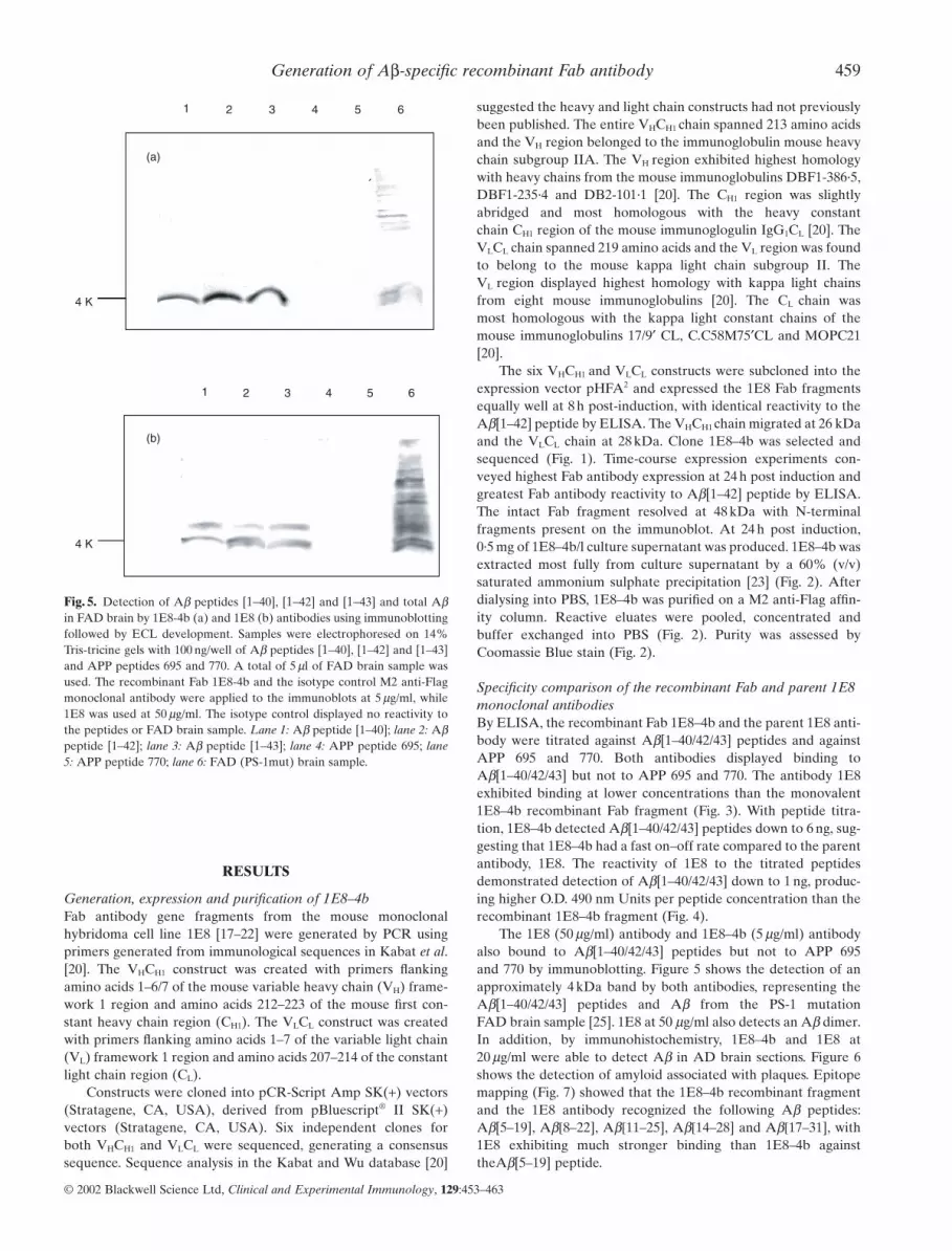
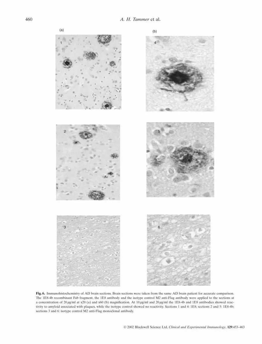
The 1E8 (50 mg/ml) antibody and 1E8–4b (5 mg/ml) antibodyalso bound to Ab[1–40/42/43] peptides but not to APP 695 and 770 by immunoblotting. Figure 5 shows the detection of anapproximately 4 kDa band by both antibodies, representing theAb[1–40/42/43] peptides and Ab from the PS-1 mutation FAD brain sample [25]. 1E8 at 50 mg/ml also detects an Ab dimer.In addition, by immunohistochemistry, 1E8–4b and 1E8 at20 mg/ml were able to detect Ab in AD brain sections. Figure 6shows the detection of amyloid associated with plaques. Epitopemapping (Fig. 7) showed that the 1E8–4b recombinant fragmentand the 1E8 antibody recognized the following Ab peptides:Ab[5–19], Ab[8–22], Ab[11–25], Ab[14–28] and Ab[17–31], with1E8 exhibiting much stronger binding than 1E8–4b againsttheAb[5–19] peptide.
4 K
(a)
4 K
1 2 3 4 5 6
1 2 3 4 5 6
(b)
Fig. 5. Detection of Ab peptides [1–40], [1–42] and [1–43] and total Abin FAD brain by 1E8-4b (a) and 1E8 (b) antibodies using immunoblottingfollowed by ECL development. Samples were electrophoresed on 14%Tris-tricine gels with 100 ng/well of Ab peptides [1–40], [1–42] and [1–43]and APP peptides 695 and 770. A total of 5 ml of FAD brain sample wasused. The recombinant Fab 1E8-4b and the isotype control M2 anti-Flagmonoclonal antibody were applied to the immunoblots at 5 mg/ml, while1E8 was used at 50 mg/ml. The isotype control displayed no reactivity tothe peptides or FAD brain sample. Lane 1: Ab peptide [1–40]; lane 2: Abpeptide [1–42]; lane 3: Ab peptide [1–43]; lane 4: APP peptide 695; lane5: APP peptide 770; lane 6: FAD (PS-1mut) brain sample.
Fig. 6. Immunohistochemistry of AD brain sections. Brain sections were taken from the same AD brain patient for accurate comparison.The 1E8-4b recombinant Fab fragment, the 1E8 antibody and the isotype control M2 anti-Flag antibody were applied to the sections at a concentration of 20 mg/ml at x20 (a) and x60 (b) magnification. At 10 mg/ml and 20 mg/ml the 1E8-4b and 1E8 antibodies showed reac-tivity to amyloid associated with plaques, while the isotype control showed no reactivity. Sections 1 and 4: 1E8; sections 2 and 5: 1E8-4b;sections 3 and 6: isotype control M2 anti-Flag monoclonal antibody.
Generation of Ab-specific recombinant Fab antibody 461
Despite procedures for pre-mortem AD diagnosis (i.e. neurologi-cal examination, ELISA-based tests, risk assessment by familyhistory/genetic predisposition to AD) post-mortem immunohis-tological brain tissue examination is the definitive AD diagnostic[26,27]. Generating antibodies specific for key AD markers isimportant for ante-mortem reagent development to diagnose AD rapidly and accurately, eliminating stringent tests and permitting therapy prior to neurodegeneration [26]. There is currently no cure for AD and although therapies such as
cholinesterase inhibitors are reported to improve cognitive func-tion, disease progression continues [28]. Ideal therapeutics shouldprevent AD progression and restore normal function. Antibodiesmay clear amyloid from brain [17] and combined with 5 mer b-sheet breaker peptide tags [29], neurotrophic tags [30] and Absolubilizing substances [31], therapeutic ‘magic bullets’ couldensue. We have produced the recombinant Fab antibody, 1E8–4b,and performed comparitive binding studies with the parent antibody, 1E8.
To correct for the differences in size and number of valenciesbetween 1E8 and 1E8–4b, the antibodies should be compared on
0 1 2
O.D. 490 nm
0 1 2
O.D. 490 nm
1E8
(a)
(b)
1E8-4b
Fig. 7. Epitope mapping of 1E8 (a) and 1E8-4b (b). The pepset encompasses the human APP695[589–652] sequence and consists of anoverlapping set of 15-mer linear peptides, incrementing by 3 amino acids per peptide. APP[597–640] amino acid residues represent theAb[1–43] peptide sequence and are underlined.
462 A. H. Tammer et al.
the development of the double-antibody capture ELISA as reported inJayasena et al. [18]. The mRNA and purified 1E8 antibodies from the 1E8hybridoma, cultured in Dr Underwood’s laboratory in the Department ofPathology at the University of Melbourne, were used for this study. Theauthors also thank Ms Tina Cardamone for her assistance with immuno-histochemical staining, Dr Genevieve Evin for supplying the PS-1 muta-tion brain sample, Ms Denise Galatis for her advice and Dr Bob Irving forproviding reagents and advice.
2 Ebly EM, Parhad IM, Hogan DB, Fung TS. Prevalence and types ofdementia in the very old: results from the Canadian study of Healthand Aging. Neurology 1994; 44:1593–600.
3 Hardy J. Amyloid, the presenilins and Alzheimer’s disease. TrendsNeurosci 1997; 20:154–9.
4 Gravina SA, Ho L, Eckman CB et al. Amyloid b protein (Ab) inAlzheimer’s disease brain. Biochemical and immunocytochemicalanalysis with antibodies specific for forms ending at Abeta 40 or A beta42 (43). J Biol Chem 1995; 270:7013–6.
5 Roher AE, Lowenson JD, Clarke S et al. Structural alterations in the peptide backbone of b-amyloid core protein may account for itsdeposition and stability in Alzheimer’s disease. J Biol Chem 1993;268:3072–83.
6 Mattson MP. Cellular actions of beta-amyloid precursor protein and its soluble and fibrillogenic derivatives. Physiol Rev 1997; 77:1081–132.
7 Roher AE, Kuo YM, Kokjohn KM, Emmerling MR, Gracon S.Amyloid and lipids in the pathology of Alzheimer disease. Amyloid1999; 6:136–45.
8 Jarrett JT, Lansbury PT. Seeding ‘one-dimensional crystallization’ ofamyloid. A pathogenic mechanism in Alzheimer’s disease and scrapie?Cell 1993; 73:1055–8.
9 Mann DM, Iwatsubo T, Cairns NJ et al. Amyloid beta protein (Abeta)deposition in Chromosome 14 linked Alzheimer’s disease. Predomi-nance of Abeta42 (43). Ann Neurol 1996; 40:149–56.
10 Hock C, Golombowski S, Muller-Spahn F et al. Histological markersin nasal mucosa of patients with Alzheimer’s disease. Eur Neurol 1998;39:111–8.
11 Shoji M, Matsubara E, Kanai M et al. Combination assay of CSF Tau,Ab1–40 and Ab1-42 (43) as a biochemical marker of Alzheimer’sdisease. J Neurol Sci 1998; 158:134–40.
12 Vanmechelen E, Vanderstichele H. Alzheimer tau test and detergentcellulase made by genetic engineering. J Biotechnol 1998; 66:229–33.
14 Scheuner D, Eckman C, Jensen M et al. Secreted amyloid beta-proteinsimilar to that in the senile plaques of Alzheimer’s disease is increasedin vivo by the presenilin 1 and 2 and APP mutations linked to famil-ial Alzheimer’s disease. Nat Med 1996; 2:864–70.
15 Mehta PD, Dalton AJ, Mehta SP, Kim KS, Sersen EA, Wisniewski HM.Increased plasma amyloid b protein 1–42 levels in Down syndrome.Neurosci Lett 1998; 241:13–6.
16 Ida N, Hartmann T, Pantel J et al. Analysis of heterogeneous bA4 pep-tides in human cerebrospinal fluid and blood by a newly developedsensitive Western blot assay. J Biol Chem 1996; 271:22908–14.
17 Schenk D, Barbour R, Dunn W et al. Immunization with amyloid-betaattenuates Alzheimer-disease-like pathology in the PDAPP mouse.Nature 1999; 400:173–7.
18 Jayasena ULHR, Gribble SK, McKenzie A, Beyreuther K, MastersCL, Underwood JR. Identification of structural variations in the car-boxyl terminus of Alzheimer’s disease-associated betaA4. Clin ExpImmunol 2001; 124:297–305.
a 1 : 1 mole ratio. As 1E8 is an intact, divalent IgG1 of 160 kDaand 1E8–4b is a monovalent Fab of 48 kDa, the amount of 1E8needed relative to 1E8–4b is: (160 kDa/48 kDa)/2 valencies = 1·67times more 1E8 than 1E8–4b to give a 1 : 1 mole ratio. Hence10 mg/ml of 1E8 is equivalent to 6 mg/ml of 1E8–4b to have thesame number of moles. In ELISA experiments using a highepitope density of 50 ng/well Ab[1–40/42/43] peptides and titratedantibody (Fig. 3), 1E8–4b did not perform as well as 1E8 on amole to mole ratio, although both antibodies are quite sensitive.1E8 produced a signal down to 20 ng/ml and produced a curvewith a steep linear slope of 1·5 from low to high antibody dilu-tions. In contrast, 1E8–4b produced a signal down to 200 ng/mlwith a decreased linear slope of 0·4. As bivalently bound antibodydissociates slower than univalently bound antibody, apparentduring ELISA washing steps [32], the characteristic curve for 1E8may have arisen (Fig. 3). High-affinity antibodies generallyproduce steeper-sloped titration curves, suggesting a lower affin-ity for 1E8–4b [33,34]. At the same antibody concentration, with1·7 times less moles of 1E8 than 1E8–4b, 1E8 detected lowerlevels of Ab (1·5 ng/well Ab) than 1E8–4b (6 ng/well Ab). Theslower titration of 1E8 suggested stronger binding of 1E8 to theAb peptides than 1E8–4b and perhaps a slower dissociation ratedue to divalency or higher affinity [32]. Presence of the Flag tagon 1E8–4b was expected to produce an enhanced signal for the1E8–4b ELISA due to the additional M2 anti-Flag monoclonalantibody detection step [13].
By immunohistochemistry, 1E8–4b and 1E8 detected Abin AD brain sections, which was localized to plaques [4].Immunoblotting depicted reactivity to Ab[1–40/42/43] peptidesby 1E8 and 1E8–4b. In addition, in FAD brain characterized witha novel PS-1 mutation [25] we detected a 4 kDa fragment corre-sponding to Ab. Immunoblotting and ELISA results conveyedthat 1E8 and 1E8–4b did not react with APP 695 or APP 770, suggesting Ab specificity by these antibodies. Pepset epitopemapping showed specific binding of 1E8–4b and 1E8 to Ab aminoacid residues 17–22. Removal of Ab[17–19] obliterated bindingdespite the presence of [20–31]. Stronger binding was seen whenAb[20–22] was included with Ab[17–19]. 1E8–4b showed aweaker response to Ab[17–19] than 1E8 due perhaps to alteredconformation or unique requirements of only Ab[18,19/19] plus[20–22]. Overall, 1E8 and 1E8–4b contained a specific epitopeencompassing Ab[17,18,18–22].
On a mole : mole ratio, 1E8 showed greater reactivity than1E8–4b with Ab[1–40/42/43] but the specificity of 1E8–4b and 1E8 was nearly identical. Improved detection of Ab in CSF and plasma requires an antibody of higher affinity than 1E8 or 1E8–4b. With several rounds of affinity selection, 1E8–4b may attain this characteristic [13] with diagnostic benefits for AD.
The use of recombinant technology for antibody productionin the diagnosis and therapy of AD is relatively unexplored andthe need to examine recombinant antibody design and applica-tion should be pursued. The recombinant Fab fragment 1E8–4bproduced in this study, is the first recombinant Fab fragment tobe described that is reactive with the AD-related Ab protein.
ACKNOWLEDGEMENTS
The authors thank Drs Steven Holmes and Carol Gray of SmithKlineBeecham for the 1E8 hybridoma cell line which was provided in 1995 for
Generation of Ab-specific recombinant Fab antibody 463
19 Kettleborough CA, Saldanha J, Ansell KH, Bendig MM. Optimizationof primers for cloning libraries of mouse immunoglobulin genes usingthe polymerase reaction. Eur J Immunol 1993; 23:206–11.
20 Kabat EA, Wu TT, Perry HM, Gottesman KS, Foeller C. Sequences ofproteins of immunological interest, 5th edn. Bethesda, MD: NationalInstitutes of Health, 1991.
21 Dolezal O, Coia G, Guthrie RE, Lilley GG, Hudson PJ. Escherichiacoli expression of a bifunctional Fab peptide epitope reagent for the rapid diagnosis of HIV-1 and HIV-2. Immunotechnology 1995;1:197–209.
22 Hoogenboom HR, Griffiths AD, Johnson KS, Chiswell DJ, Hudson P,Winter G. Multi-subunit proteins on the surface of filamentous phage:methodologies for displaying antibody (Fab) heavy and light chains.Nuc Acids Res 1991; 19:4133–7.
23 Coppola G, Underwood J, Cartwright G, Hearn MTW. Comparison ofmethods for the purification of mouse monoclonal immunoglobulin Mautoantibodies. J Chromatogr 1989; 426:269.
24 Sambrook J, Fritsch EF, Maniatis T. Molecular cloning, a laboratorymanual, 2nd edn. New York: Nolan C, 1989.
25 Smith MJ, Gardner RJ, Knight MA et al. Early-onset Alzheimer’sdisease caused by a novel mutation at codon 219 of the presenilin-gene.Neuroreport 1999; 10:503–7.
26 Arai H. Biological markers for the clinical diagnosis of Alzheimer’sdisease. Tohoku J Exp Med 1996; 179:65–79.
28 Hecker J. Alzheimer’s disease: the advent of effective therapy. AustNZ J Med 1998; 28:765–71.
29 Soto C. Alzheimer’s and prion disease as disorders of protein confor-mation: implications for the design of novel therapeutic approaches. J Mol Med 1999; 77:412–8.
30 Birkhauser MH, Strnad J, Kampf C, Bahro M. Oestrogens andAlzheimer’s disease. Int J Geriatr Psychiatry 2000; 15:600–9.
31 Janciauskiene S, de Frutos PG, Carlemalm E, Dahlback B, Eriksson S.Inhibition of Alzheimer b-peptide fibril formation by serum amyloidP component. J Biol Chem 1995; 270:26041–4.
32 Mason DW, Williams AF. The kinetics of antibody binding to mem-brane antigens in solution and at the cell surface. Biochem J 1980;187:1–20.
33 Lew AM. The effect of epitope density and antibody affinity on ELISAas analysed by monoclonal antibodies. J Immunol Meth 1984; 72:171.
34 Nimmo GR, Lew AM, Stanley CM, Steward MW. Influence of antibody affinity on the performance of different antibody assays. J Immunol Meth 1984; 72:177.