Generation of follicular helper T cells is mediated by IL-21 but independent of TH1, TH2 or TH17 lineages Roza I. Nurieva 1 , Yeonseok Chung 1 , Daehee Hwang 2 , Xuexian O. Yang 1 , Hong Soon Kang 3 , Li Ma 4 , Yi-hong Wang 1 , Stephanie S. Watowich 1 , Anton M. Jetten 3 , Qiang Tian 4 , and Chen Dong 1 1Department of Immunology, University of Texas M. D. Anderson Cancer Center and Graduate School of Biomedical Sciences, Houston, TX 77030, USA 2School of Interdisciplinary Bioscience and Bioengineering & Department of Chemical Engineering, Pohang University of Science and Technology, Kyungbuk 790-784, Republic of Korea 3Cell Biology Section, LRB, National Institutes of Health, NIEHS, Research Triangle Park, NC 27709, USA 4Institute for Systems Biology, Seattle, WA 98103, USA Abstract After activation, CD4+ helper T (TH) cells differentiate into distinct effector lineages. Although CXCR5+ follicular TH (T FH ) cells are important in humoral immunity, their developmental regulation is unclear. Here we show that T FH cells have a distinct gene expression profile from other effector T cells and develop in vivo independent of the TH1 or TH2 lineages. T FH cell generation is regulated by B7h expressed on B cells and, similar to TH17 cell development, is dependent on IL-21, IL-6 and STAT3. However, differentiation of T FH cells, unlike TH17 cells, does not require TGFβ signaling or TH17-specific orphan nuclear receptors RORα and RORγ in vivo. Finally, naïve T cells activated in vitro in the presence of IL-21 but not TGFβ signaling preferentially acquire T FH gene expression and function to promote germinal center reactions in vivo. This study thus demonstrates T FH as a distinct lineage of effector TH differentiation. Introduction Naïve CD4 + helper T (TH) cells, upon encountering their cognate antigens presented on professional antigen-presenting cells (APC), differentiate into effector cells that are characterized by their distinct cytokine production profiles and immune regulatory functions (Dong and Flavell, 2000; Glimcher and Murphy, 2000). TH1 cells produce IFNγ and regulate antigen presentation and immunity against intracellular pathogens, whereas TH2 cells produce IL-4, IL-5 and IL-13, and mediate humoral responses and immunity against parasites. IFNγ and IL-4 are not only the key effector cytokines but also mediate the differentiation of TH1 and TH2 cells, respectively. Recently, TH17, a third subset of TH cells, has been identified, which produce IL-17, IL-17F and IL-22 and regulate inflammatory responses by tissue cells (Bettelli et al., 2007; Dong, 2006; Reiner, 2007; Weaver et al., 2006). TH17 differentiation, at least in mouse, is initiated Correspondence should be addressed to C.D. ([email protected]) or R.I.N. ([email protected]) . Publisher's Disclaimer: This is a PDF file of an unedited manuscript that has been accepted for publication. As a service to our customers we are providing this early version of the manuscript. The manuscript will undergo copyediting, typesetting, and review of the resulting proof before it is published in its final citable form. Please note that during the production process errors may be discovered which could affect the content, and all legal disclaimers that apply to the journal pertain. NIH Public Access Author Manuscript Immunity. Author manuscript; available in PMC 2009 July 1. Published in final edited form as: Immunity. 2008 July ; 29(1): 138–149. doi:10.1016/j.immuni.2008.05.009. NIH-PA Author Manuscript NIH-PA Author Manuscript NIH-PA Author Manuscript
Transcript
Generation of follicular helper T cells is mediated by IL-21 butindependent of TH1, TH2 or TH17 lineages
Roza I. Nurieva1, Yeonseok Chung1, Daehee Hwang2, Xuexian O. Yang1, Hong SoonKang3, Li Ma4, Yi-hong Wang1, Stephanie S. Watowich1, Anton M. Jetten3, Qiang Tian4, andChen Dong1
1Department of Immunology, University of Texas M. D. Anderson Cancer Center and Graduate School ofBiomedical Sciences, Houston, TX 77030, USA
2School of Interdisciplinary Bioscience and Bioengineering & Department of Chemical Engineering, PohangUniversity of Science and Technology, Kyungbuk 790-784, Republic of Korea
3Cell Biology Section, LRB, National Institutes of Health, NIEHS, Research Triangle Park, NC 27709, USA
4Institute for Systems Biology, Seattle, WA 98103, USA
AbstractAfter activation, CD4+ helper T (TH) cells differentiate into distinct effector lineages. AlthoughCXCR5+ follicular TH (TFH) cells are important in humoral immunity, their developmentalregulation is unclear. Here we show that TFH cells have a distinct gene expression profile from othereffector T cells and develop in vivo independent of the TH1 or TH2 lineages. TFH cell generation isregulated by B7h expressed on B cells and, similar to TH17 cell development, is dependent on IL-21,IL-6 and STAT3. However, differentiation of TFH cells, unlike TH17 cells, does not require TGFβsignaling or TH17-specific orphan nuclear receptors RORα and RORγ in vivo. Finally, naïve T cellsactivated in vitro in the presence of IL-21 but not TGFβ signaling preferentially acquire TFH geneexpression and function to promote germinal center reactions in vivo. This study thus demonstratesTFH as a distinct lineage of effector TH differentiation.
IntroductionNaïve CD4+ helper T (TH) cells, upon encountering their cognate antigens presented onprofessional antigen-presenting cells (APC), differentiate into effector cells that arecharacterized by their distinct cytokine production profiles and immune regulatory functions(Dong and Flavell, 2000; Glimcher and Murphy, 2000). TH1 cells produce IFNγ and regulateantigen presentation and immunity against intracellular pathogens, whereas TH2 cells produceIL-4, IL-5 and IL-13, and mediate humoral responses and immunity against parasites. IFNγand IL-4 are not only the key effector cytokines but also mediate the differentiation of TH1and TH2 cells, respectively.
Recently, TH17, a third subset of TH cells, has been identified, which produce IL-17, IL-17Fand IL-22 and regulate inflammatory responses by tissue cells (Bettelli et al., 2007; Dong,2006; Reiner, 2007; Weaver et al., 2006). TH17 differentiation, at least in mouse, is initiated
Correspondence should be addressed to C.D. ([email protected]) or R.I.N. ([email protected]) .Publisher's Disclaimer: This is a PDF file of an unedited manuscript that has been accepted for publication. As a service to our customerswe are providing this early version of the manuscript. The manuscript will undergo copyediting, typesetting, and review of the resultingproof before it is published in its final citable form. Please note that during the production process errors may be discovered which couldaffect the content, and all legal disclaimers that apply to the journal pertain.
NIH Public AccessAuthor ManuscriptImmunity. Author manuscript; available in PMC 2009 July 1.
Published in final edited form as:Immunity. 2008 July ; 29(1): 138–149. doi:10.1016/j.immuni.2008.05.009.
by TGFβ and IL-6 (Bettelli et al., 2006; Mangan et al., 2006; Veldhoen et al., 2006a), possiblyvia regulating the chromatin remodeling of the Il17-Il17f locus (Akimzhanov et al., 2007).While IL-6 is necessary for TH17 differentiation (Korn et al., 2007; Yang et al., 2007), IL-21was recently reported as an autocrine factor induced by IL-6 to regulate TH17 differentiation(Korn et al., 2007; Nurieva et al., 2007a; Zhou et al., 2007). On the other hand, TGFβ signalinghas also been clearly demonstrated to mediate TH17 differentiation in vivo (Bettelli et al.,2006; Mangan et al., 2006; Veldhoen et al., 2006b), and activated T cells may serve as animportant sources of TGFβ for this regulation (Li et al., 2007). TH17 development is dependenton STAT3 (Laurence et al., 2007; Yang et al., 2007), which functions to upregulate theexpression of two TH17-specific orphan nuclear receptors RORγt and RORα that ultimatelydetermines TH17 terminal differentiation (Ivanov et al., 2006; Yang et al.).
A fundamental function of TH cells is to provide “help” to B cells and regulate theirproliferation and immunoglobulin class-switching, especially in the germinal center structures.TH1 and TH2 cells have been shown to regulate B cell responses to some extents. For example,IFNγ regulates IgG2a production while IL-4 is critical in IgE class-switching. However, anadditional TH subset called follicular helper T (TFH) cells are recently found to be present ingerminal centers and are characterized by their expression of CXCR5 (Vinuesa et al., 2005b).Although activated T cells may transiently express CXCR5, TFH cells appear to have morestable expression of this chemokine receptor. These cells are thought to regulate humoralimmunity, especially germinal center reactions. Consistent with this notion, CXCR5 has beenshown to be important for proper T and B cell localization in immune responses and antibodyproduction (Haynes et al., 2007; Junt et al., 2005). In addition to CXCR5, other markers havebeen also reported for TFH cells, such as ICOS costimulatory receptor, IL-21 cytokine andBcl-6 transcription factor. ICOS was found essential for generation of TFH cells in vivo (Akibaet al., 2005). In addition to TH17 cells, IL-21 is also expressed in TFH cells and may serve asan important regulator of humoral responses by TFH cells. IL-21 directly regulates B cellproliferation and class-switching; IL-21R deficiency results in defective antibody responsesand impaired germinal center formation (Spolski and Leonard, 2008). On the other hand,sanroque mice, which have a mutation in a RING-type E3 ubiquitin ligase, Roquin, developedspontaneous autoantibody production and lupus-like autoimmunity, associated with greatlyincreased numbers of CXCR5+ CD4 T cells and enhanced expression of IL-21 and ICOS(Vinuesa et al., 2005a).
Despite their potential importance in humoral immunity and immunopathology, thedevelopmental regulation of TFH cells and their relationship with other TH subsets is unclear,particularly considering the fact that IL-21 is expressed by both TH17 and TFH cells. A previousstudy revealed that human TFH cells express distinct genes from TH1 or TH2 cells (Chtanovaet al., 2004). Here we show that mouse TFH cells have a divergent gene expression profile fromTH1, TH2 and TH17 cells, and develop in vivo independent of these lineages. IL-21, IL-6 andSTAT3 are critical in the generation of TFH cells. Moreover, T cells activated in vitro in thepresence of IL-21 but without TGFβ signaling preferentially acquire TFH gene expression andfunction to promote humoral immunity in vivo. Thus, our data indicate that TFH cells representa distinct TH lineage and suggest a reciprocal relationship between the TFH and TH17 lineages.
ResultsDistinct gene expression profile of TFH cells
As a first step toward understanding TFH cell regulation, we compared the gene expressionprofiles of TH1, TH2 and TH17 cells differentiated in vitro with in-vivo generated TFH cells.Taking advantage of the co-expression of BTLA by CXCR5+ TFH cells (Supplementary Figure1), CD4+CD44hiCXCR5+BTLA+ were FACS sorted from splenocytes of C57BL/6 mice 7days after immunization with KLH. These cells as well as TH1, TH2 and TH17 cells were
Nurieva et al. Page 2
Immunity. Author manuscript; available in PMC 2009 July 1.
restimulated with anti-CD3 for 4 hours and subject to gene profiling analysis in duplicatesusing Affimetrix gene chips. The microarray data were normalized using GCRMA and thegenes whose expression levels were changed across the TH1, TH2, TH17 and TFH cells werethen selected using a False Discovery Rate (FDR) estimation method. Then, the expressionlevels of 8350 probe-sets showing differential expressions among the four types of cells wereused for hierarchical clustering, which revealed that TFH cells have a very distinct geneexpression profile (Figure 1A).
To confirm the above results, we performed real-time RT-PCR analysis on multiple subset-specific genes. The data indicate that TFH cells did not express the typical markers for TH1(IFNγ and T-bet) or TH2 (IL-4 and GATA3) cells (Figure 1B), consistent with a previous reporton human TFH cells (Chtanova et al., 2004). Although TFH cells shared IL-21 expression withTH17 cells, they did not express IL-17, IL-17F, IL-22 or RORγt (Figure 1B). Instead, similarto their human counterparts, (Chtanova et al., 2004; Kim et al., 2004), mouse TFH cells expressmRNAs for CXCR5 as well as Bcl-6 (Figure 1B). In addition, TFH cells preferentiallyexpressed mRNAs for IL-6R and IL-6st (gp130) and also upregulated the expression of IL-21R(Figure 1B), suggesting possible regulation of TFH cells by IL-6 and IL-21. Moreover,consistent with human TFH cells (Chtanova et al., 2004) and a recent report indicating thatTFH cells express PD-1 protein (Haynes et al., 2007), PD-1 mRNA was highly upregulated inTFH cells compare to other TH subsets (data not shown).
To substantiate the above results, we also measured cytokine secretion of purified TFH cellsafter they were activated ex vivo with anti-CD3 and anti-CD28 for 24 hours. High levels ofIL-21 but not IL-4, IL-10, IFNγ or IL-17 were observed in TFH cells (Figure 1C). In addition,purified TFH cells were activated with KLH and irradiated splenic APC. Consistent with aboveresults, TFH cells preferentially produced IL-21, but not TH1, TH2 and TH17 cytokines(Supplementary Figure 3). Furthermore, intracellular analysis on CXCR5+ and CXCR5- cellsfollowing PMA and ionomycin restimulation also revealed that TFH cells did not expressIFNγ or IL-17 (Figure 1D). These results indicate that TFH cells are distinct from TH1, TH2and TH17 cells in their gene expression and cytokine production.
Naïve T cell differentiation into TFH cells is independent of TH1 and TH2 lineagesPrevious (Chtanova et al., 2004) and our current gene expression analysis on human and mouseTFH cells, respectively, suggested that they are distinct from TH1 and TH2 cells. However, itwas not clear if generation of TFH cells is distinct from TH1 or TH2 differentiation. To addressthis question, we transferred FACS sorted naïve CD4+ T cells (CXCR5-) from OT-II TcRtransgenic mice (CD45.2+, Figure 2A) into C57BL/6 recipients (CD45.1+) followed byimmunization with Ova protein in CFA. 7 days later, we examined splenic CD45.1+ andCD45.2+ CD4 cells, and found a substantial increase in the frequency of CXCR5+CD4+ cells(Figure 2A). Interestingly, treatment of recipient mice with antibodies to IL-4 and IFNγ didnot reduce the proportion of CXCR5+CD4+ cells (Figure 2A), suggesting that TFH celldevelopment is independent of TH1 or TH2 development. In addition, donorCD44hiCXCR5+ and CD44hiCXCR5- OT-II cells from immunized mice were purified andreal-time RT-PCR analysis of TFH specific genes were performed. CXCR5+ T cells weregenerated in the presence of blocking antibodies to IL-4 and IFNγ exhibited gene expressionpatterns comparable to those from control group (Figure 2A), supporting that these CXCR5+cells were indeed TFH cells.
To substantiate the above finding, we also immunized mice deficient in IL-4, IFNγ STAT6 orSTAT4 and their appropriate controls with KLH. 7 days after immunization, we did not detectany defect in TFH cells or PNA+ germinal center B cells in the knockout animals compared towild-type controls (Figure 2B-D, Supplementary Figure 4A-C). To confirm the above results,we also measured cytokine secretion of purified CXCR5+ cells from IL-4- and IFNγ-deficient
Nurieva et al. Page 3
Immunity. Author manuscript; available in PMC 2009 July 1.
mice and their controls after they were activated ex vivo with anti-CD3 and anti-CD28 for 24hours. TFH cells IL-4- or IFNγ-deficient mice produced similar level of IL-21, but did notexpress IL-4, IL-10, IFNγ or IL-17 (Supplementary Figure 4D), supporting that they wereTFH cells. Thus, we conclude that CXCR5+CD4+ TFH cells develop independent of the TH1and TH2 lineages.
B7h expressed on B cells regulates the generation of TFH cellsInducible costimulator (ICOS) is the third member of the CD28 family with an important rolein regulation of T-dependant Ab responses and germinal center reactions (Dong and Nurieva,2003). ICOS was previously shown to be expressed at high levels on human tonsillular CXCR5+ T cells within the light zone of germinal centers and efficiently supported theimmunoglobulin production (Breitfeld et al., 2000; Schaerli et al., 2000). In addition, ICOSdeficiency in human and mouse resulted in significantly reduced numbers of TFH cell,indicating an essential role of ICOS in the differentiation of CXCR5+ CD4 T cells (Akiba etal., 2005; Bossaller et al., 2006b). Consistently, we also observed reduced percentages ofCXCR5+CD4+ cell and decreased expression of IL-21 but not IFNγ in our B7h germlineknockout animals (Nurieva et al., 2003b) following KLH immunization (Figure 3A). TFH cellsare regarded as regulators of the germinal center reaction by providing help to activated B cellsthat also express CXCR5. Since B cells constitutively express B7h, we asked whether thegeneration of TFH cells may require B cell help via engagement of ICOS receptor on T cells.We thus bred mice carrying the B7h conditional flox (f) allele (Nurieva et al., 2003b) withCD19-cre mice (Rickert et al., 1995). In B7h f/f mice carrying the CD19-cre (B7h BKO), incomparison to those without cre expression, there was efficient deletion of the B7h gene in Bcells, similar to those in B7h germline knockout mice (Figure 3B). We then immunized B7hBKO mice with KLH and found that absence of B7h in B cells led to a greatly reduced frequencyof CXCR5+CD4+ cells (Figure 3C). Similarly, when CD45.1+ OT-II cells were transferredinto B7h BKO mice, the numbers of CD45.1+ TFH cells were also greatly reduced followingOva peptide immunization (data not shown). In addition to CXCR5 expression, we found thatthe expression of IL-21 was also greatly reduced in B7h BKO mice, whereas IFNγ expressionwas elevated in these mice (Figure 3C). Moreover, PNA+ germinal center B cells were greatlyreduced in these animals (Figure 3C and E). KLH-specific IgG production was also reduced(Figure 3D). Overall, these data indicate that B cell expression of B7h is necessary for IL-21production and for the generation of TFH cells and appropriate antibody responses, indicatingthat TFH cell differentiation is regulated by B cells.
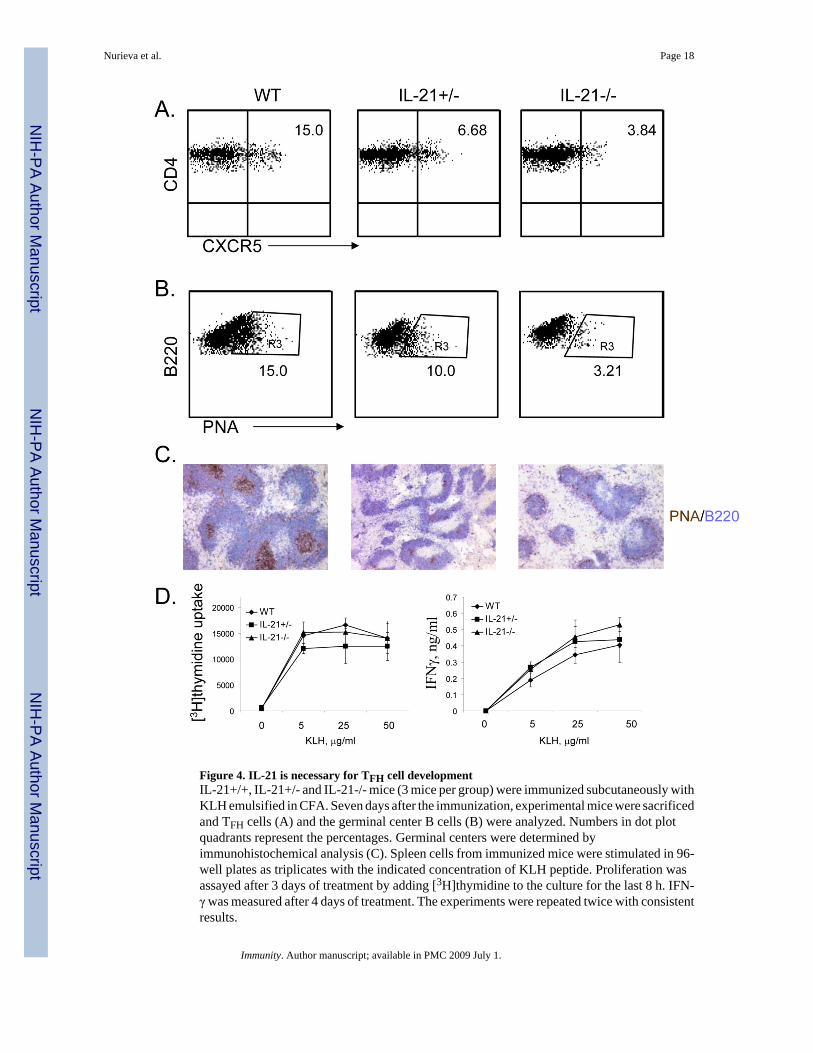
IL-21 and IL-6 are required for generation of TFH cells, which is dependent on STAT3IL-21 has been recently shown to be induced by IL-6 and to autoregulate its own expressionduring TH17 differentiation (Nurieva et al., 2007a). In addition, TFH T cells producedsignificantly greatrt amount of IL-21 compared to TH1 and TH2 subsets (Chtanova et al.,2004), and induced the differentiation of autologous B cells into Ig-secreted plasma cellsthrough IL-21 (Bryant et al., 2007). Since IL-21 is also expressed in TFH cells, we assessed ifIL-21 is important for TFH cell generation. IL-21+/+, +/- and -/- mice were immunized withKLH and splenic TFH cells were analyzed in these mice. IL-21+/- mice exhibited reducednumber of TFH cells, which was further reduced in IL-21-/- mice (Figure 4A). In addition,PNA+ germinal center B cells were also greatly reduced in IL-21-/- mice (Figure 4B-C). Incontrast, CD4 T cells from IL-21+/- and -/- mice showed normal proliferation and IFNγexpression after re-stimulation with KLH ex vivo. (Figure 5D). Thus, these results indicatethat IL-21 is necessary for TFH cell development.
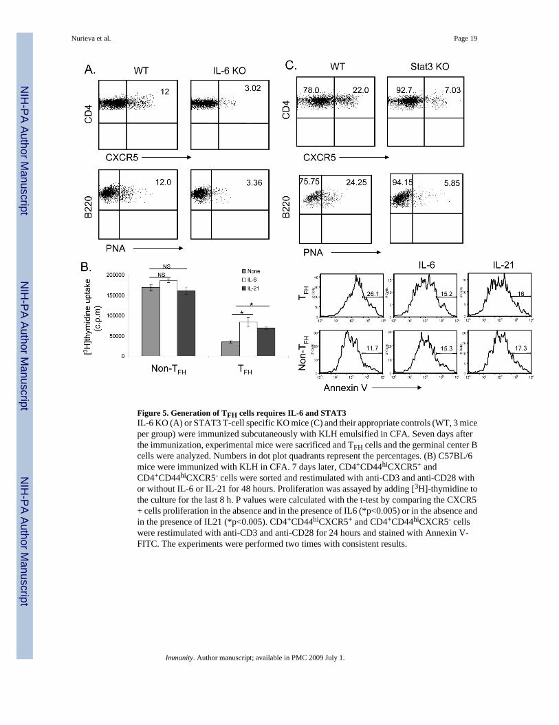
Since IL-6 induces IL-21 expression, we also tested TFH cell generation in mice lacking IL-6.It has been previously that IL-6 deficient mice showed reduced germinal centers and antigenspecific Ig-production (IgG) (Kopf et al., 1998). Compared to the wild-type controls, IL-6-/-
Nurieva et al. Page 4
Immunity. Author manuscript; available in PMC 2009 July 1.
mice exhibited greatly reduced numbers of TFH cells and germinal center B cells (Figure 5A),indicating that IL-6 is also necessary for TFH cell generation.
Since TFH cells expressed high levels of IL-21R, IL-6R and IL-6st (Figure 1B), we alsoexamined if IL-6 and IL-21 signaling regulates TFH cells. C57BL/6 mice were immunized withKLH and CD4+CD44hi T cells with or without CXCR5 expression were FACS sorted andrestimulated with anti-CD3 and anti-CD28 in the absence or presence of IL-21 or IL-6.Compared to non-TFH cells, CXCR5+ T cells showed a significant reduction in proliferationwhen activated with anti-CD3 and anti-CD28. Treatment of CXCR5+ T cells with IL-6 or IL-21significantly enhanced their proliferation (Figure 5B), suggesting that IL-6 and IL-21preferentially regulate TFH cells. Furthermore, we stained restimulated CXCR5+ andCXCR5- cells with Annexin V. We found that a significant portion of CXCR5+ cells underwentapoptosis compare to CXCR5- (Figure 5B). However, IL-6 or IL-21 treatment reducedapoptosis of TFH cells.
Since both IL-6 and IL-21 signal through STAT3, we analyzed TFH cell generation in STAT3flox/flox mice (Takeda et al., 1999) bred with CD4-cre mice (Lee et al., 2001). The deletionof STAT3 gene in CD4+ thymocytes was found to be complete (data not shown). When weimmunized these mice as well as their controls with KLH, the numbers of CXCR5+ TFH cellswere found to be greatly reduced in the absence of STAT3 (Figure 5C). Moreover, STAT3deficiency in T cells also led to defective germinal center B cell generation (Figure 5C). KLH-specific IgG and IgM production was also reduced in the absence of STAT3 in T cells(Supplementary Figure 5).
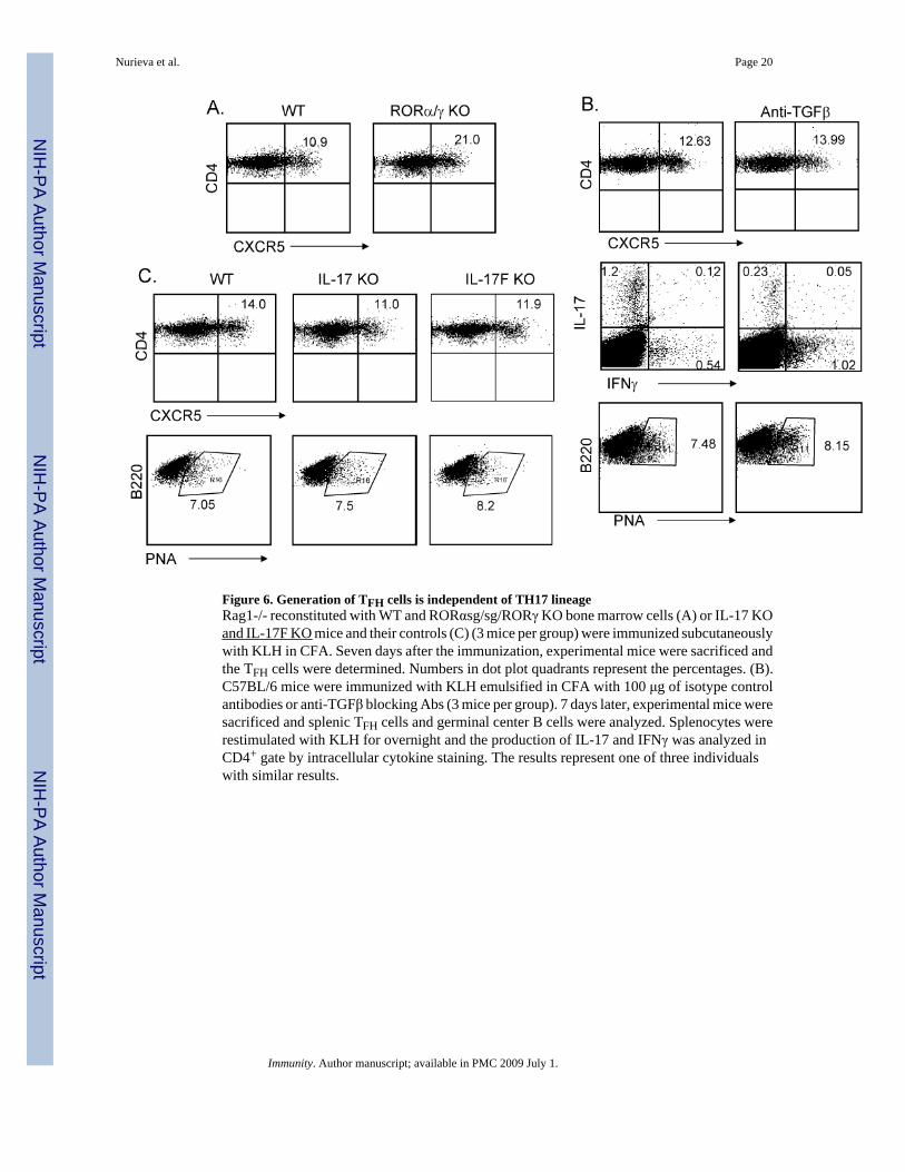
TFH cell generation is independent of TH17 differentiation or functionThe above results indicate that the IL-6-IL-21 axis and STAT3 transcription factor arenecessary for TFH cell generation, which is similar to the regulation of TH17 differentiation.To test whether the development of TFH cells is dependent or independent upon the TH17lineage, we utilized Rag1-/- mice reconstituted with bone marrow cells from wild-type, RORα st/st or ROR αst/stROR α-/- mice. We recently reported that the latter mice were completelyimpaired in TH17 differentiation in vitro and in vivo (Yang et al., 2008b). However, upon KLHimmunization, these mice deficient in both RORα and RORγ in lymphocytes developed moreCXCR5+ cells in spleen (Figure 6A), indicating that TFH cells can be generated in the absenceof TH17 development. Moreover, PNA+ germinal center B cells were not affected in theseanimals (Supplementary Figure 6B).
Since TH17 differentiation requires also TGFβ in addition to IL-6 or IL-21, we examinedwhether TGFβ signaling is required for TFH cell generation. C57BL/6 mice were immunizedwith KLH in the absence or presence of anti-TGFβ blocking antibody as previously described(Veldhoen et al., 2006b). While IL-17 expression was substantially decreased in anti-TGFβ-treated mice, CXCR5 expression was not (Figure 6B), indicating that TGFβ signaling is notessential for TFH cell generation. Moreover, PNA expression on B cells was not affected either(Figure 6B).
To understand if TFH cell generation requires TH17 function, we also immunized micedeficient in IL-17 or IL-17F. Lack of IL-17 or IL-17F did not significantly reduce the numberof TFH cells in spleen or the number of germinal center B cells (Figure 6C, SupplementaryFigure 6B). IL-17 or IL-17F is thus not essential in the generation of TFH cells in vivo.
IL-21 in the absence of TGFβ initiates TFH cell differentiationOur results thus far suggest that although IL-6 and IL-21 are required for both TFH and TH17differentiation, these two subsets appear to have distinct genetic program and differ in their
Nurieva et al. Page 5
Immunity. Author manuscript; available in PMC 2009 July 1.
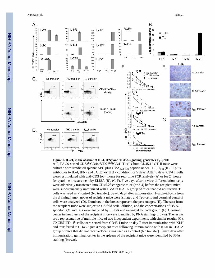
dependency on TGFβ signaling. Since TFH cells have never been generated in vitro, we nextassessed whether IL-21 is sufficient to drive TFH cell development in the absence of TGFβsignaling. Naïve OT-II cells were activated by Ova peptide and splenic APC in the absence(neutral condition) or presence of IL-21, TGFβ and antibodies to IL-4 and IFNγ (TH17condition) or IL-21 plus antibodies to IL-4, IFNγ and TGFβ. 5 days later, the activated T cellswere extensively washed and restimulated with anti-CD3 for 4 hours and their gene expressionwas assessed by real-time RT-PCR. As expected, cells cultured under TH17 condition highlyexpressed TH17-specific genes, including IL-17, IL-17F, IL-22, RORα and RORγt (Figure7A). In contrast, T cells treated with IL-21 in the absence of TGFβ signaling upregulated genesthat are specifically expressed in TFH cells, including CXCR5, Bcl-6, IL-6R and IL-6st (Figure7A). They also upregualted IL-21R expression but did not express TH17 genes (Figure 7A).PD-1 was also highly expressed in these cells (data not shown). To confirm this result, cellsupernatants were measured 24 hours after restimulation for cytokine secretion by ELISA.IL-21 in the absence of TGFβ signaling did not support IL-17 expression, but the resultingcells expressed high levels of IL-21 (Figure 7B), supporting that IL-21 expression isindependent of TGFβ signaling.
To test the function of these TFH-like cells in vivo, we transferred OT-II cells (CD45.1+)activated under neutral condition or with IL-21 plus blocking antibodies to IFNγ, IL-4 andTGFβ into recipient mice (CD45.2+) followed by immunization with Ova protein. Comparedto mice receiving no cells or T cells activated under neutral condition, the recipients of IL-21-treated cells exhibited greatly increased CD45.1+ TFH cells (Figure 7C). Interestingly, host Tcells in these mice also had approximately 4 times TFH cells compared to those receiving Tcells activated under neutral condition (Figure 7C). The transferred cells also promoted Ova-specific antibody production and germinal center reactions (Figure 7D-F). As a control for theabove experiment, we sorted CD45.1+ CXC5+CD44hi cells from KLH immunized mice andtransferred them into recipient mice (CD45.2+) followed by immunization with KLH. Similarto in vitro generated TFH cells, in vivo generated CXCR5+ cells also promoted a significantincrease in PNA+ germinal center B cells (Figure 7G). These results indicate that IL-21, in theabsence of TH1, TH2 and TH17 differentiation, initiates TFH cell development.
DiscussionFollowing antigenic activation, naïve TH cells differentiate into distinct effector subsets. Afundamental function of TH cells is to “help” humoral immunity. Although strongly implicatedin antibody production and germinal center reactions, the ontogeny of TFH cells has beenunclear. In the current study, we find that TFH cells are distinct from TH1, TH2 or TH17 cellsin their gene expression and developmental regulation. Generation of TFH cells requires IL-21,IL-6 and STAT3.
Although implicated in humoral immunity and antibody-mediated autoimmunity, theregulation of TFH cell development is unclear. ICOS-B7h interaction has been well establishedin the literature to regulate humoral immunity and germinal center reactions (Dong andNurieva, 2003). This costimulatory pathway was also found important in generation of TFHcells in mouse (Akiba et al., 2005). Interestingly, ICOS deficiency in human patients alsocauses a severe reduction of TFH cells (Bossaller et al., 2006a). Conversely, impaired negativeregulation of ICOS by Roquin E3 ubiquitin ligase led to increased numbers of CXCR5+ TFHcells and IL-21 hyperproduction (Vinuesa et al., 2005a; Yu et al., 2007). However, themechanism by which ICOS/B7h regulates TFH cell development has not been clear. In ourcurrent study, we find that ICOS-B7h interaction is necessary for IL-21 expression by T cells.ICOS may thus regulate TFH cells through production of IL-21. A recent study by Kim et alhas revealed IL-21 regulation by calcium signaling and NFAT factors (Kim et al., 2005). Wepreviously showed that ICOS, together with TcR and CD28, increases the expression of
Nurieva et al. Page 6
Immunity. Author manuscript; available in PMC 2009 July 1.
NFATc1 through a PI-3 kinase-Itk-calcium pathway (Nurieva et al., 2007b; Nurieva et al.,2003a). ICOS may act through NFATc1 to regulate IL-21 expression. Since IL-21 alsoregulates TH17 differentiation, an IL-21 defect may account for the impairment in IL-17expression in the ICOS-deficient animals (Dong and Nurieva, 2003). Moreover, using a B cell-specific B7h knockout mouse, we show that B7h expression on B cells is required for thegeneration of TFH cells and IL-21 expression. This result not only substantiates the importanceof ICOS-B7h interaction in TFH cell development, but also suggests an important function ofB cells in vivo as APCs in the generation or maintenance of TFH cells. Consistent with ourfindings, Ebert et al previously showed that human B cells regulate TFH cell phenotypes (Ebertet al., 2004).
Previous gene expression analysis has revealed that human TFH cells are distinct from TH1and TH2 cells (Chtanova et al., 2004; Kim et al., 2004). Our current study also reveals thatTFH cells do not produce TH1 or TH2 cytokines. Interestingly, although human TFH cellsappear to express IL-10 and CXCL13, a ligand for CXCR5 (Chtanova et al., 2004; Ebert et al.,2004; Kim et al., 2004; Kim et al., 2001), mouse TFH cells do not. What accounts for thisspecies difference would need further investigation. Moreover, we found that naïve T cellsdifferentiate into TFH cells in vivo, which is independent of IFNγ, IL-4 and STAT6. From thesedata, we conclude that TFH cells develop independent of TH1 and TH2 lineages.
On the other hand, TFH cells share common regulators with TH17 cells. Both subsets expressIL-21 and their development depends similarly on IL-6, IL-21 and STAT3. However, TFH cellsdiffer from TH17 cells in the following aspects. First, they are distinct in their gene expressionprofiles. Second, TFH cells do not produce IL-17, IL-17F or IL-22. Most importantly, TFH celldevelopment does not require RORα or RORγt. Thus, we believe that TFH cells developindependent of the TH17 lineage. TH17 development in mouse is not only mediated by theIL-6-IL-21 axis, but also by TGFβ. We show here that IL-21 can be induced in T cellsindependent of TGFβ signaling. T cells activated in the presence of IL-21 but in the absenceof IL-4, IFNγ and TGFβ signaling produced IL-21 but not IL-4, IFNγ, IL-17, IL-17F or IL-22.Furthermore, these cells acquired expression of CXCR5, Bcl-6, IL-6R and IL-6st, genes thatexpressed by in vivo generated TFH cells, suggesting that TFH cells may be generated in vitrounder the above condition. Moreover, these TFH-like cells generated in vitro preferentiallyexpressed CXCR5 in vivo and functioned to promote humoral immunity, similar to in vivo-generated TFH cells. Furthermore, TGFβ signaling, although required for IL-17 expression invivo, is not essential for TFH cells, indicating a reciprocal relationship of TFH and TH17 cells.Interestingly, Bcl-6 has recently been shown as a repressor of TGFβ-SMAD signaling (Wanget al., 2008), suggesting that lack of TGFβ signaling may favor TFH cell development. Theseresults further support that TFH cells development is independent of TH1, TH2 and TH17 cells,and IL-21 serves as critical factor for generation of this lineage. Interestingly, in vitro generatedTFH cells also enhanced the TFH cell generation in recipient mice, suggesting that IL-21 mayfunction in a paracrine fashion to regulate TFH cell development.
In summary, our current study has extensively characterized the developmental regulation ofTFH cells. Our data indicate that TFH cells are distinct in their gene expression and immunefunction and develop via a pathway that is dependent on IL-21 or IL-6 but independent of TH1,TH2 or TH17 lineages (Supplemental Figure 7). In mice defective in TFH cells, there existedstill detectable amounts of antigen-specific antibodies, suggesting that other TH subsets mayindependently regulate the humoral immunity. This knowledge may help us to find ways totreat antibody-mediated autoimmune diseases.
Nurieva et al. Page 7
Immunity. Author manuscript; available in PMC 2009 July 1.
IL-4-, IFNγ- and IL-6-deficient mice on C57BL/6 background and STAT6 and STAT4-deficient mice on BALB/c background were purchased from Jackson Laboratories and C57BL/6, B6.SJL (CD45.1) and BALB/c mice were used as controls. RORαst/st, RORαst/st RORγ-/-
and wild-type bone marrow chimeras were generated as described (Yang et al., 2008b). Stat3fl/fl mice (Takeda et al., 1999) were bred with CD4-Cre mice provided by Dr. ChristopherWilson (Lee et al., 2001). IL-21 knockout mice on 129×C57BL/6 F1 mixed background wereobtained from NIH Mutant Mouse Regional Resource Centers (MMRRC) (Nurieva et al.,2007a). B7h BKO mice were created by breeding B7H flox mice (Nurieva et al., 2003b) withCD19-cre mice (Rickert et al., 1995). IL-17- and IL-17F- mice were recently generated in ourlab (Yang et al., 2008a). Mice were housed in the SPF animal facility at M. D. Anderson CancerCenter and the animal experiments were performed at the age of 6-10 weeks using protocolsapproved by Institutional Animal Care and Use Committee.
T cell differentiationDifferentiation of OT-II cells in Figure 1A-B was performed as previously described (Chunget al., 2006). CD62LhiCD44loCD25negCD4+ T cells were isolated and cultured with irradiatedsplenic APC plus OVA323-339 peptide (10 μg/ml) (TH0 condition) and in the presence ofpolarizing reagents as the following for 5 days: 10 μg/ml of anti-IL-4 (11B11) and 10 ng/mlof IL-12 for TH1; 10 μg/ml of anti-IFNγ (XMG 1.2) and 10 ng/ml of IL-4 for TH2; 30 ng/mlof IL-6, 5 ng/ml of TGFβ, 50 ng/ml IL-23, 10 μg/ml of anti-IL-4 and 10 μg/ml of anti-IFNγfor TH17. In Figure 7A, the cytokine stimuli for TH17 differentiation were 100 ng/ml of IL-21,5 ng/ml of TGFβ, and 10 μg/ml of anti-IL-4 and 10 μg/ml of anti-IFNγ, and for generation ofTFH cells were 50 ng/ml of IL-21, 10 μg/ml anti-IFNγ, 10 μg/ml anti-IL-4 and 20 μg/ml anti-TGF-β (1D11) neutralizing Abs. IL-4, IL-6, IL-12 and TGFβ were purchased from Peprotech.IL-21, IL-23 and anti-TGF-β (1D11) neutralizing Abs were purchased from R&D. Tocharacterized the in vitro-differentiated CD4 T cells under these conditions, these cells wererestimulated with plate-bound anti-CD3 Ab (5 μg/ml) for 4hours for real-time PCR analysis,or for 24 hours for cytokine measurement by ELISA.
Keyhole Limpet Hemocyanin (KLH) ImmunizationVarious strains of mice (6-8 wk old; three per group) were immunized with KLH (0.5 mg/ml)emulsified in CFA (0.5 mg/ml) at the base of the tail (100 μl each mouse). In Figure 6 for localblockade of TGF-β, 100 μg anti-TGFβ (1D11) was included in the emulsion; the control groupreceived 100 μg isotype control antibodies. Seven days after immunization, these mice weresacrificed and analyzed individually. The germinal center B cells were determined by stainingwith FITC-labeled PNA (Pharmingen) and PerCP-labeled anti-B220 mAb (Pharmingen). TheTFH cell induction was determined by staining with PerCP-labeled anti-CD4 mAb(Pharmingen) and biotinylated anti-CXCR5 mAb (Pharmingen), followed by APC-labeledstreptavidin (Jackson ImmunoResearch Laboratories, Inc.). In some experiments, sera fromimmunized mice were collected, and antigen-specific IgM, and IgG antibodies were measuredby using ELISA. Briefly, serum samples were added in a 3-fold serial dilution onto platesprecoated with 10 μg/ml KLH or Ova protein. Antigen-specific antibodies were detected withbiotinylated goat antimouse IgM or rat anti-mouse IgG antibodies (Southern BiotechnologyAssociates). To analyze the role of B7h in regulation of T cell responses in vivo (Fig. 3A andC), spleen cells from KLH-immunized mice were stimulated in 96-well plates as triplicateswith or without KLH. Effector cytokines (IFN-γ and IL-21) were analyzed 4 days later byELISA (Pharmingen). In Figure 6 B, spleen cells from immunized mice were restimulated with50 μg KLH for 24 h. In the final 5 h, Golgi-stop (BD Bioscience) was added and IL-17- and
Nurieva et al. Page 8
Immunity. Author manuscript; available in PMC 2009 July 1.
IFNγ-producing cells were analyzed using a BD CytoFix/CytoPerm intracellular staining kit(BD Bioscience).
Statistical analysis of microarray dataThe DNA microarray analysis was carried out at the Institute for Systems Biology microarraycore facility using Affymetrix Mouse 430 2.0 chips. The total RNA samples were labeledaccording to manufacturer’s instruction using One-Cycle Target Labeling method, whichconsists of oligo-dT primed cDNA synthesis followed by in vitro transcription that incorporatesbiotinylated nucleotides. The microarray data were normalized using GCRMA (Zhijin et al.,2004). We then selected the genes whose expression levels were changed across the TH1, TH2,TH17, and TFH cells using a False Discovery Rate (FDR) estimation method (Storey andTibshirani, 2001): 1) one-way ANOVA was performed to compute unadjusted p-value for thefour classes of cells; 2) the number of non-differentially expressed probe-sets (m0) wasestimated as 2 × the number of the probe-sets with p-value>0.5 (m0= 25088); 3) the expectednumber of false positives under the complement null hypothesis E(V0) was estimated for agiven ANOVA F-statistic value by performing 500 times of resampling; and 4) FDR was finallyestimated as m0/m × E(V0)/R where m is the total number of the probe-sets (m=45037) and Ris the number of the genes being selected using the given ANOVA F-statistic value (FDRestimated for various F-statistic value is shown in Supplementary Figure 2). Then, theexpression levels of 8350 probe-sets showing differential expressions among the four types ofcells (FDR = 0.1, corresponding to an unadjusted p-value = 0.033 from one-way ANOVA)were used for hierarchical clustering (Euclidian distance metric and Ward Minimum VarianceLinkage) and PCA.
Adoptive transfer studyCD4+ T cells from OT-II mice (CD45.2) were intravenously transferred into C57BL/6(CD45.1+) mice (3×10⁶ cells/mouse) (3 groups; 3 mice per group). 2 groups of recipient micewere immunized subcutaneously with 100 μg Ova protein emulsified in CFA and treated witha 300 μg of control rat Ig or anti-IFNγ and anti-IL-4 mAbs at the time of immunization (day0) and on days 2 and 4. Seven days after the immunization, experimental mice were sacrificedand splenic CD45.1+ and CD45.2+ CD4 cells were stained with biotinylated anti-CXCR5mAb, followed by APC-labeled streptavidin. In Figure 7C-E, FACS-sorted naïve(CD4+CD62LhiCD44-CD25-) T cells from CD45.1+ OT-II mice were activated under TH0 orTFH condition, washed and intravenously transferred into C57BL/6 (CD45.2+) mice (4×10⁶
cells/mouse) and the recipient mice were subcutaneously immunized with 100 μg OVA proteinemulsified in IFA. A group of C57BL/6 mice that did not receive T cells was used as a control(No transfer). In Fig. 7G, CXCR5+CD44hi cells were sorted from B6.SJL (CD45.1) miceimmunized with KLH. These cells were transferred into C57BL/6 (CD45.2+) mice (5×10⁶
cells/mouse) (3 mice per group). Second group did not receive cells. All mice were immunizedsubcutaneously with 1000 μg KLH. Seven days after immunzation, lymphoid cells from thedraining lymph nodes of the recipient mice were isolated and stained with FITC-labeled anti-CD45.1 mAb and PerCP-labeled anti-CD4 mAb plus biotinylated anti-CXCR5 mAb, followedby APC-labeled streptavidin, or stained with FITC-labeled PNA and PerCP-labeled anti-B220mAb.
Quantitative real-time PCRTotal RNA was prepared from T cells using TriZol regent (Invitrogen). cDNA were synthesizedusing Superscript reverse transcriptase and oligo(dT) primers (Invitrogen) and gene expressionwas examined with a Bio-Rad iCycler Optical System using iQ™ SYBR green real-time PCRkit (Bio-Rad Laboratories, Inc.). The data were normalized to β-actin reference. The followingprimer pair for Bcl-6, IL-6R, IL-6st, and CXCR5 was used: Bcl6 forward:
Nurieva et al. Page 9
Immunity. Author manuscript; available in PMC 2009 July 1.
CACACCCGTCCATCATTGAA, reverse: TGTCCTCACGGTGCCTTTTT; IL6R forward:GGTGGCCCAGTACCAATGC, reverse: GGACCTGGACCACGTGCT; CXCR5 forward:ACTCCTTACCACAGTGCACCTT, reverse: GGAAACGGGAGGTGAACCA; IL-6stforward: ATT TGT GTG CTG AAG GAG GC, reverse: AAA GGA CAG GAT GTT GCAGG. The primers for IL-21, IL-17, IL-17F, IL-4, IFNγ, RORγ, RORα, T-bet, GATA3, and β-actin were previously described (Yang et al., 2007).
Immunohistochemical analysisFresh mouse spleen tissues were embedded in OCT and frozen with isopentane in Histobath.Tissue blocks were sliced 6 μm with cryotome. Slides were fixed with acetone cold. Purifiedanti-mouse CD4 or biotin labeled anti-mouse PNA were applied as primary Ab following withbiotinylated anti-rat secondary Ab and avidin-peroxidase complex reagent. Novared was usedas substrate. For double staining, biotin-labeled B220 was applied and following with avidin-aklinphosphotase complex reagent. Vector blue was used as substrate. CD4 and B220 are fromBD Pharmagen. PNA and other reagent are from Vector Laboratary. Slides for PNA stainingwere count stained with Hematoxlin.
Supplementary MaterialRefer to Web version on PubMed Central for supplementary material.
AcknowledgementsWe thank B. Marzolf in Institute for Systems Biology for his assistance in microarray analysis, Drs. Robert Rickertand Robert Carter for the CD19-cre mice, Dr. Chris Wilson for the CD4-cre mice, Dr. Yue Wang for advice and theDong lab members for their help. The work is supported by research grants from NIH (to CD), an Intramural ResearchProgram of the NIEHS, NIH (to AMJ) and MD Anderson Cancer Center and the Gillson Longenbaugh Foundation(to SSW). RN is a recipient of a Scientist Development Grant from the American Heart Association. CD is a TrustFellow of the MD Anderson Cancer Center, a Cancer Research Institute Investigator and an American LungAssociation Career Investigator.
ReferencesAkiba H, Takeda K, Kojima Y, Usui Y, Harada N, Yamazaki T, Ma J, Tezuka K, Yagita H, Okumura
K. The Role of ICOS in the CXCR5+ Follicular B Helper T Cell Maintenance In Vivo. J Immunol2005;175:2340–2348. [PubMed: 16081804]
Akimzhanov AM, Yang XO, Dong C. Chromatin remodeling of interleukin-17 (IL-17)-IL-17F cytokinegene locus during inflammatory helper T cell differentiation. J Biol Chem 2007;282:5969–5972.[PubMed: 17218320]
Bettelli E, Carrier Y, Gao W, Korn T, Strom TB, Oukka M, Weiner HL, Kuchroo VK. Reciprocaldevelopmental pathways for the generation of pathogenic effector TH17 and regulatory T cells. Nature2006;441:235–238. [PubMed: 16648838]
Bettelli E, Oukka M, Kuchroo VK. T(H)-17 cells in the circle of immunity and autoimmunity. NatImmunol 2007;8:345–350. [PubMed: 17375096]
Bossaller L, Burger J, Draeger R, Grimbacher B, Knoth R, Plebani A, Durandy A, Baumann U, SchlesierM, Welcher AA, et al. ICOS Deficiency Is Associated with a Severe Reduction of CXCR5+CD4Germinal Center Th Cells. J Immunol 2006a;177:4927–4932. [PubMed: 16982935]
Bossaller L, Burger J, Draeger R, Grimbacher B, Knoth R, Plebani A, Durandy A, Baumann U, SchlesierM, Welcher AA, et al. ICOS deficiency is associated with a severe reduction of CXCR5+CD4 germinalcenter Th cells. J Immunol 2006b;177:4927–4932. [PubMed: 16982935]
Breitfeld D, Ohl L, Kremmer E, Ellwart J, Sallusto F, Lipp M, Forster R. Follicular B helper T cellsexpress CXC chemokine receptor 5, localize to B cell follicles, and support immunoglobulinproduction. J Exp Med 2000;192:1545–1552. [PubMed: 11104797]
Nurieva et al. Page 10
Immunity. Author manuscript; available in PMC 2009 July 1.
Bryant VL, Ma CS, Avery DT, Li Y, Good KL, Corcoran LM, de Waal Malefyt R, Tangye SG. Cytokine-mediated regulation of human B cell differentiation into Ig-secreting cells: predominant role of IL-21produced by CXCR5+ T follicular helper cells. J Immunol 2007;179:8180–8190. [PubMed: 18056361]
Chtanova T, Tangye SG, Newton R, Frank N, Hodge MR, Rolph MS, Mackay CR. T Follicular HelperCells Express a Distinctive Transcriptional Profile, Reflecting Their Role as Non-Th1/Th2 EffectorCells That Provide Help for B Cells. J Immunol 2004;173:68–78. [PubMed: 15210760]
Chung Y, Yang X, Chang SH, Ma L, Tian Q, Dong C. Expression and regulation of IL-22 in the IL-17-producing CD4+ T lymphocytes. Cell Res 2006;16:902–907. [PubMed: 17088898]
Dong C. Diversification of T-helper-cell lineages: finding the family root of IL-17-producing cells. NatRev Immunol 2006;6:329–334. [PubMed: 16557264]
Dong C, Flavell RA. Cell fate decision: T-helper 1 and 2 subsets in immune responses. Arthritis Res2000;2:179–188. [PubMed: 11094427]
Dong C, Nurieva RI. Regulation of immune and autoimmune responses by ICOS. J Autoimmun2003;21:255–260. [PubMed: 14599850]
Ebert, Lisa M.; Horn, Michael P.; Lang, Alois B.; Moser, B. B cells alter the phenotype and function offollicular-homing CXCR5+ T cells. Eur J Immunol 2004;34:3562–3571. [PubMed: 15549776]
Glimcher LH, Murphy KM. Lineage commitment in the immune system: the T helper lymphocyte growsup. Genes Dev 2000;14:1693–1711. [PubMed: 10898785]
Haynes NM, Allen CDC, Lesley R, Ansel KM, Killeen N, Cyster JG. Role of CXCR5 and CCR7 inFollicular Th Cell Positioning and Appearance of a Programmed Cell Death Gene-1High GerminalCenter-Associated Subpopulation. J Immunol 2007;179:5099–5108. [PubMed: 17911595]
Ivanov II, McKenzie BS, Zhou L, Tadokoro CE, Lepelley A, Lafaille JJ, Cua DJ, Littman DR. The orphannuclear receptor RORgammat directs the differentiation program of proinflammatory IL-17+ Thelper cells. Cell 2006;126:1121–1133. [PubMed: 16990136]
Junt T, Fink K, Forster R, Senn B, Lipp M, Muramatsu M, Zinkernagel RM, Ludewig B, Hengartner H.CXCR5-Dependent Seeding of Follicular Niches by B and Th Cells Augments Antiviral B CellResponses. J Immunol 2005;175:7109–7116. [PubMed: 16301613]
Kim CH, Lim HW, Kim JR, Rott L, Hillsamer P, Butcher EC. Unique gene expression program of humangerminal center T helper cells. Blood 2004;104:1952–1960. [PubMed: 15213097]
Kim CH, Rott LS, Clark-Lewis I, Campbell DJ, Wu L, Butcher EC. Subspecialization of CXCR5+ Tcells: B helper activity is focused in a germinal center-localized subset of CXCR5+ T cells. J ExpMed 2001;193:1373–1381. [PubMed: 11413192]
Kim H-P, Korn LL, Gamero AM, Leonard WJ. Calcium-dependent Activation of Interleukin-21 GeneExpression in T Cells. J Biol Chem 2005;280:25291–25297. [PubMed: 15879595]
Kopf M, Herren S, Wiles MV, Pepys MB, Kosco-Vilbois MH. Interleukin 6 influences germinal centerdevelopment and antibody production via a contribution of C3 complement component. J Exp Med1998;188:1895–1906. [PubMed: 9815267]
Korn T, Bettelli E, Gao W, Awasthi A, Jager A, Strom TB, Oukka M, Kuchroo VK. IL-21 initiates analternative pathway to induce proinflammatory TH17 cells. Nature 2007;448:484–487. [PubMed:17581588]
Laurence A, Tato CM, Davidson TS, Kanno Y, Chen Z, Yao Z, Blank RB, Meylan F, Siegel R,Hennighausen L, et al. Interleukin-2 Signaling via STAT5 Constrains T Helper 17 Cell Generation.Immunity 2007;26:371–381. [PubMed: 17363300]
Lee PP, Fitzpatrick DR, Beard C, Jessup HK, Lehar S, Makar KW, Perez-Melgosa M, Sweetser MT,Schlissel MS, Nguyen S, et al. A critical role for Dnmt1 and DNA methylation in T cell development,function, and survival. Immunity 2001;15:763–774. [PubMed: 11728338]
Li MO, Wan YY, Flavell RA. T Cell-Produced Transforming Growth Factor-[beta]1 Controls T CellTolerance and Regulates Th1- and Th17-Cell Differentiation. Immunity 2007;26:579–591.[PubMed: 17481928]
Mangan PR, Harrington LE, O’Quinn DB, Helms WS, Bullard DC, Elson CO, Hatton RD, Wahl SM,Schoeb TR, Weaver CT. Transforming growth factor-beta induces development of the T(H)17lineage. Nature 2006;441:231–234. [PubMed: 16648837]
Nurieva et al. Page 11
Immunity. Author manuscript; available in PMC 2009 July 1.
Nurieva R, Yang XO, Martinez G, Zhang Y, Panopoulos AD, Ma L, Schluns K, Tian Q, Watowich SS,Jetten AM, Dong C. Essential autocrine regulation by IL-21 in the generation of inflammatory Tcells. Nature 2007a;448:480–483. [PubMed: 17581589]
Nurieva RI, Chuvpilo S, Wieder ED, Elkon KB, Locksley R, Serfling E, Dong C. A Costimulation-Initiated Signaling Pathway Regulates NFATc1 Transcription in T Lymphocytes. J Immunol 2007b;179:1096–1103. [PubMed: 17617602]
Nurieva RI, Duong J, Kishikawa H, Dianzani U, Rojo JM, Ho I, Flavell RA, Dong C. Transcriptionalregulation of th2 differentiation by inducible costimulator. Immunity 2003a;18:801–811. [PubMed:12818161]
Nurieva RI, Mai XM, Forbush K, Bevan MJ, Dong C. B7h is required for T cell activation, differentiation,and effector function. Proc Natl Acad Sci U S A 2003b;100:14163–14168. [PubMed: 14615582]
Reiner SL. Development in motion: helper T cells at work. Cell 2007;129:33–36. [PubMed: 17418783]Rickert RC, Rajewsky K, Roes J. Impairment of T-cell-dependent B-cell responses and B-l cell
development in CD19-deficient mice. Nature 1995;376:352–355. [PubMed: 7543183]Schaerli P, Willimann K, Lang AB, Lipp M, Loetscher P, Moser B. CXC chemokine receptor 5 expression
defines follicular homing T cells with B cell helper function. J Exp Med 2000;192:1553–1562.[PubMed: 11104798]
Spolski R, Leonard WJ. Interleukin-21: Basic Biology and Implications for Cancer and Autoimmunity.Annu Rev Immunol 2008;26
Storey, JD.; Tibshirani, R. Estimating the Positive False Discovery Rate Under Dependence, withApplications to DNA Microarrays. 2001. Department of Statistics, Stanford University; 2001.
Takeda K, Clausen BE, Kaisho T, Tsujimura T, Terada N, Forster I, Akira S. Enhanced Th1 Activity andDevelopment of Chronic Enterocolitis in Mice Devoid of Stat3 in Macrophages and Neutrophils.Immunity 1999;10:39–49. [PubMed: 10023769]
Veldhoen M, Hocking RJ, Atkins CJ, Locksley RM, Stockinger B. TGFbeta in the context of aninflammatory cytokine milieu supports de novo differentiation of IL-17-producing T cells. Immunity2006a;24:179–189. [PubMed: 16473830]
Veldhoen M, Hocking RJ, Flavell RA, Stockinger B. Signals mediated by transforming growth factor-beta initiate autoimmune encephalomyelitis, but chronic inflammation is needed to sustain disease.Nat Immunol 2006b;7:1151–1156. [PubMed: 16998492]
Vinuesa CG, Cook MC, Angelucci C, Athanasopoulos V, Rui L, Hill KM, Yu D, Domaschenz H, WhittleB, Lambe T, et al. A RING-type ubiquitin ligase family member required to repress follicular helperT cells and autoimmunity. Nature 2005a;435:452–458. [PubMed: 15917799]
Vinuesa CG, Tangye SG, Moser B, Mackay CR. FOLLICULAR B HELPER T CELLS IN ANTIBODYRESPONSES AND AUTOIMMUNITY. Nature Reviews Immunology Nat Rev Immunol 2005b;5:853–865.
Wang D, Long J, Dai F, Liang M, Feng X-H, Lin X. BCL6 Represses Smad Signaling in TransformingGrowth Factor-B Resistance. Cancer Research. 2008
Weaver CT, Harrington LE, Mangan PR, Gavrieli M, Murphy KM. Th17: an effector CD4 T cell lineagewith regulatory T cell ties. Immunity 2006;24:677–688. [PubMed: 16782025]
Yang X, Chang SH, Park H, Nurieva R, Shah B, Acero L, Wang Y, Schluns KS, Broaddus RR, Zhu Z,Dong C. Regulation of inflammatory responses by IL-17F. J Exp Med. 2008aIn press
Yang XO, Panopoulos AD, Nurieva R, Chang SH, Wang D, Watowich SS, Dong C. STAT3 regulatescytokine-mediated generation of inflammatory helper T cells. J Biol Chem 2007;282:9358–9363.[PubMed: 17277312]
Yang XO, Pappu BP, Nurieva R, Akimzhanov A, Kang HS, Chung Y, Ma L, Shah B, Panopoulos AD,Schluns KS, et al. T Helper 17 Lineage Differentiation Is Programmed by Orphan Nuclear ReceptorsROR[alpha] and ROR[gamma]. Immunity 2008b;28:29–39. [PubMed: 18164222]
Yu D, Tan AH, Hu X, Athanasopoulos V, Simpson N, Silva DG, Hutloff A, Giles KM, Leedman PJ,Lam KP, et al. Roquin represses autoimmunity by limiting inducible T-cell co-stimulator messengerRNA. Nature 2007;450:299–303. [PubMed: 18172933]
Zhijin W, Rafael AI, Robert G, Francisco M-M, Forrest S. A Model-Based Background Adjustment forOligonucleotide Expression Arrays. Journal of the American Statistical Association 2004;99:909–917.
Nurieva et al. Page 12
Immunity. Author manuscript; available in PMC 2009 July 1.
Zhou L, Ivanov II, Spolski R, Min R, Shenderov K, Egawa T, Levy DE, Leonard WJ, Littman DR. IL-6programs TH-17 cell differentiation by promoting sequential engagement of the IL-21 and IL-23pathways. Nat Immunol 2007;8:967–974. [PubMed: 17581537]
Nurieva et al. Page 13
Immunity. Author manuscript; available in PMC 2009 July 1.
NIH
-PA Author Manuscript
NIH
-PA Author Manuscript
NIH
-PA Author Manuscript
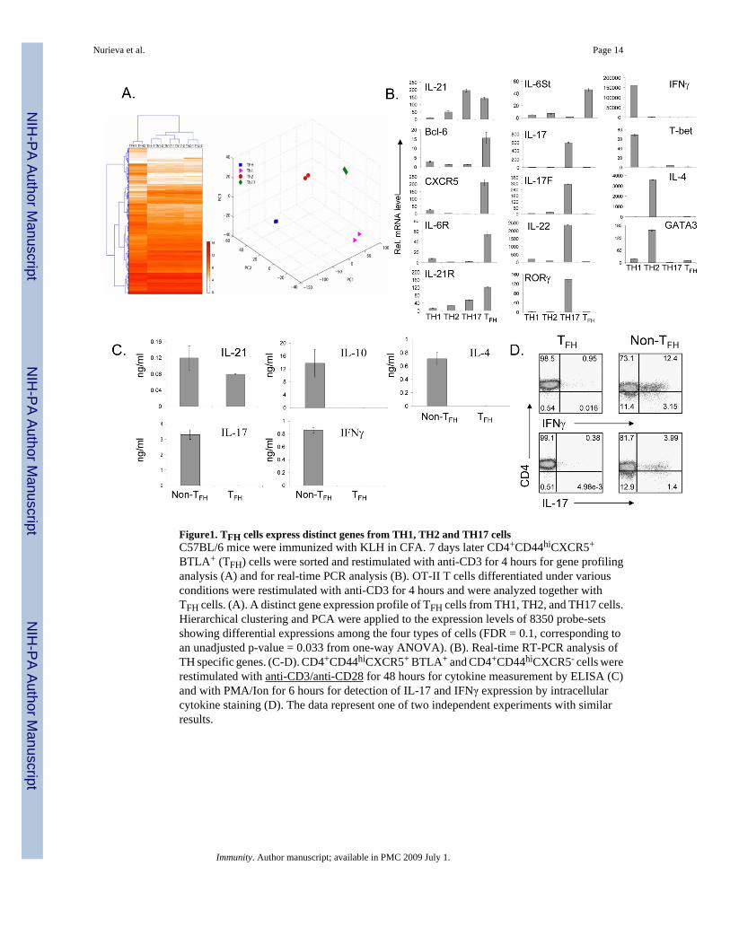
Figure1. TFH cells express distinct genes from TH1, TH2 and TH17 cellsC57BL/6 mice were immunized with KLH in CFA. 7 days later CD4+CD44hiCXCR5+
BTLA+ (TFH) cells were sorted and restimulated with anti-CD3 for 4 hours for gene profilinganalysis (A) and for real-time PCR analysis (B). OT-II T cells differentiated under variousconditions were restimulated with anti-CD3 for 4 hours and were analyzed together withTFH cells. (A). A distinct gene expression profile of TFH cells from TH1, TH2, and TH17 cells.Hierarchical clustering and PCA were applied to the expression levels of 8350 probe-setsshowing differential expressions among the four types of cells (FDR = 0.1, corresponding toan unadjusted p-value = 0.033 from one-way ANOVA). (B). Real-time RT-PCR analysis ofTH specific genes. (C-D). CD4+CD44hiCXCR5+ BTLA+ and CD4+CD44hiCXCR5- cells wererestimulated with anti-CD3/anti-CD28 for 48 hours for cytokine measurement by ELISA (C)and with PMA/Ion for 6 hours for detection of IL-17 and IFNγ expression by intracellularcytokine staining (D). The data represent one of two independent experiments with similarresults.
Nurieva et al. Page 14
Immunity. Author manuscript; available in PMC 2009 July 1.
NIH
-PA Author Manuscript
NIH
-PA Author Manuscript
NIH
-PA Author Manuscript
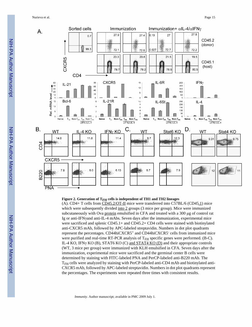
Figure 2. Generation of TFH cells is independent of TH1 and TH2 lineages(A). CD4+ T cells from CD45.2/OT-II mice were transferred into C57BL/6 (CD45.1) micewhich were subsequently divided into 2 groups (3 mice per group). Mice were immunizedsubcutaneously with Ova protein emulsified in CFA and treated with a 300 μg of control ratIg or anti-IFNγand anti-IL-4 mAbs. Seven days after the immunization, experimental micewere sacrificed and splenic CD45.1+ and CD45.2+ CD4 cells were stained with biotinylatedanti-CXCR5 mAb, followed by APC-labeled streptavidin. Numbers in dot plot quadrantsrepresent the percentages. CD44hiCXCR5+ and CD44hiCXCR5- cells from immunized micewere purified and real-time RT-PCR analysis of TFH specific genes were performed. (B-C).IL-4 KO, IFNγ KO (B), STAT6 KO (C) and STAT4 KO (D) and their appropriate controls(WT, 3 mice per group) were immunized with KLH emulsified in CFA. Seven days after theimmunization, experimental mice were sacrificed and the germinal center B cells weredetermined by staining with FITC-labeled PNA and PerCP-labeled anti-B220 mAb. TheTFH cells were analyzed by staining with PerCP-labeled anti-CD4 mAb and biotinylated anti-CXCR5 mAb, followed by APC-labeled streptavidin. Numbers in dot plot quadrants representthe percentages. The experiments were repeated three times with consistent results.
Nurieva et al. Page 15
Immunity. Author manuscript; available in PMC 2009 July 1.
NIH
-PA Author Manuscript
NIH
-PA Author Manuscript
NIH
-PA Author Manuscript
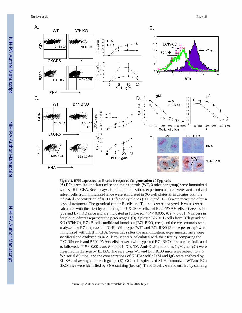
Figure 3. B7H expressed on B cells is required for generation of TFH cells(A) B7h germline knockout mice and their controls (WT, 3 mice per group) were immunizedwith KLH in CFA. Seven days after the immunization, experimental mice were sacrificed andspleen cells from immunized mice were stimulated in 96-well plates as triplicates with theindicated concentration of KLH. Effector cytokines (IFN-γ and IL-21) were measured after 4days of treatment. The germinal center B cells and TFH cells were analyzed. P values werecalculated with the t-test by comparing the CXCR5+ cells and B220/PNA+ cells between wild-type and B7h KO mice and are indicated as followed: * P < 0.005; #, P < 0.001. Numbers indot plot quadrants represent the percentages. (B). Splenic B220+ B cells from B7h germlineKO (B7hKO), B7h B-cell conditional knockout (B7h BKO, cre+) and the cre- controls wereanalyzed for B7h expression. (C-E). Wild-type (WT) and B7h BKO (3 mice per group) wereimmunized with KLH in CFA. Seven days after the immunization, experimental mice weresacrificed and analyzed as in A. P values were calculated with the t-test by comparing theCXCR5+ cells and B220/PNA+ cells between wild-type and B7h BKO mice and are indicatedas followed: ** P < 0.001; ##, P < 0.001. (C). (D). Anti-KLH antibodies (IgM and IgG) weremeasured in the sera by ELISA. The sera from WT and B7h BKO mice were subject to a 3-fold serial dilution, and the concentrations of KLH-specific IgM and IgG were analyzed byELISA and averaged for each group. (E). GC in the spleens of KLH-immunized WT and B7hBKO mice were identified by PNA staining (brown). T and B cells were identified by staining
Nurieva et al. Page 16
Immunity. Author manuscript; available in PMC 2009 July 1.
NIH
-PA Author Manuscript
NIH
-PA Author Manuscript
NIH
-PA Author Manuscript
with anti-CD4 (red) and anti-B220 (blue) Abs. The data represent at least three independentexperiments with consistent results.
Nurieva et al. Page 17
Immunity. Author manuscript; available in PMC 2009 July 1.
NIH
-PA Author Manuscript
NIH
-PA Author Manuscript
NIH
-PA Author Manuscript
Figure 4. IL-21 is necessary for TFH cell developmentIL-21+/+, IL-21+/- and IL-21-/- mice (3 mice per group) were immunized subcutaneously withKLH emulsified in CFA. Seven days after the immunization, experimental mice were sacrificedand TFH cells (A) and the germinal center B cells (B) were analyzed. Numbers in dot plotquadrants represent the percentages. Germinal centers were determined byimmunohistochemical analysis (C). Spleen cells from immunized mice were stimulated in 96-well plates as triplicates with the indicated concentration of KLH peptide. Proliferation wasassayed after 3 days of treatment by adding [3H]thymidine to the culture for the last 8 h. IFN-γ was measured after 4 days of treatment. The experiments were repeated twice with consistentresults.
Nurieva et al. Page 18
Immunity. Author manuscript; available in PMC 2009 July 1.
NIH
-PA Author Manuscript
NIH
-PA Author Manuscript
NIH
-PA Author Manuscript
Figure 5. Generation of TFH cells requires IL-6 and STAT3IL-6 KO (A) or STAT3 T-cell specific KO mice (C) and their appropriate controls (WT, 3 miceper group) were immunized subcutaneously with KLH emulsified in CFA. Seven days afterthe immunization, experimental mice were sacrificed and TFH cells and the germinal center Bcells were analyzed. Numbers in dot plot quadrants represent the percentages. (B) C57BL/6mice were immunized with KLH in CFA. 7 days later, CD4+CD44hiCXCR5+ andCD4+CD44hiCXCR5- cells were sorted and restimulated with anti-CD3 and anti-CD28 withor without IL-6 or IL-21 for 48 hours. Proliferation was assayed by adding [3H]-thymidine tothe culture for the last 8 h. P values were calculated with the t-test by comparing the CXCR5+ cells proliferation in the absence and in the presence of IL6 (*p<0.005) or in the absence andin the presence of IL21 (*p<0.005). CD4+CD44hiCXCR5+ and CD4+CD44hiCXCR5- cellswere restimulated with anti-CD3 and anti-CD28 for 24 hours and stained with Annexin V-FITC. The experiments were performed two times with consistent results.
Nurieva et al. Page 19
Immunity. Author manuscript; available in PMC 2009 July 1.
NIH
-PA Author Manuscript
NIH
-PA Author Manuscript
NIH
-PA Author Manuscript
Figure 6. Generation of TFH cells is independent of TH17 lineageRag1-/- reconstituted with WT and RORαsg/sg/RORγ KO bone marrow cells (A) or IL-17 KOand IL-17F KO mice and their controls (C) (3 mice per group) were immunized subcutaneouslywith KLH in CFA. Seven days after the immunization, experimental mice were sacrificed andthe TFH cells were determined. Numbers in dot plot quadrants represent the percentages. (B).C57BL/6 mice were immunized with KLH emulsified in CFA with 100 μg of isotype controlantibodies or anti-TGFβ blocking Abs (3 mice per group). 7 days later, experimental mice weresacrificed and splenic TFH cells and germinal center B cells were analyzed. Splenocytes wererestimulated with KLH for overnight and the production of IL-17 and IFNγ was analyzed inCD4+ gate by intracellular cytokine staining. The results represent one of three individualswith similar results.
Nurieva et al. Page 20
Immunity. Author manuscript; available in PMC 2009 July 1.
NIH
-PA Author Manuscript
NIH
-PA Author Manuscript
NIH
-PA Author Manuscript
Figure 7. IL-21, in the absence of IL-4, IFNγ and TGF-b signaling, generates TFH cellsA-F, FACS-sorted CD62hiCD44loCD25negCD4+ T cells from CD45.1+ OT-II mice werecultured with irradiated splenic APC plus OVA323-339 peptide under TH0, TFH (IL-21 plusantibodies to IL-4, IFNγ and TGFβ) or TH17 condition for 5 days. After 5 days, CD4 T cellswere restimulated with anti-CD3 for 4 hours for real-time PCR analysis (A) or for 24 hoursfor cytokine measurement by ELISA (B). (C-F). Five days after in vitro differentiation, cellswere adoptively transferred into CD45.2+ congenic mice (n=3-4) before the recipient micewere subcutaneously immunized with OVA in IFA. A group of mice that did not receive Tcells was used as a control (No transfer). Seven days after immunzation, lymphoid cells fromthe draining lymph nodes of recipient mice were isolated and TFH cells and germinal center Bcells were analyzed (D). Numbers in the boxes represent the percentages. (E). The sera fromthe recipient mice were subject to a 3-fold serial dilution, and the concentrations of OVA-specific IgM and IgG were analyzed by ELISA and averaged for each group. (F). Germinalcenter in the spleens of the recipient mice were identified by PNA staining (brown). The resultsare a representative of multiple mice of two independent experiments with similar results. (G).CXCR5+CD44hi cells were sorted from CD45.1 mice on day 7 after immunization with KLHand transferred to CD45.2 (n=3) recipient mice following immunization with KLH in CFA. Agroup of mice that did not receive T cells was used as a control (No transfer). Seven days afterimmunization, germinal center in the spleens of the recipient mice were identified by PNAstaining (brown).
Nurieva et al. Page 21
Immunity. Author manuscript; available in PMC 2009 July 1.