33
Grand Rounds Presentation SUNY Downstate Medical Center 10.23.14 Nora Silverman MD, PhD
| Date post: | 24-Feb-2023 |
| Category: |
Documents |
| Upload: | khangminh22 |
| View: | 0 times |
| Download: | 0 times |
Grand Rounds Presentation SUNY Downstate Medical Center
10.23.14
Nora Silverman MD, PhD
Patient Presentation • HPI: 51 BF with POH ‘poor vision’ in both eyes since childhood. Patient
recently diagnosed Glc (s) and presents for review of OCT ON. No acute
changes in vision and no new ocular complaints . ROS (-‐)
• PMH: none
• Meds: none
• Gtts: none
• PSH: none
• FH: (-‐) for glc or blindness
• SH: (-‐) x 3
Exam
• dVAsc: 20/40 PH to 20/30 OD; 20/50-‐2 PH to 20/25-‐2
• Mrx: + 2.0 -‐2.5 x 150 to 20/30 OD; +2.0, -‐1.75 x 20 to
20/20 OS
• Pupils: 4-‐2 no APD
• EOMS full
• CVF full • Tapp: 10/10 at 5:30 pm
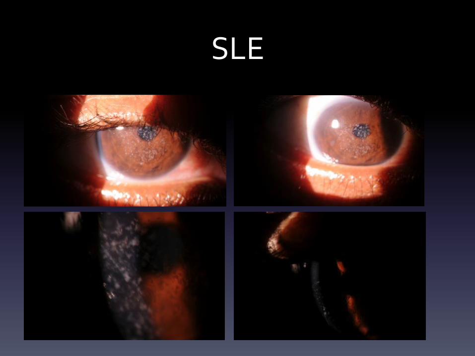
SLE
Exam, cont’d • SLE:
– LLA: MGD OU
– C/S: White and Quiet OU
– K: Centrally-‐located bilateral infiltrates within the stroma **Present since childhood
– AC: Deep and Quiet OU
– Iris/Pupil: Flat, Round and Reactive OU
– Lens: trace-‐1+ ns OU
• DFE:
– Vitreous: clear OU
– C/D: 0.75 s/p with thinning inferiorly OU
– Mac: flat OU
– V/P: Vessels WNL, no heme, holes or tears
Differential Diagnosis
• Granular Dystrophy
• Macular Dystrophy
• Lattice Dystrophy
• Schnyder Corneal Dystrophy
• Salzman’s Nodular Corneal Degeneration
• Infections-‐ Strep viridans, Haemophilus, Enterococcus
• Staph Hypersensitivity
• DIAGNOSIS: Granular Dystrophy
Dystrophy vs Degeneration
• Corneal Dystrophy vs Corneal Degeneration
– Dystrophies:
• Genetic (usually Aut Dom) with onset in childhood/early adulthood
• Not associated with systemic disease
• Bilateral
• Centrally located within the cornea
• Typically involve only one layer of the cornea
– Degenerations:
• Progressive (onset typically after age 40)
• Usually unrelated to family history or genetic predisposition
• Commonly associated with systemic disease (rheumatologic, infectious)
• Usually unilateral, asymmetric if bilateral, and peripherally located on the cornea
• Can involve one or multiple layers of the cornea
• Often associated with neovascularization
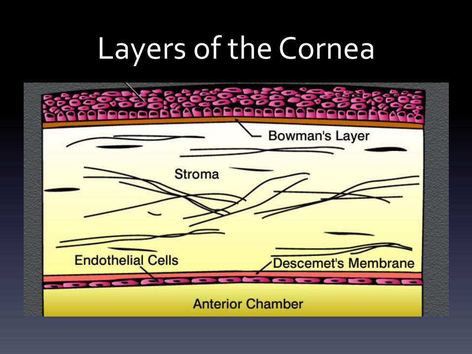
Layers of the Cornea
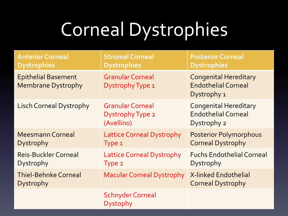
Corneal Dystrophies Anterior Corneal Dystrophies
Stromal Corneal Dystrophies
Posterior Corneal Dystrophies
Epithelial Basement Membrane Dystrophy
Granular Corneal Dystrophy Type 1
Congenital Hereditary Endothelial Corneal Dystrophy 1
Lisch Corneal Dystrophy Granular Corneal Dystrophy Type 2 (Avellino)
Congenital Hereditary Endothelial Corneal Dystrophy 2
Meesmann Corneal Dystrophy
Lattice Corneal Dystrophy Type 1
Posterior Polymorphous Corneal Dystrophy
Reis-‐Buckler Corneal Dystrophy
Lattice Corneal Dystrophy Type 2
Fuchs Endothelial Corneal Dystrophy
Thiel-‐Behnke Corneal Dystrophy
Macular Corneal Dystrophy
X-‐linked Endothelial Corneal Dystrophy
Schnyder Corneal Dystophy
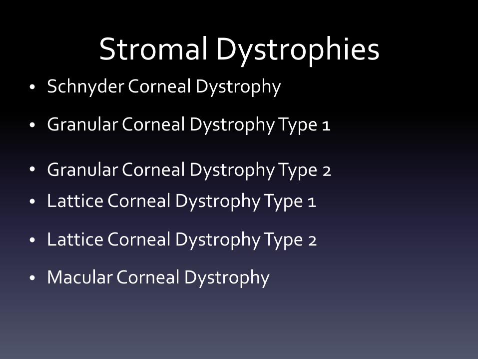
Stromal Dystrophies • Schnyder Corneal Dystrophy
• Granular Corneal Dystrophy Type 1
• Granular Corneal Dystrophy Type 2 • Lattice Corneal Dystrophy Type 1
• Lattice Corneal Dystrophy Type 2
• Macular Corneal Dystrophy
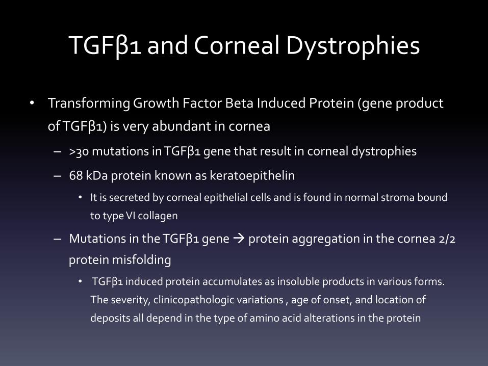
TGFβ1 and Corneal Dystrophies
• Transforming Growth Factor Beta Induced Protein (gene product
of TGFβ1) is very abundant in cornea
– >30 mutations in TGFβ1 gene that result in corneal dystrophies
– 68 kDa protein known as keratoepithelin
• It is secreted by corneal epithelial cells and is found in normal stroma bound
to type VI collagen
– Mutations in the TGFβ1 gene à protein aggregation in the cornea 2/2
protein misfolding
• TGFβ1 induced protein accumulates as insoluble products in various forms.
The severity, clinicopathologic variations , age of onset, and location of
deposits all depend in the type of amino acid alterations in the protein
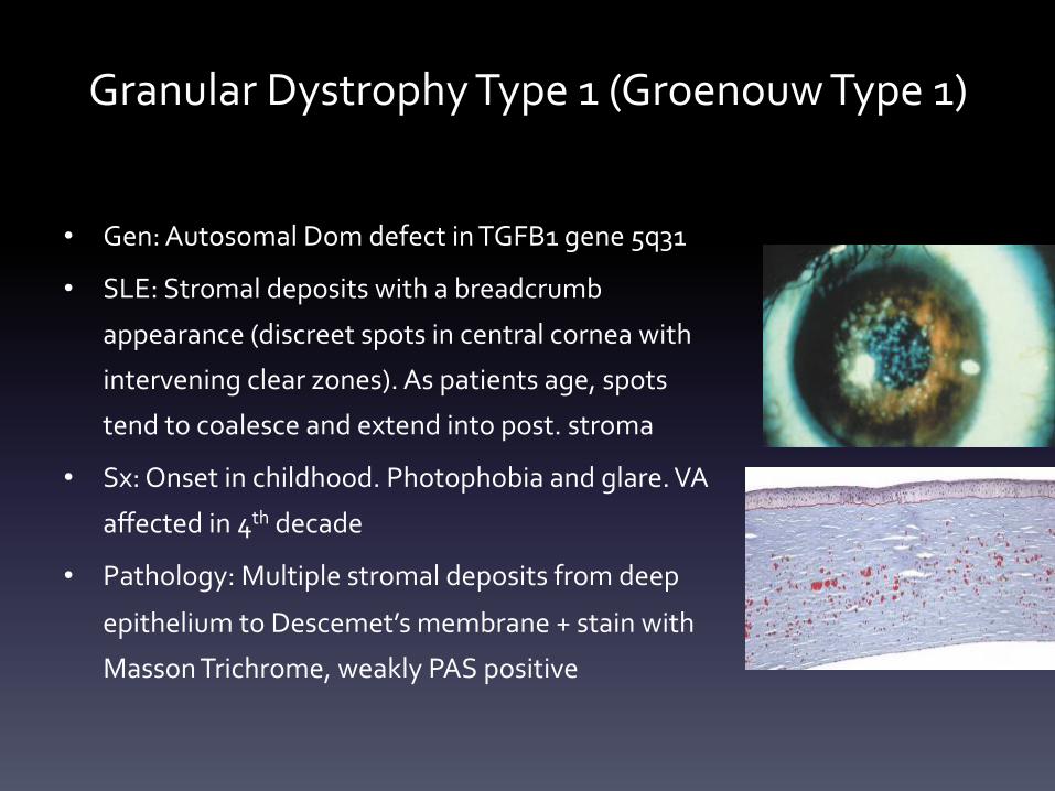
Granular Dystrophy Type 1 (Groenouw Type 1)
• Gen: Autosomal Dom defect in TGFB1 gene 5q31
• SLE: Stromal deposits with a breadcrumb
appearance (discreet spots in central cornea with
intervening clear zones). As patients age, spots
tend to coalesce and extend into post. stroma
• Sx: Onset in childhood. Photophobia and glare. VA
affected in 4th decade
• Pathology: Multiple stromal deposits from deep
epithelium to Descemet’s membrane + stain with
Masson Trichrome, weakly PAS positive
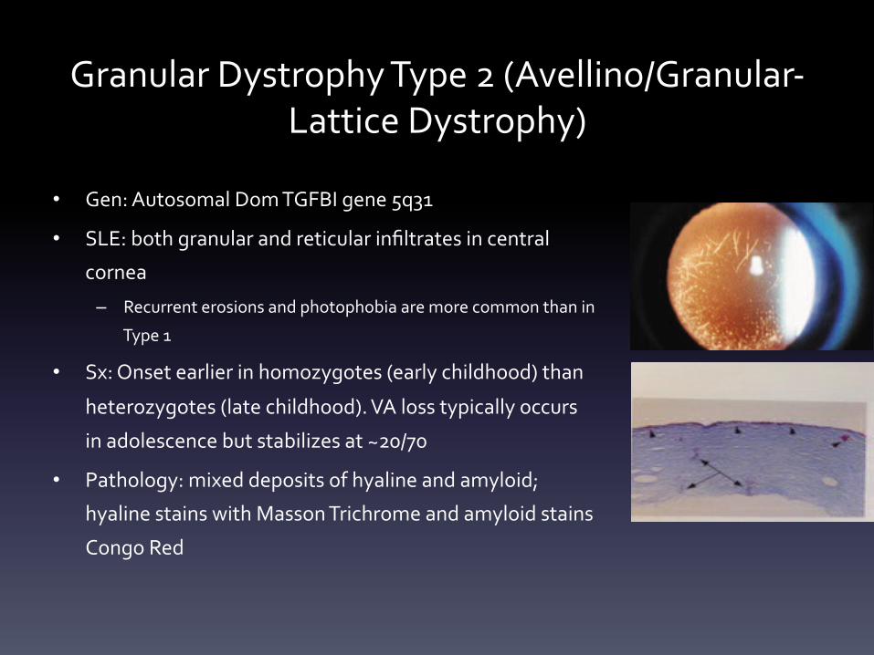
Granular Dystrophy Type 2 (Avellino/Granular-‐Lattice Dystrophy)
• Gen: Autosomal Dom TGFBI gene 5q31
• SLE: both granular and reticular infiltrates in central
cornea
– Recurrent erosions and photophobia are more common than in
Type 1
• Sx: Onset earlier in homozygotes (early childhood) than
heterozygotes (late childhood). VA loss typically occurs
in adolescence but stabilizes at ~20/70
• Pathology: mixed deposits of hyaline and amyloid;
hyaline stains with Masson Trichrome and amyloid stains
Congo Red
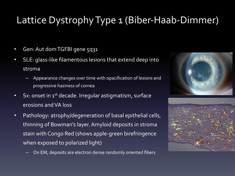
• Gen: Aut dom TGFBI gene 5q31
• SLE: glass-‐like filamentous lesions that extend deep into
stroma
– Appearance changes over time with opacification of lesions and
progressive haziness of cornea
• Sx: onset in 1st decade. Irregular astigmatism, surface
erosions and VA loss
• Pathology: atrophy/degeneration of basal epithelial cells,
thinning of Bowman’s layer. Amyloid deposits in stroma
stain with Congo Red (shows apple-‐green birefringence
when exposed to polarized light)
– On EM, deposits are electron dense randomly oriented fibers
Lattice Dystrophy Type 1 (Biber-‐Haab-‐Dimmer)
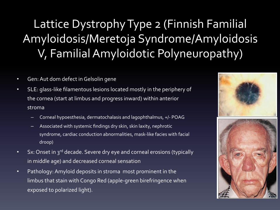
Lattice Dystrophy Type 2 (Finnish Familial Amyloidosis/Meretoja Syndrome/Amyloidosis
V, Familial Amyloidotic Polyneuropathy)
• Gen: Aut dom defect in Gelsolin gene
• SLE: glass-‐like filamentous lesions located mostly in the periphery of
the cornea (start at limbus and progress inward) within anterior
stroma
– Corneal hypoesthesia, dermatochalasis and lagophthalmus, +/-‐ POAG
– Associated with systemic findings dry skin, skin laxity, nephrotic
syndrome, cardiac conduction abnormalities, mask-‐like facies with facial
droop)
• Sx: Onset in 3rd decade. Severe dry eye and corneal erosions (typically
in middle age) and decreased corneal sensation
• Pathology: Amyloid deposits in stroma most prominent in the
limbus that stain with Congo Red (apple-‐green birefringence when
exposed to polarized light).
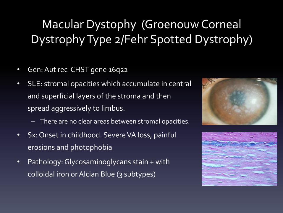
• Gen: Aut rec CHST gene 16q22
• SLE: stromal opacities which accumulate in central
and superficial layers of the stroma and then
spread aggressively to limbus.
– There are no clear areas between stromal opacities.
• Sx: Onset in childhood. Severe VA loss, painful
erosions and photophobia
• Pathology: Glycosaminoglycans stain + with
colloidal iron or Alcian Blue (3 subtypes)
Macular Dystophy (Groenouw Corneal Dystrophy Type 2/Fehr Spotted Dystrophy)
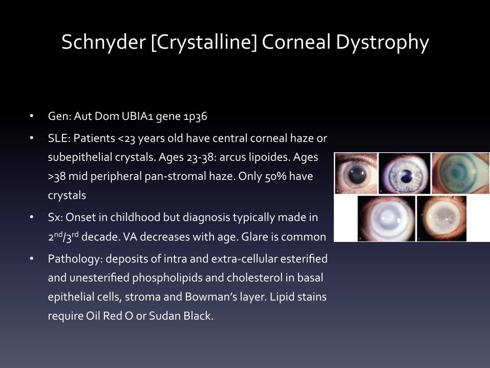
Schnyder [Crystalline] Corneal Dystrophy
• Gen: Aut Dom UBIA1 gene 1p36
• SLE: Patients <23 years old have central corneal haze or
subepithelial crystals. Ages 23-‐38: arcus lipoides. Ages
>38 mid peripheral pan-‐stromal haze. Only 50% have
crystals
• Sx: Onset in childhood but diagnosis typically made in
2nd/3rd decade. VA decreases with age. Glare is common
• Pathology: deposits of intra and extra-‐cellular esterified
and unesterified phospholipids and cholesterol in basal
epithelial cells, stroma and Bowman’s layer. Lipid stains
require Oil Red O or Sudan Black.
Epidemiology
• Men and women affected equally (except
Fuchs, which affects women: men 4:1)
• Depending on subtype, presentation ranges from early childhood to mid-‐adulthood
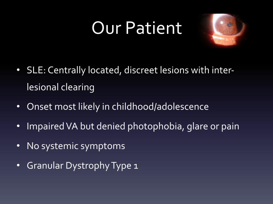
Our Patient
• SLE: Centrally located, discreet lesions with inter-‐
lesional clearing
• Onset most likely in childhood/adolescence
• Impaired VA but denied photophobia, glare or pain
• No systemic symptoms
• Granular Dystrophy Type 1
Management of Corneal Dystrophies
• **Depends on the patient’s symptoms
• Initial tx: non-‐surgical – Lubricating drops, Restasis, or autologous serum therapy
– Punctal plugs
– Bandage Contact Lenses +/-‐ antibiotics for recurrent erosions
– Muro 128
Surgical Management
• PTK: Phototherapeutic Keratectomy
– Excimer laser removes superficial opacities (recurrent epithelial
erosion syndrome), smoothes the corneal surface and allows
epithelium to re-‐adhere more tightly
– Smoother stromal surface improves post-‐operative corneal
clarity and decreases scarring
• Deep opacities that cause significant visual disturbances may require corneal transplantation
– Lamellar or full thickness

Corneal Transplant
• Partial Thickness CT: ALK, DALK
– ALK (Automated Lamellar Keratoplasty): endothelium is retained: risk of rejection may be decreased, and
healing may be faster
• Best for keratoconus and partial thickness corneal scars
– DALK (Deep Anterior Lamellar Keratoplasty): epithelium and stroma are removed leaving the endothelium
and Descemet’s membrane left intact
• Full Thickness Corneal Transplantation (Penetrating Keratoplasty) – PKP
– Most frequently performed corneal transplant – entire cornea removed and replaced with a donor cornea
• Intralase Enabled Keratoplasty (IEK) **new
– Both full thickness (PK) and Anterior Lamellar Transplants (ALK and DALK) can be assisted with intralase
femtosecond laser
• Rather than preparing the corneal transplant graft with traditional trephine, both the patient and recipient corneas are
fashioned with the laser
– Benefit: personalized edge shapes which may induce stronger healing of the transplant and thus faster recovery of vision

Tailored Surgical Options • Granular Corneal Dystrophies: because visual impairment is typically not severe,
corneal grafting does not need to be done until the disease is advanced
– PKP +/-‐ phototherapeutic keratectomy is successful usually for 30 months. Recurrence is
common in the superficial portions of the graft
– LASIK is contraindicated in GD type 2
• Lattice Corneal Dystrophies: PKP has high success rate
• Macular Corneal Dystrophy: because entire stroma is affected, lamellar kp is
insufficient, and PKP is more successful
• Schnyder Corneal Dystrophy: phototherapeutic keratectomy for sub-‐epithelial
crystals and PKP for pan-‐stromal involvement.
Data on Recurrence of CDs • Recurrence of Granular Dystrophy tends to be superficial (usually
centrally located and epithelial)
– Time of recurrence is 13-‐73 months following surgical intervention and there
has not been a consistently apparent sig diff between PK and LK
• Patanelli et al (2014): case study
– 28 M Granular Corneal Dystrophy Type 1 who underwent DALK
– Recurrence 3 yrs post-‐op : full-‐thickness PKP was performed and cornea was
sent for pathology
– Recurrence of granular deposits was entirely within the stroma (as opposed to
anterior/epithelial)
– ** host keratocytes are a source of recurrence
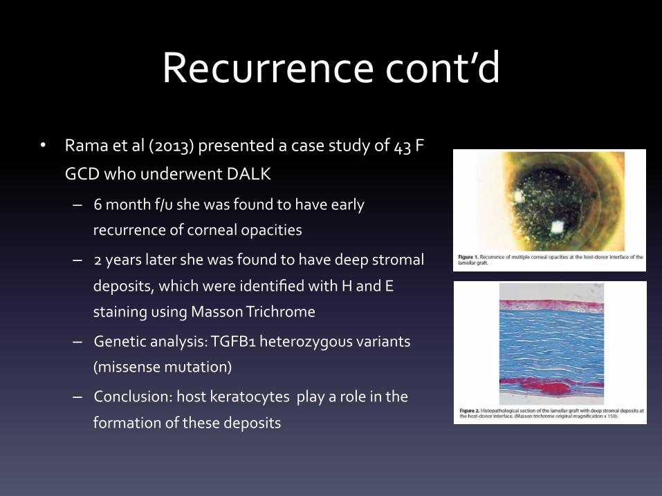
Recurrence cont’d
• Rama et al (2013) presented a case study of 43 F
GCD who underwent DALK
– 6 month f/u she was found to have early
recurrence of corneal opacities
– 2 years later she was found to have deep stromal
deposits, which were identified with H and E
staining using Masson Trichrome
– Genetic analysis: TGFB1 heterozygous variants (missense mutation)
– Conclusion: host keratocytes play a role in the formation of these deposits
Recurrence cont’d • Cheng et al (2013) compared the therapeutic effects of PKP and
DALK on patients with Macular Corneal Dystrophy (retrospective,
comparative study of 78 eyes)
• Results: best corrected VA was much better in PKP group
compared to DALK group at 1,2,3 and5 years
• Incidence of complications at 1 yr: 21% PKP and 4.8% DALK
• Rate of recurrence in PK group was 17.5%and 42.9% in DALK
group (5x higher than PK)
• Selection of PK vs DALK should be patient-‐tailored
Are Preservatives Precipitants in some Corneal Dystrophies?
• Formation of amyloid fibrils is accelerated by surfactants such as SDS (sodium
dodecyl sulfate)
• Most eye drops contain BAC, which is a cationic surfactant
• Kato et al (2013) used 3 types of synthetic peptides containing different amino
acid varieties of the keratoepithelin sequence
• The time course of spontaneous amyloid fibrillation and seed dependent fibril
elongation were monitored in the presence of various concentrations of BAC or
SDS
• Results indicates that both BAC and SDS accelerated fibrillation of all synthetic
peptides
• Eye drops containing BAC may deteriorate corneal dystrophies
What happened to Our Patient?
• Best corrected dVAcc 20/30 and 20/20. The patient was started on AT for symptomatic
relief
• Surgical options were discussed as a potential
treatment in the future if VA becomes
significantly impaired
Reflective Practice • This case demonstrated the importance of a thorough
ophthalmic exam and diagnostic workup and allowed
me to learn more about a rare disease entity and its
complications.
• This case also allowed me to evaluate the literature for
the differential diagnoses of this disease entity while
keeping in my mind my patient’s expectations
Core Competencies
• Patient Care: The case involved thorough patient care and careful attention to the patient’s past
medical history. Once diagnosed the patient received proper management and follow up care.
• Medical Knowledge: This presentation allowed me to review the presentation, differential
diagnosis, proper evaluation/workup and different treatment options for corneal dystrophies
• Practice-‐Based Learning and Improvement: This presentation included a current literature search
of current studies in the roles of TGFbeta genes and corneal dystrophies
• Interpersonal and Communication Skills: The patient was treated with respect and every effort was
made to communicate with the patient in a timely manner.
• Professionalism: The patient was diagnosed in a timely manner. She was informed of her diagnosis
and explained current treatment options.
• Systems Based Practice: The patient was discussed with several cornea specialists in order to
facilitate proper management
References
• http://www.clspectrum.com/articleviewer.aspx?articleID=12400
• http://www.cornealdystrophyfoundation.org/html/cd_defined.html
• http://eyewiki.aao.org/Corneal_Stromal_Dystrophies
• http://www.corneasociety.org/sites/default/files/publications/ic3d_class_cornealdystrophies.pdf
• http://www.vision-‐institute.com/new-‐jersey/vision-‐disorders/ptk.htm
• http://www.vision-‐institute.com/new-‐jersey/vision-‐disorders/cornea-‐transplant.htm
• http://www.mayoclinic.org/tests-‐procedures/cornea-‐transplant/basics/risks/prc-‐20014357
• Recurrence of Granular Corneal Dystrophy Type 1 Deposits within Host Stroma After Non-‐Descemet Baring
Anterior Lamellar Keratoplasty. Patanelli SM, Herzlich A, Yeaney G, Ching ST. Cornea 2014 Epub.
• Clinical and Genetic Aspects of the TGFB1-‐associated Corneal Dystrophies. Lakshminaryan R et al. Ocular Sur
2014 Oct; 12 (4) 234-‐251
• Mutation in Transforming Growth Factor Beta Induced Protein Associated with Granular Corneal Dystrophy Type 1
Reduces the Proteolytic Susceptibility through Local Structural Stabilization . Underhaug et al. Biochimica et
Biophysica Acta 2013. Dec; 1834 (12):2812-‐22.
References cont’d • Benzalkonium Chloride Accelerates the Formation of the Amyloid Fibrils of Corneal Dystrophy-‐
associated Peptides. Kato et al, The Journal of Biological Chemistry. 2013 Aug; 288 (35):25109-‐18.
• Recurrence of Granular Corneal Dystrophy Type 1 Deposits within Host Stroma After Non-‐
Descemet Baring Anterior Lamellar Keratoplasty. Patanelli SM, Herzlich A, Yeaney G, Ching ST.
Cornea 2014 Epub.
• Clinical and Genetic Aspects of the TGFB1-‐associated Corneal Dystrophies. Lakshminaryan R et al.
Ocular Sur 2014 Oct; 12 (4) 234-‐251
• Mutation in Transforming Growth Factor Beta Induced Protein Associated with Granular Corneal
Dystrophy Type 1 Reduces the Proteolytic Susceptibility through Local Structural Stabilization .
Underhaug et al. Biochimica et Biophysica Acta 2013. Dec; 1834 (12):2812-‐22.
• Unusual Early Recurrence of Granular Dystrophy After Deep Anterior Lamellar Keratoplasty: case
report. Rama et al. Arq Bras Ofthalmol. 2013 Mar-‐Apr; 76(2): 126-‐8
• The IC3D Classification of the Corneal Dystrophies. Cornea, Dec 2008; 27 (Suppl2): S1-‐83
Thank You
• Patient
• Dr Allison Rizzuti
• Dr Kichiemon Asoma


