Helper T Cell Epitope-Mapping Reveals MHC-Peptide Binding Affinities That Correlate with T Helper Cell Responses to Pneumococcal Surface Protein A Rajesh Singh 1 , Shailesh Singh 2 , Praveen K. Sharma 2 , Udai P. Singh 3 , David E. Briles 4 , Susan K. Hollingshead 4 , James W. Lillard, Jr. 1 * 1 Department of Microbiology, Biochemistry, and Immunology, Morehouse School of Medicine, Atlanta, Georgia, United States of America, 2 Department of Microbiology and Immunology, University of Louisville School of Medicine, Louisville, Kentucky, United States of America, 3 Department of Pathology, Microbiology and Immunology, University of South Carolina School of Medicine, Columbia, South Carolina, United States of America, 4 Department of Microbiology, University of Alabama at Birmingham School of Medicine, Birmingham, Alabama, United States of America Abstract Understanding the requirements for protection against pneumococcal carriage and pneumonia will greatly benefit efforts in controlling these diseases. Several proteins and polysaccharide capsule have recently been implicated in the virulence of and protective immunity against Streptococcus pneumonia. Pneumococcal surface protein A (PspA) is highly conserved among S. pneumonia strains, inhibits complement activation, binds lactoferrin, elicits protective systemic immunity against pneumococcal infection, and is necessary for full pneumococcal virulence. Identification of PspA peptides that optimally bind human leukocyte antigen (HLA) would greatly contribute to global vaccine efforts, but this is hindered by the multitude of HLA polymorphisms. Here, we have used an experimental data set of 54 PspA peptides and in silico methods to predict peptide binding to HLA and murine major histocompatibility complex (MHC) class II. We also characterized spleen- and cervical lymph node (CLN)-derived helper T lymphocyte (HTL) cytokine responses to these peptides after S. pneumonia strain EF3030-challenge in mice. Individual, yet overlapping peptides, 15 amino acids in length revealed residues 199 to 246 of PspA (PspA 199–246 ) consistently caused the greatest IFN-c, IL-2, IL-5 and proliferation as well as moderate IL-10 and IL-4 responses by ex vivo stimulated splenic and CLN CD4 + T cells isolated from S. pneumonia strain EF3030-challeged F 1 (B6 6 BALB/c) mice. IEDB, RANKPEP, SVMHC, MHCPred, and SYFPEITHI in silico analysis tools revealed peptides in PspA 199–246 also interact with a broad range of HLA-DR, -DQ, and -DP allelles. These data suggest that predicted MHC class II-peptide binding affinities do not always correlate with T helper (Th) cytokine or proliferative responses to PspA peptides, but when used together with in vivo validation can be a useful tool to choose candidate pneumococcal HTL epitopes. Citation: Singh R, Singh S, Sharma PK, Singh UP, Briles DE, et al. (2010) Helper T Cell Epitope-Mapping Reveals MHC-Peptide Binding Affinities That Correlate with T Helper Cell Responses to Pneumococcal Surface Protein A. PLoS ONE 5(2): e9432. doi:10.1371/journal.pone.0009432 Editor: Jo ¨ rg Hermann Fritz, University of Toronto, Canada Received September 10, 2009; Accepted February 2, 2010; Published February 25, 2010 Copyright: ß 2010 Singh et al. This is an open-access article distributed under the terms of the Creative Commons Attribution License, which permits unrestricted use, distribution, and reproduction in any medium, provided the original author and source are credited. Funding: This study was supported by funds from the National Institutes of Health Grants AI057808, GM09248, MD00525, and RR03034. The funders had no role in study design, data collection and analysis, decision to publish, or preparation of the manuscript. Competing Interests: The authors have declared that no competing interests exist. * E-mail: [email protected]Introduction Pneumococcal pneumonia is the most common cause of childhood deaths in the developing world and among the top ten causes of death in aged populations worldwide; recently, antibiotic-resistant S. pneumonia strains have emerged [1,2,3,4]. Hence, vaccines against these strains are greatly needed. This study characterizes the HTL epitopes of a candidate pneumococ- cal vaccine antigen, PspA, which is a highly conserved, cell wall- associated surface protein that plays a major role in pneumococcal virulence by binding human lactoferrin and interferes with com- plement deposition on the bacterial surface [5]. During the course of invasive disease, antibodies against PspA peak during the convalescent phase, but CD4 + T cell help is required for optimal protective immune responses to PspA [6,7]. A central event in the adaptive immune response to invasive microorganisms is the specific recognition of processed antigens bound to the peptide-binding region of MHC class II molecules on the surface of antigen-presenting cells. These peptide antigens are subsequently detected by the T cell receptor (TCR) of CD4 + T cells, which proliferate, secrete cytokines, and differentiate into antigen-specific Th effector cells. To induce protective immunity, HTL epitopes contained in synthetic peptide vaccines must: (i) match those naturally presented to the immune system during infection, (ii) be recognized by the majority of the human popu- lation, and (iii) induce an appropriate effector immune response to eliminate the pathogen of interest. Single epitope-based vaccines may, however, have drawbacks. For example, the mono-specificity of the induced immune response might miss the emergence of sequence mutants that would potentially escape the vaccine’s protective effect [8]. It is also unlikely that T cells from genetically distinct populations would recognize, and respond to a single peptide epitope. These obstacles are secondary to the wide-ranging polymor- phisms of HLA molecules that present antigenic peptides to T cells. Indeed, a unique set of epitopes from a given protein antigen will be presented to T cells of an individual bearing hundreds of unique HLA molecules. Additionally, some HLA molecules may PLoS ONE | www.plosone.org 1 February 2010 | Volume 5 | Issue 2 | e9432
Transcript
Helper T Cell Epitope-Mapping Reveals MHC-PeptideBinding Affinities That Correlate with T Helper CellResponses to Pneumococcal Surface Protein ARajesh Singh1, Shailesh Singh2, Praveen K. Sharma2, Udai P. Singh3, David E. Briles4, Susan K.
Hollingshead4, James W. Lillard, Jr.1*
1 Department of Microbiology, Biochemistry, and Immunology, Morehouse School of Medicine, Atlanta, Georgia, United States of America, 2 Department of Microbiology
and Immunology, University of Louisville School of Medicine, Louisville, Kentucky, United States of America, 3 Department of Pathology, Microbiology and Immunology,
University of South Carolina School of Medicine, Columbia, South Carolina, United States of America, 4 Department of Microbiology, University of Alabama at Birmingham
School of Medicine, Birmingham, Alabama, United States of America
Abstract
Understanding the requirements for protection against pneumococcal carriage and pneumonia will greatly benefit efforts incontrolling these diseases. Several proteins and polysaccharide capsule have recently been implicated in the virulence ofand protective immunity against Streptococcus pneumonia. Pneumococcal surface protein A (PspA) is highly conservedamong S. pneumonia strains, inhibits complement activation, binds lactoferrin, elicits protective systemic immunity againstpneumococcal infection, and is necessary for full pneumococcal virulence. Identification of PspA peptides that optimallybind human leukocyte antigen (HLA) would greatly contribute to global vaccine efforts, but this is hindered by themultitude of HLA polymorphisms. Here, we have used an experimental data set of 54 PspA peptides and in silico methods topredict peptide binding to HLA and murine major histocompatibility complex (MHC) class II. We also characterized spleen-and cervical lymph node (CLN)-derived helper T lymphocyte (HTL) cytokine responses to these peptides after S. pneumoniastrain EF3030-challenge in mice. Individual, yet overlapping peptides, 15 amino acids in length revealed residues 199 to 246of PspA (PspA199–246) consistently caused the greatest IFN-c, IL-2, IL-5 and proliferation as well as moderate IL-10 and IL-4responses by ex vivo stimulated splenic and CLN CD4+ T cells isolated from S. pneumonia strain EF3030-challeged F1
(B66BALB/c) mice. IEDB, RANKPEP, SVMHC, MHCPred, and SYFPEITHI in silico analysis tools revealed peptides in PspA199–246
also interact with a broad range of HLA-DR, -DQ, and -DP allelles. These data suggest that predicted MHC class II-peptidebinding affinities do not always correlate with T helper (Th) cytokine or proliferative responses to PspA peptides, but whenused together with in vivo validation can be a useful tool to choose candidate pneumococcal HTL epitopes.
Citation: Singh R, Singh S, Sharma PK, Singh UP, Briles DE, et al. (2010) Helper T Cell Epitope-Mapping Reveals MHC-Peptide Binding Affinities That Correlate withT Helper Cell Responses to Pneumococcal Surface Protein A. PLoS ONE 5(2): e9432. doi:10.1371/journal.pone.0009432
Editor: Jorg Hermann Fritz, University of Toronto, Canada
Received September 10, 2009; Accepted February 2, 2010; Published February 25, 2010
Copyright: � 2010 Singh et al. This is an open-access article distributed under the terms of the Creative Commons Attribution License, which permitsunrestricted use, distribution, and reproduction in any medium, provided the original author and source are credited.
Funding: This study was supported by funds from the National Institutes of Health Grants AI057808, GM09248, MD00525, and RR03034. The funders had no rolein study design, data collection and analysis, decision to publish, or preparation of the manuscript.
Competing Interests: The authors have declared that no competing interests exist.
proliferation and IL-10 responses (CLN..spleen), IFN-c, IL-2
and IL-4 secretion (spleen..CLN) and IL-5 expression
(spleen#CLN) largely in response to PspA peptides 19, 20, 21,
and 22. Moreover, PspA peptides 21 and 22 mounted compar-
atively high proliferation responses, 20 and 21 induced consistently
high IFN-c and IL-2 responses, and 19, 20, and 21 caused IL-10,
IL-4 and IL-5 responses by HTLs isolated from Pneumococci-
exposed mice.
Predicted PspA Peptide-MHC Class II Alleles BindingAffinities and Correlation with Proliferation and CytokineSecretion Responses
PspA peptides 19, 20, 21, and 22 mounted significant HTL
responses, and displayed strong predictive binding affinities to
numerous HLA-DR, -DQ, and -DP as well as I-Ab and I-Ed
haplotypes. This is best illustrated by viewing a 3-dimensional plot of
the proliferation index as well as IFN-c, IL-10, IL-2, IL-4, and/or IL-
5 responses compared with MHC allele binding affinities (Figures 8
and 9). PspA peptide-specific T cell proliferation and IFN-c, IL-10,
IL-2, IL-4 and IL-5 secretion by CLN and splenic CD4+ T cells from
S. pneumonia strain EF3030-challenged mice was higher than the naıve
group. In general, CLN HTLs from mice previously challenged with
S. pneumonia strain EF3030) secreted high levels of IFN-c, IL-2, IL-4,
IL-5 and IL-10 as well as enhanced proliferation in response to PspA
peptides (19, 20.21, 22) stimulation.
PspA peptides 19, 20, 21, and 22 were predicted to bind I-Ab/I-
Ad, I-Ab/I-Eb, I-Ab and I-Ab/I-Ad, respectively, with
IC50,500 nM. From these, PspA peptide 20 was predicted to
have marginal binding affinities to I-Ab and I-Eb with IC50 = 485
and 493 nM, respectively. This also corresponded with relatively
high IL-10 responsiveness. Spleen-derived CD4+ T cells secreted
significant amounts of IFN-c, IL-2, IL-4 and IL-5 as well as
proliferated in response to PspA peptides 19, 20, 21, and 22 (i.e.,
PspA199–246) stimulation from mice previously challenged with S.
pneumonia strain EF3030. Peptide 20 or 23 stimulation of splenic
HTLs resulted in comparatively high secretion of IL-10. Similar to
PspA peptide 20, peptide 23 was predicted to have moderate I-Ab
and I-Eb binding affinity i.e., IC50 = 452 and 412 nM, respectively.
Peptides that induced spleen-derived CD4+ T cells to secrete high
levels of Th1 (IFN-c/IL-2) and Th2 (IL-4/IL-5) cytokines also
correlated with relatively high MHC binding affinities. It is
important to note that several PspA peptides predicted to tightly
bind I-A and/or I-E alleles did not always correspond with
elevated cytokine secretion (e.g., peptides 6, 18, 30, and 53).
Table 1. Overlapping PspA peptides and antigenic region description.
Peptide Antigenic epitope region Peptide Antigenic epitope region
01-MNKKKMILTSLASVA Leader 28-TIAAKKAELEKTEAD Region B
02-ASVAILGAGFVASQP Leader 29-TEADLKKAVNEPEKP Region B
03-ASQPTVVRAEESPVA Leader/Region A 30-PEKPAPAPETPAPEA Region B/C
04-SPVASQSKAEKDYDA Region A 31-APEAPAEQPKPAPAP Region C
05-DYDAAKKDAKNAKKA Region A 32-APAPQPAPAPKPEKP Region C
06-AKKAVEDAQKALDDA Region A 33-PEKPAEQPKPEKTDD Region C
07-LDDAKAAQKKYDEDQ Region A 34-KTDDQQAEEDYARRS Region C
08-DEDQKKTEEKAALEK Region A 35-ARRSEEEYNRLTQQQ Region C
09-ALEKAASEEMDKAVA Region A 36-TQQQPPKAEKPAPAP Region C
10-KAVAAVQQAYLAYQQ Region A 37-APAPKTGWKQENGMW Region C
11-AYQQATDKAAKDAAD Region A 38-NGMWYFYNTDGSMAT Region C
12-DAADKMIDEAKKREE Region A* 39-SMATGWLQNNGSWYY Region C
13-KREEEAKTKFNTVRA Region A* 40-SWYYLNSNGAMATGW Region C
14-TVRAMVVPEPEQLAE Region A* 41-ATGWLQYNGSWYYLN Region C
15-QLAETKKKSEEAKQK Region A* 42-YYLNANGAMATGWAK Region C
16-AKQKAPELTKKLEEA Region A* 43-GWAKVNGSWYYLNAN Region C
17-LEEAKAKLEEAEKKA Region A* 44-LNANGAMATGWLQYN Region C
18-EKKATEAKQKVDAEE Region A* 45-LQYNGSWYYLNANGA Region C
19-DAEEVAPQAKIAELE Region A* 46-ANGAMATGWAKVNGS Region C
20-AELENQVHRLEQELK Region A* 47-VNGSWYYLNANGAMA Region C
21-QELKEIDESESEDYA Region A*/B 48-GAMATGWLQYNGSWY Region C
22-EDYAKEGFRAPLQSK Region B 49-GSWYYLNANGAMATG Region C
23-LQSKLDAKKAKLSKL Region B 50-MATGWAKVNGSWYYL Region C
24-LSKLEELSDKIDELD Region B 51-WYYLNANGAMATGWV Region C
25-DELDAEIAKLEDQLK Region B 52-TGWVKDGDTWYYLEA Region C
26-DQLKAAEENNNVEDY Region B 53-YLEASGAMKASQWFK Region C
27-VEDYFKEGLEKTIAA Region B 54-QWFKVSDKWYYVNGL Region C
Individual, yet overlapping, Streptococcus pneumonia strain R6 PspA peptides, 15 amino acids in length were used in ex vivo and in silico assays. The antigenic epitoperegions based on homologous alignment of PspA amino acid sequences from other strains were previously described as leader, A, A*, B, and C regions [20].doi:10.1371/journal.pone.0009432.t001
PspA HTL Epitopes
PLoS ONE | www.plosone.org 3 February 2010 | Volume 5 | Issue 2 | e9432
Figure 1. Modular PspA amino acid sequence showing regions of predicted immunogenicity and secondary structure. Major domainsof PspA are indicated. The aligned amino acid sequence shows the previously defined PspA windows A, A*, B and C. The PspA amino acid (AA)sequence was used to predict helical (H), coiled (C), a strand (E), b turns (t), and asparagine endopeptidase sites (N).doi:10.1371/journal.pone.0009432.g001
PspA HTL Epitopes
PLoS ONE | www.plosone.org 4 February 2010 | Volume 5 | Issue 2 | e9432
Table 2. Overview of PspA peptide predicted binding affinities to MHC class II alleles.
*Dashes (–) represent the predicted affinity of peptides that poorly (i.e., IC50.500 nM) bind mouse I-Ab, I-Eb, I-Ad, or I-Ed alleles. Similarly, absent HLA alleles are thosethat poorly (i.e., IC50.500 nM) bind the corresponding peptide.doi:10.1371/journal.pone.0009432.t002
Table 2. Cont.
PspA HTL Epitopes
PLoS ONE | www.plosone.org 6 February 2010 | Volume 5 | Issue 2 | e9432
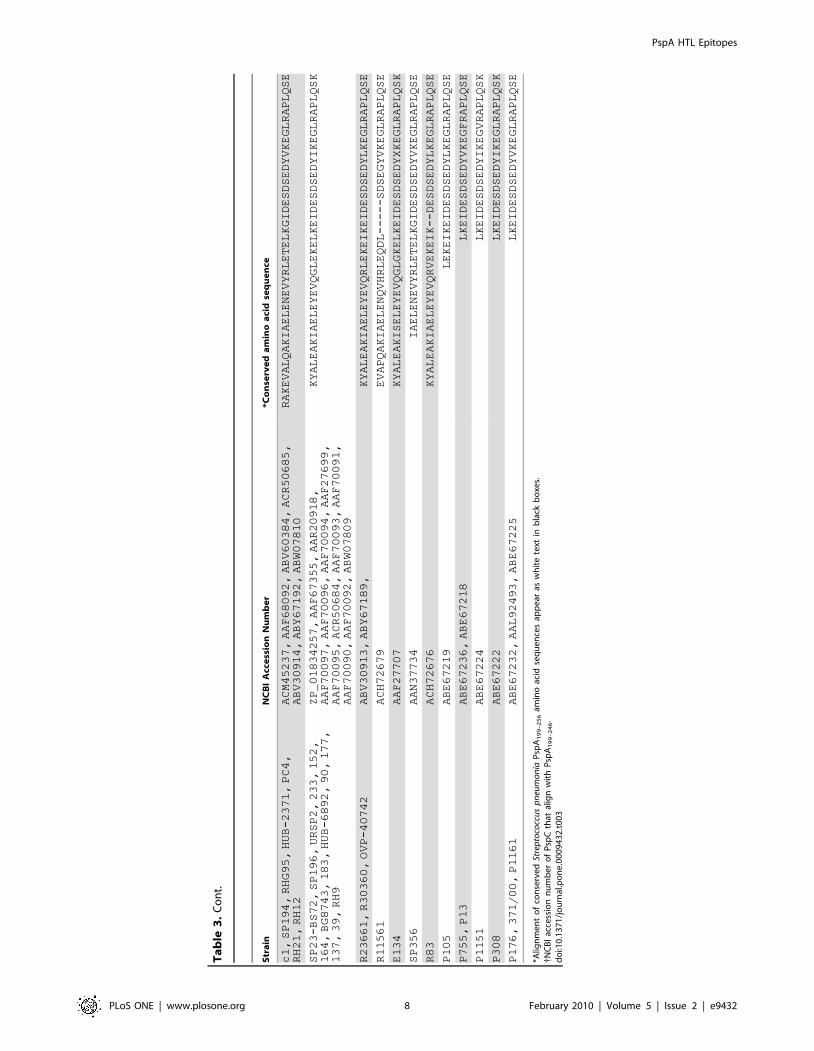
Ta
ble
3.
Alig
nm
en
to
fP
spA
19
9–
25
6am
ino
acid
seq
ue
nce
sfr
om
fam
ily1
Pn
eu
mo
cocc
ist
rain
s.
Str
ain
NC
BI
Acc
ess
ion
Nu
mb
er
*Co
nse
rve
da
min
oa
cid
seq
ue
nce
D39/R6
NP_357715
DAEEVAPQAKIAELENQVHRLEQELKEIDESESEDYAKEGFRAPLQSK
WU2
AAF27710
EVAPQAKIAELENQVHRLEQELKEIDESESEDYAKEGFRAPLQSK
195
AAF68105
EEVAPQAKIAELENQVHRLEQELKEIDESDSEDYIKEGFRAPLQSE
SP19
AAF68093
AEEVAPQAKIAELENQVHKLEQKLKEIDESDSEDYVKEGFRAPLQSE
CGSP14,
R41
YP_001834837,
ABY67182
EVAPQAKIAELENQVHRLEQDLKDINESDSEDYVKEGLRAPLQSE
RHG79,
OVP-41721
ABY67197,
ACR50702
HAEEVAPQVKIAELENQVHKLEQKLKEIDESDSEDYVKEGLRAPLQSE
EF3030
to
be
determined
..
EVALQAKIAELENQVHRLETELKEIDESDSEDYVKEGLRVPLQSE
c2,
OVP-43533,
OVP-42723,
OVP-43431,
R24729,
DBL5,
HUB-6893,
St
371/00
ACM45238,
ACR50689,
ACR50693,
ACR50694,
ABY67184,
AAF27706,
ACR50678,
ABR53733
HAKEVAPQAKIAELENQVHRLEQDLKDINESDSEDYVKEGLRAPLQSE
L81905,
RH5,
BG9739,
MC-247
AAF27705,
ABV60383,
AAF27700,
ACR50682
RAKEVVLQAKIAELENEVHKLEQKLKEIDESDSEDYVKEGFRAPLQSE
70585
YP_002739507
RAKEVALQAKIAELENEVHRLETKLKEIDESDSEDYVKEGLRAPLQSE
AC94
AAF27698
RAKEVALQAKIAELENEVHRLETELKEIDESDSEDYVKEGLRVPLQSE
SP6-BS73,
EF6796,
BG9163,
RHG63
ZP_01819322,
AAF27709,
AAF27711,
ABY67195
EVALQAKIAELEYEVQRLEKELEEINESDSEDYAKEGFRAPLQSK
SP18-BS74
ZP_01829602
HAEEVVPQAKIAELENEVQKLEKDLKEIDESDSEDYVKEGLRAPLQSE
SP200,
MC-332,
SP221
AAF67354,
ACR50683,
AAF68099
RAKEVALQAKIAELENQVHRLETELKEIDESDSEDYVKEGLRVPLQSE
BG8838,
R30318
AAF27703,
ABW07806
HAEEVVPQAKIAELENEVQKLEKDLKEIDESDSEDYVKEGLRAPLQSE
R30397,
R171,
BG6692
ABV60382,
ACH72677,
AAF27704
HAEEVVPQAKIAELENEVQKLEKDLKEIDESDSEDYVKEGLRAPLQSE
HUB-7682
ACR50697
RAKEVALQAKIAELENEVHRLETKLKEIDESDSEDYVKEGLRAPLQSE
130
AAF68103
HAEEVVPQAKIAELENEVQKLEKDLKEIDESASEDYVKEGLRAPLQSE
R30318
ABW07807
RAKEVALQAKIAELENEVHRLETKLKETDESDSEDYVKEGLRAPLQSE
OVI-2328
ACR50701
HAKEVVPQAKIAELENEVQKLEKDLKEIDESDSEDYVKEGLRAPLQSE
*CDC1873-00,
ST858,
*SP6-BS73,
*EF6796,
ST860,
*SRF10,
SP23-BS72,
*g5,
E134,
BG9163
ZP_02709307
{ ,ABN71686,
ZP_01820249
{ ,AAD00184
{ ,ABN71687,
AAF73809
{ ,ZP_01835080,
AAF73801
{ ,AAF13457,
AAF13460
DAEEYALEAKIAELEYEVQRLEKELKEIDESDSEDYLKEGLRAPLQSK
232
AAF68104
HAEEVVPQAKIAELENEVQKLEKDLKEIDESASEDYVKEGLRAPLQSE
P1031,
R30087
YP_002737416,
ABY67187
RAKEVALQAKIAELENEVHRLETKLKETDESDSEDYVKEGLRAPLQSE
CDC3059-06
ZP_02717970
HAEEVAPQAKIAELEHEVQKLEKALKEIGESDSEDYVKEGLRAPLQSE
OVP-42725
ACR50703
LFLQAKIAELENEVHKLEQKLKEIDESDSEDYVKEGFRAPLQSE
PN124
AAN37735
AKIAELENQVHRLEQDLKDINESDSEDYVKEGFRAPLQSE
DBL6A
AAF27701
RAKEVVLQAQIAELENEVHKLEPKLKEIDESDSEDYVKEGFRAPLQSE
St
435/96
AAL92492
HAEEVAPQAKIAELEHEVQKLEKALKEIDESDSEDYVKEGLRAPLQFE
EF10197
AAF27708
RAKEVVLHAKLAELENEVHKLDQKLKEIDESDSEDYVKEGFRAPLQSE
R402
ABY67181
HAEEVAPQAKIAELEHEVQKLEKALKEIDESDSEDYVKEGLRAPLQFE
DBL1
AAF27702
RAKEVALQAKIAELENEVYRLETELKGIDESDSEDYVKEGLRAPLQSE
HUB-4197,
237
ACR50680,
AAF68102
HAEEVAPQAKIAELEHEVQKLEKALKEIDESDSEDYVKEGLRAPLQFE
PspA HTL Epitopes
PLoS ONE | www.plosone.org 7 February 2010 | Volume 5 | Issue 2 | e9432
Str
ain
NC
BI
Acc
ess
ion
Nu
mb
er
*Co
nse
rve
da
min
oa
cid
seq
ue
nce
c1,
SP194,
RHG95,
HUB-2371,
PC4,
RH21,
RH12
ACM45237,
AAF68092,
ABV60384,
ACR50685,
ABV30914,
ABY67192,
ABW07810
RAKEVALQAKIAELENEVYRLETELKGIDESDSEDYVKEGLRAPLQSE
SP23-BS72,
SP196,
URSP2,
233,
152,
164,
BG8743,
183,
HUB-6892,
90,
177,
137,
39,
RH9
ZP_01834257,
AAF67355,
AAR20918,
AAF70097,
AAF70096,
AAF70094,
AAF27699,
AAF70095,
ACR50684,
AAF70093,
AAF70091,
AAF70090,
AAF70092,
ABW07809
KYALEAKIAELEYEVQGLEKELKEIDESDSEDYIKEGLRAPLQSK
R23661,
R30360,
OVP-40742
ABV30913,
ABY67189,
KYALEAKIAELEYEVQRLEKEIKEIDESDSEDYLKEGLRAPLQSE
R11561
ACH72679
EVAPQAKIAELENQVHRLEQDL-----SDSEGYVKEGLRAPLQSE
E134
AAF27707
KYALEAKISELEYEVQGLGKELKEIDESDSEDYXKEGLRAPLQSK
SP356
AAN37734
IAELENEVYRLETELKGIDESDSEDYVKEGLRAPLQSE
R83
ACH72676
KYALEAKIAELEYEVQRVEKEIK--DESDSEDYLKEGLRAPLQSE
P105
ABE67219
LEKEIKEIDESDSEDYLKEGLRAPLQSE
P755,
P13
ABE67236,
ABE67218
LKEIDESDSEDYVKEGFRAPLQSE
P1151
ABE67224
LKEIDESDSEDYIKEGVRAPLQSK
P308
ABE67222
LKEIDESDSEDYIKEGLRAPLQSK
P176,
371/00,
P1161
ABE67232,
AAL92493,
ABE67225
LKEIDESDSEDYVKEGLRAPLQSE
*Alig
nm
en
to
fco
nse
rve
dSt
rep
toco
ccu
sp
neu
mo
nia
Psp
A1
99
–2
56
amin
oac
idse
qu
en
ces
app
ear
asw
hit
ete
xtin
bla
ckb
oxe
s.{N
CB
Iac
cess
ion
nu
mb
er
of
Psp
Cth
atal
ign
wit
hP
spA
19
9–
24
6.
do
i:10
.13
71
/jo
urn
al.p
on
e.0
00
94
32
.t0
03
Ta
ble
3.
Co
nt.
PspA HTL Epitopes
PLoS ONE | www.plosone.org 8 February 2010 | Volume 5 | Issue 2 | e9432
Figure 2. Proliferation responses of PspA peptide-specific systemic and mucosal CD4+ T cells during pneumococcal carriage. Spleenand cervical lymph node (CLN) lymphocytes were isolated from F1 (B66Balb/c) mice, 28 days after intranasal challenge with Streptococcus pneumoniastrain EF3030 (&) and naıve (%). CD4+ T cells were incubated with 1 mM of PspA peptide (15 amino acid peptides that overlapped every 11 residues)plus mitomycin C-treated naıve syngeneic feeder cells, for 3 days, at a ratio of 5:16106 cells. Proliferation was measured by BrdU incorporation, whichwas measured by ELISA. The data presented are the mean OD450. Experimental groups consisted of 10 mice. The results were expressed as the mean6 the standard error mean (SEM) of the response from 3 replicate determinations of three independent experiments.doi:10.1371/journal.pone.0009432.g002
Figure 3. PspA peptide-specific IFN-c secretion by CD4+ T cell following pneumococcal challenge. Groups of 10 F1 (B66Balb/c) mice wereintranasally challenged with 107 CFUs of S. pneumonia strain EF3030 in a 15 ml volume of Ringer’s solution. Spleen and cervical lymph node (CLN) lymphocyteswere isolated from mice, 28 days after intranasal challenge with Streptococcus pneumonia strain EF3030 (&) and naıve (%). CD4+ T cells were incubated with1 mM of PspA peptide (15 amino acid peptides that overlapped every 11 residues) plus mitomycin C-treated naıve syngeneic feeder cells, for 3 days, at a ratio of5:16106 cells. The results were expressed as the mean 6 the standard error mean (SEM) of IFN-c supernatant levels from 3 replicate determinations of threeindependent experiments. IFN-c production of cultured supernatants was determined by Luminex capable of detecting .2 pg/ml of IFN-c.doi:10.1371/journal.pone.0009432.g003
PspA HTL Epitopes
PLoS ONE | www.plosone.org 9 February 2010 | Volume 5 | Issue 2 | e9432
previously challenged with S. pneumonia strain EF3030. Another
Th1 cytokine, IFN-c, is required for protective pneumococcal
immunity [47]. CD4+ T cells from S. pneumonia strain EF3030-
challenged mice secreted significant amounts of IFN-c following ex
vivo PspA peptide re-stimulation. IFN-c blockade accelerated the
death of animals during pneumococcal infection [48], whereas
treatment of mice with IFN-c enhanced the survival of mice [49].
However, confounding studies suggest that too much IFN-c and
too little IL-10 can inhibit pneumococcal clearance during S.
pneumonia infection that is secondary to influenza virus infection
[50].
IL-10 has been suggested to be both deleterious and important
for pneumococcal immunity. On one hand, administration of anti-
IL-10 antibody was shown to enhance pneumococcal immunity
[51], while others showed this Th2-associated cytokine is critical
for MARCO-1 expression and subsequent pneumococcal clear-
ance [50]. We show that PspA199–246 stimulates pneumococcal
strain EF3030-primed CD4+ T cells to secrete IL-10. Interestingly,
HTLs from CLN mounted IL-10 responses to more peptides, than
similar cells isolated from the spleen. Perhaps this contributes to
establishing pneumococcal carriage by supporting selective
pneumococcal clearance by CLN..spleen antigen-presenting
cells after stimulation with CD4+ T cell-derived IL-10, whereas
IFN-c-secreting HTLs might support spleen..CLN macrophag-
es activation and/or internalization of S. pneumonia.
In the absence of IL-10, a marked increase in pro-inflammatory
cytokines is induced during pneumococcal infection [52]. To this
end, IL-10 plays an indispensable role in mucous cell metaplasia
and hyperplasia. IL-10 attenuates the proinflammatory cytokine
response and its absence hampers effective clearance of the
infection, and reduces survival of pneumococcal infection [53]. We
have shown that CCL5 inhibition resulted in lower IFN-c-
secreting CD4+ T cells and significantly more PspA-specific IL-10-
producing CD4+ T cells, which corresponded with the transition
from pneumococcal carriage to lethal pneumonia [45,54]. Thus,
the precise contribution of IL-10 in pneumococcal immunity
remains uncertain, but the preponderance of the evidence suggests
excessive IL-10 responses play a deleterious role in pneumococcal
immunity, but moderate levels of this cytokine are required for
optimal adaptive (humoral) immune responses to S. pneumonia and
reduced mucosal hyperplasia.
An effective intranasal conjugate pneumococcal vaccine using
interleukin-12 (IL-12) as a mucosal adjuvant induced protection
and increased expression of lung and splenic IFN-c and IL-10
mRNAs and protected mice from lethal challenge [55]. Thus,
interplay and requirement of the HTL-derived IFN-c and IL-10 in
pneumococcal carriage and pneumonia will require further study.
In addition, the adjuvants or cytokines, e.g., IL-12, required by
antigen presenting cells to promote IFN-c and IL-10 secreting,
PspA-specific T cells will be addressed in the future.
Some studies suggest that Th2 cytokines do not support optimal
pneumococcal immunity. Mice primed to mount Th2 cell responses
followed by pneumococcal infection showed an increase in the
number of Pneumococci and an increase in sinus inflammation than
compared to naive or Th1 -primed groups [56]. IL-4 plays a central
role in directing the development of the Th2 phenotype and IL-4
responses in lung have been associated with an increased risk to
pneumococcal infection [57]. While IL-4 does not stimulate T cell
proliferation, it induces the growth of lymphoblasts [58]. IL-5 was
originally defined as a Th2 cell-derived cytokine that triggers B cell
Figure 4. PspA peptide-specific IL-2 secretion by CD4+ T cell following pneumococcal challenge. Groups of 10 F1 (B66Balb/c) mice wereintranasally challenged with 107CFUs of S. pneumonia strain EF3030 in a 15 ml volume of Ringer’s solution. Spleen and Cervical lymph node (CLN)lymphocytes were isolated from mice, 28 days after intranasal challenge with Streptococcus pneumoniae strain EF3030 (&) and naıve (%). CD4+ T cellswere incubated with 1 mM of PspA peptide (15 amino acid peptides that overlapped every 11 residues) plus mitomycin C-treated naıve syngeneicfeeder cells, for 3 days, at a ratio of 5:16106 cells. The results were expressed as the mean 6 the standard error mean (SEM) of IL-2 supernatant levelsfrom 3 replicate determinations of three independent experiments. IL-2 production of cultured supernatants was determined by Luminex capable ofdetecting .2 pg/ml of IL-2.doi:10.1371/journal.pone.0009432.g004
PspA HTL Epitopes
PLoS ONE | www.plosone.org 10 February 2010 | Volume 5 | Issue 2 | e9432
activation and differentiation into plasma cells [59]. PspA199–249-
specific HTLs from S. pneumonia strain EF3030-challenged mice
secreted significant amounts of IL-4 (spleen..CLN) and IL-5
(spleen#CLN) largely in response to PspA peptides 19, 20, 21, and
22. However, the uncertain role of IL-4 and IL-5 in pneumococcal
cellular immunity makes correlations of these cytokines with
protective immunity difficult.
The role of Th17 cells in pneumococcal immunity has not been
extensively studied. However, recent reports suggest that IL-17A
supports antibody responses to pneumococcal capsular polysac-
charides [60]. Mice lacking the IL-17A receptor or mice with
neutrophil depletion are more susceptible to pneumococci [61].
Additional studies on the role of HTL-derived IL-17 would greatly
contribute to the field and will be required to understand how
secretion of this cytokine correlates with pneumococcal immunity.
While the precise role of peptide MHC class II interactions that
determine protective pneumococcal immunity are not known, this
study addresses important questions that are relevant to MHC
polymorphisms and antigen responsiveness. A number of studies
have definitively proven a cause and effect relationship between
human MHC genes and resistance to infection [62,63] as well as
autoimmune diseases [64]. I-A, which is highly homologous to
HLA-DQ [65], typically restricts antigen-specific CD4+ T cells in
mice, whereas I-E (homologous to HLA-DR) [66,67,68] has been
reported to control non-responsiveness through antigen-specific
suppressor cells [69]. Further studies will be required to determine
whether I-E or I-A as well as DQ or DR molecules might be
involved in pneumococcal antigen non-responsiveness or cytokine
secretion in mouse or man, respectively. To this end, many of the
PspA peptides were predicted to bind I-A, while relatively few
were predicted to bind I-E. These studies support the use of in silico
and in vivo methods to validate T cell responsiveness to PspA
peptide-based vaccines.
Materials and Methods
AnimalsFemale F1 (B66Balb/c) mice, aged 8 to 12 weeks, contain
MHC class II haplotype and corresponding TCR diversity that
approaches those seen in man [70,71] and were purchased from
Jackson Laboratories. All mice were housed in horizontal laminar
flow cabinets free of microbial pathogens. Routine antibody
screening for a large panel of pathogens and routine histological
analysis of organs and tissues were performed to insure that mice
were pathogen free.
S. pneumonia Strain EF3030 Growth and ChallengeS. pneumonia capsular strain EF3030 was among the human
isolates of capsular group 19 that were previously examined and
found to be relatively non-invasive in mice [72]. Pneumococci
were grown in Todd Hewitt broth and stored frozen in aliquots at
280uC, in 20% glycerol, in sterile lactated Ringer’s solution
(Ringer’s) (Abbott Labs, North Chicago, IL) [73,74]. To establish
nasal carriage, Pneumococci were introduced into groups of mice
(8 to 12 week old) by nasal administration. The animals were
anesthetized with ketamine (100 mg/ml) and xylazine (20 mg/ml),
mixed at a 4:1 (vol/vol) ratio. The anesthesia mixture was injected
intramuscularly into the right hamstring muscle at a dose of
100 mg of ketamine per kg of body weight. After anesthesia was
established, the mice were inoculated with approximately 107
Figure 5. PspA peptide-specific IL-10 secretion by CD4+ T cell following pneumococcal challenge. Groups of 10 F1 (B66Balb/c) micewere intranasally challenged with 107CFUs of S. pneumonia strain EF3030 in a 15 ml volume of Ringer’s solution. Spleen and Cervical lymph node(CLN) lymphocytes were isolated from mice, 28 days after intranasal challenge with Streptococcus pneumonia strain EF3030 (&) and naıve (%). CD4+ Tcells were incubated with 1 mM of PspA peptide (15 amino acid peptides that overlapped every 11 residues) plus mitomycin C-treated naıvesyngeneic feeder cells, for 3 days, at a ratio of 5:16106 cells. The results were expressed as the mean 6 the standard error mean (SEM) of IL-10supernatant levels from 3 replicate determinations of three independent experiments. IL-10 production of cultured supernatants was determined byLuminex capable of detecting .2 pg/ml of IL-10.doi:10.1371/journal.pone.0009432.g005
PspA HTL Epitopes
PLoS ONE | www.plosone.org 11 February 2010 | Volume 5 | Issue 2 | e9432
Figure 6. PspA peptide-specific IL-4 secretion by CD4+ T cell following pneumococcal challenge. Groups of 10 F1 (B66Balb/c) mice wereintranasally challenged with 107CFUs of S. pneumonia strain EF3030 in a 15 ml volume of Ringer’s solution. Spleen and Cervical lymph node (CLN) lymphocyteswere isolated from mice, 28 days after intranasal challenge with Streptococcus pneumonia strain EF3030 (&) and naıve (%). CD4+ T cells were incubated with1 mM of PspA peptide (15 amino acid peptides that overlapped every 11 residues) plus mitomycin C-treated naıve syngeneic feeder cells, for 3 days, at a ratio of5:16106 cells. The results were expressed as the mean 6 the standard error mean (SEM) of IL-4 supernatant levels from 3 replicate determinations of threeindependent experiments. IL-4 production of cultured supernatants was determined by Luminex capable of detecting .2 pg/ml of IL-4.doi:10.1371/journal.pone.0009432.g006
Figure 7. PspA peptide-specific IL-5 secretion by CD4+ T cell following pneumococcal challenge. Groups of 10 F1 (B66Balb/c) mice wereintranasally challenged with 107CFUs of S. pneumonia strain EF3030 in a 15 ml volume of Ringer’s solution. Spleen and Cervical lymph node (CLN) lymphocyteswere isolated from mice, 28 days after intranasal challenge with Streptococcus pneumonia strain EF3030 (&) and naıve (%). CD4+ T cells were incubated with1 mM of PspA peptide (15 amino acid peptides that overlapped every 11 residues) plus mitomycin C-treated naıve syngeneic feeder cells, for 3 days, at a ratio of5:16106 cells. The results were expressed as the mean 6 the standard error mean (SEM) of IL-5 supernatant levels from 3 replicate determinations of threeindependent experiments. IL-5 production of cultured supernatants was determined by Luminex capable of detecting .2 pg/ml of IL-5.doi:10.1371/journal.pone.0009432.g007
PspA HTL Epitopes
PLoS ONE | www.plosone.org 12 February 2010 | Volume 5 | Issue 2 | e9432
colony forming units (CFU) of S. pneumonia strain EF3030 in 15 ml
of Ringer’s solution using a 25-gauge ball-tipped gavage needle
[75]. Experimental groups consisted of 10 mice and studies were
repeated 3 times. The guidelines proposed by the committee for
the Care of Laboratory Animal Resources Commission of Life
Sciences - National Research Council were followed to minimize
animal pain and distress. All procedures involving mice were
approved by the Morehouse School of Medicine Committees
(IACUC).
Pneumococcal Antigens54 overlapping peptides, spanning the entire length of S.
pneumonia strain D39/R6 PspA protein sequence (NCBI Accession
# NP_357715), starting with the first 15 residues at the N-
terminus, was synthesized by the multipin synthesis method by
Chiron Mimotopes Peptide Systems. Peptides overlapped by four
amino acids (Table 1) and were acetylated at the N- terminus and
ended with a COOH-terminal. Purity of these peptides was
approximately 95%. The peptides were dissolved in a mixture (v/
v) of 75% dimethyl sulfoxide and 25% water, to a concentration of
70 mM, divided into small aliquots and stored frozen at 280uC.
Tissue Collection and Cell IsolationMice were sacrificed by CO2 inhalation to collect spleen and
CLNs for single cell isolation of lymphocytes 28 days following S.
pneumonia strain EF3030 challenge. Individual single cell suspen-
sions of spleen and CLNs were collected and prepared by
aseptically removing tissues and passage through a sterile wire
screen. Unpooled CD4+ T cells were further separated by
OctoMACSTM (Miltenyi Biotec) using negative selection. Re-
maining (non-CD4+) cells, were used as accessory feeder cells for
antigen peptide-specific stimulation assays after mitomycin C
(Sigma-Aldrich) treatment.
Cytokine Quantitation by LuminexTM AnalysisPurified CD4+ T cells and mitomycin C-treated feeder cells
were cultured at a density of 56106 and 106 cells per ml,
respectively, in complete medium containing 1 mM of each PspA
peptide at 37uC in 5% CO2. For the assessment of cytokine
production, 100 mL of culture supernatants from 96-well flat
bottom plates (Corning Glass Works) were harvested 3 days after
ex vivo PspA peptide stimulation to determine the levels of IL-10
and IFN-c secreted by CD4+ T cells. phorbol-12-myristate-13-
acetate (PMA) 1 mg/ml was used as a positive control , ovalbumin
(1 mg/ml) and medium only is used as negative control to reduce
the background reading. Supernatant cytokine levels were
determined by the BeadlyteTM mouse multi-cytokine detection
(Bio-Rad). Briefly, filter bottom ELISA plates were rinsed with
100 mL of Bio-plex assay buffer and liquid was removed using a
MilliporeTM Multiscreen Separation Vacuum Manifold System set
at 5 mm Hg. Analyte beads in assay buffer were added to the wells
followed by 50 mL of serum or standard solution. The plates were
Figure 8. 3D plot of Th1/Th2 cytokine secretion relative to proliferation or I-A/I-E predicted peptide-binding by cervical lymphnode-derived CD4+ T cells. The panels summarize IFN-c, IL-10, IL-2, IL-4, IL-5 and proliferation responses of PspA peptide-specific CD4+ T cellsisolated from cervical lymph nodes of F1 (B66Balb/c) mice, 28 days after S. pneumonia strain EF3030- challenge and predicted I-A or I-E bindingaffinities. Y-axis and X-axis indicate the concentration (ng/ml) of IFN-c and IL-10, IL-2, IL-4, IL-5 respectively, secreted by PspA peptide-stimulatedCD4+ T cells. The Z- axis represents the predicted I-A or I-E binding affinities (Kd). PspA peptides 19, 20, 21 and 22 appear as white circles, whileremaining peptides are open circles.doi:10.1371/journal.pone.0009432.g008
PspA HTL Epitopes
PLoS ONE | www.plosone.org 13 February 2010 | Volume 5 | Issue 2 | e9432
incubated for 30 minutes at room temperature with continuous
shaking (at setting #3) using a Lab-LineTM Instrument Titer Plate
Shaker. The filter bottom plates were washed, as before, and
centrifuged at 3006g for 30 seconds. Subsequently, 50 mL of anti-
mouse IL-10 or IFN-c antibody-biotin reporter solution was added
in each well, after which the plates were incubated with continuous
shaking for 30 min followed by centrifugation and washing. Next,
50 mL streptavidin-phycoerythrin (PE) solution was added, and the
plates were incubated with continuous shaking for 10 min at RT.
125 mL of Bio-plex assay buffer was added, and BeadlyteTM
readings were measured using a LuminexTM System and
calculated using Bio-plexTM software (Bio-Rad). The cytokine
BeadlyteTM assays were capable of detecting .5 pg/mL for each
analyte.
Cell ProliferationLymphocyte proliferation was measured by a 5-Bromo-29-
deoxy uridine (BrdU) absorption and detection (Roche Diagnos-
tics). In brief, purified CD4+ T cells were cultured at a density of
56106 cells/mL, with 106 mitomycin C-treated feeder cells/mL in
complete medium containing 1 mM of PspA peptide at 37uC in
5% CO2. After 2 days of ex vivo antigen stimulation, cells were
transferred to polystyrene 96 well plates (Corning Glass Work).
10 mL of BrdU labeling solution (10 mM final concentration per
well) were added and incubated for 18 hours at 37uC with 5%
CO2. The cells were then fixed and incubated with 100 mL of
nuclease in each well for 30 minute at 37uC. The cells were
washed with complete media and incubated with BrdU-POD
solution for 30 minute at 37uC. BrdU incorporation was
developed with an 2,29–azino-bis 3- ethylbenzthiazoline-6-sulfonic
acid (ABTS) solution and optical density (OD) was read at
450 nm. The proliferation index (PI) was calculated as follows.
Antigen-specific CD4+ T cell proliferation was obtained by
stored at IEDB were used to estimate performance. Because the
Figure 9. 3D plot of Th1/Th2 cytokine secretion relative to proliferation or I-A/I-E predicted peptide-binding by spleen- derivedCD4+ T cells. The panels summarize IFN-c, IL-10, IL-2, IL-4, IL-5 and proliferation responses of PspA peptide-specific CD4+ T cells isolated from spleenof F1 (B66Balb/c) mice, 28 days after S. pneumonia strain EF3030-challenge and predicted I-A or I-E binding affinities. Y-axis and X-axis indicate theconcentration (ng/ml) of IFN-c and IL-10, IL-2, IL-4,IL-5 respectively, secreted by PspA peptide-stimulated CD4+ T cells. The Z-axis represents thepredicted I-A or I-E binding affinities (Kd). PspA peptides 19, 20, 21 and 22 appear as white circles, while remaining peptides are open circles.doi:10.1371/journal.pone.0009432.g009
PspA HTL Epitopes
PLoS ONE | www.plosone.org 14 February 2010 | Volume 5 | Issue 2 | e9432
binding of peptides to MHC class II molecules is not dependent on
exact size, derivation of MHC class II ARB matrices followed an
iterative procedure. For the first iterative step, a matrix was
generated from a set of nine-residue core sequences randomly
obtained from each peptide sequence in the training set. For
subsequent cycles, nine-residue core sequences were used to
generate a matrix. The overall binding affinity of a peptide was
predicted using the highest scoring nine-residue core sequence. For
the SYFPEITHI prediction, we patched each testing peptide with
three glycine residues at both ends before evaluation for prediction.
This was recommended by the creators of SYFPEITHI method to
ensure that all potential binders were correctly presented to the
prediction algorithm. For all other methods, the original tested
peptides were submitted directly for prediction. Peptide sequences
were sent to web servers one at a time and predictions were
extracted from the server’s response. To assign a single prediction
for peptides longer than nine amino acids in the context of tools
predicting the affinity of 9 core-binding regions, we took the highest
affinity prediction of all possible 9-mers within the longer peptide as
the prediction result. For each MHC class II molecule whose
binding can be predicted by three or more algorithms, the top three
methods were selected that gave the best performance. For each
method, peptides were tested and ranked by their scores with higher
ranks for better binders. For each tested peptide, three ranks from
different methods were taken and the median rank was taken
as the consensus score. Peptides were classified into binders
(IC50,500 nM) and nonbinders (IC50$500 nM), as practical
cutoffs.
StatisticsData are expressed as the mean 6 SEM and compared using a
two-tailed student’s t-test or an unpaired Mann Whitney U test.
The results were analyzed using Microsoft Excel for Macintosh
computers and were considered statistically significant if p values
were less than 0.01. When cytokine or antibody levels were below
the detection limit (BD), they were recorded as one-half the lower
detection limit for statistical analysis.
Acknowledgments
The content of this manuscript benefited from many fruitful conversations
with members of the Morehouse School of Medicine and the University of
Alabama at Birmingham
Author Contributions
Conceived and designed the experiments: JL. Performed the experiments:
RS SS PKS UPS. Analyzed the data: RS SH JL. Contributed reagents/
materials/analysis tools: DB SH JL. Wrote the paper: RS JL.
References
1. Nasrin D, Collignon PJ, Wilson EJ, Pilotto LS, Douglas RM (1999) Antibiotic
resistance in Streptococcus pneumoniae isolated from children. Journal of Paediatrics
& Child Health 35: 558–561.
2. Hooper DC (2001) Emerging mechanisms of fluoroquinolone resistance.
Emerging Infectious Diseases 7: 337–341.
3. Morita JY, Zell ER, Danila R, Farley MM, Hadler J, et al. (2002) Association
between antimicrobial resistance among pneumococcal isolates and burden of
invasive pneumococcal disease in the community. Clinical Infectious Diseases
9. Brooks-Walter A, Briles DE, Hollingshead SK (1999) The pspC gene of
Streptococcus pneumoniae encodes a polymorphic protein, PspC, which elicits cross-
reactive antibodies to PspA and provides immunity to pneumococcal
bacteremia. Infection & Immunity 67: 6533–6542.
10. Sturniolo T, Bono E, Ding J, Raddrizzani L, Tuereci O, et al. (1999) Generation
of tissue-specific and promiscuous HLA ligand databases using DNA micro-
arrays and virtual HLA class II matrices.[see comment]. Nature Biotechnology
17: 555–561.
11. Cunha-Neto E (1999) MHC-restricted antigen presentation and recognition:
constraints on gene, recombinant and peptide vaccines in humans. Brazilian
Journal of Medical & Biological Research 32: 199–205.
12. Rammensee HG (1995) Chemistry of peptides associated with MHC class I and
class II molecules. Current Opinion in Immunology 7: 85–96.
13. Meister GE, Roberts CG, Berzofsky JA, De Groot AS (1995) Two novel T cell
epitope prediction algorithms based on MHC-binding motifs; comparison of
predicted and published epitopes from Mycobacterium tuberculosis and HIV protein
sequences. Vaccine 13: 581–591.
14. Kang H, Mansel RE, Jiang WG (2005) Genetic manipulation of stromal cell-
derived factor-1 attests the pivotal role of the autocrine SDF-1-CXCR4 pathway
in the aggressiveness of breast cancer cells. International Journal of Oncology 26:
1429–1434.
15. Reche PA, Glutting JP, Zhang H, Reinherz EL (2004) Enhancement to the
RANKPEP resource for the prediction of peptide binding to MHC molecules
using profiles. Immunogenetics 56: 405–419.
16. Donnes P, Kohlbacher O (2006) SVMHC: a server for prediction of MHC-binding peptides. Nucleic Acids Research 34: W194–197.
17. Gu Y, Filippi MD, Cancelas JA, Siefring JE, Williams EP, et al. (2003)
Hematopoietic cell regulation by Rac1 and Rac2 guanosine triphosphatases.
Science 302: 445–449.
18. Parry CS (2008) Flanking p10 contribution and sequence bias in matrix basedepitope prediction: revisiting the assumption of independent binding pockets.
BMC Struct Biol 8: 44.
19. Briles DE, Hollingshead SK, Paton JC, Ades EW, Novak L, et al. (2003)Immunizations with Pneumococcal Surface Protein A and Pneumolysin Are
Protective against Pneumonia in a Murine Model of Pulmonary Infection with
Streptococcus pneumoniae. Journal of Infectious Diseases 188: 339–348.
20. Hollingshead SK, Becker R, Briles DE (2000) Diversity of PspA: mosaic genesand evidence for past recombination in Streptococcus pneumoniae. Infect Immun 68:
5889–5900.
21. McGuffin LJ, Bryson K, Jones DT (2000) The PSIPRED protein structure
prediction server. Bioinformatics 16: 404–405.
22. Fuchs PF, Alix AJ (2005) High accuracy prediction of beta-turns and their typesusing propensities and multiple alignments. Proteins 59: 828–839.
23. Manoury B, Hewitt EW, Morrice N, Dando PM, Barrett AJ, et al. (1998) An
asparaginyl endopeptidase processes a microbial antigen for class II MHC
presentation. Nature 396: 695–699.
24. Reche PA, Reinherz EL (2003) Sequence variability analysis of human class Iand class II MHC molecules: functional and structural correlates of amino acid
polymorphisms. Journal of Molecular Biology 331: 623–641.
25. Corte G, Damiani G, Calabi F, Fabbi M, Bargellesi A (1981) Analysis of HLA-DR polymorphism by two-dimensional peptide mapping. Proceedings of the
National Academy of Sciences of the United States of America 78: 534–538.
26. Shackelford DA, Strominger JL (1980) Demonstration of structural polymor-
phism among HLA-DR light chains by two-dimensional gel electrophoresis.Journal of Experimental Medicine 151: 144–165.
Degradation of mouse invariant chain: roles of cathepsins S and D and theinfluence of major histocompatibility complex polymorphism. J Exp Med 186:
549–560.
35. Castellino F, Zappacosta F, Coligan JE, Germain RN (1998) Large proteinfragments as substrates for endocytic antigen capture by MHC class II
molecules. J Immunol 161: 4048–4057.36. Manoury B, Mazzeo D, Fugger L, Viner N, Ponsford M, et al. (2002)
Destructive processing by asparagine endopeptidase limits presentation of a
dominant T cell epitope in MBP. Nat Immunol 3: 169–174.37. Chen JM, Rawlings ND, Stevens RA, Barrett AJ (1998) Identification of the
active site of legumain links it to caspases, clostripain and gingipains in a newclan of cysteine endopeptidases. FEBS Lett 441: 361–365.
38. Chen JM, Dando PM, Rawlings ND, Brown MA, Young NE, et al. (1997)Cloning, isolation, and characterization of mammalian legumain, an asparaginyl
endopeptidase. J Biol Chem 272: 8090–8098.
39. Blum JS, Cresswell P (1988) Role for intracellular proteases in the processing andtransport of class II HLA antigens. Proc Natl Acad Sci U S A 85: 3975–3979.
40. Amigorena S, Webster P, Drake J, Newcomb J, Cresswell P, et al. (1995)Invariant chain cleavage and peptide loading in major histocompatibility
complex class II vesicles. J Exp Med 181: 1729–1741.
41. Antoniou AN, Blackwood SL, Mazzeo D, Watts C (2000) Control of antigenpresentation by a single protease cleavage site. Immunity 12: 391–398.
42. Watts C (2001) Antigen processing in the endocytic compartment. Curr OpinImmunol 13: 26–31.
43. McDaniel LS, Yother J, Vijayakumar M, McGarry L, Guild WR, et al. (1987)Use of insertional inactivation to facilitate studies of biological properties of
pneumococcal surface protein A (PspA). Journal of Experimental Medicine 165:
Differential PsaA-, PspA-, PspC-, and PdB-specific immune responses in amouse model of pneumococcal carriage. Infection & Immunity 73: 1006–1013.
46. McDaniel LS, Ralph BA, McDaniel DO, Briles DE (1994) Localization ofprotection-eliciting epitopes on PspA of Streptococcus pneumoniae between amino
acid residues 192 and 260. Microbial Pathogenesis 17: 323–337.47. Pomeroy C, Rubins JB (1997) Role of gamma interferon in the pathogenesis of
modulates pneumococcal immunity and carriage. Journal of Immunology 176:2346–2356.
55. Khan AQ, Shen Y, Wu Z-Q, Wynn TA, Snapper CM (2002) Endogenous pro-and anti-inflammatory cytokines differentially regulate an in vivo humoral
response to Streptococcus pneumoniae. Infection & Immunity 70: 749–761.
56. Yu X, Sperling A, Blair C, Thompson K, Naclerio R (2004) Antigen stimulationof TH2 cells augments acute bacterial sinusitis in mice. J Allergy Clin Immunol
114: 328–334.
57. Kang CI, Rouse MS, Patel R, Kita H, Juhn YJ (2009) Allergic airwayinflammation and susceptibility to pneumococcal pneumonia in a murine model with
real-time in vivo evaluation. Clin Exp Immunol 156: 552–561.
58. Murtaugh MP, Johnson CR, Xiao Z, Scamurra RW, Zhou Y (2009) Speciesspecialization in cytokine biology: is interleukin-4 central to the T(H)1-T(H)2
paradigm in swine? Dev Comp Immunol 33: 344–352.
59. Kouro T, Takatsu K (2009) IL-5- and eosinophil-mediated inflammation: fromdiscovery to therapy. Int Immunol 21: 1303–1309.
60. Malley R, Srivastava A, Lipsitch M, Thompson CM, Watkins C, et al. (2006)
Antibody-independent, interleukin-17A-mediated, cross-serotype immunity topneumococci in mice immunized intranasally with the cell wall polysaccharide.
Infect Immun 74: 2187–2195.
61. Lu YJ, Gross J, Bogaert D, Finn A, Bagrade L, et al. (2008) Interleukin-17Amediates acquired immunity to pneumococcal colonization. PLoS Pathog 4:
e1000159.
62. Fuller-Espie SL, Murphy GA, Brett SJ, Lechler RI (1997) Quantitative but not
qualitative variation in MHC class II alters CD4 interaction and influences T