120
Typhoid and paratyphoid fever in Jakarta, Indonesia Epidemiology and risk factors Albert M. Vollaard
| Date post: | 13-May-2023 |
| Category: |
Documents |
| Upload: | independent |
| View: | 0 times |
| Download: | 0 times |

Typhoid and paratyphoid fever in Jakarta, Indonesia
Epidemiology and risk factors
Albert M. Vollaard
18439 Vollaard 14-12-2004 08:24 Pagina 1

18439 Vollaard 14-12-2004 08:24 Pagina 2

Typhoid and paratyphoid fever
in Jakarta, Indonesia
Epidemiology and risk factors
P R O E F S C H R I F T
Ter verkrijging van
de graad van Doctor aan de Universiteit Leiden,
op gezag van de Rector Magnificus Dr. D.D. Breimer,
hoogleraar in de faculteit der Wiskunde en
Natuurwetenschappen en die der Geneeskunde,
volgens besluit van het College voor Promoties
te verdedigen op dinsdag 25 januari 2005
klokke 16.15 uur
door
Albert Meint Vollaard
geboren te Veenendaal in 1970
18439 Vollaard 14-12-2004 08:24 Pagina 3
Promotiecommissie
Promotor: Prof. Dr. J.T. van Dissel (Universiteit Leiden)
Co-promotores: Prof. Dr. S. Widjaja (Atma Jaya Catholic University, Jakarta)
Prof. Dr. Ch. Surjadi (Atma Jaya Catholic University, Jakarta)
Referent: Prof. Dr. P. Speelman (Universiteit van Amsterdam)
Leden: Prof. Dr. J.W.M. van der Meer (Radboud Universiteit Nijmegen)
Prof. Dr. J.P. Vandenbroucke (Universiteit Leiden)
Prof. Dr. A.M. Deelder (Universiteit Leiden)
Financial support for the publication of this thesis
by GlaxoSmithKline and Pfizer is gratefully acknowledged.
ISBN 90-9019002-3
Printed by Febodruk, Enschede, the Netherlands
Graphic design Jan Kleingeld, Leiden, the Netherlands
Copyright © 2004 by Albert M. Vollaard, Den Haag, the Netherlands
18439 Vollaard 14-12-2004 08:24 Pagina 4

Contents
General introduction 7
Outline of the thesis 20
Chapter 1 Identification of typhoid fever and paratyphoid 27
fever cases at presentation in outpatient clinics
in Jakarta, Indonesia
Chapter 2 Risk factors for typhoid and paratyphoid fever 43
in Jakarta, Indonesia
Chapter 3 Risk factors for transmission of food borne illness in 61
restaurants and street vendors in Jakarta, Indonesia
Chapter 4 A survey of the supply and bacteriologic quality of 75
drinking water and sanitation in Jakarta, Indonesia
Chapter 5 Helicobacter pylori infection and typhoid fever 89
in Jakarta, Indonesia
General discussion 101
Nederlandse samenvatting 110
Acknowledgements 117
About the author 119
18439 Vollaard 14-12-2004 08:24 Pagina 5

Pursue him to his house, and pluck him thence;
Lest his infection, being of catching nature,
Spread further.
William Shakespeare, Coriolanus, act iii, scene 1
18439 Vollaard 14-12-2004 08:24 Pagina 6

Typhoid and paratyphoid fever – together often called enteric fever – constitute a serious
health threat worldwide. In developing countries 21 million patients suffer from typhoid
fever annually and more than 200 000 typhoid fever patients die every year.1 Paratyphoid
fever is also a global health burden, but its incidence is about ten times less than typhoid.
Most Dutch physicians will deal sporadically with enteric fever patients, because enteric
fever is virtually non-existent in the Netherlands since more than half a century. In fact,
many of the cases in hospitals in the Netherlands are travelers coming from Indonesia.
At the turn of the 19th century the picture in the Netherlands was quite different.
Incidence rates of more than 50/100 000 population-year were reported, that rapidly
declined to 0.2/100 000 population-year in 1967 due to improvements in drinking water
supply, pasteurization of milk and identification of chronic carriers.2 In the Indonesian
archipelago (para)typhoid fever is still an endemic disease. In consequence, studies were
needed to understand the reason for its frequent occurrence. An increased understanding
could lead to better and cost-effective control strategies implemented by public health
authorities.
The presented compilation of articles in this PhD-thesis has a specific focus on
Indonesia, because in a scientific collaboration Indonesian and Dutch physicians –
including the author – participated in a typhoid fever research project in Jakarta from
February 2001 until October 2003.
Typhoid feverBacterial aspectsBacterium: Salmonella enterica serotype Typhi (S. typhi) is a Gram-negative rod and a member
of the Enterobacteriaceae. In the 19th century several infectious diseases were dubbed
“typhus”. “Typhos” in Greek means smoke, in which resonates both the delirious state
commonly observed in typhoid fever, and the miasmatic theory, i.e., “malicious vapours
as cause of disease”, that dominated conceptual thinking about the origin of febrile ill-
nesses in those days. The dispute on the cause of the specific and lethal variant “typhus
abdominalis” or “typhoid fever” was only settled in 1880 with the discovery of the bacterium
responsible for infection by three independent investigators: Eberth, Klebs and Koch.
The genus derives its name however from another investigator, Salmon, who together
with Smith discovered a related serotype in 1885: Salmonella choleraesuis. After the initial
discovery of bacteria in intestinal tissues followed the isolation of bacteria in stools, urine
and blood, explaining the pathogenesis and transmission of the disease. Robert Koch
7
General introduction
18439 Vollaard 14-12-2004 08:24 Pagina 7
8
deserves the credit for being the first to describe the concept of convalescent or even
“healthy” carriers in the transmission of typhoid fever.3
Antigen structure: Bacteria of the Salmonella genus and other related organisms share
several antigens with S. typhi. Relatively S. typhi-specific antigenic features are the
somatic lipopolysaccharide antigens O9 and O12, protein flagellar antigen Hd and the
polysaccharide capsular antigen Vi. Vi-negative strains have been described 4 and also
a distinct flagellar antigen Hj was detected in circulating strains in Indonesia.5
Genetics: In 2001 the complete genome sequence of a S. typhi strain was determined
and published in Nature 6, which elucidated many individual features of this highly host-
adapted bacterium. A remarkable colinearity with genomes of E.coli and S. enterica serotype
Typhimurium was observed, which led to the assumption that S. typhi is a “recent”
offspring of an ancestral E. coli.7 Two major differences have been observed: 11 large
insertions unique for S. typhi that are called salmonella pathogenicity islands (SPI),
combined with multiple smaller insertions scattered in the genome, and 204 so-called
pseudogenes. The acquired insertions are important for the survival, host-specificity and
pathogenicity of S. typhi in man. The pseudogenes, of which interestingly more than half
are inactivated by mutations, are ancestral genes that presumably have lost their relevance
for bacterial survival in a wide variety of hosts, because of the adaptation of S. typhi to
the human host only.
Strain typing: Salmonella family members can be distinguished by the agglutination cha-
racteristics of members as was first described for S. typhi by Widal in 1896. With the
discovery of antibiotics and consequent rise of antibiotic resistance of strains in the
1960s, also different strains within the S. typhi-group could be distinguished using anti-
biotic susceptibility tests. The introduction of (bacterio)phage-typing of S.typhi has been
helpful in epidemiological surveillance and has been refined in recent years by the use of
pulsed field gel electrophoresis, ribotyping and amplified fragment length polymorphism
fingerprinting.8-12
Infective dose: In optimal conditions S. typhi undergoes division in less than half an
hour. Prior multiplication of bacteria in the intestine is not a necessary step in the deve-
lopment of typhoid fever.13 Therefore, the ingested dose is the decisive momentum in the
infection. Experiments in the 1960s demonstrated the required dose for infection:
at least 1000 bacteria.14 High numbers of ingested bacteria resulted in higher attack rates
implicating a linear dose-response curve with respect to the logarithmic dose, starting
with attack rates of 10-20% at a dose of 103 organisms. The inoculum-size is also associated
with the length of the incubation period, as was illustrated by the longer incubation periods
in waterborne outbreaks of typhoid fever. The dilution and lack of growth of bacteria in
water result in lower bacterial concentrations.14 The intensity of excretion in carriers is
variable, but up to 450 million organisms per gram faeces have been determined in the
18439 Vollaard 14-12-2004 08:24 Pagina 8

stool of a paratyphoid fever carrier.13 Excreted bacteria in water do not multiply, but
can survive for substantial periods depending on the temperature and amount of organic
matter in water. In sewage survival of at least 2 weeks is reported.
In food, however, bacteria can multiply to high numbers and subsequently overcome
acquired immunization due to prior infection or the protective effect of vaccination.15
Milk, (ice)cream, meat products, salads and coconut milk are good culture media and
before pasteurization dairy products were often implicated in typhoid transmission.
Direct person-to-person spread of bacteria is rare, but transmission in homosexual
contact is documented.16
PathogenesisGeneral life cycle: The mapping of the genome of S. typhi has been essential for the gro-
wing understanding of the unique host-adaptation of the bacterium: humans are the only
host. Next to this host-specificity another feature is characteristic of S. typhi: its ability to
survive and even multiply in the human host inside the macrophages that are responsible
for the first line of defense against invaders. Even so, the roadmap of infection should
begin in the gastro-intestinal tract after ingestion of a sufficient number of bacteria in
food or water, i.e., the minimum infective dose.14 Gastric acid is the first barrier to over-
come and a reduced production of gastric acid, for example due to antacids, Helicobacter
pylori gastritis, chronic atrophic gastritis or gastrectomy, might understandably lead to
an increased susceptibility for disease by allowing the passage of high numbers of
S. typhi, as is explained in the fifth chapter of this thesis.17
Inside the small intestine S. typhi bacteria attach to intestinal cells. Both enterocytes and
microfold- or M-cells overlying the Peyer’s patches are the porte-d’entrée of bacteria into
the circulation of the human host. The S. typhi-specific interaction with the enterocytes
depends on the expression of the cystic fibrosis transmembrane conductance regulator
(CFTR) on the surface of the enterocytes.18 CFTR interacts with bacterial LPS and factors
from S. typhi are able to upregulate the CFTR levels on the enterocytic membrane leading
to enhanced bacterial ingestion and submucosal translocation.19 The type III secretion
apparatus of the bacteria, encoded within SPI-1, injects signaling components into the
enterocytes in order to modify the cytoskeletal and vacuolar organization of the host cell
to trigger invasion.20 Passage through the intestinal mucosa in membrane-bound vacuoles
enables S. typhi to reach the lymphatic circulation in the lamina propria and the draining
mesenterial lymph nodes. After reaching the blood circulation via the thoracic duct the
bacteria are filtered from the circulation and sequestered inside the phagocytic cells of
the liver, spleen and bone marrow. On the SPI-2 pathogenicity island of S. typhi the SpiC
gene encodes the inhibition of phagosome-lysosome fusion, which enables S. typhi to
9
General introduction
18439 Vollaard 14-12-2004 08:24 Pagina 9

10
survive and even multiply inside the macrophage. After an incubation period of 6-21 days
secondary dissemination could occur causing disease symptoms associated with systemic
infection and also re-infection of the Peyer’s patches due to excretion of bacteria in bile.
This re-infection could result in ulceration and necrosis of the previously primed Peyer’s
patches culminating in intestinal hemorrhage or perforation.21 However, not all subjects
infected with S. typhi develop symptoms, because the eventual outcome is influenced by
interacting factors related to the bacterium, the host and antimicrobial agents.
Bacterial factors: An increase in the ingested dose leads to a higher attack rate and shor-
ter incubation period.14 However, the total number of bacteria ingested seems not to be
associated with the severity of disease, suggesting an on-off mechanism of disease instead
of an dose-response curve as found in other salmonelloses.22 An increased virulence of
the bacteria as determined by the presence of the Vi-antigen and mutations resulting in
fluoroquinolone resistance 23 was found to be associated with severe typhoid.
Host factors: Typhoid in young children may follow a mild course.24,25 Following the
roadmap of infection multiple sites can be identified where insufficiencies in the defense
mechanisms could lead to increased susceptibility or severity of disease. A decreased
gastric acid production or gastrectomy has already been mentioned. Other factors are
related to the immune response, because S. typhi induces macrophages to produces
cytokines. The cytokine-mediated signaling of immune cells is responsible for clinical
manifestation of typhoid fever such as fever, altered consciousness, hepatic dysfunction,
renal failure, intestinal necrosis, thrombosis and shock. In some patients an increased
production of proinflammatory cytokines (TNF-α, IL-1β and IL-6) and cytokine antago-
nists (IL-1 receptor antagonist and soluble TNF-α receptor) has been demonstrated.26,27
Consequently, circulating cytokine levels are associated with severity and response to
treatment. 28 The acute stage of typhoid fever results in depressed TNF-α and IL-1βrelease and consequently in delayed recovery.29
Polymorphisms in the genes encoding the nRAMP (natural-resistance-associated macro-
phage protein) are not associated with resistance to typhoid, even though in murine
models this mechanism proved to be important for bacterial survival.30
The influence of genes of the major histocompatibility complex class II and III loci, enco-
ding TNF-α and lymphotoxin-a, on typhoid fever susceptibility has been studied and asso-
ciations of different haplotypes with disease susceptibility and resistance were demon-
strated. 31,32 Future studies will examine whether the genetic polymorphisms associated
with increased susceptibility to other salmonelloses play a role in typhoid fever as well.33
The presence of anti-S. typhi antibodies does not prevent previously infected individuals
from recurrence of infection when they are challenged with high inocula.34,35
Carrier state: The gall bladder could be invaded after the secondary dissemination of
bacteria as discussed above. Salmonella bacteria are capable of survival in gall and could
18439 Vollaard 14-12-2004 08:24 Pagina 10
turn into permanent inhabitants of the gall bladder in case of favorable conditions,
such as stones. Their ability to produce a biofilm might help them to evade the immune
system.36 Four percent of patients with acute infections, most of them female patients
and especially in presence of gall stones, continue to excrete bacteria for prolonged periods
of time. The continuous excretion of bile soiled with bacteria is the likely mechanism
required to permit the survival of S. typhi in the human population, because during many
years the carriers may constitute a potential source of infection for immunologically naive
humans. The first identified carrier in the USA was Mary Mallon, the infamous cook in
New York, better known as ‘Typhoid Mary’. After causing several micro-epidemics in
New York in the beginning of the 20th century, she was quarantined for life in a tubercu-
losis colony on North Brother Island until she died in 1938 from a non-related stroke.
Chronic typhoid and paratyphoid fever carriers have an increased risk of cancer of the
gallbladder and biliary tract.37-40 However, this risk may be confounded by the associa-
tion of gallstones and malignancies of the hepatobiliary tract.
DiagnosisCulture: Diagnosis of typhoid fever requires culture of bacteria in bone marrow, blood
(i.e., in the first week and lower chance of recovery from blood in the second to third
week, sensitivity 60-80 percent), stool (i.e., end of first week with highest number of
bacteria in second week), bile 41, urine (positive in a quarter to one-third of cases in the
first weeks) and rose spots.42 The bacterial loads in humans are low: in blood 1 bacterium
per mL was measured of which 66% lies inside phagocytic cells, whereas in bone marrow
10 bacteria per mL were isolated.23,43 However, the ratio of bacteria in blood versus bone
marrow depends on the duration of illness; in the first week of illness this ratio is
approximately 1, but later in illness the likelihood to isolate bacteria from bone marrow
is greater than from blood, especially after antibiotic treatment.44
Serology: After the initial discovery of the agglutination of bacteria in blood of infected
patients in 1896 by Widal, little progress in serologic diagnosis has been made. The sim-
plicity of the Widal test has been hard to match even though the limitations of this test
became apparent in endemic regions.45,46 Major drawbacks for the use of the Widal test
are: false-positivity in healthy individuals living in regions of endemicity, cross-reactivity
with other Enterobacteriaceae, the choice of a cutoff titre signifying acute infection, particu-
larly low sensitivity in the first week of infection, reduced sensitivity after antibiotic treat-
ment, false-positivity after immunization with attenuated strains and differences in anti-
gen preparation or laboratory standards. Efforts have been made to develop new simple
serodiagnostic methods to replace the Widal test and some of them have been evaluated
in clinical setting. The Typhidot and Typhidot-M (Malaysian Biodiagnostic Research) is
11
General introduction
18439 Vollaard 14-12-2004 08:24 Pagina 11
12
a dot enzyme immunoabsorbent (dotEIA) assay which detects antibodies to a presumably
S.typhi specific antigen – an outer membrane protein of 50 kD – and has been tested in
case-control studies.47-49 A recent, innovative rapid-test, Tubex (distributor IDL,
Sweden), which allows detection of antibody production to O9-somatic antigen has been
compared to the Widal test and showed a better sensitivity.50,51 A non-commercial proto-
type dipstick assay for the detection of IgM-antibodies against S.typhi was developed by
the Dutch Royal Tropical Institute and tested.52,53
Newer methods using PCR were not very useful, since it only reached sensitive levels with
10 bacteria per mL, whereas bacterial numbers in blood of patients are frequently lower.54
A sensitivity of 75% and specificity of 92% for detection of chronic carriers using Vi anti-
body titers of 1/160 were found in a study in Chile.55
Clinical presentationSymptoms of disease: After replication inside the macrophages in spleen, liver, bone
marrow and Peyer’s patches during the incubation period of 6-21 days, S. typhi bacteria
are released from these cells and the dissemination is accompanied by progressive fever,
chills, headache, malaise, anorexia, nausea, abdominal discomfort, a dry cough or myal-
gia.4 The onset of illness after the dissemination is usually insidious with a characteristic
stepladder increase of fever reaching 39-400C after 5 days. Consequently, prolonged fever
is commonly the presenting symptom in health care facilities. Although gastro-intestinal
symptoms such as abdominal discomfort, diarrhoea or constipation may occur in
patients, absence of gastro-intestinal symptoms is common in typhoid fever. The latter
was demonstrated in a diarrheal diseases surveillance in Jakarta where in only 0.3% of the
acute diarrhoeal patients S. typhi was isolated in stools.56 Also so-called pathognomonic
symptoms such as relative bradycardia, rose spots appearing at the end of the first week of
illness or a coated tongue are frequently absent. From the observations of physicians in the
pre-antibiotic era several stages in the course of typhoid fever could be distinguished.13
After the initial week showing increasing fever and malaise, the second week is characte-
rized by apathy, anorexia, abdominal discomfort, increased weakness and continuous
high fever. In the second week splenomegaly and hepatomegaly become prominent.
This may culminate in the feared typhoid state or “toxic typhoid” in the third week in
which the patient becomes increasingly lethargic. In this week also the complications
of gastro-intestinal bleeding from necrotized Peyer’s patches or perforation could occur.
The latter were responsible for the mortality rates of 10-24% in the pre-antibiotic era.
If the typhoid patient survived the first 3 weeks a gradual decrease of fever could be
observed in the 4th week.
Physical examination of typhoid fever patients should include inspection of the tongue,
18439 Vollaard 14-12-2004 08:24 Pagina 12
the skin to detect rose spots and abdominal palpitation to detect hepato- or splenomegaly.
Laboratory examination shows normal to reduced leukocytes.57
Although all above-mentioned symptoms could occur in typhoid fever, most decisive for
the development of symptoms and complications is the delay of antibiotic treatment.
Development of symptoms could also be age-related and in literature severity of disease is
assumed to be less in young children.58 Symptomless infection in children has also been
demonstrated by sampling of stools and blood.24 Recent reports also mention a higher
virulence of MDR-strains causing higher bacterial loads in blood and bone marrow, a
more pronounced clinical presentation of typhoid fever and increased mortality.23,59
Complications: Three complications of typhoid fever are well known: relapse (in about
10% of typhoid fever patients), haemorrhage (in up to 10% of patients) and perforation
(in 0.7-4.7%).13
Unfortunate patients may experience a relapse of fever after initial recovery. This second
fever episode or relapse of typhoid fever is usually less severe and results from a secondary
outburst of S. typhi bacteria from the bone marrow. The fever-free interval between the
two episodes can range from 8 to 40 days.60 Treatment of typhoid fever with chloramphe-
nicol did not markedly lower relapse rates.
Ulceration of Peyer’s patches could result in erosion of an enteric blood vessel and subse-
quent intestinal haemorrhage. The most serious and life threatening complication is per-
foration of the intestinal wall of the terminal ileum, which requires surgical intervention
and treatment of peritonitis.
Several other sites of infection than the Peyer’s patches, spleen or liver are documented in
typhoid patients. Since antibiotic treatment became available most of these complications
are not seen nowadays. ‘Pneumo-typhoid’ may occur due to S. typhi infiltration of the
lungs. Myocarditis is regarded to occur quite often. Christie mentions a study describing
evidence of myocarditis in 12.6% of patients examined post-mortem. Other infrequent
complications such as pyelonephritis (‘nephro-typhoid’), meningitis and periostitis have
been described in less than 2% of typhoid fever patients.13
Nowadays, the case-fatality rate of typhoid fever is less than 1% and is predominantly
influenced by delay in instituting effective antibiotic treatment.4
TreatmentAntibiotic treatment: One year after chloramphenicol was isolated from the Streptomyces
venezuelae from soil in Venezuela and a compost heap in Illinois this new antibiotic proved
to reduce typhoid mortality dramatically 61, making 1948 the starting point of a new stage
in the symbiosis between humans and S. typhi. The widespread use inevitably led to deve-
lopment of antibiotic resistance in the 1970s in many endemic countries.62 Towards the
13
General introduction
18439 Vollaard 14-12-2004 08:24 Pagina 13
14
end of the 1980s this IncHi plasmid-encoded antibiotic resistance involved ampicillin and
co-trimoxazole as well and these strains were dubbed multidrug-resistant (MDR). The
fluoroquinolones gave temporary relief for the next decade until from 1993 on nalidixic
acid resistance and low-level resistance to fluoroquinolones was reported in Vietnam,
Pakistan and Tajikistan with an inferior clinical response or even treatment failure.63-67
Interestingly, this trend is also observed for other Enterobacteriaceae.68 Multi-drug resistance
could originate from clonal dissemination of individual resistant strains or transfer of
plasmids to multiple strains.69-71
Currently several antibiotics are used for treatment of typhoid fever. In areas such as
Indonesia where S.typhi is susceptible to the standard first-line antibiotics, i.e., chloram-
phenicol, cotrimoxazole and ampicillin, these cheap drugs provide adequate treatment
(our study).72,73 Interestingly, reappearance of susceptibility to chloramphenicol has been
observed in regions where earlier resistance was common.74-76 In other regions where
the prevalence of multidrug resistance was high, fluoroquinolones are the recommended
treatment.77 In case of decreased susceptibility for fluoroquinolones treatment with intra-
venous third generation cefalosporines or azitromycin is the last refuge 78 until typhoid
fever might once again regain its well-known mortality and morbidity rates from the past.
Evaluation of the effects of the mentioned antibiotics should include several parameters:
reduction of mortality and complications, toxicity of the administered antibiotic, required
duration of treatment, fever clearance, low faecal carriage rates at the end of treatment to
limit spread by convalescent cases, and the prevention of relapse.4
Chloramphenicol: With the introduction of chloramphenicol mortality rates dropped
dramatically to 2% from earlier rates of 10-24%, but interestingly relapse and carrier rates
after treatment for 2 weeks were not influenced by treatment. Defervescence occurs on
average on the 5th day of treatment. Relapse and faecal carriage rates at the end of treat-
ment are 5.6 and 5.9%, respectively. In especially Caucasians irreversible aplastic anaemia
has occasionally been observed which led to the abolition of chloramphenicol for the
treatment of typhoid fever in developed countries.
Beta-lactam antibiotics: Ampicillin and amoxicillin have similar fever clearance rates
of 6.4 days and also 2 weeks of treatment are advised. Relapse and fecal-carriage rates
are 2.2 and 4.1%, respectively.4 These drugs are considered safe for the treatment of
pregnant typhoid fever patients.79
Cotrimoxazole: Recent surveillance data in the SENTRY Program 80 demonstrated that
S. typhi retained 94.9-100% susceptibility to cotrimoxazole worldwide. Although the
duration of treatment is equal compared to chloramphenicol, fewer capsules are required
for treatment and patient’s adherence to treatment might consequently be stimulated.
Fever clearance time is roughly equal to chloramphenicol, but relapse and fecal-carriage
rates at the end of treatment are slightly lower: 1.7 and 3.5%, respectively.
18439 Vollaard 14-12-2004 08:24 Pagina 14

Fluoroquinolones: Until low-level resistance to fluoroquinolones was noticed these drugs
seemed a gift from pharmaceutical heaven. Shorter fever clearance times of 2-4 days and
lower relapse rates were observed that could be related to the good penetration quality
into macrophages.4 The good penetration in bile resulted in reduced periods of faecal
carriage of convalescent carriers. Even short courses of 5-7 days or less appeared to suffice
for treatment.81,82 Long discussions about the toxicity of fluoroquinolones on cartilage
formation in young children, as was observed in animal tests with beagle dogs, have
resulted in the cautious introduction of these effective antibiotics in the treatment of
typhoid fever (and other febrile illnesses) in children. After several studies it became clear
that in humans cartilage toxicity or growth impairment is not associated with fluoroqui-
nolone treatment.83,84 The antibiotic susceptibility of S. typhi is still very different from
that of serotypes such as Salmonella typhimurium DT 104, which contains chromosomally-
encoded multi-drug resistance.80 However, full resistance to fluoroquinolones has already
been noticed 85 and remains a frightening scenario, since the expensive intra-venous
alternatives might be one bridge too far for treatment of typhoid fever in poor countries.
Cephalosporines: Resistance to extended-spectrum cephalosporins has been reported in
Salmonella typhimurium, but prevalence is low (max. 1.2%).80,86,87 In S. typhi strains
resistance to ceftriaxone is very rare.88 The fever clearance time of one week with ceftria-
xone and cefixime is somewhat slower than with fluoroquinolones. Rates of treatment
failure were 5-10%, relapse rates were 3-6% and fecal-carriage rates less than 3%.89-91
Azitromycin: For the macrolide azitromycin cure rates of 95% have been reported after
5-7 days of treatment. Fever clearance occurred after 4-6 days of treatment and both
relapse and fecal carriage rates were less than 3 percent.91-94
Treatment of chronic carriers: Since S. typhi bacteria reside in the gallbladder or bile
ducts of chronic carriers, good penetration of antibiotic agents in bile is required.
Prolonged courses of ampicillin or cotrimoxazole of 3 months have been tried 4, but
shorter courses of ciprofloxacin 750 mg b.i.d. during 28 days yielded better cure rates
of 92%.95 In presence of gallstones cholecystecomy is recommended.
EpidemiologyGlobal incidence: WHO’s estimates on the incidence of typhoid fever (21.7 million cases
annually)1 are seriously hampered by the incompleteness of epidemiological data from
developing countries. Evidence for increased typhoid susceptibility in HIV-positive indivi-
duals is conflicting 4,96 but major outbreaks of disease in Africa might occur. As was
clearly shown by the eradication of typhoid fever in developed countries by the introduction
of safe water supply and adequate sanitary provisions 2,97,98, the end of the symbiosis of
S. typhi and man may be near providing that developing countries tackle water supply
15
General introduction
18439 Vollaard 14-12-2004 08:24 Pagina 15
16
and human waste disposal efficiently.
Risk factors: Two scenarios that require risk factor analysis can be defined. First, outbreaks
demand the quick determination of sources of infection to prevent further spread.99-102
Second, in endemic regions an assessment of the role and weight of all contributing risk
factors is needed to focus the scarce resources on the most prominent factors. Several
studies have been carried out in (South-East) Asia to describe the epidemiology of
typhoid fever. Independently associated risk factors suggesting waterborne transmission
were: drinking water at the work-site 103; drinking of non-boiled spring water 104; drin-
king of tap water 100; drinking water from other sources than the municipal water net-
work 105 and drinking of non-boiled water or water from outside taps.99 Independently
associated risk factors suggesting food borne transmission were: consumption of ice-
cream 103,106; eating food from roadside cabins in summer months 103 and eating from
food stalls. 107 Other independently associated risk factors were: taking antimicrobials
in the 2 weeks preceding the onset of symptoms 103; crowded living conditions, poor
kitchen hygiene and poor garbage handling 100; recent contact with typhoid fever and
low economic level 108; poor hand washing hygiene 17,105,107; living in houses with open
sewers, and being unemployed or having a part-time job 105 and being a single student,
washing clothes, and living in larger households.107
Although these studies gave insight on predominant local routes of transmission of
typhoid, e.g., piped water or other sources of water, street food, poor hygiene and low
socio-economic status, the methodology of the studies differed to a large extent. Most
cases were included in hospitals, but different inclusion criteria were used, i.e., blood
culture confirmation, clinical suspicion or serological tests. Also the selection methods
of the control-groups were diverse: matched hospital controls with or without fever
and/or matched neighbourhood controls, which might have influenced the outcome of
the risk estimates for typhoid fever in these studies.
Prevention strategiesPublic health interventions: The introduction of drinking water treatment and construction
of water mains to reduce the possibility of contact of human waste with drinking water
in the beginning of the 20th century in the US and Europe did most for the reduction of
the incidence of typhoid fever.97,109 Ironically, connection to water mains also opened
the opportunities for massive typhoid outbreaks when central contamination of drinking
water sources occurred.99 The initiation of governmental public health initiatives to track
down chronic carriers to isolate them from food preparation did the rest for the contain-
ment.2,110 Antibiotic treatment was introduced after most improvements in public sanita-
tion were achieved and helped to reduce patient suffering and to eliminate the role of
18439 Vollaard 14-12-2004 08:24 Pagina 16
chronic carriers in developed countries. In many developing countries the quality of
drinking water, sanitation and public health facilities is poor and transmission of typhoid
is hard to reduce.
Vaccination: An important interim regime might be immunization as long as water supply
and sanitation are inadequate, especially in the case of epidemics of fluoroquinolone-
resistant strains.111 Heat-killed whole cell bacteria were used for control of epidemics
since 1900. Introduction of heat-phenol killed and acetone-dried whole cell vaccines in
the 1960s demonstrated a protective efficacy of 51-66% and 79-88%, respectively.112
Considerable decreases of typhoid incidence and the appearance of herd-immunity have
been documented.113 The growing dissatisfaction with frequent systemic side-effects
resulted in the introduction of live, attenuated mutants, such as oral vaccine Ty21a, with
50-90% protective efficacy. However, the elaborate 3 dosage-regime and possible risk
of infection in AIDS patients gave way to the most commonly used single-injection
Vi-vaccine with 55-75% protection for at least two years.112 The current development
of a Vi-vaccine conjugated to nontoxic recombinant Pseudomonas aeruginosa exotoxin
A (Vi-rEPA) has shown promising results in prevention of (severe) disease and stimula-
tion of antibody response also in children less than 2 years of age.114
Paratyphoid feverParatyphoid fever is caused by Salmonella paratyphi A, B (S. schotmuelleri) or C (S. hirschfeldii).
The incidence of paratyphoid fever caused by one of these 3 bacteria seems to be geogra-
phically determined. In the Netherlands S. paratyphi A was very infrequently diagnosed
and most notably among immigrants or sailors in the first half of the 20th century,
whereas S. paratyphi B was endemic.2 Also Christie referred mostly to the latter infection
in the section on typhoid and paratyphoid fever in his excellent book.13 In developing
countries S. paratyphi A infections are more frequently diagnosed.115
Paratyphoid fever in enteric fever: Enteric fever is caused in 5-15% by paratyphoid
bacteria.116 Recent reports from India, Nepal and also our study in Jakarta show a relative
increase of enteric fever caused by paratyphoid fever due to S. paratyphi A.117-119
Whether the growing importance of paratyphoid fever is due to a worldwide downward
trend of typhoid fever 1 and a consequent proportional increase of paratyphoid fever or
due to an absolute increase in the incidence of paratyphoid fever, is not clear. Most likely
is that changes in risk factors for disease, e.g., by improvement of drinking water or sani-
tary provisions, could have decreased the relative burden of typhoid fever compared to
that of paratyphoid fever. Also, since paratyphoid fever is mostly transmitted by food, the
growing dependency of the urban population in the developing world on street food may
have contributed to some extent.
17
General introduction
18439 Vollaard 14-12-2004 08:24 Pagina 17

18
Transmission: Paratyphoid fever is usually a human disease with a human source, but
rare infections of S. paratyphi B in cows have been described.120
Symptoms: Paratyphoid fever caused by S. paratyphi B has a milder course than typhoid
fever. Also, symptomless excreters are thought to be commoner than in typhoid fever.13
In systemic infection duration of fever is shorter and occurrence of complications is less.
Paratyphoid fever could also cause gastro-enteritis-like symptoms, comparable to other
non-typhoidal Salmonella infections.121 Infection with S. paratyphi A could have the same
clinical course as typhoid fever as was demonstrated in our study as well.119
Treatment: In contrast to typhoid fever standard antibiotics mostly suffice for treatment
of paratyphoid fever. However, an increase in the prevalence of MDR-S. paratyphi strains -
even to nalidix acid - has recently been reported.115,122,123
Vaccination: In the whole cell vaccines that contained killed bacteria also S. paratyphi A
and B were included. The later typhoid vaccines – parenteral Vi and oral Ty21a – did not
include cross-linking antigens, with the exception of Vi, that is shared by S. typhi and
S. paratyphi C. Whether vaccines are needed for the control of the spread of paratyphoid
fever 116 or programs to improve food safety and preparation hygiene, should be decided
after determination of the incidence rates of paratyphoid fever by use of local surveillance
data.
Typhoid and paratyphoid fever in IndonesiaTyphoid fever is endemic in Indonesia. A vaccination trial in Sumatra established an inci-
dence of typhoid fever of 810/100 000 population-year in the placebo group.124 The same
study found an incidence of paratyphoid fever of 189/100 000 population-year. In a
surveillance-study in Jakarta S. typhi was responsible for a small percentage of diarrheal
episodes in patients (0.3%), but gastro-intestinal symptoms are not the predominant
clinical symptoms in typhoid fever 56 (this thesis). High rates of faecal carriage of non-
typhoidal Salmonella species of up to 8% have been detected, but S. typhi was not isolated
in the screened healthy population.125
In contrast to other Asian countries S. typhi strains in Indonesia are susceptible to most
first-line standard drugs. 72,73 Several studies have been done to determine the hetero-
geneity or clustering of S. typhi strains among countries in Southeast Asia, that could
explain why Indonesian typhoid fever patients seem to suffer more frequently from
neuro-psychiatric manifestations and higher mortality rates than patients in other coun-
tries.126 Evaluation of variable-number tandem repeat profiles of isolates by use of
Multiplex PCR showed that most of the isolates in one country were different from the
isolates from all other countries, and that a high level of heterogeneity could be observed
among isolates from within a country.8 Evaluation of isolates by use of pulsed field gel
18439 Vollaard 14-12-2004 08:24 Pagina 18

electrophoresis demonstrated that identical or very similar PFGE patterns are shared by
isolates from Indonesia, Malaysia and Thailand. Due to migrant workers extensive move-
ment of strains among Southeast Asian countries could be expected, which would explain
the similarity of PFGE patterns of Indonesian strains and those from other countries.10
Although these data demonstrate that Indonesia-specific S. typhi strains might circulate,
none of the studies so far has correlated genetic profiles or specific protein bands with
severity of illness.127 Interestingly, the j-flagellar antigen appears to be more prevalent in
Indonesian strains and may be associated with a milder course of disease.5 In agreement
with the hypothesis of cross-border travel, significant genetic homogeneity among
S. paratyphi A isolates from Pakistan and Indonesia has been observed.115
Two risk factor studies have been carried out in Indonesia: in Ujung Pandang (Sulawesi)
and Semarang (Java).105,107 The latter study compared 75 blood culture-confirmed
typhoid fever cases with 75 neighbourhood controls and identified poor housing and
inadequate food and personal hygiene as risk factors, such as the lack of connection to
the water mains, living in houses with open sewers and rarely washing hands before
eating. The study in Ujung Pandang was a hospital-based study, used other inclusion
criteria for cases (i.e., clinical presentation and Widal test confirmation) and identified
poor hand-washing hygiene as a risk factor and also street food consumption.
These studies demonstrated that all distinctive routes of transmission of typhoid fever
could play a role in Indonesia, i.e., person-to-person spread within households by poor
personal hygiene, and spread at community-level by inadequate drinking water supply
and sanitation, and purchase of contaminated street foods. Evaluation of these factors in
every endemic situation is essential for the public health agencies and municipal authori-
ties to target the predominant routes of transmission in order to control the spread of
disease.
19
General introduction
18439 Vollaard 14-12-2004 08:24 Pagina 19
20
Outline of the thesis
- Introduction on typhoid and paratyphoid fever
In the introduction the bacterial cause of typhoid fever is discussed: bacterial aspects,
pathogenesis, diagnosis, treatment, epidemiology and prevention are reviewed to
enhance understanding of the subjects raised in the articles. Similarly, paratyphoid fever
is discussed.
- Indonesia and (para)typhoid fever
In this chapter the available data on typhoid and paratyphoid fever from Indonesia are
briefly reviewed.
- Chapter I: Identification of typhoid fever and paratyphoid fever cases at presentation
in outpatient clinics in Jakarta, Indonesia
The first article is the description of the surveillance study in East Jakarta in which
typhoid and paratyphoid fever patients were identified. Specific patient characteristics
are evaluated and compared with that of non-enteric fever patients to develop an index-
of-suspicion for local physicians, which could help them to target empiric treatment
to suspected enteric fever patients.
- Chapter II: Risk factors for typhoid and paratyphoid fever in Jakarta, Indonesia
The second article deals with the risk factors of personal hygiene, water supply and
quality, and eating habits for typhoid and paratyphoid fever in the study area, because
the identification and determination of the contribution of risk factors are essential for
the development of effective control strategies.
- Chapter III: Risk factors for transmission of food borne illness in restaurants and
street vendors in Jakarta, Indonesia
This chapter describes the identification of the determinants in the transmission of
food borne diseases, such as (para)typhoid fever, in commercial food handling in
restaurants, food stalls and pushcarts.
- Chapter IV: A survey of the supply and bacteriologic quality of drinking water and
sanitation in Jakarta, Indonesia
In this chapter different drinking water sources are compared, and sanitary conditions
evaluated to identify transmission routes for waterborne diarrheal diseases in Jakarta.
- Chapter V: Helicobacter pylori infection and typhoid fever in Jakarta, Indonesia
The final article determines the association of enteric fever and Helicobacter pylori
infection of the stomach as a possible host-dependent predisposing factor.
- General discussion
In this section the chapters will be evaluated and summarized.
18439 Vollaard 14-12-2004 08:24 Pagina 20

References
1. Crump JA, Luby SP, Mintz ED. The global burden of typhoid fever. Bull World Health Organ 2004: 82:346-353.
2. Taams JD. Gegevens over tyfus en paratyfus-B in Nederland, Amsterdam en Rotterdam in deze eeuw. PhD-thesis. Amsterdam: 1968.
3. Mortimer PP. Mr N the milker, and Dr Koch’s concept of the healthy carrier. Lancet 1999; 353:1354-6.
4. Parry CM, Hien TT, Dougan G, White NJ, Farrar JJ. Typhoid fever. N Engl J Med 2002; 347:1770-82.
5. Grossman DA, Witham ND, Burr DH, Lesmana M, Rubin FA, Schoolnik GK et al. Flagellar serotypes of Salmonella typhi in
Indonesia: relationships among motility, invasiveness, and clinical illness. J Infect Dis 1995; 171:212-6.
6. Parkhill J, Dougan G, James KD, Thomson NR, Pickard D, Wain J et al. Complete genome sequence of a multiple drug resistant
Salmonella enterica serovar Typhi CT18. Nature 2001; 413:848-52.
7. Kidgell C, Reichard U, Wain J, Linz B, Torpdahl M, Dougan G et al. Salmonella typhi, the causative agent of typhoid fever, is
approximately 50,000 years old. Infect Genet Evol 2002; 2:39-45.
8. Liu Y, Lee MA, Ooi EE, Mavis Y, Tan AL, Quek HH. Molecular typing of Salmonella enterica serovar typhi isolates from various
countries in Asia by a multiplex PCR assay on variable-number tandem repeats. J Clin Microbiol 2003; 41:4388-94.
9. Thong KL, Cordano AM, Yassin RM, Pang T. Molecular analysis of environmental and human isolates of Salmonella typhi.
Appl Environ Microbiol 1996; 62:271-4.
10. Thong KL, Puthucheary S, Yassin RM, Sudarmono P, Padmidewi M, Soewandojo E et al. Analysis of Salmonella typhi isolates
from Southeast Asia by pulsed-field gel electrophoresis. J Clin Microbiol 1995; 33:1938-41.
11. Nair S, Poh CL, Lim YS, Tay L, Goh KT. Genome fingerprinting of Salmonella typhi by pulsed-field gel electrophoresis for
subtyping common phage types. Epidemiol Infect 1994; 113:391-402.
12. Nair S, Schreiber E, Thong KL, Pang T, Altwegg M. Genotypic characterization of Salmonella typhi by amplified fragment length
polymorphism fingerprinting provides increased discrimination as compared to pulsed-field gel electrophoresis and ribotyping.
J Microbiol Methods 2000; 41:35-43.
13. Christie AB. Infectious Diseases: Epidemiology and Clinical Practice. 4th edition. Edinburgh: Churchill Livingstone; 1987.
14. Blaser MJ, Newman LS. A review of human salmonellosis: I. Infective dose. Rev Infect Dis 1982; 4:1096-106.
15. Hornick RB, Greisman SE, Woodward TE, DuPont HL, Dawkins AT, Snyder MJ. Typhoid fever: pathogenesis and immunologic
control. N Engl J Med 1970; 283:686-91.
16. Owen WF, Jr. Sexually transmitted diseases and traumatic problems in homosexual men. Ann Intern Med 1980; 92:805-8.
17. Bhan MK, Bahl R, Sazawal S, Sinha A, Kumar R, Mahalanabis D et al. Association between Helicobacter pylori infection and
increased risk of typhoid fever. J Infect Dis 2002; 186:1857-60.
18. Pier GB, Grout M, Zaidi T, Meluleni G, Mueschenborn SS, Banting G et al. Salmonella typhi uses CFTR to enter intestinal
epithelial cells. Nature 1998; 393:79-82.
19. Lyczak JB, Pier GB. Salmonella enterica serovar typhi modulates cell surface expression of its receptor, the cystic fibrosis trans
membrane conductance regulator, on the intestinal epithelium. Infect Immun 2002; 70:6416-23.
20. Finlay BB, Heffron F, Falkow S. Epithelial cell surfaces induce Salmonella proteins required for bacterial adherence and invasion.
Science 1989; 243:940-3.
21. Everest P, Wain J, Roberts M, Rook G, Dougan G. The molecular mechanisms of severe typhoid fever. Trends Microbiol 2001; 9:316-20.
22. Glynn JR, Hornick RB, Levine MM, Bradley DJ. Infecting dose and severity of typhoid: analysis of volunteer data and examination
of the influence of the definition of illness used. Epidemiol Infect 1995; 115:23-30.
21
18439 Vollaard 14-12-2004 08:24 Pagina 21
22
23. Wain J, Diep TS, Ho VA, Walsh AM, Nguyen TT, Parry CM et al. Quantitation of bacteria in blood of typhoid fever patients and
relationship between counts and clinical features, transmissibility, and antibiotic resistance. J Clin Microbiol 1998; 36:1683-7.
24. Ferreccio C, Levine MM, Manterola A, Rodriguez G, Rivara I, Prenzel I et al. Benign bacteremia caused by Salmonella typhi and
paratyphi in children younger than 2 years. J Pediatr 1984; 104:899-901.
25. Mahle WT, Levine MM. Salmonella typhi infection in children younger than five years of age. Pediatr Infect Dis J 1993; 12:627-31.
26. Butler T, Ho M, Acharya G, Tiwari M, Gallati H. Interleukin-6, gamma interferon, and tumor necrosis factor receptors in typhoid
fever related to outcome of antimicrobial therapy. Antimicrob Agents Chemother 1993; 37:2418-21.
27. Bhutta ZA, Mansoorali N, Hussain R. Plasma cytokines in paediatric typhoidal salmonellosis: correlation with clinical course
and outcome. J Infect 1997; 35:253-6.
28. Keuter M, Dharmana E, Gasem MH, van d, V, Djokomoeljanto R, Dolmans WM et al. Patterns of proinflammatory cytokines and
inhibitors during typhoid fever. J Infect Dis 1994; 169:1306-11.
29. House D, Chinh NT, Hien TT, Parry CP, Ly NT, Diep TS et al. Cytokine release by lipopolysaccharide-stimulated whole blood from
patients with typhoid fever. J Infect Dis 2002; 186:240-5.
30. Dunstan SJ, Ho VA, Duc CM, Lanh MN, Phuong CX, Luxemburger C et al. Typhoid fever and genetic polymorphisms at the
natural resistance-associated macrophage protein 1. J Infect Dis 2001; 183:1156-60.
31. Dunstan SJ, Stephens HA, Blackwell JM, Duc CM, Lanh MN, Dudbridge F et al. Genes of the class II and class III major
histocompatibility complex are associated with typhoid fever in Vietnam. J Infect Dis 2001; 183:261-8.
32. Dharmana E, Joosten I, Tijssen HJ, Gasem MH, Indarwidayati R, Keuter M et al. HLA-DRB1*12 is associated with protection
against complicated typhoid fever, independent of tumour necrosis factor alpha. Eur J Immunogenet 2002; 29:297-300.
33. Ottenhoff TH, Verreck FA, Lichtenauer-Kaligis EG, Hoeve MA, Sanal O, van Dissel JT. Genetics, cytokines and human infectious
disease: lessons from weakly pathogenic mycobacteria and salmonellae. Nat Genet 2002; 32:97-105.
34. Levine MM, Tacket CO, Sztein MB. Host-Salmonella interaction: human trials. Microbes Infect 2001; 3:1271-9.
35. Hornick RB, Greisman SE, Woodward TE, DuPont HL, Dawkins AT, Snyder MJ. Typhoid fever: pathogenesis and immunologic
control. 2. N Engl J Med 1970; 283:739-46.
36. Prouty AM, Schwesinger WH, Gunn JS. Biofilm formation and interaction with the surfaces of gallstones by Salmonella spp.
Infect Immun 2002; 70:2640-9.
37. Caygill CP, Hill MJ, Braddick M, Sharp JC. Cancer mortality in chronic typhoid and paratyphoid carriers. Lancet 1994; 343:83-4.
38. Welton JC, Marr JS, Friedman SM. Association between hepatobiliary cancer and typhoid carrier state. Lancet 1979; 1:791-4.
39. el Zayadi A, Ghoneim M, Kabil SM, el Tawil A, Sherif A, Selim O. Bile duct carcinoma in Egypt: possible etiological factors.
Hepatogastroenterology 1991; 38:337-40.
40. Mellemgaard A, Gaarslev K. Risk of hepatobiliary cancer in carriers of Salmonella typhi. J Natl Cancer Inst 1988; 80:288.
41. Hoffman SL, Punjabi NH, Rockhill RC, Sutomo A, Rivai AR, Pulungsih SP. Duodenal string-capsule culture compared with
bone-marrow, blood, and rectal-swab cultures for diagnosing typhoid and paratyphoid fever. J Infect Dis 1984; 149:157-61.
42. Gilman RH, Terminel M, Levine MM, Hernandez-Mendoza P, Hornick RB. Relative efficacy of blood, urine, rectal swab,
bone-marrow, and rose-spot cultures for recovery of Salmonella typhi in typhoid fever. Lancet 1975; 1:1211-3.
43. Wain J, Pham VB, Ha V, Nguyen NM, To SD, Walsh AL et al. Quantitation of bacteria in bone marrow from patients with
typhoid fever: relationship between counts and clinical features. J Clin Microbiol 2001; 39:1571-6.
44. Gasem MH, Dolmans WM, Isbandrio BB, Wahyono H, Keuter M, Djokomoeljanto R. Culture of Salmonella typhi and Salmonella
paratyphi from blood and bone marrow in suspected typhoid fever. Trop Geogr Med 1995; 47:164-7.
45. Levine MM, Grados O, Gilman RH, Woodward WE, Solis-Plaza R, Waldman W. Diagnostic value of the Widal test in areas
endemic for typhoid fever. Am J Trop Med Hyg 1978; 27:795-800.
18439 Vollaard 14-12-2004 08:24 Pagina 22

46. Parry CM, Hoa NT, Diep TS, Wain J, Chinh NT, Vinh H et al. Value of a single-tube widal test in diagnosis of typhoid fever in
Vietnam. J Clin Microbiol 1999; 37:2882-6.
47. Ismail A, Kader ZS, Kok-Hai O. Dot enzyme immunosorbent assay for the serodiagnosis of typhoid fever. Southeast Asian J Trop
Med Public Health 1991; 22:563-6.
48. Choo KE, Davis TM, Ismail A, Tuan Ibrahim TA, Ghazali WN. Rapid and reliable serological diagnosis of enteric fever:
comparative sensitivity and specificity of Typhidot and Typhidot-M tests in febrile Malaysian children. Acta Trop 1999; 72:175-83.
49. Choo KE, Oppenheimer SJ, Ismail AB, Ong KH. Rapid serodiagnosis of typhoid fever by dot enzyme immunoassay in an endemic
area. Clin Infect Dis 1994; 19:172-6.
50. House D, Wain J, Ho VA, Diep TS, Chinh NT, Bay PV et al. Serology of typhoid fever in an area of endemicity and its relevance to
diagnosis. J Clin Microbiol 2001; 39:1002-7.
51. Lim PL, Tam FC, Cheong YM, Jegathesan M. One-step 2-minute test to detect typhoid-specific antibodies based on particle
separation in tubes. J Clin Microbiol 1998; 36:2271-8.
52. Gasem MH, Smits HL, Goris MG, Dolmans WM. Evaluation of a simple and rapid dipstick assay for the diagnosis of typhoid
fever in Indonesia. J Med Microbiol 2002; 51:173-7.
53. Hatta M, Mubin H, Abdoel T, Smits HL. Antibody response in typhoid fever in endemic Indonesia and the relevance of serology
and culture to diagnosis. Southeast Asian J Trop Med Public Health 2002; 33:742-51.
54. Song JH, Cho H, Park MY, Na DS, Moon HB, Pai CH. Detection of Salmonella typhi in the blood of patients with typhoid fever by
polymerase chain reaction. J Clin Microbiol 1993; 31:1439-43.
55. Lanata CF, Levine MM, Ristori C, Black RE, Jimenez L, Salcedo M et al. Vi serology in detection of chronic Salmonella typhi
carriers in an endemic area. Lancet 1983; 2:441-3.
56. Oyofo BA, Subekti D, Tjaniadi P, Machpud N, Komalarini S, Setiawan B et al. Enteropathogens associated with acute diarrhea in
community and hospital patients in Jakarta, Indonesia. FEMS Immunol Med Microbiol 2002; 34:139-46.
57. Ross IN, Abraham T. Predicting enteric fever without bacteriological culture results. Trans R Soc Trop Med Hyg 1987; 81:374-7.
58. Lin FY, Vo AH, Phan VB, Nguyen TT, Bryla D, Tran CT et al. The epidemiology of typhoid fever in the Dong Thap Province,
Mekong Delta region of Vietnam. Am J Trop Med Hyg 2000; 62:644-8.
59. Bhutta ZA. Impact of age and drug resistance on mortality in typhoid fever. Arch Dis Child 1996; 75:214-7.
60. Wain J, Hien TT, Connerton P, Ali T, Parry CM, Chinh NT et al. Molecular typing of multiple-antibiotic-resistant Salmonella
enterica serovar Typhi from Vietnam: application to acute and relapse cases of typhoid fever. J Clin Microbiol 1999; 37:2466-72.
61. van den Bergh ET, Gasem MH, Keuter M, Dolmans MV. Outcome in three groups of patients with typhoid fever in Indonesia
between 1948 - 1990. Trop Med Int Health 1999; 4:211-5.
62. Lampe RM, Mansuwan P, Duangmani C. Letter: Chloramphenicol—resistant typhoid. Lancet 1974; 1:623-4.
63. Asna SM, Haq JA, Rahman MM. Nalidixic acid-resistant Salmonella enterica serovar Typhi with decreased susceptibility to
ciprofloxacin caused treatment failure: a report from Bangladesh. Jpn J Infect Dis 2003; 56:32-3.
64. Aarestrup FM, Wiuff C, Molbak K, Threlfall EJ. Is it time to change fluoroquinolone breakpoints for Salmonella spp.?
Antimicrob Agents Chemother 2003; 47:827-9.
65. Oteo J, Aracil B, Alos JI, Gomez-Garces JL. High rate of resistance to nalidixic acid in Salmonella enterica: its role as a marker of
resistance to fluoroquinolones. Clin Microbiol Infect 2000; 6:273-6.
66. Threlfall EJ, Ward LR, Skinner JA, Smith HR, Lacey S. Ciprofloxacin-resistant Salmonella typhi and treatment failure.
Lancet 1999; 353:1590-1.
67. Wain J, Hoa NT, Chinh NT, Vinh H, Everett MJ, Diep TS et al. Quinolone-resistant Salmonella typhi in Viet Nam: molecular basis
of resistance and clinical response to treatment. Clin Infect Dis 1997; 25:1404-10.
23
General introduction
18439 Vollaard 14-12-2004 08:24 Pagina 23

24
68. Livermore DM, James D, Reacher M, Graham C, Nichols T, Stephens P et al. Trends in fluoroquinolone (ciprofloxacin) resistance
in enterobacteriaceae from bacteremias, England and Wales, 1990-1999. Emerg Infect Dis 2002; 8:473-8.
69. Thong KL, Bhutta ZA, Pang T. Multidrug-resistant strains of Salmonella enterica serotype typhi are genetically homogenous and
coexist with antibiotic-sensitive strains as distinct, independent clones. Int J Infect Dis 2000; 4:194-7.
70. Connerton P, Wain J, Hien TT, Ali T, Parry C, Chinh NT et al. Epidemic typhoid in Vietnam: molecular typing of multiple-
antibiotic-resistant Salmonella enterica serotype typhi from four outbreaks. J Clin Microbiol 2000; 38:895-7.
71. Mirza S, Kariuki S, Mamun KZ, Beeching NJ, Hart CA. Analysis of plasmid and chromosomal DNA of multidrug-resistant
Salmonella enterica serovar typhi from Asia. J Clin Microbiol 2000; 38:1449-52.
72. Isbandrio BB, Gasem MH, Dolmans WM, Hoogkamp-Korstanje JA. Comparative activities of three quinolones and seven
comparison standard drugs against Salmonella typhi from Indonesia. J Antimicrob Chemother 1994; 33:1055-6.
73. Tjaniadi P, Lesmana M, Subekti D, Machpud N, Komalarini S, Santoso W et al. Antimicrobial resistance of bacterial pathogens
associated with diarrheal patients in Indonesia. Am J Trop Med Hyg 2003; 68:666-70.
74. Wasfy MO, Frenck R, Ismail TF, Mansour H, Malone JL, Mahoney FJ. Trends of multiple-drug resistance among Salmonella
serotype Typhi isolates during a 14-year period in Egypt. Clin Infect Dis 2002; 35:1265-8.
75. Rodrigues C, Shenai S, Mehta A. Enteric fever in Mumbai, India: the good news and the bad news. Clin Infect Dis 2003; 36:535.
76. Sood S, Kapil A, Das B, Jain Y, Kabra SK. Re-emergence of chloramphenicol-sensitive Salmonella typhi. Lancet 1999; 353:1241-2.
77. Chandra R, Srinivasan S, Nalini P, Rao RS. Multidrug resistant enteric fever. J Trop Med Hyg 1992; 95:284-7.
78. Bhutta ZA. Therapeutic aspects of typhoidal salmonellosis in childhood: the Karachi experience. Ann Trop Paediatr 1996;
16:299-306.
79. Seoud M, Saade G, Uwaydah M, Azoury R. Typhoid fever in pregnancy. Obstet Gynecol 1988; 71:711-4.
80. Stephen JM, Toleman MA, Walsh TR, Jones RN. Salmonella bloodstream infections: report from the SENTRY Antimicrobial
Surveillance Program (1997-2001). Int J Antimicrob Agents 2003; 22:395-405.
81. Vinh H, Wain J, Vo TN, Cao NN, Mai TC, Bethell D et al. Two or three days of ofloxacin treatment for uncomplicated
multidrug-resistant typhoid fever in children. Antimicrob Agents Chemother 1996; 40:958-61.
82. Nelwan RH, Hendarwanto, Zulkarnain I, Gunawan J, Supandiman I, Yusuf H et al. A comparative study of short course
ciprofloxacin treatment in typhoid and paratyphoid fever. Drugs 1995; 49 Suppl 2:463-5.
83. Doherty CP, Saha SK, Cutting WA. Typhoid fever, ciprofloxacin and growth in young children. Ann Trop Paediatr 2000; 20:297-303.
84. Burkhardt JE, Walterspiel JN, Schaad UB. Quinolone arthropathy in animals versus children. Clin Infect Dis 1997; 25:1196-204.
85. Rodrigues C, Mehta A, Andrews R, Joshi VR. Clinical resistance to ciprofloxacin in Salmonella typhi. J Assoc Physicians India 1998;
46:323-4.
86. Threlfall EJ, Fisher IS, Berghold C, Gerner-Smidt P, Tschape H, Cormican M et al. Antimicrobial drug resistance in isolates of
Salmonella enterica from cases of salmonellosis in humans in Europe in 2000: results of international multi-centre surveillance.
Euro Surveill 2003; 8:41-5.
87. Rossi A, Lopardo H, Woloj M, Picandet AM, Marino M, Galds M et al. Non-typhoid Salmonella spp. resistant to cefotaxime.
J Antimicrob Chemother 1995; 36:697-702.
88. Saha SK, Talukder SY, Islam M, Saha S. A highly ceftriaxone-resistant Salmonella typhi in Bangladesh. Pediatr Infect Dis J 1999;
18:387.
89. Smith MD, Duong NM, Hoa NT, Wain J, Ha HD, Diep TS et al. Comparison of ofloxacin and ceftriaxone for short-course
treatment of enteric fever. Antimicrob Agents Chemother 1994; 38:1716-20.
90. Cao XT, Kneen R, Nguyen TA, Truong DL, White NJ, Parry CM. A comparative study of ofloxacin and cefixime for treatment of
typhoid fever in children. The Dong Nai Pediatric Center Typhoid Study Group. Pediatr Infect Dis J 1999; 18:245-8.
18439 Vollaard 14-12-2004 08:24 Pagina 24
91. Frenck RW, Jr., Nakhla I, Sultan Y, Bassily SB, Girgis YF, David J et al. Azithromycin versus ceftriaxone for the treatment of
uncomplicated typhoid fever in children. Clin Infect Dis 2000; 31:1134-8.
92. Girgis NI, Butler T, Frenck RW, Sultan Y, Brown FM, Tribble D et al. Azithromycin versus ciprofloxacin for treatment of
uncomplicated typhoid fever in a randomized trial in Egypt that included patients with multidrug resistance.
Antimicrob Agents Chemother 1999; 43:1441-4.
93. Chinh NT, Parry CM, Ly NT, Ha HD, Thong MX, Diep TS et al. A randomized controlled comparison of azithromycin and ofloxacin
for treatment of multidrug-resistant or nalidixic acid-resistant enteric fever. Antimicrob Agents Chemother 2000; 44:1855-9.
94. Butler T, Sridhar CB, Daga MK, Pathak K, Pandit RB, Khakhria R et al. Treatment of typhoid fever with azithromycin versus
chloramphenicol in a randomized multicentre trial in India. J Antimicrob Chemother 1999; 44:243-50.
95. Ferreccio C, Morris JG, Jr., Valdivieso C, Prenzel I, Sotomayor V, Drusano GL et al. Efficacy of ciprofloxacin in the treatment of
chronic typhoid carriers. J Infect Dis 1988; 157:1235-9.
96. Gotuzzo E, Frisancho O, Sanchez J, Liendo G, Carrillo C, Black RE et al. Association between the acquired immunodeficiency
syndrome and infection with Salmonella typhi or Salmonella paratyphi in an endemic typhoid area. Arch Intern Med 1991; 151:381-2.
97. Hornick RB. Selective primary health care: strategies for control of disease in the developing world. XX. Typhoid fever.
Rev Infect Dis 1985; 7:536-46.
98. Goh KT. Enteric infections in Singapore with special reference to typhoid. Southeast Asian J Trop Med Public Health 1978; 9:433-9.
99. Mermin JH, Villar R, Carpenter J, Roberts L, Samaridden A, Gasanova L et al. A massive epidemic of multidrug-resistant typhoid
fever in Tajikistan associated with consumption of municipal water. J Infect Dis 1999; 179:1416-22.
100. King CC, Chen CJ, You SL, Chuang YC, Huang HH, Tsai WC. Community-wide epidemiological investigation of a typhoid
outbreak in a rural township in Taiwan, Republic of China. Int J Epidemiol 1989; 18:254-60.
101. Olsen SJ, Kafoa B, Win NS, Jose M, Bibb W, Luby S et al. Restaurant-associated outbreak of Salmonella typhi in Nauru:
an epidemiological and cost analysis. Epidemiol Infect 2001; 127:405-12.
102. Yoon J, Segal-Maurer S, Rahal JJ. An Outbreak of Domestically Acquired Typhoid Fever in Queens, NY. Arch Intern Med 2004;
164:565-7.
103. Luby SP, Faizan MK, Fisher-Hoch SP, Syed A, Mintz ED, Bhutta ZA et al. Risk factors for typhoid fever in an endemic setting,
Karachi, Pakistan. Epidemiol Infect 1998; 120:129-38.
104. Swaddiwudhipong W, Kanlayanaphotporn J. A common-source water-borne outbreak of multidrug-resistant typhoid fever in a
rural Thai community. J Med Assoc Thai 2001; 84: 1513-7.
105. Gasem MH, Dolmans WM, Keuter MM, Djokomoeljanto RR. Poor food hygiene and housing as risk factors for typhoid fever in
Semarang, Indonesia. Trop Med Int Health 2001; 6:484-90.
106. Bhan MK, Bahl R, Sazawal S, Sinha A, Kumar R, Mahalanabis D et al. Association between Helicobacter pylori infection and
increased risk of typhoid fever. J Infect Dis 2002; 186:1857-60.
107. Velema JP, van Wijnen G, Bult P, van Naerssen T, Jota S. Typhoid fever in Ujung Pandang, Indonesia—high-risk groups and
high-risk behaviours. Trop Med Int Health 1997; 2:1088-94.
108. Luxemburger C, Chau MC, Mai NL, Wain J, Tran TH, Simpson JA et al. Risk factors for typhoid fever in the Mekong delta,
southern Viet Nam: a case-control study. Trans R Soc Trop Med Hyg 2001; 95:19-23.
109. Cvjetanovic B, Grab B, Uemura K. Epidemiological model of typhoid fever and its use in the planning and evaluation of
antityphoid immunization and sanitation programmes. Bull World Health Organ 1971; 45:53-75.
110. Goh KT. Imported communicable diseases in Singapore. Ann Acad Med Singapore 1984; 13:127-35.
111. Tarr PE, Kuppens L, Jones TC, Ivanoff B, Aparin PG, Heymann DL. Considerations regarding mass vaccination against typhoid
fever as an adjunct to sanitation and public health measures: potential use in an epidemic in Tajikistan.
Am J Trop Med Hyg 1999; 61:163-70.
25
General introduction
18439 Vollaard 14-12-2004 08:24 Pagina 25
26
112. Engels EA, Falagas ME, Lau J, Bennish ML. Typhoid fever vaccines: a meta-analysis of studies on efficacy and toxicity.
BMJ 1998; 316:110-6.
113. Bodhidatta L, Taylor DN, Thisyakorn U, Echeverria P. Control of typhoid fever in Bangkok, Thailand, by annual immunization of
schoolchildren with parenteral typhoid vaccine. Rev Infect Dis 1987; 9:841-5.
114. Lin FY, Ho VA, Khiem HB, Trach DD, Bay PV, Thanh TC et al. The efficacy of a Salmonella typhi Vi conjugate vaccine in
two-to-five-year-old children. N Engl J Med 2001; 344:1263-9.
115. Goh YL, Puthucheary SD, Chaudhry R, Bhutta ZA, Lesmana M, Oyofo BA et al. Genetic diversity of Salmonella enterica serovar
Paratyphi A from different geographical regions in Asia. J Appl Microbiol 2002; 92:1167-71.
116. Arya SC, Sharma KB. Urgent need for effective vaccine against Salmonella paratyphi A, B and C. Vaccine 1995; 13:1727-8.
117. Sood S, Kapil A, Dash N, Das BK, Goel V, Seth P. Paratyphoid fever in India: An emerging problem. Emerg Infect Dis 1999; 5:483-4.
118. Tankhiwale SS, Agrawal G, Jalgaonkar SV. An unusually high occurrence of Salmonella enterica serotype paratyphi A in patients
with enteric fever. Indian J Med Res 2003; 117:10-2.
119. Shlim DR, Schwartz E, Eaton M. Clinical Importance of Salmonella Paratyphi A Infection to Enteric Fever in Nepal. J Travel Med
1995; 2:165-8.
120. George JTA, Wallace JG, Morrison HR, Harbourne JF. Paratyphoid in man and cattle. BMJ 1972; 3:208-11.
121. Thisyakorn U, Mansuwan P, Taylor DN. Typhoid and paratyphoid fever in 192 hospitalized children in Thailand. Am J Dis Child
1987; 141:862-5.
122. Hirose K, Tamura K, Watanabe H. Screening method for Salmonella enterica serovar Typhi and serovar Paratyphi A with
reduced susceptibility to fluoroquinolones by PCR-restriction fragment length polymorphism. Microbiol Immunol 2003; 47:161-5.
123. Threlfall EJ, Fisher IS, Berghold C, Gerner-Smidt P, Tschape H, Cormican M et al. Trends in antimicrobial drug resistance in
Salmonella enterica serotypes Typhi and Paratyphi A isolated in Europe, 1999-2001. Int J Antimicrob Agents 2003; 22:487-91.
124. Simanjuntak CH, Paleologo FP, Punjabi NH, Darmowigoto R, Soeprawoto, Totosudirjo H et al. Oral immunisation against
typhoid fever in Indonesia with Ty21a vaccine. Lancet 1991; 338:1055-9.
125. Gracey M, Iveson JB, Sunoto, Suharyono. Human salmonella carriers in a tropical urban environment. Trans R Soc Trop Med Hyg
1980; 74:479-82.
126. Hoffman SL, Punjabi NH, Kumala S, Moechtar MA, Pulungsih SP, Rivai AR et al. Reduction of mortality in chloramphenicol-
treated severe typhoid fever by high-dose dexamethasone. N Engl J Med 1984; 310:82-8.
127. Franco A, Gonzalez C, Levine OS, Lagos R, Hall RH, Hoffman SL et al. Further consideration of the clonal nature of Salmonella
typhi: evaluation of molecular and clinical characteristics of strains from Indonesia and Peru. J Clin Microbiol 1992; 30:2187-90.
18439 Vollaard 14-12-2004 08:24 Pagina 26
1
Identification of typhoid and paratyphoid fever cases at
presentation in outpatient clinics in Jakarta, Indonesia
Albert M. Vollaard 1 Soegianto Ali 2 Suwandhi Widjaja 3
Henri A.G.H. van Asten 4 Leo G. Visser 1
Charles Surjadi 5 Jaap T. van Dissel 1
1 Dept. Infectious Diseases, Leiden University Medical Center, the Netherlands2 Dept. Biology, Medical Faculty Atma Jaya Catholic University, Jakarta, Indonesia
3 Dept. Internal Medicine, Atma Jaya Catholic University, Jakarta, Indonesia4 Institute for International Health, University Medical Center Nijmegen, the Netherlands
5 Center for Health Research, Atma Jaya Catholic University, Jakarta, Indonesia
Trans R Soc Trop Med Hyg, in press
Reprinted with permission
18439 Vollaard 14-12-2004 08:24 Pagina 27
Abstract
Objective: In Jakarta, Indonesia, over eighty percent of patients with typhoid fever or
paratyphoid fever are treated in outpatient setting. We evaluated the clinical presentation
of (para)typhoid fever to develop a clinical prediction rule that may help focus empiric
antibiotic treatment to cases with suspected (para)typhoid fever rather than all febrile
patients, or refer patients for additional diagnostic tests.
Methods: Standardized interviews were obtained from 59 blood culture-confirmed
typhoid, 23 paratyphoid fever and 259 non-enteric fever outpatients, who were identified
in a community-based prospective passive surveillance study.
Results: Decisions on empiric antibiotic treatment and advice on hygiene measures in
patients with suspected (para)typhoid fever should take into account: duration of fever,
absence of cough, and chills in the first week of fever, and, in the second week of illness
delirium. This prediction rule will increase the likelihood of (para)typhoid fever from 1 : 10
in the first week to at most 2 : 3 in the second week of a febrile illness. However, the
clinical prediction rule cannot be used as absolute screening method, because of the low
sensitivity of presenting symptoms in (para)typhoid. A lack of these symptoms may sug-
gest absence of (para)typhoid fever in a febrile outpatient, but is less useful in identifying
(para)typhoid cases. Furthermore, paratyphoid fever could not be distinguished clinically
from typhoid fever.
Conclusion: Clinical symptoms alone cannot provide certainty whether a febrile patient
suffers from (para)typhoid fever or another febrile illness, and a robust clinical prediction
rule to help focus empiric antibiotic therapy and replace the more definite blood culture
method could not be proposed.
Chapter 1
18439 Vollaard 14-12-2004 08:24 Pagina 28
Introduction
Typhoid fever constitutes a serious public health problem in developing countries with
approximately 16 million cases and 600 000 deaths per year worldwide (Pang et al., 1998).
Also paratyphoid fever is an endemic disease in developing countries, but its incidence
fever is lower than that of typhoid fever (ratio 1 : 5-20)(Arya and Sharma, 1995).
Diagnosis of typhoid and paratyphoid fever requires culture of blood, bone marrow,
stools or urine to confirm growth of Salmonella typhi or S. paratyphi A, B or C. However, in
developing countries culture facilities are expensive and mostly confined to hospitals,
and because most typhoid patients are diagnosed and treated in outpatient setting, the
insensitive Widal test or a diagnosis based on clinical presentation are predominantly
applied in the diagnostic process.
A correct diagnosis followed by directed antibiotic treatment are required to shorten
duration of illness, to prevent complications and to monitor the spread of disease at com-
munity-level. A unique feature in the transmission chain of typhoid fever is the continued
excretion of bacteria in stools in a small proportion of patients (i.e., about 4%) during
years after the acute infection, i.e., the chronic carriers (Parry et al., 2002; Christie, 1987).
Typhoid fever will therefore remain endemic as long as hygiene, water and sanitation are
inadequate and carrier detection and treatment are not effectively carried out (Cvjetanovic
et al., 1971).
Typhoid fever is difficult to differentiate clinically from other causes of fever, because its
clinical presentation consists of non-specific symptoms such as fever, chills, headache,
malaise, anorexia, nausea, abdominal discomfort, a dry cough or myalgia (Parry et al.,
2002). Only in the later phase of illness, more specific physical signs such as rose spots
and splenomegaly may be observed. Comparative data on the clinical presentation of
(para)typhoid fever and non-enteric fever in outpatient setting are scarce because most
data is derived from hospitalized patients (Yew et al., 1991; Ross and Abraham 1987;
Butler et al., 1991). In developing countries 60-90% of typhoid fever patients are treated
as outpatients (Parry et al., 2002).
When all patients with a prolonged fever were treated as (para)typhoid fever patients,
without use of blood culture for confirmation of (para)typhoid fever, the empiric treat-
ment would inevitably include many febrile patients without S. (para)typhi infection. At
the level of the individual patient this may imply unnecessary exposure to antibiotic
agents in case of a viral cause of febrile illness (e.g., dengue). In addition, the isolation
of bacteria is essential for determination of antibiotic susceptibility of bacteria to target
adequate treatment and monitor spread of increasingly common multi-drug resistant
strains (Rowe et al., 1997). Also at the community-level a correct diagnosis is required to
monitor the transmission chain of typhoid fever and to determine clusters of patients and
29
Identification of typhoid and paratyphoid fever outpatients
18439 Vollaard 14-12-2004 08:24 Pagina 29
Chapter 1
local transmission routes. Detection of sources of infection related to, for instance,
recent typhoid fever in household contacts (Luxemburger et al., 2001), commercial food
handlers (Reeve and Dwyer, 1995) or contaminated drinking water sources (Mermin et
al., 1999; Swaddiwudhipong and Kanlayanaphotporn, 2001) is essential to design effective
preventive measures for the containment of disease. Any successful disease surveillance
starts with adequate diagnostic methods.
Considering the costs of cultures and predominant outpatient treatment of typhoid fever,
a clinical prediction rule would be useful to limit the use of cultures to those febrile out-
patients with a high index-of-suspicion for (para)typhoid fever, that would still allow
correct identification of (para)typhoid patients and adequate public health monitoring.
We initiated a community-based prospective passive-surveillance study in Jakarta, in
which 1019 consecutive patients with fever for 3 or more days were enrolled, as described
(Vollaard et al., 2004). We compared the clinical presentation of (para)typhoid fever out-
patients with that of outpatients with other causes of fever in order to predict or exclude
(para)typhoid fever. The latter would efficiently target blood culture confirmation of
diagnosis to those outpatients with a high index-of-suspicion for (para)typhoid fever or
target effective empiric treatment in case culture facilities are absent.
Materials and methodsFebrile illnesses surveillance: An eastern district of 10.6 km2 in Jakarta, Jatinegara, with
262 699 registered inhabitants was selected as study area, because of its varied socio-
economic conditions and good access to public community health centers (puskesmas).
A passive surveillance study was established involving all 4 hospitals in (immediate vici-
nity of ) the area, all 12 puskesmas, and 8 of the 13 small local private outpatients clinics,
as was described elsewhere (Vollaard et al., 2004). The puskesmas provide medical care
for low-income residents in Indonesia. In the fee of $ 0.35 three days of treatment is
included, but cultures or Widal tests are not part of the usual diagnostic practice.
This study was ethically approved by the Indonesian National Institute of Health Research
and Development (Litbangkes) and the provincial authorities.
Patients: Eligible patients were individuals living in the study area who consulted one of
the participating health care facilities because of self-reported fever for 3 or more conse-
cutive days. Dependent on the age, 5-10 mL of blood was collected into culture bottles
(aerobic) containing antibiotic absorbing resins (Bactec, Becton Dickinson, USA) that
were provided to the centres free of charge. Cases were defined as eligible patients with
blood culture-confirmed S. typhi or S. paratyphi infection. Non-enteric fever patients ful-
filled the same criteria, except that the blood cultures showed no growth or other bacteria
as cause of fever. Malaria could be excluded in the differential diagnosis of prolonged
30
18439 Vollaard 14-12-2004 08:24 Pagina 30

fever, because it is not transmitted in Jakarta. The drug of choice for the treatment of
suspected typhoid fever and other causes of febrile illness was left to the discretion of
the referring physician.
Household visits: The households of cases and every second non-matched non-enteric
fever patient (i.e., fever control) were visited within 1 month after the febrile episode that
led to blood culture. This selection of non-enteric fever patients was introduced to obtain
cases and controls in a ratio of about 1 : 4, to maximize statistical power while comparing
cases and controls. Only the first reported eligible patient from a household was interviewed.
Cases and controls were interviewed by trained medical graduates using standardized
questionnaires. The presence of symptoms of disease was verified with household mem-
bers. Delirium was defined as impaired consciousness, diarrhoea as three or more loose
stools per day, constipation as absence of defecation during 3 or more days and cough as
either productive or dry cough in the course of disease. When cases or controls were less
than 13 years of age, the mother or guardian was interviewed. At the household visit a
single stool sample was collected by the subjects in a vial with Cary Blair transport
medium that was transported and processed within 24 hours after collection.
From all participants or from parents or guardians a written informed consent was obtai-
ned at the household visit.
Laboratory methods: Blood culture vials from outpatient facilities were transported on
the day of collection to the central reference lab of Mitra Internasional, one of the partici-
pating private hospitals. Blood and stool culture methods are described elsewhere
(Vollaard et al., 2004).
Statistical methods: Data from questionnaires was entered twice using Epi6 software
(CDC, Atlanta, USA), validated and imported in SPSS version 11. Unpaired t-tests (nor-
mally distributed variables) or Mann Whitney U tests (not-normally distributed variables)
were used to test numerical variables. Significance levels were p-values < 0.05. Measures
of association were expressed as odds ratios with their respective confidence limits
(OR [95% CI ]) when categorical exposures were explored. To control for confounding a
multivariate analysis was performed by the use of logistic regression with a variable selec-
tion using a forward likelihood ratio test with the significantly associated variables from
the univariate analysis. The sensitivity (i.e., the probability that a patient with enteric
fever presents with a certain symptom), specificity (i.e., the probability that a non-enteric
fever patient would not present with that symptom), positive predictive value (PPV, the
probability that a fever patient presenting with that symptom has enteric fever) and nega-
tive predictive value (NPV, the probability that a patient without that symptom would have
another disease than enteric fever) were calculated for presenting symptoms of cases and
fever controls. Combinations of symptoms in individual patients were separately compared
and predictive values calculated. The PPV of symptoms can be calculated taking into
31
Identification of typhoid and paratyphoid fever outpatients
18439 Vollaard 14-12-2004 08:24 Pagina 31

Chapter 1
account the prevalence of (para)typhoid fever in the patient-population being investigated
(p), by use of the formula: PPV = {sensitivity x p} / {(sensitivity x p) + [(1-specificity)(1-p)]}.
Negative predictive value = {specificity x (1-p)} / {[specificity x (1-p)] + [(1-sensitivity) x p]}.
For the calculation of p at a duration of fever of x days: p = {cases with fever for x days} /
{(cases with fever for x days) + 2 x (febrile controls with fever for x days)}. The factor 2 for
the febrile controls was introduced because of the 1 : 2 selection from the non-enteric
fever patients for interviews.
ResultsFebrile illness surveillance: Blood cultures were obtained from 1019 consecutive patients
with fever for 3 or more days from June 11th 2001 - February 4th 2003, yielding 88 S. typhi
(8.6%), 26 S. paratyphi A (2.6%), 11 (1%) other bacterial pathogens (7 S. aureus, and K.
pneumoniae and Streptococcus spp. in 2 patients each) and 894 negative cultures (Figure 1).
Three (3%) of S. typhi and none of the S. paratyphi A strains were resistant to chloram-
phenicol, ampicillin and cotrimoxazole. Only 113 (11%) patients were included in the
hospitals, the other fever outpatients were included in the puskesmas: 717 (70%), and
private practices: 189 (19%). The relative number of (para)typhoid fever among all febrile
patients was identical in the patients enrolled by the various health care providers
(p = 0.81). None of the (para)typhoid fever patients died.
Interviews in household visits: The eligible 114 cases and 450 fever controls (i.e., every
second non-enteric fever patient) were interviewed in a household visit, if they had not
died or moved, if they were the first patient in their household, their address could be tra-
ced, and did not refuse, as was described elsewhere (Figure 1). Subjects were interviewed
median 24 days (IQR 21-29) after taking of the blood culture. At the household visit stool
cultures were obtained from 95% of the cases and 94% of the fever controls. Two fever
controls had a positive stool culture (1 S. typhi and 1 S. paratyphi A) and were therefore
excluded from further analysis. In stools from 5 fever controls (2%) non-typhoidal
Salmonellae were cultured. Three (5%) of the 65 sampled typhoid fever patients and 1 (4%)
of the 23 sampled paratyphoid fever patients were still excreting Salmonella bacteria in
their stools 4 weeks after blood culture.
Outpatient characteristics: The characteristics of the 59 typhoid and 23 paratyphoid fever
outpatients and 259 non-enteric fever controls that had been included in outpatient facili-
ties were compared (Table 1). The median age (IQR) of the (para)typhoid fever cases was
19 (11-26) and of controls 20 (13-30) years. No significant differences in age or sex-ratio
were observed (Table 1). The median time of residence (IQR) in the study area was 6
(1-13) and 11 (3-21) years for cases and fever controls, respectively. Inclusion of
(para)typhoid fever and non-enteric fever outpatients had been in the puskesmas (63 cases
32
18439 Vollaard 14-12-2004 08:24 Pagina 32
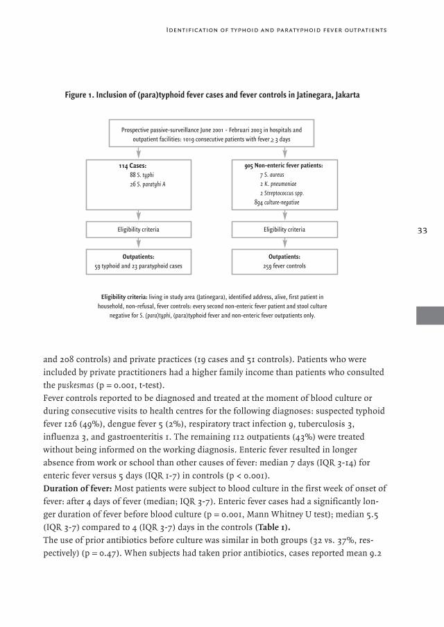
and 208 controls) and private practices (19 cases and 51 controls). Patients who were
included by private practitioners had a higher family income than patients who consulted
the puskesmas (p = 0.001, t-test).
Fever controls reported to be diagnosed and treated at the moment of blood culture or
during consecutive visits to health centres for the following diagnoses: suspected typhoid
fever 126 (49%), dengue fever 5 (2%), respiratory tract infection 9, tuberculosis 3,
influenza 3, and gastroenteritis 1. The remaining 112 outpatients (43%) were treated
without being informed on the working diagnosis. Enteric fever resulted in longer
absence from work or school than other causes of fever: median 7 days (IQR 3-14) for
enteric fever versus 5 days (IQR 1-7) in controls (p < 0.001).
Duration of fever: Most patients were subject to blood culture in the first week of onset of
fever: after 4 days of fever (median; IQR 3-7). Enteric fever cases had a significantly lon-
ger duration of fever before blood culture (p = 0.001, Mann Whitney U test); median 5.5
(IQR 3-7) compared to 4 (IQR 3-7) days in the controls (Table 1).
The use of prior antibiotics before culture was similar in both groups (32 vs. 37%, res-
pectively) (p = 0.47). When subjects had taken prior antibiotics, cases reported mean 9.2
33
Figure 1. Inclusion of (para)typhoid fever cases and fever controls in Jatinegara, Jakarta
Eligibility criteria: living in study area (Jatinegara), identified address, alive, first patient in household, non-refusal, fever controls: every second non-enteric fever patient and stool culture
negative for S. (para)typhi, (para)typhoid fever and non-enteric fever outpatients only.
Prospective passive-surveillance June 2001 - Februari 2003 in hospitals and outpatient facilities: 1019 consecutive patients with fever >_ 3 days
114 Cases:88 S. typhi26 S. paratyhi A
905 Non-enteric fever patients:7 S. aureus2 K. pneumoniae2 Streptococcus spp.
894 culture-negative
Eligibility criteria Eligibility criteria
Outpatients:59 typhoid and 23 paratyphoid cases
Outpatients:259 fever controls
Identification of typhoid and paratyphoid fever outpatients
18439 Vollaard 14-12-2004 08:24 Pagina 33
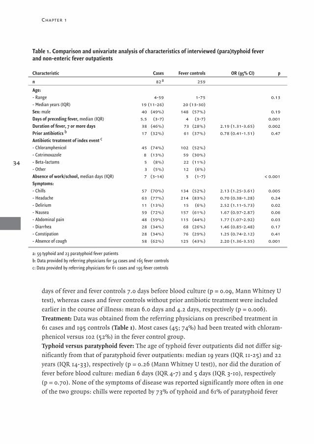
days of fever and fever controls 7.0 days before blood culture (p = 0.09, Mann Whitney U
test), whereas cases and fever controls without prior antibiotic treatment were included
earlier in the course of illness: mean 6.0 days and 4.2 days, respectively (p = 0.006).
Treatment: Data was obtained from the referring physicians on prescribed treatment in
61 cases and 195 controls (Table 1). Most cases (45; 74%) had been treated with chloram-
phenicol versus 102 (52%) in the fever control group.
Typhoid versus paratyphoid fever: The age of typhoid fever outpatients did not differ sig-
nificantly from that of paratyphoid fever outpatients: median 19 years (IQR 11-25) and 22
years (IQR 14-33), respectively (p = 0.26 (Mann Whitney U test)), nor did the duration of
fever before blood culture: median 6 days (IQR 4-7) and 5 days (IQR 3-10), respectively
(p = 0.70). None of the symptoms of disease was reported significantly more often in one
of the two groups: chills were reported by 73% of typhoid and 61% of paratyphoid fever
34
Chapter 1
Table 1. Comparison and univariate analysis of characteristics of interviewed (para)typhoid fever and non-enteric fever outpatients
Characteristic Cases Fever controls OR (95% CI) p
n 82 a 259
Age:- Range 4-59 1-75 0.13- Median years (IQR) 19 (11-26) 20 (13-30)Sex: male 40 (49%) 148 (57%) 0.19Days of preceding fever, median (IQR) 5.5 (3-7) 4 (3-7) 0.001Duration of fever, 7 or more days 38 (46%) 73 (28%) 2.19 (1.31-3.65) 0.002Prior antibiotics b 17 (32%) 61 (37%) 0.78 (0.41-1.51) 0.47Antibiotic treatment of index event c
- Chloramphenicol 45 (74%) 102 (52%)- Cotrimoxazole 8 (13%) 59 (30%)- Beta-lactams 5 (8%) 22 (11%)- Other 3 (5%) 12 (6%)Absence of work/school, median days (IQR) 7 (3-14) 5 (1-7) < 0.001Symptoms:- Chills 57 (70%) 134 (52%) 2.13 (1.25-3.61) 0.005- Headache 63 (77%) 214 (83%) 0.70 (0.38-1.28) 0.24- Delirium 11 (13%) 15 (6%) 2.52 (1.11-5.73) 0.02- Nausea 59 (72%) 157 (61%) 1.67 (0.97-2.87) 0.06- Abdominal pain 48 (59%) 115 (44%) 1.77 (1.07-2.92) 0.03- Diarrhea 28 (34%) 68 (26%) 1.46 (0.85-2.48) 0.17- Constipation 28 (34%) 76 (29%) 1.25 (0.74-2.12) 0.41- Absence of cough 58 (62%) 125 (43%) 2.20 (1.36-3.55) 0.001
a: 59 typhoid and 23 paratyphoid fever patients b: Data provided by referring physicians for 54 cases and 165 fever controlsc: Data provided by referring physicians for 61 cases and 195 fever controls
18439 Vollaard 14-12-2004 08:24 Pagina 34
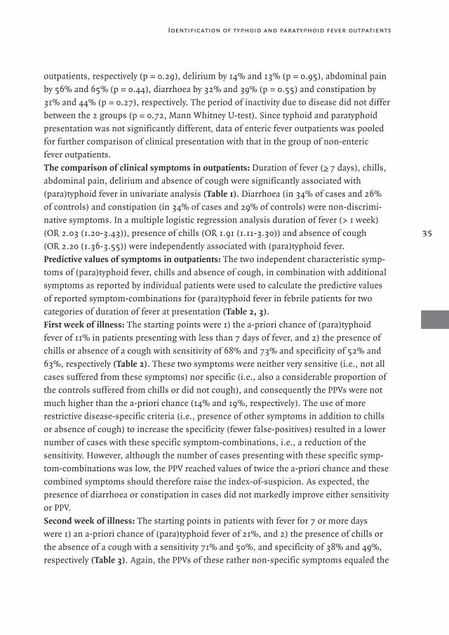
outpatients, respectively (p = 0.29), delirium by 14% and 13% (p = 0.95), abdominal pain
by 56% and 65% (p = 0.44), diarrhoea by 32% and 39% (p = 0.55) and constipation by
31% and 44% (p = 0.27), respectively. The period of inactivity due to disease did not differ
between the 2 groups (p = 0.72, Mann Whitney U-test). Since typhoid and paratyphoid
presentation was not significantly different, data of enteric fever outpatients was pooled
for further comparison of clinical presentation with that in the group of non-enteric
fever outpatients.
The comparison of clinical symptoms in outpatients: Duration of fever (>_ 7 days), chills,
abdominal pain, delirium and absence of cough were significantly associated with
(para)typhoid fever in univariate analysis (Table 1). Diarrhoea (in 34% of cases and 26%
of controls) and constipation (in 34% of cases and 29% of controls) were non-discrimi-
native symptoms. In a multiple logistic regression analysis duration of fever (> 1 week)
(OR 2.03 (1.20-3.43)), presence of chills (OR 1.91 (1.11-3.30)) and absence of cough
(OR 2.20 (1.36-3.55)) were independently associated with (para)typhoid fever.
Predictive values of symptoms in outpatients: The two independent characteristic symp-
toms of (para)typhoid fever, chills and absence of cough, in combination with additional
symptoms as reported by individual patients were used to calculate the predictive values
of reported symptom-combinations for (para)typhoid fever in febrile patients for two
categories of duration of fever at presentation (Table 2, 3).
First week of illness: The starting points were 1) the a-priori chance of (para)typhoid
fever of 11% in patients presenting with less than 7 days of fever, and 2) the presence of
chills or absence of a cough with sensitivity of 68% and 73% and specificity of 52% and
63%, respectively (Table 2). These two symptoms were neither very sensitive (i.e., not all
cases suffered from these symptoms) nor specific (i.e., also a considerable proportion of
the controls suffered from chills or did not cough), and consequently the PPVs were not
much higher than the a-priori chance (14% and 19%, respectively). The use of more
restrictive disease-specific criteria (i.e., presence of other symptoms in addition to chills
or absence of cough) to increase the specificity (fewer false-positives) resulted in a lower
number of cases with these specific symptom-combinations, i.e., a reduction of the
sensitivity. However, although the number of cases presenting with these specific symp-
tom-combinations was low, the PPV reached values of twice the a-priori chance and these
combined symptoms should therefore raise the index-of-suspicion. As expected, the
presence of diarrhoea or constipation in cases did not markedly improve either sensitivity
or PPV.
Second week of illness: The starting points in patients with fever for 7 or more days
were 1) an a-priori chance of (para)typhoid fever of 21%, and 2) the presence of chills or
the absence of a cough with a sensitivity 71% and 50%, and specificity of 38% and 49%,
respectively (Table 3). Again, the PPVs of these rather non-specific symptoms equaled the
35
Identification of typhoid and paratyphoid fever outpatients
18439 Vollaard 14-12-2004 08:24 Pagina 35

Chapter 1
36
Table 2. Sensitivity, specificity and predictive values (%) of clinical symptoms for (para)typhoid feverin outpatients with fever < 7 days
Symptoms Sensitivity Specificity PPV NPV
A-priori chance 11
Chills 68 52 14 93Chills, absence of cough 50 78 21 93Chills, absence of cough, abdominal pain 30 91 28 92Chills, absence of cough, abdominal pain, diarrhoea or constipation 23 93 28 91Chills, absence of cough, delirium 5 98 25 90Chills, absence of cough, abdominal pain, delirium a 0 100 - 89Chills, abdominal pain 36 77 16 91Chills, abdominal pain, diarrhoea or constipation 27 84 17 91Chills, abdominal pain, delirium b 2 100 100 90Absence of cough 73 63 19 95Absence of cough, abdominal pain 34 84 20 92Absence of cough, abdominal pain, diarrhoea or constipation 25 91 26 91Absence of cough, delirium 7 97 23 90Absence of cough, abdominal pain, delirium c 0 99 0 89
A-priori chance of (para)typhoid fever in febrile patients = 11% (44 cases and 2 x 185 fever controls) a: Combination reported by none of the cases and controlsb: Combination reported by none of the controlsc: Combination reported by none of the cases and by 2 controls
Table 3. Sensitivity, specificity and predictive values (%) of clinical symptoms for (para)typhoid fever in outpatients with fever >_ 7 days
Symptoms Sensitivity Specificity PPV NPV
A-priori chance 21
Chills 71 38 23 84Chills, absence of cough 34 74 25 81Chills, absence of cough, abdominal pain 29 86 35 82Chills, absence of cough, abdominal pain, diarrhoea or constipation 18 92 37 81Chills, absence of cough, delirium 8 99 60 80Chills, absence of cough, abdominal pain, delirium a 8 100 100 81Chills, abdominal pain 55 66 30 85Chills, abdominal pain, diarrhoea or constipation 34 77 28 82Chills, abdominal pain, delirium 11 96 40 81Absence of cough 50 49 20 79Absence of cough, abdominal pain 39 78 32 83Absence of cough, abdominal pain, diarrhoea or constipation 29 85 33 82Absence of cough, delirium 11 96 40 81Absence of cough, abdominal pain, delirium 11 99 67 81
A-priori chance = 21% (38 cases and 2 x 73 fever controls)a: Combination reported by none of the controls
18439 Vollaard 14-12-2004 08:24 Pagina 36

a-priori chance. Also, the presence of additional symptoms reduced both the number of
cases and controls presenting with these symptom-combinations with similar trends for
the specificity (i.e., up), sensitivity (i.e., down) and PPV (i.e., up). Delirium in patients
with fever >_ 7 days in addition to other symptoms would increase the likelihood of
(para)typhoid fever to at least 2 times the a-priori chance (up to 67% and in 1 combina-
tion to 100%).
Use of clinical symptoms for screening and further diagnostic tests: When culture facili-
ties are available in (immediate vicinity of ) a health care facility, suspected typhoid fever
patients should be subject to blood culture. Whether the clinical symptoms of febrile
patients can be used as a screening test to decide on further diagnostic steps (i.e., blood
culture or not) depends on a high sensitivity of the symptom-combinations in individual
patients. No true enteric fever patient should be missed and therefore wide inclusion cri-
teria should guarantee a high sensitivity and subsequent entrance to further diagnostic
steps, even if that meant that the blood of a proportion of non-enteric fever patients, with
similar symptoms as cases, would be cultured as well. The highest sensitivity was found
in the first week of illness: absence of a cough (73%) (Table 2).
However, application of this presentation as a criterion for further diagnostic testing
would mean that 27% of the cases would be missed and also that 37% of the non-enteric
fever patients would be cultured (specificity 63%).
None of symptom(-combination)s yielded a sufficiently high detection rate of true
(para)typhoid cases due to the limited sensitivity. Therefore, the clinical presentation
cannot be used as a screening instrument for (para)typhoid fever to decide whether an
individual patient should additionally be subject to blood culture for the confirmation
of (para)typhoid fever or not.
Use of clinical symptoms for empiric treatment of (para)typhoid fever: When culture
facilities are not available, empiric treatment should be started in patients with suspected
typhoid fever to avoid the serious complications of (para)typhoid fever (i.e., hemorrhage
and perforation) and advice on hygiene should be given to patients to reduce the risk of
further spread. For that purpose the predictive values of symptom-combinations (Table 2, 3)
are useful, because physicians could check for symptoms in febrile patients to increase
the index-of-suspicion for (para)typhoid fever (Figure 2, expressed as ratios).
A first clue should be the duration of fever, because the a-priori chance increased twofold
in patients with fever >_ 7 days (from 11% to 21%). In the first week of fever the absence of
cough with or without chills would increase the probability of (para)typhoid fever in a
febrile patient twofold as well (from 11% to 19-21%). The presence of other symptoms in
addition to absence of cough and/or chills did not increase the PPV any further in the first
week, and are therefore not very helpful to increase the index-of-suspicion. For patients
with 7 or more days of fever delirium in combination with other symptoms increased the
37
Identification of typhoid and paratyphoid fever outpatients
18439 Vollaard 14-12-2004 08:24 Pagina 37
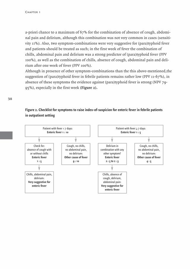
Chapter 1
a-priori chance to a maximum of 67% for the combination of absence of cough, abdomi-
nal pain and delirium, although this combination was not very common in cases (sensiti-
vity 11%). Also, two symptom-combinations were very suggestive for (para)typhoid fever
and patients should be treated as such; in the first week of fever the combination of
chills, abdominal pain and delirium was a strong predictor of (para)typhoid fever (PPV
100%), as well as the combination of chills, absence of cough, abdominal pain and deli-
rium after one week of fever (PPV 100%).
Although in presence of other symptom-combinations than the this above-mentioned,the
suggestion of (para)typhoid fever in febrile patients remains rather low (PPV 11-67%), in
absence of these symptoms the evidence against (para)typhoid fever is strong (NPV 79-
95%), especially in the first week (Figure 2).
38
Figure 2. Checklist for symptoms to raise index-of-suspicion for enteric fever in febrile patients in outpatient setting
Patient with fever < 7 days:Enteric fever 1 : 10
Check for:absence of cough with
or without chillsEnteric fever
1 : 5
Chills, abdominal pain,delirium:
Very suggestive for enteric fever
Cough, no chills, no abdominal pain,
no delirium:Other cause of fever
9 : 10
Delirium in combination with any
other symptom?Enteric fever2 : 5 to 2 : 3
Chills, absence of cough, delirium, abdominal pain:
Very suggestive for enteric fever
Cough, no chills, no abdominal pain,
no delirium: Other cause of fever
4 : 5
Patient with fever >_ 7 days:Enteric fever 1 : 5
18439 Vollaard 14-12-2004 08:24 Pagina 38

Discussion
In our recent surveillance study in the Jatinegara district, Jakarta, we observed that 88%
of (para)typhoid fever patients was treated in outpatient setting in Jakarta, making an
evaluation of (para)typhoid fever outpatients relevant. Paratyphoid fever could not be
distinguished clinically from typhoid fever. Our study confirmed quantitatively that in
outpatients (para)typhoid fever presentation is relatively similar to other febrile illnesses.
In consequence, due to the limited sensitivity of symptom-combinations the clinical pre-
sentation cannot be used as a screening method for further diagnostic tests and therefore
all febrile patients should be subject to blood culture (or serological tests) for the confir-
mation of (para)typhoid fever. When culture facilities are absent decisions on empiric
treatment and advice on hygiene measures should be influenced by the duration of fever
of patients, absence of cough with or without chills in the first week of fever, and deli-
rium in the second week, to increase the likelihood of (para)typhoid fever in a febrile
outpatient from 1 : 10 in the first week to at most 2 : 3 for the latter combination,
although numbers of cases presenting as such are low.
Some issues regarding the inclusion of patients need consideration. In our surveillance
study Bactec blood cultures were used for confirmation of (para)typhoid fever. The effec-
tive neutralization of antibiotics by resins in Bactec bottles (Koontz et al., 1991) reduced
the interference of prior antibiotics, and accordingly the relative number of patients using
antibiotics before blood culture was equal in both groups. Because 75% of the patients in
our study was included in the first week of illness, the lower sensitivity of the Widal test
compared to blood culture would have been reduced even more by the short duration of
illness (Parry et al., 1999). Furthermore, to minimize misclassification of controls, since
the sensitivity of blood culture for confirmation of (para)typhoid fever never reaches 100
percent, faeces was cultured in all subjects at the household visit and two stool culture-
positive fever controls were excluded from the analysis. With respect to the use of a
questionnaire, although recall-bias cannot be ruled out because of the interval of 3 weeks
between blood culture and interview, it would have affected both groups equally.
Moreover, most of the fever controls thought they had been treated for typhoid fever or
were not informed on the final diagnosis, consequently reducing recall-bias. Finally, the
inclusion criterion of three or more days of fever might have affected the inclusion of
children, who may have an atypical presentation of typhoid fever (Ferreccio et al., 1984).
Consequently, in contrast to reports from India and Bangladesh (Sinha et al., 1999; Saha
et al., 2001) few cases were young children and the presented clinical guideline is therefore
predominantly applicable in adolescent and adult fever patients.
Most of the controls had been treated empirically without final diagnosis. This treatment
strategy is an accurate reflection of the conditions of physicians in outpatient health centres
39
Identification of typhoid and paratyphoid fever outpatients
18439 Vollaard 14-12-2004 08:24 Pagina 39

Chapter 1
in many developing countries. In absence of laboratory facilities likelihood estimates are
applied that are based on local endemicity levels of typhoid and other febrile illnesses,
and the clinical presentation of febrile patients. The latter was explored extensively in our
study, but the former might differ in other settings and consequently influence the gene-
ral applicability of our findings, because we showed that the interpretation of clinical pre-
sentation to predict (para)typhoid fever in a febrile patient also depends on the local
endemicity levels of the prevailing febrile illnesses, i.e., the a-priori chance of finding
(para)typhoid fever as a cause of fever. When the a-priori chance, i.e., the relative number
of enteric fever patients in a patient-population, increases, also the PPV of symptoms will
increase. Predictive values are therefore more useful for decisions on empiric treatment
than the sensitivity of symptoms or odds ratios for symptoms in the total group of
patients, because the latter are unaffected by the a-priori chance. The prior chance of
having typhoid fever in our study was 8.6% among all patients with fever for three or
more days, comparable to rates from other surveillance studies for typhoid fever using
the same inclusion criteria: 4.6-8.5% (Lin et al., 2000; Saha et al., 2001; Sinha et al., 1999;
Simanjuntak et al., 1991; Bodhidatta et al., 1987), but was shown to increase in patients
with fever for at least one week. Our study demonstrated that in daily medical practice in
outpatient setting the index-of-suspicion for (para)typhoid fever should be raised in ado-
lescent patients who report prolonged fever, chills, absence of cough and especially deli-
rium after one week of fever, but that these symptoms cannot be used for the screening of
patients for further diagnostic testing due to the low sensitivity of symptoms.
The treatment of (para)typhoid fever in Jakarta with relatively cheap, standard antibiotics
such as chloramphenicol, ampicillin and cotrimoxazole, still is a valid option, since only
3% of the S. typhi and none of the S. paratyphi A strains were multi-drug resistant, in line
with other reports from Indonesia (Isbandrio et al., 1994; Tjaniadi et al., 2003; Oyofo et
al., 2002). The accessibility of the low-priced puskesmas and early and adequate treatment
in these outpatient centres was likely the cause of the absence of deaths in the case-group
and the low rate of hospitalization.
Non-treatment of (para)typhoid cases would increase complication rates of haemorrhage
and perforation and also lead to infection of other household contacts if hygiene is poor
(Luxemburger et al., 2001). As was shown in stool cultures after 3-4 weeks in treated
(para)typhoid case subjects, 5% of the cases continued to excrete bacteria, enabling latter
transmission route. Adequate treatment and public health measures in case of suspected
enteric fever are therefore essential. The evaluated symptoms allow an increase in the cor-
rect identification of (para)typhoid fever patients from an a-priori chance of 11% in febrile
patients to 67% at most, when we disregard the few cases with the very rare combination
of all chills, abdominal pain and delirium with or without absence of cough. However, in
settings without culture facilities the empiric treatment of (para)typhoid fever will inevi-
40
18439 Vollaard 14-12-2004 08:24 Pagina 40
tably include a substantial number of patients with other causes of fever, because a con-
clusive clinical picture could be not distilled from our data that may help physicians to
restrict the prescription of antibiotics to those patients with a high probability of
(para)typhoid fever. Whereas most symptom-combinations were not very useful for the
identification of enteric fever patients, in absence of these symptoms other causes for
fever in a febrile outpatient are probable in at least 4 out of 5 patients.
We focussed on the clinical presentation of outpatients and did not include laboratory
parameters such as white blood cell or thrombocyte count or Widal test in the prediction
model (Ross and Abraham, 1987). The additional information that can be obtained using
lab parameters in patients with a high index-of-suspicion as identified in our study needs
further validation in outpatient centres with laboratory equipment.
In conclusion, as long as culture facilities are limited (and costs inhibitive), and sensitive
serological tests are not available, many febrile patients without S. (para)typhi infection
will receive empiric treatment for suspected (para)typhoid fever. Although chills, deli-
rium, abdominal pain, and/or absence of a cough in adolescent patients with prolonged
fever should raise the index-of-suspicion of (para)typhoid fever, no conclusive clinical
picture could be presented to differentiate (para)typhoid fever from other febrile illnesses.
Culture or serological tests are therefore essential in the diagnosis and in health surveil-
lance. A reduction of the number of cultures in febrile patients could not be proposed,
because of the limited sensitivity of the symptoms in (para)typhoid fever compared to
other causes of fever. A follow-up visit after 3-5 days of antibiotic treatment of suspected
(para)typhoid fever is recommended to decide on prolongation of treatment for patients
with delayed fever-clearance, because after administration of standard antibiotics fever
due to typhoid fever should reside after 3-5 days of treatment (Parry et al., 2002). Also
instruction on adequate hygiene in the first weeks after treatment should be emphasised.
AcknowledgementsThis study was funded by a grant through the Royal Netherlands Academy of Sciences
and Arts (KNAW). The study group likes to thank dr. Wil Dolmans for his help in the
preparatory stage of this project. We thank the physicians, nurses, and technicians of the
participating health centres in Jakarta for their cooperation: Mitra Internasional hospital
and its reference microbiology lab, Budhi Asih hospital, St. Carolus hospital,
Persahabatan hospital, all puskesmas in Jatinegara, the local private practitioners, and the
Microbiology department of Atma Jaya University Hospital. This study could not have
been done without our research-assistants: Billy Hunsinger, Ferry Kandaw, Rinny
Listyani, Meily, Vea Noveria, Carmelita Ridwan, Min Ali Sugiharto, Lidwina Sutikno,
Mariana Tasman, and Lily Yaputra.
41
Identification of typhoid and paratyphoid fever outpatients
18439 Vollaard 14-12-2004 08:24 Pagina 41
42
References
1. Arya SC, Sharma KB (1995) Urgent need for effective vaccine against Salmonella paratyphi A, B and C. Vaccine 13, 1727-1728.
2. Bodhidatta L, Taylor DN, Thisyakorn U, Echeverria P (1987) Control of typhoid fever in Bangkok, Thailand, by annual
immunization of schoolchildren with parenteral typhoid vaccine. Rev Infect Dis 9, 841-845.
3. Butler T, Islam A, Kabir I, Jones PK (1991) Patterns of morbidity and mortality in typhoid fever dependent on age and gender:
review of 552 hospitalized patients with diarrhea. Rev Infect Dis 13, 85-90.
4. Christie AB (1987) Infectious Diseases: Epidemiology and Clinical Practice, fourth ed. Churchill Livingstone, Edinburgh.
5. Cvjetanovic B, Grab B, Uemura K (1971) Epidemiological model of typhoid fever and its use in the planning and evaluation of
antityphoid immunization and sanitation programmes. Bull World Health Organ 45, 53-75.
6. Ferreccio C, Levine MM, Manterola A et al. (1984) Benign bacteremia caused by Salmonella typhi and paratyphi in children
younger than 2 years. J Pediatr 104, 899-901.
7. Isbandrio BB, Gasem MH, Dolmans WM, Hoogkamp-Korstanje JA (1994) Comparative activities of three quinolones and seven
comparison standard drugs against Salmonella typhi from Indonesia. J Antimicrob Chemother 33, 1055-1056.
8. Koontz FP, Flint KK, Reynolds JK, Allen SD (1991) Multicenter comparison of the high volume (10 ml) NR BACTEC PLUS system
and the standard (5 ml) NR BACTEC system. Diagn Microbiol Infect Dis 14, 111-118.
9. Lin FY, Vo AH, Phan VB et al. (2000) The epidemiology of typhoid fever in the Dong Thap Province, Mekong Delta region of
Vietnam. Am J Trop Med Hyg 62, 644-648.
10. Luxemburger C, Chau MC, Mai NL et al. (2001) Risk factors for typhoid fever in the Mekong delta, southern Viet Nam:
a case-control study. Trans R Soc Trop Med Hyg 95, 19-23.
11. Mermin JH, Villar R, Carpenter J et al. (1999) A massive epidemic of multidrug-resistant typhoid fever in Tajikistan associated
with consumption of municipal water. J Infect Dis 179, 1416-1422.
12. Oyofo BA, Subekti D, Tjaniadi P et al. (2002) Enteropathogens associated with acute diarrhea in community and hospital
patients in Jakarta, Indonesia. FEMS Immunol Med Microbiol 34, 139-146.
13. Pang T, Levine MM, Ivanoff B, Wain J, Finlay BB (1998) Typhoid fever- important issues still remain. Trends Microbiol 6, 131-133.
14. Parry CM, Hien TT, Dougan G, White NJ, Farrar JJ (2002) Typhoid fever. N Engl J Med 347, 1770-1782.
15. Parry CM, Hoa NT, Diep TS et al. (1999) Value of a single-tube widal test in diagnosis of typhoid fever in Vietnam.
J Clin Microbiol 37, 2882-2886.
16. Reeve PA, Dwyer DE (1995) An outbreak of typhoid associated with a “lafet” in Vanuatu. Med J Aust 162, 55-56.
17. Ross IN, Abraham T (1987) Predicting enteric fever without bacteriological culture results. Trans R Soc Trop Med Hyg 81, 374-377.
18. Rowe B, Ward LR, Threlfall EJ (1997) Multidrug-resistant Salmonella typhi: a worldwide epidemic. Clin Infect Dis 24 Suppl 1,
S106-S109.
19. Saha SK, Baqui AH, Hanif M et al. (2001) Typhoid fever in Bangladesh: implications for vaccination policy. Pediatr Inf Dis J 20, 521-524.
20. Simanjuntak CH, Paleologo FP, Punjabi NH et al. (1991) Oral immunisation against typhoid fever in Indonesia with
Ty21a vaccine. Lancet 338, 1055-1059.
21. Sinha A, Sazawal S, Kumar R et al. (1999) Typhoid fever in children aged less than 5 years. Lancet 354, 734-737.
22. Swaddiwudhipong W, Kanlayanaphotporn J (2001) A common-source water-borne outbreak of multidrug-resistant typhoid
fever in a rural Thai community. J Med Assoc Thai 84, 1513-1517.
23. Tjaniadi P, Lesmana M, Subekti D et al. (2003) Antimicrobial resistance of bacterial pathogens associated with diarrheal
patients in Indonesia. Am J Trop Med Hyg 68, 666-670.
24. Vollaard AM, Ali S., van Asten AGH et al. (2004) Risk factors for typhoid and paratyphoid fever in Jakarta, Indonesia.
JAMA 291: 2607-2615.
25. Yew FS, Chew SK, Goh KT, Monteiro EH, Lim YS (1991) Typhoid fever in Singapore: a review of 370 cases.
J Trop Med Hyg 94, 352-357.
Chapter 1
18439 Vollaard 14-12-2004 08:24 Pagina 42
2
Risk factors for typhoid and paratyphoid fever in Jakarta, Indonesia
Albert M. Vollaard 1 Soegianto Ali 2
Henri A.G.H. van Asten 3 Suwandhi Widjaja 4
Leo G. Visser 1 Charles Surjadi 5 Jaap T. van Dissel 1
1 Dept. Infectious Diseases, Leiden University Medical Center, the Netherlands2 Dept. Biology, Medical Faculty Atma Jaya Catholic University, Jakarta, Indonesia
3 Institute for International Health, University Medical Center Nijmegen, the Netherlands4 Dept. Internal Medicine, Atma Jaya Catholic University, Jakarta, Indonesia
5 Center for Health Research, Atma Jaya Catholic University, Jakarta, Indonesia
JAMA 2004; 291: 2607-2615
Copyrighted © (2004), American Medical Association. All rights reserved.
18439 Vollaard 14-12-2004 08:24 Pagina 43
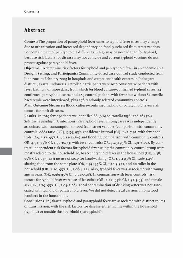
Abstract
Context: The proportion of paratyphoid fever cases to typhoid fever cases may change
due to urbanization and increased dependency on food purchased from street vendors.
For containment of paratyphoid a different strategy may be needed than for typhoid,
because risk factors for disease may not coincide and current typhoid vaccines do not
protect against paratyphoid fever.
Objective: To determine risk factors for typhoid and paratyphoid fever in an endemic area.
Design, Setting, and Participants: Community-based case-control study conducted from
June 2001 to February 2003 in hospitals and outpatient health centers in Jatinegara
district, Jakarta, Indonesia. Enrolled participants were 1019 consecutive patients with
fever lasting 3 or more days, from which 69 blood culture–confirmed typhoid cases, 24
confirmed paratyphoid cases, and 289 control patients with fever but without Salmonella
bacteremia were interviewed, plus 378 randomly selected community controls.
Main Outcome Measures: Blood culture–confirmed typhoid or paratyphoid fever; risk
factors for both diseases.
Results: In 1019 fever patients we identified 88 (9%) Salmonella typhi and 26 (3%)
Salmonella paratyphi A infections. Paratyphoid fever among cases was independently
associated with consumption of food from street vendors (comparison with community
controls: odds ratio [OR], 3.34; 95% confidence interval [CI], 1.41-7.91; with fever con-
trols: OR, 5.17; 95% CI, 2.12-12.60) and flooding (comparison with community controls:
OR, 4.52; 95% CI, 1.90-10.73; with fever controls: OR, 3.25; 95% CI, 1.31-8.02). By con-
trast, independent risk factors for typhoid fever using the community control group were
mostly related to the household, ie, to recent typhoid fever in the household (OR, 2.38;
95% CI, 1.03-5.48); no use of soap for handwashing (OR, 1.91; 95% CI, 1.06-3.46);
sharing food from the same plate (OR, 1.93; 95% CI, 1.10-3.37), and no toilet in the
household (OR, 2.20; 95% CI, 1.06-4.55). Also, typhoid fever was associated with young
age in years (OR, 0.96; 95% CI, 0.94-0.98). In comparison with fever controls, risk
factors for typhoid fever were use of ice cubes (OR, 2.27; 95% CI, 1.31-3.93) and female
sex (OR, 1.79; 95% CI, 1.04-3.06). Fecal contamination of drinking water was not asso-
ciated with typhoid or paratyphoid fever. We did not detect fecal carriers among food
handlers in the households.
Conclusions: In Jakarta, typhoid and paratyphoid fever are associated with distinct routes
of transmission, with the risk factors for disease either mainly within the household
(typhoid) or outside the household (paratyphoid).
Chapter 2
18439 Vollaard 14-12-2004 08:24 Pagina 44
Introduction
Typhoid fever, a food- and waterborne disease caused by Salmonella enterica serotype Typhi
(S. typhi), is a serious public health problem in developing countries that claims 600 000
lives every year.1 Paratyphoid fever, caused by Salmonella paratyphi A, B, or C, has a disease
presentation similar to that of typhoid fever, but its incidence is reportedly about one tenth
that of typhoid (ratio, 1:10-20).2-3 In developing countries the identification of risk factors
and relevant route of transmission for a disease such as typhoid fever is essential for the
development of rational control strategies. Resources could consequently be allocated to
where they count most, e.g., to the construction or expansion of water distribution
networks or sewage systems, chlorination of drinking water, ensurance of food safety,
hygiene education, mass vaccination campaigns, and/or the identification of carriers
within or outside the households of patients.
Risk factors for typhoid fever have been identified in several epidemiologic studies sug-
gesting either waterborne 4-8 or food borne transmission.7,9-11 Whether these factors coin-
cide with those for paratyphoid fever has not been determined. The assumption is that in
paratyphoid fever, a higher dose of bacteria is required for infection than in typhoid fever;
consequently, food is implicated as the major vehicle for transmission of paratyphoid fever,
since Salmonella bacteria can multiply in food.12 Comparison of the transmission of both
diseases is becoming increasingly relevant, because recent reports have demonstrated an
increasing occurrence of paratyphoid fever.3,13 It is not clear whether this is due to incom-
pleteness of epidemiologic data in endemic countries or to a downward trend in the inci-
dence of typhoid fever 1,14 and a consequent relative or absolute increase in the incidence of
paratyphoid fever. In consequence, however, public health measures may well be refocused.
In particular, recent interest in mass immunization as a control strategy in regions of ende-
micity needs to be reconsidered if the incidence of typhoid fever is decreasing and para-
typhoid fever is on the rise, because current typhoid fever vaccines (i.e., parenteral Vi and
oral Ty21a vaccine) do not protect against paratyphoid fever.2
In this community-based case-control study in an endemic area in East Jakarta, Indonesia,
we compared case patients having paratyphoid and typhoid fever with random community
controls to identify hygienic practices, eating habits, and environmental and household
characteristics that could elucidate prevailing transmission routes. For this purpose we
also examined the microbiological quality of drinking water and cultured stools of intra-
household food handlers to detect transient or chronic carriers. A second control group
composed of patients with non-enteric fever was used for comparison and confirmation
of the results. Patients with typhoid fever, paratyphoid fever, and non-enteric fever were
identified in a prospective passive-surveillance study involving hospitals and outpatient
health centers in the study area.
45
Risk factors for typhoid and paratyphoid fever
18439 Vollaard 14-12-2004 08:24 Pagina 45
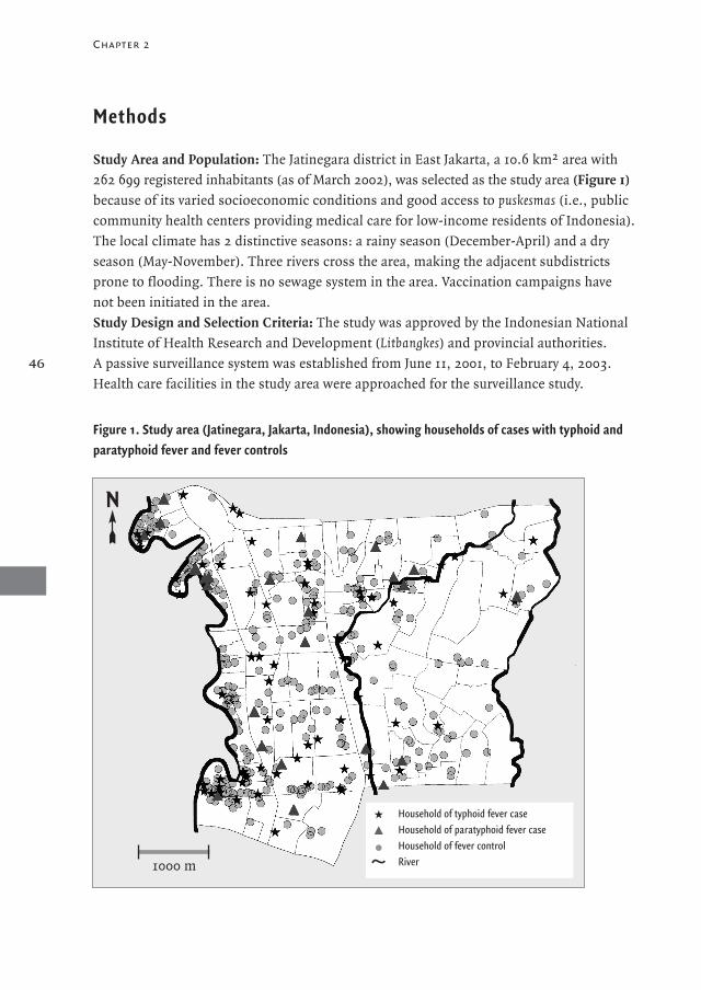
Chapter 2
Methods
Study Area and Population: The Jatinegara district in East Jakarta, a 10.6 km2 area with
262 699 registered inhabitants (as of March 2002), was selected as the study area (Figure 1)
because of its varied socioeconomic conditions and good access to puskesmas (i.e., public
community health centers providing medical care for low-income residents of Indonesia).
The local climate has 2 distinctive seasons: a rainy season (December-April) and a dry
season (May-November). Three rivers cross the area, making the adjacent subdistricts
prone to flooding. There is no sewage system in the area. Vaccination campaigns have
not been initiated in the area.
Study Design and Selection Criteria: The study was approved by the Indonesian National
Institute of Health Research and Development (Litbangkes) and provincial authorities.
A passive surveillance system was established from June 11, 2001, to February 4, 2003.
Health care facilities in the study area were approached for the surveillance study.
46
Figure 1. Study area (Jatinegara, Jakarta, Indonesia), showing households of cases with typhoid andparatyphoid fever and fever controls
Household of typhoid fever caseHousehold of paratyphoid fever caseHousehold of fever controlRiver1000 m
N
18439 Vollaard 14-12-2004 08:24 Pagina 46
Those participating included all 4 hospitals in the immediate vicinity, 8 of the 13 additional
small private outpatient clinics in the area, and all 12 puskesmas. A fee of US $0.35 covers
3 days of antibiotic treatment, but cultures or Widal tests are not part of the usual dia-
gnostic practice in puskesmas. Eligible patients were individuals living in the study area
who consulted one of the participating health care facilities because of self-reported fever
for 3 or more consecutive days. A single blood specimen for culture was collected from
each eligible patient. Depending on the age of the patient, 5 to 10 mL of blood was collec-
ted into blood culture vials (aerobic) containing antibiotic-absorbing resins (Bactec;
Becton Dickinson, Franklin Lakes, NJ) that were provided to the centers by the study
group free of charge.
Cases were eligible patients with blood culture–confirmed S. typhi or S. paratyphi infection.
All cases were subject to a household visit within a month after the febrile episode that
prompted the blood culture.
Blood cultures of patients with non-enteric fever showed either no growth or bacteria
other than S. typhi or S. paratyphi as cause of fever. Malaria could be excluded in the diffe-
rential diagnosis of prolonged fever, because transmission does not occur in Jakarta.
Every second consecutive patient with non-enteric fever was selected as a fever control
and visited. Also, during the surveillance, community controls were randomly selected
within a random household in every third rukun tetangga (i.e., the smallest administrative
unit of 40-60 area households) of a total of 1140 rukun tetanggas. When a community
control reported fever in the 30 days preceding the interview or refused participation, the
house on alternating sides of the initially selected household was approached. The selec-
tion of both groups of controls was nonmatched for age, sex, or neighborhood (i.e.,
residence in 1 of the 8 subdistricts of Jatinegara) to limit selection bias and prevent over-
matching. Four controls from both groups for every case of enteric fever were selected to
increase statistical power.
Household Visits and Sample Collection: Cases and controls were interviewed by trained
medical school graduates, using a standardized questionnaire that included the known
risk factors from previous studies and questions from a questionnaire that was used in a
similar risk factor study, which had been locally tested and validated.6 Written informed
consent was provided by all participants at the household visit. To prevent the overrepre-
sentation of multiple-case households, only 1 patient (i.e., the first reported case or fever
control) per household was interviewed. If cases or controls were younger than 13 years,
the mother or guardian was interviewed. No time frame for hygiene behavior and food
habits was mentioned, because it aimed at the description of usual practice. A household
was defined as a dwelling whose inhabitants ate from the same pot. Flooding was defined
as inundation of the house of a participant in the 12 months preceding the interview.
47
Risk factors for typhoid and paratyphoid fever
18439 Vollaard 14-12-2004 08:24 Pagina 47

Chapter 2
Intrahousehold food handlers were defined as individuals preparing meals for cases or
controls 3 or more times a week. A single stool sample of 2 g was collected from all
cases, controls, and their intrahousehold food handlers in a vial with Cary-Blair transport
medium and samples were processed within 24 hours after collection. Water samples of
150 mL directly from the source of running drinking water were collected in the house-
holds of 62 typhoid and 20 paratyphoid cases, 341 community controls, and 233 fever
controls using World Health Organization guidelines.15
Laboratory Methods: Blood culture vials from outpatient facilities were transported on
the day of collection to Mitra Internasional, one of the participating private hospitals with
a microbiology laboratory certified by the International Organization for Standardization.
Blood cultures were incubated for up to 7 days. Samples demonstrating growth were
plated on blood agar medium. Salmonella typhi or S. paratyphi A were identified by use of
agglutination antisera (Polyvalent, D, Vi, H, and Paratyphi A; Murex Biotech Ltd,
Dartford, England) and biochemical tests (Microbact; Medvet Diagnostics, Adelaide,
Australia). Susceptibility against chloramphenicol, ampicillin, cotrimoxazole, and cipro-
floxacin was tested by disk diffusion on Mueller-Hinton agar. Stool samples were cultured
for Salmonella bacteria using selenite enrichment broth (Oxoid Ltd, Hampshire, England).
Suspected colonies as identified by visual inspection were plated on xylose-lysine-desoxy-
cholate agar and Salmonella-Shigella agar, and on triple sugar iron agar, SIM (sulphide
and indole production and motility) medium, and Simmons citrate (Oxoid). Bacterial
identification was identical to that for bacteria from blood cultures.
Samples from the sources of drinking water were transported on ice and processed
within 6 hours after collection at the Nusantara Water Centre.15 In samples from piped
water the bactericidal effect of chlorine during transport was neutralized by 0.1 mL of
10% sodium thiosulphate. Water samples were examined for total and fecal coliforms by
use of most probable number method.15 Fecal contamination was defined as a most pro-
bable number index for fecal coliforms of 1/100 mL or greater.
Statistical Methods: Data from the questionnaires were entered twice using EpiInfo 6.04b
software (US Centers for Disease Control and Prevention, Atlanta, Ga), validated, and
imported into SPSS version 11.5 (SPSS Inc, Chicago, Ill) for statistical analysis. After the
first 3 months of surveillance, an interim analysis was performed and the needed sample
size was calculated; a minimum sample size of 80 enteric fever cases (assuming 4 times
as many fever controls) was required to detect significant associations (P<.05) between
key exposure variables and outcome, with a power of 0.80. Normally and nonnormally
distributed numerical variables were analyzed using t tests and Mann-Whitney U tests,
respectively. Measures for association were expressed as odds ratios (ORs) for disease
with their 95% confidence intervals (CIs) for categorical variables. To control for con-
founding, a multivariate analysis was performed using logistic regression with a forward
48
18439 Vollaard 14-12-2004 08:24 Pagina 48

likelihood ratio test with the significantly associated variables from the bivariate analysis
and potential confounders (e.g., age, sex, income, and neighborhood residence).16 Sex
and income were also included in the bivariate analysis; age and neighborhood residence
were not. Effect modification by interaction of age, sex, or income was tested, but these
terms were not significantly associated and did not change the ORs of associated variables.
The attributable risk of each independently associated variable from the multivariate
analysis was calculated.17
ResultsSurveillance Study : During the study period 1019 consecutive patients with fever lasting
3 or more days were included. We identified 88 S. typhi and 26 S. paratyphi A infections.
In 905 patients with non-enteric fever, 11 had bacteremia of another cause (Staphylococcus
aureus [n = 7], Klebsiella pneumoniae [n = 2], and Streptococcus spp [n = 2]), whereas the
remaining 894 patients were culture-negative (Figure 2). Most of the patients were trea-
ted in the puskesmas (n = 717 [70%]), and fewer patients in hospitals (n = 113 [11%]) and
outpatient clinics (n = 189 [19%]). The relative number of patients with typhoid or para-
typhoid fever among febrile patients was similar for all health care centers (P = .81).
Typhoid and paratyphoid fever accounted for 114 (11%) of the febrile episodes identified.
Twenty-three percent (26/114) of enteric fevers were paratyphoid fever. Three (3%) of the
88 S. typhi strains were resistant to chloramphenicol, ampicillin, and cotrimoxazole;
all S. paratyphi A strains were susceptible to these antibiotics.
Patients with typhoid and paratyphoid fever reported a median of 4 days (interquartile
range [IQR], 3-7) of fever before blood cultures were taken. This period was similar to
that in patients with non-enteric fever (median, 4 days; IQR, 3-54). The age of all patients
enrolled in the surveillance study ranged from 1 to 76 years (3-59 years for patients with
enteric fever and 1-76 years for those with non-enteric fever). The number of enteric fever
cases enrolled in the dry season was higher than that in the rainy season (ratio, 7:3) and
this ratio was similar (P>.05) in patients with non-enteric fever (ratio, 6:4). Referring
physicians reported prior use of antibiotics in 26 patients (23%) with typhoid or para-
typhoid fever and in 200 patients (22%) with nonenteric fever (P = .86).
Household Visits: In total, 69 typhoid fever cases, 24 paratyphoid fever cases, 289 fever
controls, and 378 community controls were available for analysis (Figure 2). Not all of
the cases and fever controls could be interviewed. Two fever controls died. Three cases
(3%) and 8 fever controls (2%) were secondary patients from households in which only
the first patient was interviewed to prevent overrepresentation of these households. Five
cases (4%) and 47 fever controls (10%) were not living in the study area. Some addresses
could not be found or patients had migrated out of the area (13 [11%] and 79 [18%] for
49
Risk factors for typhoid and paratyphoid fever
18439 Vollaard 14-12-2004 08:24 Pagina 49
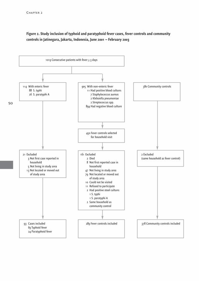
Chapter 2
Figure 2. Study inclusion of typhoid and paratyphoid fever cases, fever controls and community controls in Jatinegara, Jakarta, Indonesia, June 2001 – February 2003
1019 Consecutive patients with fever >_ 3 days
114 With enteric fever88 S. typhi26 S. paratyphi A
21 Excluded3 Not first case reported in
household5 Not living in study area
13 Not located or moved out of study area
93 Cases included69 Typhoid fever24 Paratyphoid fever
905 With non-enteric fever11 Had positive blood culture:
7 Staphylococcus aureus2 Klebsiella pneumoniae2 Streptococcus spp.
894 Had negative blood culture
450 Fever controls selected for household visit
161 Excluded2 Died8 Not first reported case in
household47 Not living in study area79 Not located or moved out
of study area10 Could not be visited11 Refused to participate
2 Had positive stool culture:1 S. typhi1 S. paratyphi A
2 Same household as community control
289 Fever controls included
380 Community controls
2 Excluded (same household as fever control)
378 Community controls included
50
18439 Vollaard 14-12-2004 08:24 Pagina 50
cases and fever controls, respectively). Due to manpower constraints, 10 fever controls
(2%) could not be visited; 11 fever controls (2%) but none of the remaining cases refused
cooperation. Two fever controls had positive stool culture results (for S. typhi [n = 1] and
S. paratyphi A [n = 1]) at the household visit and were therefore excluded from the analysis.
Enteric fever cases and fever controls were visited a median of 24 (IQR, 21-29) days after
the blood culture. Fever controls reported to be diagnosed and treated for the following
diagnoses: suspected typhoid fever (n = 126 [44%]), dengue fever (n = 11 [4%]), respira-
tory tract infections (n = 10 [3%]), tuberculosis (n = 3 [1%]), influenza (n = 3 [1%]),
gastroenteritis (n = 2), urinary tract infection (n = 1), and encephalitis (n = 1); 132
patients (46%) were not informed of the working diagnosis.
During the study period, 380 random households in the study area community were visi-
ted; 289 (76%) of the community controls agreed to participate at the first approach and
the remaining 91 (24%) were the neighbors from the initially selected households. From
2 households of community controls a patient with non-enteric fever was included later
in the course of the study period. These 2 households were excluded from the analysis.
Demographic Data From the Visited Cases and Controls: The median age of the typhoid
cases was 16 (range, 3-57) years; of paratyphoid cases, 22 (range, 4-59) years; of commu-
nity controls, 27 (range, 1-80) years; and of fever controls, 20 (range, 1-75) years
(Table 1). Typhoid and paratyphoid fever cases and fever controls were significantly youn-
ger than the community controls (P<.01). The age of patients with typhoid fever did not
differ significantly from that of those with paratyphoid fever (P = .12). Fever controls were
significantly more often of male sex than were community controls (P = .003 by χ2 test)
and typhoid cases (P = .03). No significant differences in the sex ratio were found when
typhoid or paratyphoid cases were compared with community controls. Compared with
the number of community controls per subdistrict, who had been included proportionally
to the size of the population, in 1 subdistrict proportionally more typhoid cases than
community controls were enrolled (P = .07), whereas in another subdistrict more patients
with paratyphoid fever were enrolled (P = .05). Within the group of patients with enteric
fever itself, no significant overrepresentation of any subdistrict was found in the com-
parison of patients with typhoid and paratyphoid fever (P = .37).
Risk Factors for Typhoid and Paratyphoid Fever: Risk factors for typhoid and paratyphoid
fever in comparison with community and fever controls are shown in Table 1. Compared
with paratyphoid cases the typhoid cases were more often female, lived in more crowded
conditions, were more frequently from a lower income category, more frequently reported
recent typhoid fever among household contacts in the preceding 12 months, used ice
cubes more often, shared food more often, and observed poor handwashing hygiene.
Flooding and eating food purchased from street vendors were more frequently reported
by patients with paratyphoid fever than by those with typhoid fever. Among the 2 control
51
Risk factors for typhoid and paratyphoid fever
18439 Vollaard 14-12-2004 08:24 Pagina 51
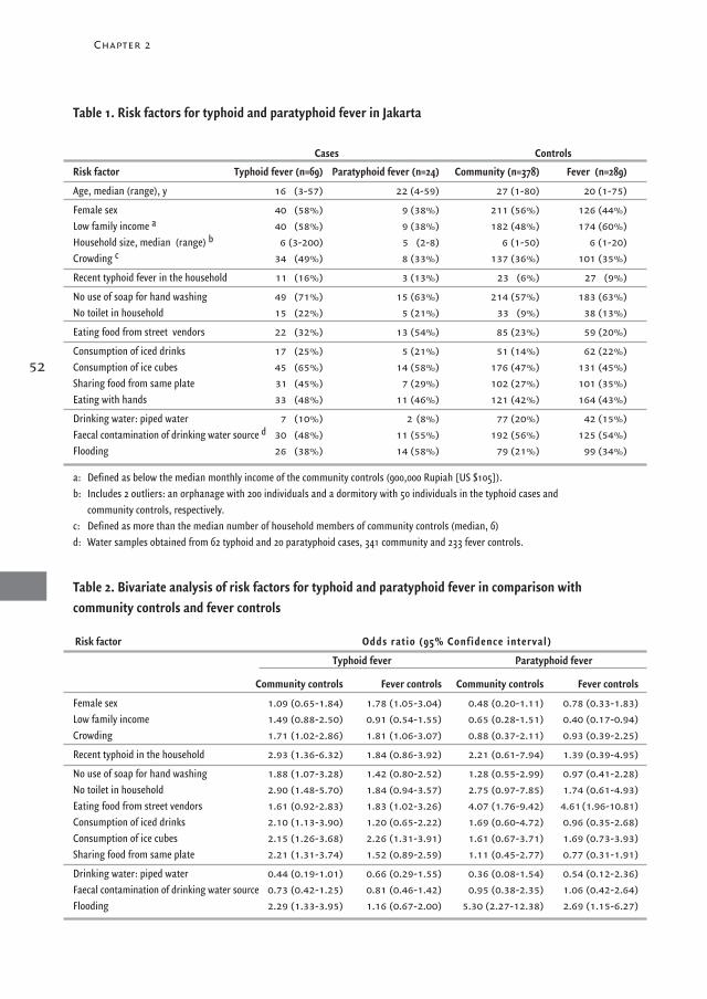
Chapter 2
52
Risk factor Typhoid fever (n=69) Paratyphoid fever (n=24) Community (n=378) Fever (n=289)
Age, median (range), y 16 (3-57) 22 (4-59) 27 (1-80) 20 (1-75)
Female sex 40 (58%) 9 (38%) 211 (56%) 126 (44%)Low family income a 40 (58%) 9 (38%) 182 (48%) 174 (60%)Household size, median (range) b 6 (3-200) 5 (2-8) 6 (1-50) 6 (1-20)Crowding c 34 (49%) 8 (33%) 137 (36%) 101 (35%)
Recent typhoid fever in the household 11 (16%) 3 (13%) 23 (6%) 27 (9%)
No use of soap for hand washing 49 (71%) 15 (63%) 214 (57%) 183 (63%)No toilet in household 15 (22%) 5 (21%) 33 (9%) 38 (13%)
Eating food from street vendors 22 (32%) 13 (54%) 85 (23%) 59 (20%)
Consumption of iced drinks 17 (25%) 5 (21%) 51 (14%) 62 (22%)Consumption of ice cubes 45 (65%) 14 (58%) 176 (47%) 131 (45%)Sharing food from same plate 31 (45%) 7 (29%) 102 (27%) 101 (35%)Eating with hands 33 (48%) 11 (46%) 121 (42%) 164 (43%)
Drinking water: piped water 7 (10%) 2 (8%) 77 (20%) 42 (15%)Faecal contamination of drinking water source d 30 (48%) 11 (55%) 192 (56%) 125 (54%)Flooding 26 (38%) 14 (58%) 79 (21%) 99 (34%)
a: Defined as below the median monthly income of the community controls (900,000 Rupiah [US $105]).b: Includes 2 outliers: an orphanage with 200 individuals and a dormitory with 50 individuals in the typhoid cases and
community controls, respectively.c: Defined as more than the median number of household members of community controls (median, 6)d: Water samples obtained from 62 typhoid and 20 paratyphoid cases, 341 community and 233 fever controls.
Community controls Fever controls Community controls Fever controls
Female sex 1.09 (0.65-1.84) 1.78 (1.05-3.04) 0.48 (0.20-1.11) 0.78 (0.33-1.83)Low family income 1.49 (0.88-2.50) 0.91 (0.54-1.55) 0.65 (0.28-1.51) 0.40 (0.17-0.94)Crowding 1.71 (1.02-2.86) 1.81 (1.06-3.07) 0.88 (0.37-2.11) 0.93 (0.39-2.25)
Recent typhoid in the household 2.93 (1.36-6.32) 1.84 (0.86-3.92) 2.21 (0.61-7.94) 1.39 (0.39-4.95)
No use of soap for hand washing 1.88 (1.07-3.28) 1.42 (0.80-2.52) 1.28 (0.55-2.99) 0.97 (0.41-2.28)No toilet in household 2.90 (1.48-5.70) 1.84 (0.94-3.57) 2.75 (0.97-7.85) 1.74 (0.61-4.93)Eating food from street vendors 1.61 (0.92-2.83) 1.83 (1.02-3.26) 4.07 (1.76-9.42) 4.61(1.96-10.81)Consumption of iced drinks 2.10 (1.13-3.90) 1.20 (0.65-2.22) 1.69 (0.60-4.72) 0.96 (0.35-2.68)Consumption of ice cubes 2.15 (1.26-3.68) 2.26 (1.31-3.91) 1.61 (0.67-3.71) 1.69 (0.73-3.93)Sharing food from same plate 2.21 (1.31-3.74) 1.52 (0.89-2.59) 1.11 (0.45-2.77) 0.77 (0.31-1.91)
Drinking water: piped water 0.44 (0.19-1.01) 0.66 (0.29-1.55) 0.36 (0.08-1.54) 0.54 (0.12-2.36)Faecal contamination of drinking water source 0.73 (0.42-1.25) 0.81 (0.46-1.42) 0.95 (0.38-2.35) 1.06 (0.42-2.64)Flooding 2.29 (1.33-3.95) 1.16 (0.67-2.00) 5.30 (2.27-12.38) 2.69 (1.15-6.27)
Cases Controls
Table 1. Risk factors for typhoid and paratyphoid fever in Jakarta
Table 2. Bivariate analysis of risk factors for typhoid and paratyphoid fever in comparison with community controls and fever controls
Risk factor Odds ratio (95% Confidence interval)
Typhoid fever Paratyphoid fever
18439 Vollaard 14-12-2004 08:24 Pagina 52

groups, fever controls were more often male, from a lower income group, observed
poorer handwashing hygiene, had fewer toilets and connections to the water mains in
their houses, shared food more frequently, were more likely to consume iced drinks, and
were more likely to report flooding than were community controls (Table 1).
In addition, for all interviewed participants, low income was significantly associated with
purchasing food from street vendors (OR, 1.58; 95% CI, 1.03-2.41). When ice cubes were
used, these were purchased from ice vendors by equal proportions in the groups:
41 (69%) patients with typhoid or paratyphoid fever, 107 (61%) community controls,
and 93 (71%) fever controls (P = .12).
Bivariate Analysis Risk Factors for Typhoid Fever: Bivariate analysis of risk factors comparing typhoid cases
with community controls showed the following significantly associated risk factors for
typhoid fever: crowding (>6 household members) and recent typhoid fever of household
contacts (Table 2). The association of recent typhoid fever of household contacts and
typhoid fever also remained significant in a subgroup of households with more than 6
household members: from the 34 typhoid cases, 8 (24%) reported recent typhoid fever in
a household contact, whereas from 137 community controls, 9 (7%) did (OR, 4.38; 95%
CI, 1.54-12.40). In the comparison with community controls, other significantly associated
risk factors for typhoid fever were no use of soap for handwashing, no toilet in the house-
hold, and flooding. With respect to eating habits, typhoid was not significantly associa-
ted with eating food from street vendors, but a significant association was found with
consuming iced drinks, use of ice cubes, and sharing food from the same plate. Sharing
of food occurred mostly with household contacts: 84% (26/31) of typhoid cases and 84%
(85/101) of community controls and in lower frequencies in all groups at work or school.
Female sex was associated when typhoid cases were compared with fever controls, which
was likely due to the overrepresentation of males in the fever control group (Table 2).
In the fever-control comparison crowding was associated with typhoid fever, as was
eating foods from street vendors and use of ice cubes. None of the hygiene-related risk
factors (i.e., no use of soap for handwashing, no toilet in the household) was significantly
associated with typhoid in comparison with fever controls.
Risk Factors for Paratyphoid Fever: In comparison with community controls and fever
controls, paratyphoid fever among cases was significantly associated with eating foods
from street vendors and flooding. Fever controls had a lower family income than did
patients with paratyphoid fever.
Water Examination: During the study period, 656 samples from the sources of running
drinking water of cases and controls were collected; 358 (55%) contained fecal coliforms
53
Risk factors for typhoid and paratyphoid fever
18439 Vollaard 14-12-2004 08:24 Pagina 53
Chapter 2
(median, 30; IQR, 6-250 per 100 mL). Fecal contamination of drinking water was not
significantly associated with either typhoid or paratyphoid fever in comparison with both
control groups (Table 2). Also, bacterial numbers in water samples were not significantly
different for typhoid or paratyphoid fever cases vs those for fever controls (P = .54 and
P = .90, respectively, by Mann-Whitney U test) or community controls (P = .43 and
P = .95, respectively). All respondents reported that they boiled drinking water before
consumption and that they kept water boiling for several minutes.
Food Handlers: A food handler was not present in all households of cases or controls
because some cases and controls always ate outside of the household or cooked their
own food. No S. typhi or S. paratyphi A were isolated in the single stool samples that
could be obtained from 96% of the 78 food handlers of (para)typhoid cases, 246 of
the fever controls, and 298 of the community controls, respectively.
Multivariate AnalysisResidence of participants in 1 of the 8 subdistricts was not evaluated in the bivariate ana-
lysis, but was included in the multivariate analysis as a potential confounder. In this ana-
lysis, neighborhood residence was not independently associated with either typhoid fever
or paratyphoid fever. The significant risk factors for typhoid and paratyphoid fever from
the bivariate analysis that were evaluated in the multivariate analysis are shown in Table 3.
Risk Factors for Typhoid Fever: Using the community control group, typhoid fever conti-
nued to be independently associated with hygienic practices (no use of soap for hand-
washing, sharing of food, and no toilet in the household) and recent intrahousehold
typhoid fever in the preceding 12 months. These are presented in order of decreasing
magnitude of attributable risk (Table 3). Typhoid cases were significantly younger than
community controls, suggesting that either exposure to S. typhi or susceptibility to
symptomatic infection when exposed is greater among young people.
Using the fever controls for comparison, we identified ice cubes and female sex (related
to the high percentage of male participants in the fever control group) as independent
risk factors for typhoid fever. Hygiene-related factors were not independently associated.
Risk Factors for Paratyphoid Fever: In the multivariate analysis, paratyphoid fever conti-
nued to be independently associated with eating foods from street vendors when para-
typhoid cases were compared with both control groups (Table 3). Flooding also remained
a significant risk factor for paratyphoid fever. The individual contribution of eating habits
and flooding as calculated by the attributable risk alternated in importance for both
control groups. Low income was inversely associated with paratyphoid fever in the
comparison with fever controls.
54
18439 Vollaard 14-12-2004 08:24 Pagina 54

The main finding of this study is that in Jatinegara, Jakarta, typhoid and paratyphoid fever
largely follow distinct routes of transmission. Typhoid is spread predominantly within
the household, whereas paratyphoid is mainly transmitted outside the home. No fecal
carriers among food handlers in the households were detected and there was no associa-
tion between the level of contamination of drinking water and either typhoid or paraty-
phoid fever. Apparently, S. typhi is introduced into households by convalescent cases
transiently excreting the bacterium. Consistent with this, independent risk factors for the
intrahousehold spread of typhoid were poor handwashing hygiene and sharing of food
from the same plate. On the other hand, risk factors for transmission of paratyphoid
were outside the household (i.e., flooding, consumption of foods from street vendors).
Furthermore, in this community-based passive surveillance study, paratyphoid comprised
23% of all enteric fever cases, an apparent rise in relative incidence of paratyphoid
compared with earlier studies.
55
Risk factors for typhoid and paratyphoid fever
Table 3. Multivariate analysis of independent risk factors for typhoid and paratyphoid fever incomparison with community controls and fever controls
No use of soap for handwashing 1.91 (1.06-3.46) 34 NA
Sharing food from same plate 1.93 (1.10-3.37) 22 NA
No toilet in household 2.20 (1.06-4.55) 12 NA
Recent typhoid in household 2.38 (1.03-5.48) 9 NA
Young age 0.96 (0.94-0.98) 0.99 (0.96-1.02)Flooding 1.65 (0.88-3.08) 4.52 (1.90-10.73) 45Eating food from street vendors NA 3.34 (1.41-7.91) 38Use of iced drinks 1.12 (0.55-2.26) NA
Consumption of ice cubes 1.34 (0.73-2.44) NA
Crowding 1.54 (0.88-2.72) NA
Comparison with fever controls (n=289)
Consumption of ice cubes 2.27 (1.31-3.93) 36 NA
Female sex 1.79 (1.04-3.06) 26 1.10 (0.43-2.84)Low income 0.85 (0.49-1.49) 0.28 (0.11-0.71) 49Eating food from street vendors 1.62 (0.88-2.98) 5.17 (2.12-12.60) 48Flooding NA 3.25 (1.31-8.02) 42Crowding 1.60 (0.92-2.76) NA
Abbreviations: CI, confidence interval; OR, odds ratio.NA: not significantly associated in the bivariate analysis and not included in the multivariate analysis.
Typhoid fever (n=69) Paratyphoid fever (n=24)
Risk factor OR (95% CI) Attributable Risk, % OR (95% CI) Attributable risk, %
Comparison with community controls (n=378)
Comment
18439 Vollaard 14-12-2004 08:24 Pagina 55

Chapter 2
To reach the conclusion concerning the distinct route of transmission of paratyphoid and
typhoid fever, we compared characteristics of cases with those of community controls
and fever controls. Some potential pitfalls that may affect complete recruitment of
patients in the area, and individual classification of cases and fever controls, need to be
considered. Not all eligible fever patients might have been included, although we perfor-
med blood cultures free of charge to preclude economic barriers for inclusion. Self-treat-
ment with over-the-counter antibiotics and an atypical presentation of enteric fever (e.g.,
as observed in young children) may have influenced inclusion. 18 Even so, the proportio-
nal representation of typhoid fever of 8.6% of illnesses with fever for 3 or more days is
comparable with rates in other active and passive surveillance studies for typhoid fever
using the same inclusion criteria (4.6%-8.5%). 19-23 Furthermore, the sensitivity of the
microbiological methods never reaches 100%. 24 However, because most patients with
fever were included in the first week of illness, the sensitivity of blood culture comes
close to that of quantitation in bone marrow and is superior to the Widal test. 25,26 Also,
the interference of antibiotics, which can yield false-negative results, was limited due to
this short period before inclusion and to the antibiotic-neutralizing resins in the blood
culture vials. Accordingly, equal proportions of typhoid and paratyphoid fever cases and
non-enteric fever controls had previously taken antibiotics. To further minimize misclas-
sification of fever controls, stool cultures were performed 3 to 4 weeks after blood culture
(i.e., at a time when bacteria may still be excreted in feces of patients with typhoid or
paratyphoid fever). The 2 febrile patients with negative blood culture results at inclusion,
whose stool cultures yielded S. typhi and S. paratyphi A, were accordingly excluded from
the analysis. Another potential limitation of this study concerns the screening for
Salmonella carriers by a single stool culture that might not suffice because of intermittent
excretion of the bacteria in stools. 12
The use of a representative community control group allowed us to determine the preva-
lence of risk factors in the whole population at risk. Our study demonstrates that risk
estimates from case-control studies could be affected by the selection of the control-
group used for comparison. For instance, when typhoid fever cases were compared with
community controls, most of the independent risk factors for typhoid fever were intra-
household factors (i.e., no use of soap for handwashing, sharing of food, and recent
typhoid fever in a household member), whereas those factors were not associated in the
comparison with fever controls. This suggests that hygiene practices of both cases and
fever controls were of a standard below that of community controls. In addition, partially
overlapping routes of transmission of typhoid fever and other febrile illnesses could be
interdependent and result in the demonstrated similar intrahousehold risk profile of
typhoid fever cases and fever controls with similar socioeconomic characteristics.
Food obtained from street vendors was a likely vehicle for extrahousehold transmission
56
18439 Vollaard 14-12-2004 08:24 Pagina 56

of paratyphoid fever because it contributed significantly to transmission in contrast to
hygiene-related risk factors. This is consistent with the notion that multiplication of
paratyphoid bacteria in food is required to reach a number sufficient to cause disease.
Street vendors have only limited facilities for cooled storage of foods and for washing of
hands, foods, and dishes. The low hygienic standards could therefore contribute not only
to the transmission of paratyphoid fever but of other foodborne diseases such as typhoid,
as well. 7,11,27-29 Due to the Asian economic crisis starting in 1997, the expanding urban
population became even more dependent on inexpensive food obtained from street
vendors, which may explain the relatively high proportion of paratyphoid fever in enteric
fever in Jakarta. Low-income groups more frequently ate food obtained from street
vendors than did individuals with high income, but all income groups who purchase
food from street vendors may be at risk.
In contrast to the largely extra-household transmission of paratyphoid fever, typhoid
fever was more of an intrahousehold affair introduced by recent typhoid cases in the
households and facilitated by poor hand-washing hygiene and sharing of food from the
same plate, consistent with an earlier report. 10 The association of poor handwashing
hygiene and typhoid fever was shown before in Indonesia and India. 6,9,11 A recent review
stressed the importance of the use of soap for the reduction of the incidence of diarrheal
diseases. 30 In our study we also identified a significant association between not using
soap for handwashing and all febrile illnesses (OR, 1.40; 95% CI, 1.05-1.88). The combi-
nation of poor handwashing hygiene, eating with hands, and sharing food from the same
plate can understandably facilitate transmission of typhoid, but apparently the infective
dose to allow transmission of paratyphoid is only infrequently met. Because we observed
no intrahousehold outbreaks and detected no fecal carriers among the food handlers in
the households of cases, intrahousehold person-to-person spread through convalescent
patients observing poor hygiene seems a more likely scenario than transmission by
chronic carriers among food handlers in households.
Apart from the above-mentioned risk factors, some additional observations should be
considered. First, the total number of interviewed patients with typhoid and paratyphoid
fever in our study was limited, which may have influenced the statistical power of the
analysis, especially in small subgroups, and the demonstrated associations of specific
risk factors. Second, food purchased from street vendors could be implicated as a vehicle
for transmission of typhoid as well, as shown in the bivariate analysis. Also, the con-
sumption of ice cubes obtained from street vendors might expose clients to Salmonella
bacteria because these bacteria can survive in ice. 31 Another extrahousehold location
of acquisition of typhoid fever could be public toilets, which generally lack handwashing
facilities. Third, there was an association between flooding and paratyphoid fever.
Two hypotheses may explain this association: flooding could introduce bacteria from
57
Risk factors for typhoid and paratyphoid fever
18439 Vollaard 14-12-2004 08:24 Pagina 57
Chapter 2
contaminated surface water into sources of drinking water. However, since most cases of
typhoid and paratyphoid fever occurred during the dry season, flood-related waterborne
transmission seemed not to play a major role. Alternatively, flooding may be an income-
associated geographic marker that coincides with the distribution of carriers among food
vendors in the area. This could also explain the clustering of paratyphoid fever cases in
some regions, but since community controls were nonmatched for subdistrict neighbor-
hood residence, this assumption could not be verified. Finally, although a considerable
proportion of the sources of drinking water contained fecal coliforms that were used as
indicator organisms, contamination itself was not associated with enteric fever. Dilution
of S. typhi or S. paratyphi in water might generate too low a dose to infect partially immune
residents. More likely, however, the entrenched habit of boiling drinking water from the
water mains or groundwater pumps explains the lack of an association between water
contamination and enteric fever and should certainly be continued to prevent possible
outbreaks of disease, in combination with proper storage of boiled water to prevent
domestic contamination.
In conclusion, the present findings suggest that public health policies for control of
typhoid and paratyphoid fever in Jakarta should focus on hygiene education as well as
monitoring of the street-food trade, although such strategies would have to be tested in
intervention trials to prove their value. First, instruction on proper handwashing hygiene
using soap could reduce the overall incidence of infectious diseases in Jakarta and
especially preclude transmission of typhoid fever among contacts of cases. Second,
prevention of bacterial contamination of street food and ice cubes could contribute to
containment of enteric fever, paratyphoid in particular. Follow-up of enteric fever cases,
especially among food vendors, should be prioritized to reduce the role of transient or
chronic carriers in the foodborne transmission.
If vaccination were to be considered as a means of controlling typhoid, an individualized
approach rather than mass vaccination (i.e., targeted vaccination of young household con-
tacts of cases) may be a cost-effective approach when public health resources are scarce. 32
But, because of the increasing incidence of paratyphoid fever in Jakarta, as well as readily
available antibiotic treatment and the potentially effective intervention of education to
increase appropriate handwashing, mass immunization programs for typhoid fever in
Jakarta may not be appropriate at this time.
58
18439 Vollaard 14-12-2004 08:24 Pagina 58
Acknowledgment
We thank J. P. Vandenbroucke, MD, PhD, from the Leiden University Medical Centre
(LUMC) Department of Clinical Epidemiology and Nico Nagelkerke, PhD, from the
LUMC Department of Medical Statistics for critically reviewing the document and Wil
Dolmans, MD, PhD, for his help in the preparatory stage of this project. We thank the
physicians, nurses, and technicians of the participating health centers in Jakarta for
their cooperation: Mitra Internasional hospital and microbiology laboratory, Budhi Asih,
St Carolus and Persahabatan hospital, all puskesmas in Jatinegara, the local private practi-
tioners, as well as the microbiology department of Atma Jaya University Hospital and
Nusantara Water Centre, West Jakarta. This study could not have been done without
our research assistants: Billy Hunsinger, Ferry Kandaw, Rinny Listyani, Meily, Vea
Noveria, Carmelita Ridwan, Min Ali Sugiharto, Lidwina Sutikno, Mariana Tasman,
and Lily Yaputra.
59
Risk factors for typhoid and paratyphoid fever
References
1. Pang T, Levine MM, Ivanoff B, Wain J, Finlay BB. Typhoid fever-important issues still remain. Trends Microbiol. 1998; 6:131-133.
2. Arya SC, Sharma KB. Urgent need for effective vaccine against Salmonella paratyphi A, B and C. Vaccine. 1995; 13:1727-1728.
3. Sood S, Kapil A, Dash N, Das BK, Goel V, Seth P. Paratyphoid fever in India: An emerging problem. Emerg Infect Dis. 1999; 5:483-484.
4. Swaddiwudhipong W, Kanlayanaphotporn J. A common-source water-borne outbreak of multidrug-resistant typhoid fever in a
rural Thai community. J Med Assoc Thai. 2001; 84: 1513-1517.
5. King CC, Chen CJ, You SL, Chuang YC, Huang HH, Tsai WC. Community-wide epidemiological investigation of a typhoid
outbreak in a rural township in Taiwan, Republic of China. Int J Epidemiol. 1989; 18:254-260.
6. Gasem MH, Dolmans WM, Keuter MM, Djokomoeljanto RR. Poor food hygiene and housing as risk factors for typhoid fever in
Semarang, Indonesia. Trop Med Int Health. 2001; 6:484-490.
7. Luby SP, Faizan MK, Fisher-Hoch SP, et al. Risk factors for typhoid fever in an endemic setting, Karachi, Pakistan.
Epidemiol Infect. 1998; 120:129-138.
8. Mermin JH, Villar R, Carpenter J, et al. A massive epidemic of multidrug-resistant typhoid fever in Tajikistan associated with
consumption of municipal water. J Infect Dis. 1999; 179: 1416-1422.
9. Bhan MK, Bahl R, Sazawal S, et al. Association between Helicobacter pylori infection and increased risk of typhoid fever.
J Infect Dis. 2002; 186:1857-1860.
10. Black RE, Cisneros L, Levine MM, Banfi A, Lobos H, Rodriguez H. Case-control study to identify risk factors for paediatric
endemic typhoid fever in Santiago, Chile. Bull World Health Organ. 1985; 63:899-904.
11. Velema JP, van Wijnen G, Bult P, van Naerssen T, Jota S. Typhoid fever in Ujung Pandang, Indonesia - high-risk groups and
high-risk behaviours. Trop Med Int Health. 1997; 2:1088-1094.
12. Christie AB. Infectious Diseases: Epidemiology and Clinical Practice. 4th ed. Edinburgh: Churchill Livingstone; 1987.
13. Tankhiwale SS, Agrawal G, Jalgaonkar SV. An unusually high occurrence of Salmonella enterica serotype paratyphi A in patients
with enteric fever. Indian J Med Res. 2003; 117:10-12.
18439 Vollaard 14-12-2004 08:24 Pagina 59

14. Saha MR, Dutta P, Niyogi SK, et al. Decreasing trend in the occurrence of Salmonella enterica serotype Typhi amongst
hospitalised children in Kolkata, India during 1990-2000. Indian J Med Res. 2002; 115:46-48.
15. WHO. Guidelines for Drinking-water Quality. 2nd ed. Geneva: World Health Organization; 1997.
16. Sun GW, Shook TL, Kay GL. Inappropriate use of bivariable analysis to screen risk factors for use in multivariable analysis.
J Clin Epidemiol. 1996; 49:907-916.
17. Bruzzi P, Green SB, Byar DP, Brinton LA, Schairer C. Estimating the population attributable risk for multiple risk factors using
case-control data. Am J Epidemiol. 1985; 122:904-914.
18. Ferreccio C, Levine MM, Manterola A, et al. Benign bacteremia caused by Salmonella typhi and paratyphi in children younger
than 2 years. J Pediatr 1984; 104:899-901.
19. Lin FY, Vo AH, Phan VB, Nguyen TT, et al. The epidemiology of typhoid fever in the Dong Thap Province, Mekong Delta region of
Vietnam. Am J Trop Med Hyg. 2000; 62:644-648.
20. Simanjuntak CH, Paleologo FP, Punjabi NH, et al. Oral immunisation against typhoid fever in Indonesia with Ty21a vaccine.
Lancet. 1991; 338:1055-1059.
21. Sinha A, Sazawal S, Kumar R, et al. Typhoid fever in children aged less than 5 years. Lancet. 1999; 354:734-737.
22. Saha SK, Baqui AH, Hanif M, et al. Typhoid fever in Bangladesh: implications for vaccination policy. Pediatr Infect Dis J. 2001;
20:521-524.
23. Bodhidatta L, Taylor DN, Thisyakorn U, Echeverria P. Control of typhoid fever in Bangkok, Thailand, by annual immunization of
schoolchildren with parenteral typhoid vaccine. Rev Infect Dis. 1987; 9:841-845.
24. Parry CM, Hien TT, Dougan G, White NJ, Farrar JJ. Typhoid fever. N Engl J Med. 2002; 347:1770-1782.
25. Wain J, Pham VB, Ha V, et al. Quantitation of bacteria in bone marrow from patients with typhoid fever: relationship between
counts and clinical features. J Clin Microbiol. 2001; 39: 1571-1576.
26. Parry CM, Hoa NT, Diep TS, et al. Value of a single-tube widal test in diagnosis of typhoid fever in Vietnam. J Clin Microbiol. 1999;
37:2882-6.
27. Castillo MTG, Superable JT, Magpantay RL, White ME, Dayrit MM, Saniel MC. Case-control study of resistant Salmonella typhi in
Metro Manila, Philippines. SE Asian J Trop Med Public Health (Suppl). 1995; 26:39-41.
28. Estrada-Garcia T, Cerna JF, Thompson MR, Lopez-Saucedo C. Faecal contamination and enterotoxigenic Escherichia coli in
street-vended chili sauces in Mexico and its public health relevance. Epidemiol Infect. 2002; 129:223-226.
29. Koo D, Aragon A, Moscoso V, et al. Epidemic cholera in Guatemala, 1993: transmission of a newly introduced epidemic strain by
street vendors. Epidemiol Infect. 1996; 116:121-126.
30. Curtis V, Cairncross S. Effect of washing hands with soap on diarrhoea risk in the community: a systematic review. Lancet Infect
Dis. 2003; 3:275-281.
31. Dickens DL, DuPont HL, Johnson PC. Survival of bacterial enteropathogens in the ice of popular drinks. JAMA. 1985; 253:3141-
3143.
60
Chapter 2
18439 Vollaard 14-12-2004 08:24 Pagina 60
3
Risk factors for transmission of food borne illness in
restaurants and street vendors in Jakarta, Indonesia
A.M. Vollaard 1 S. Ali 2 H.A.G.H. Van Asten 3 I. Suhariah Ismid 4
S. Widjaja 5 L.G. Visser 1 Ch. Surjadi 6 J.T. Van Dissel 1
1 Dept. Infectious Diseases, Leiden University Medical Center, the Netherlands2 Dept. Biology, Medical Faculty Atma Jaya Catholic University, Jakarta, Indonesia
3 Institute for International Health, Nijmegen University Medical Center, the Netherlands4 Dept. Parasitology, Medical Faculty, Universitas Indonesia, Jakarta, Indonesia
5 Dept. Internal Medicine, Atma Jaya Catholic University, Jakarta, Indonesia6 Center for Health Research, Atma Jaya Catholic University, Jakarta, Indonesia
Epidemiol Infect 2004, 132: 863-872
Reprinted with permission
18439 Vollaard 14-12-2004 08:24 Pagina 61

Abstract
In a previous risk factor study in Jakarta we identified purchasing street food as an
independent risk factor for paratyphoid fever. Eating from restaurants, however, was
not associated with disease.
To explain these findings we compared 128 street food vendors with 74 food handlers
from restaurants in a cross-sectional study in the same study area. Poor hand-washing
hygiene and direct hand contact with foods, male sex and low educational level were
independent characteristics of street vendors in a logistic regression analysis. Faecal
contamination of drinking water (in 65% of samples), dishwater (in 91%) and ice cubes
(in 100%) was frequent. Directly transmittable pathogens including S. typhi (n=1) and
non-typhoidal Salmonella spp. (n=6) were isolated in faeces samples in 13 (7%) vendors;
the groups did not differ, however, in contamination rates of drinking water and
Salmonella isolation rates in stools.
Poor hygiene of street vendors as compared to restaurant vendors, in combination with
faecal carriage of enteric pathogens including S. typhi, may help explain the association
found between purchasing street food and food borne illness, in particular Salmonella
infections.
Public health interventions to reduce transmission of food borne illness should focus on
general hygienic measures in street food trade, i.e., hand-washing with soap, adequate
food handling hygiene, and frequent renewal of dishwater.
Chapter 3
18439 Vollaard 14-12-2004 08:24 Pagina 62

Introduction
In a previous case-control study in Jakarta, Indonesia, we identified purchasing foods
from street vendors as an independent risk factor for (para)typhoid fever, whereas no
such association was found with eating in restaurants. 1 Similarly, in other studies in
Indonesia street food was associated with typhoid fever. 2,3 Several factors may explain
this association of street food and (para)typhoid fever, a systemic febrile illness caused
by Salmonella typhi and S. paratyphi A,B or C that only affects humans. For instance,
personal hygiene and knowledge of hygienic food preparation 4-6, faecal contamination
of basic ingredients or water used for food preparation 7 and/or isolation rates of
enteric pathogens 8, may differ between street food vendors and vendors in restaurants.
Although the possible transmission routes of enteric pathogens like Salmonella are
well-known, the relative importance of the various factors, i.e., the weak link in the
transmission chain, is uncertain but of great importance to help focus the most relevant
health intervention.
We therefore examined determinants for transmission of enteric pathogens in commercial
food handling in a cross-sectional study in Jakarta. Because of our previous findings in
the same area we compared street vendors with vendors from restaurants. In both groups
of food handlers we determined faecal isolation rates of enteric pathogens including
Salmonella spp., assessed the hygiene practices and knowledge about safe food prepara-
tion and examined water reservoirs and ice cubes used for consumption. Our findings
should be helpful to health authorities for the development of effective methods for the
containment of food borne diseases in commercial food handling especially in food stalls
and pushcarts.
Material and methodsStudy population: From 17 February until 21 May 2003 all food vendors working in the
Bidara Cina sub-district in East-Jakarta were approached by graduated medical school
students. During the study period the study area was visited daily, during daytime and
evenings, until all present food vendors were interviewed. This area of 126 hectares houses
43 829 inhabitants (December 2002) and has been subject to a typhoid fever risk factor
study as described elsewhere. 1 Ethical clearance was obtained from the Indonesian
National Institute of Health Research and Development (Litbangkes) and the local provin-
cial authorities. A written informed consent was obtained from all food vendors.
A study subject was defined as an individual working as a vendor of foods or drinks in
the study area who was physically involved in the preparation or handling of foods. All
types of units were eligible for inclusion: restaurants, food stalls, and pushcarts. Some
63
Commercial food handling and food borne illness
18439 Vollaard 14-12-2004 08:24 Pagina 63

Chapter 3
restaurants and warung (i.e., small-scale restaurants often connected to the household of
the owner) are subject to six-monthly visits by local health authorities for inspection and
education on food hygiene, but food hawkers are not visited. Food stalls are stationary
roadside facilities with or without seats. Pushcarts are mobile units that lack seating
facilities.
Questionnaires: A standardized questionnaire was used to obtain data on demographic
and socio-economic characteristics of the food vendors, recent disease history, hygiene
practice, and water sources in the units. Measure of hygiene that were assessed were:
defecation during working hours, hand washing before food preparation and after defe-
cation, the use of soap for hand washing, direct hand contact with food items, available
water sources for hand washing and dishwashing, the use of soap for dishwashing and
the frequency of renewal of dishwater, and the presence of flies on food items. Diarrhoea
was defined as three or more loose stools per day. During and following the interview
(i.e., a period of in total 30 minutes) the interviewers observed the hand washing hygiene
and food handling of the vendors to compare the given answers with the actual practice.
Any reported use of soap was verified by screening for the presence of soap in the unit.
Knowledge about safe food preparation was tested by a scoring system. Eight diseases
were mentioned: diarrhoea, typhoid fever, jaundice, worm infections, pneumonia, skin
infections, AIDS, and tuberculosis. Vendors were asked whether these illnesses could be
transmitted by food. Also knowledge about vehicles for disease transmission in food
processing was tested: i.e., flies, dirty hands, polluted water, cutting boards, traffic
fumes, and ill food handlers. For every correct answer one point was given, no point if
the answer was not known, and one point subtracted for an incorrect answer.
Sample collection: At every location 150 mL samples were collected from the water source
or container with drinking water and dishwater. If piped water was sampled, the bacteri-
cidal effect of chlorine during transport was neutralized by addition of 0.1 mL 10% sodium-
thiosulphate. Ice cubes (150 mL) were collected from cool boxes into sterile bottles. Two
stool samples were collected: two gram of faeces into a vial with Cary Blair transport
medium for bacteriological examination and ten gram of fresh stool for parasitological
examination.
Water examination: The samples were transported on ice, processed within six hours
after collection and examined for total and faecal coliform counts by use of Most
Probable Number method. 9 Serially diluted water samples were incubated in Endolactose
broth and Brilliant Green to detect specific colour changes and gas formation. Presence
of faecal coliforms (>1 MPN Index / 100 mL) was defined as faecal contamination. 9
The upper detection limit was 1600/100 mL.
Stool cultures: Stool samples were cultured in the central reference lab using Selenite
enrichment broth (Oxoid Ltd, Hampshire, England). Colonies were plated on xylose-
64
18439 Vollaard 14-12-2004 08:24 Pagina 64

lysine-desoxycholate, Salmonella Shigella agar, and on Triple Sugar Iron agar, SIM
Medium (sulphide and indole production and motility) and Simmons Citrate (Oxoid Ltd,
Hampshire, England). Salmonella bacteria were identified using agglutination anti-sera
(Polyvalent, O-9, Vi, h, paratyphi A; Murex Biotech Ltd., Dartford, England) and bioche-
mical tests (Microbact: Medvet Diagnostics, Adelaide, Australia).
Parasitologic stool examination: The second stool sample was processed within 24 hours
after collection and microscopically examined after lugol staining, Kato Katz technique,
and Harada Mori method for the detection of hookworms.
Feedback: Food vendors were informed about their water quality, instructed on safe food
preparation methods, and if necessary treated (worm infections: mebendazole, Giardia
lamblia: metronidazole). When Salmonella was isolated in stool cultures, vendors were
subject to follow-up and treatment was administered in case of repeated positive stool
cultures.
Statistical methods: Data was entered twice in EpiInfo 6.04 (CDC, Atlanta, USA), valida-
ted and imported in SPSS (SPSS Inc., Chicago, IL, USA) for analysis. T-tests were used for
evaluation of normally distributed numerical variables and Mann Whitney U-tests for not-
normally distributed numerical variables. Proportions within the group of street food
vendors and within the group of vendors from restaurants or warung were compared
using Chi square tests (χ2). Measures for association were expressed as odds ratios (OR)
with their respective confidence limits (95%-CI) for categorical exposures. To control for
confounding a multivariate analysis was performed on the significantly associated risk
factors from the bivariate analysis in a logistic regression model by forward likelihood
ratio test. For the comparison of hygiene parameters between the two groups we depen-
ded on the self-reported methods of hand-washing hygiene after defecation, but not all
food vendors reported to defecate during working hours (e.g., due to non-availability of
facilities, limited working hours per day, or to business activity). Hygiene parameters
were consequently evaluated by multivariate analysis for all food vendors, and additionally
in the sub-group of subjects who told to defecate during working hours to confirm over-
all trends. Significance levels were p-values < 0.05.
ResultsStudy population: In total 238 food vendors were found to be working in the study area.
From these 202 food vendors (85%) were interviewed. Thirty-six food vendors refused
participation: 6 worked in restaurants, 13 worked in warung, and 17 worked in roadside
stalls or pushcarts. Stool specimens could be collected from 175 of the 202 vendors; 27
(13%) refused a sample. We also collected 139 drink water samples from the 149 vendors
who offered drinking water to customers, and 172 dishwater samples. The age of food
65
Commercial food handling and food borne illness
18439 Vollaard 14-12-2004 08:24 Pagina 65
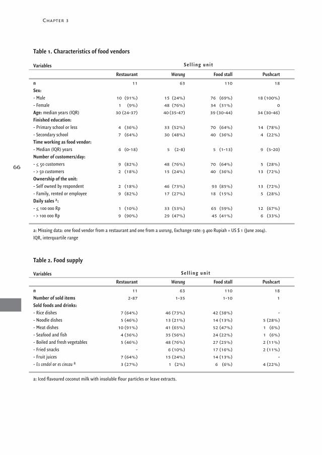
Chapter 3
66
Table 1. Characteristics of food vendors
Variables
Restaurant Warung Food stall Pushcart
n 11 63 110 18 Sex:- Male 10 (91%) 15 (24%) 76 (69%) 18 (100%)- Female 1 (9%) 48 (76%) 34 (31%) 0Age: median years (IQR) 30 (24-37) 40 (35-47) 39 (30-44) 34 (30-46)Finished education: - Primary school or less 4 (36%) 33 (52%) 70 (64%) 14 (78%)- Secondary school 7 (64%) 30 (48%) 40 (36%) 4 (22%)Time working as food vendor:- Median (IQR) years 6 (0-18) 5 (2-8) 5 (1-13) 9 (5-20)Number of customers/day:- <_ 50 customers 9 (82%) 48 (76%) 70 (64%) 5 (28%)- > 50 customers 2 (18%) 15 (24%) 40 (36%) 13 (72%)Ownership of the unit:- Self owned by respondent 2 (18%) 46 (73%) 93 (85%) 13 (72%)- Family, rented or employee 9 (82%) 17 (27%) 18 (15%) 5 (28%)Daily sales a:- <_ 100 000 Rp 1 (10%) 33 (53%) 65 (59%) 12 (67%)- > 100 000 Rp 9 (90%) 29 (47%) 45 (41%) 6 (33%)
a: Missing data: one food vendor from a restaurant and one from a warung, Exchange rate: 9 400 Rupiah = US $ 1 (June 2004). IQR, interquartile range
S e l l i n g u n i t
Table 2. Food supply
VariablesRestaurant Warung Food stall Pushcart
n 11 63 110 18Number of sold items 2-87 1-35 1-10 1Sold foods and drinks:- Rice dishes 7 (64%) 46 (73%) 42 (38%) -- Noodle dishes 5 (46%) 13 (21%) 14 (13%) 5 (28%)- Meat dishes 10 (91%) 41 (65%) 52 (47%) 1 (6%)- Seafood and fish 4 (36%) 35 (56%) 24 (22%) 1 (6%)- Boiled and fresh vegetables 5 (46%) 48 (76%) 27 (25%) 2 (11%)- Fried snacks - 6 (10%) 17 (16%) 2 (11%)- Fruit juices 7 (64%) 15 (24%) 14 (13%) -- Es cendol or es cincau a 3 (27%) 1 (2%) 6 (6%) 4 (22%)
a: Iced flavoured coconut milk with insoluble flour particles or leave extracts.
S e l l i n g u n i t
18439 Vollaard 14-12-2004 08:24 Pagina 66
vendors ranged from 18-68 years, no significant difference in age between vendors from
the four units was found (p = 0.11, ANOVA). Vendors in warung were significantly more
often female (p < 0.001, χ2) (Table 1).
Education level of the group of vendors from stalls and pushcarts was lower than that of
vendors in restaurants and warung (p = 0.03, χ2) (Table 1). For 95% of the respondents
food vending was a fulltime economic activity during six or seven days a week. Mobile
vendors proportionally served most customers per day: 72% served more than 50 custo-
mers a day. The small-scale entrepreneurs in food stalls and pushcarts tend to specialize
in food items which limits their supply to a few or single items (Table 2).
Hygiene in the grouped units: Seventy (55%) of the vendors from food stalls and push-
carts did not wash their hands before food preparation as compared with 21 (28%) of the
vendors in restaurants/warung (p < 0.001) (Table 3). Non-use of soap for hand-washing
before food preparation was reported in 79% vs. 51%, respectively (p = 0.002). Although
all vendors reported to wash their hands after defecation during working hours, non-use
of soap occurred significantly more frequent in stalls and carts than in restaurants/
warung (37% vs. 10%, p< 0.001). Direct hand contact with ready-to-eat foods occurred
more often in food stalls and pushcarts (63% vs. 36%, p < 0.001). The limited facilities
for hand- and for dishwashing were demonstrated for 86% of the pushcarts and stalls
and 58% of the warung and restaurants, because the same water reservoir was used for
both purposes (p = 0.01). Vendors reported to renew the dishwater in buckets 0-20 times
during working hours with the lowest mean frequency in the food stalls and pushcarts
(3.1 vs. 6.2, p < 0.001). In restaurants/warung, flies on ready-to-eat foods were observed
more often (p = 0.01) and ice cubes were used more often (p < 0.001). Refrigerators for
storage of ready-to-eat foods were lacking in 99% of the warung, food stalls and push-
carts and 54% of the restaurants.
Knowledge of safe food preparation and recent illness: The score for the knowledge of
safe food preparation (maximum score: 14) was not significantly different between the
two groups of units (mean score: 5.0 and 5.5 for stalls/pushcarts and restaurants/warung,
respectively: p = 0.15, t-test). Vendors most frequently indicated diarrhoea (89% of the
vendors) and least frequently AIDS (6%) as food borne illness. Ninety-one percent of the
vendors from food stalls and pushcarts and 93% from restaurants and warung were aware
that diarrhoeal diseases could be transmitted by hands (p = 0.52, χ2). In the 30 days prior
to the interview 24% of the vendors reported to have suffered from fever, and 23% of the
vendors told that they had experienced at least one diarrhoeal episode in the preceding
three months. The isolation rate of enteric pathogens and occurrence of diarrhoea in the
preceding three months was not correlated (p = 0.35, χ2). The reported occurrence of
diarrhoea did not differ between the two groups (p = 0.19): OR 0.64 (95%-CI 0.33-1.25)
(Table 3).
67
Commercial food handling and food borne illness
18439 Vollaard 14-12-2004 08:24 Pagina 67

Chapter 3
68
Table 3. Comparison of hygiene parameters between two groups of food vendors: bivariate analysis
Variable a Food stalls and pushcarts Restaurants and warung OR (95% CI) p
- n (202) 128 74
Hand-washing hygiene:- No use of soap for hand washing
after defecation (n=74 vs 63) b 27 (37%) 6 (10%) 5.46 (2.08-14.33) < 0.001- Not washing hands before
food preparation (n=128 vs 74) 70 (55%) 21 (28%) 3.05 (1.65-5.63) < 0.001- No use of soap if washing hands before
food preparation (n=58 vs 53) 46 (79%) 27 (51%) 3.69 (1.61-8.49) 0.002- Direct hand contact with
ready-to-eat food (n=128 vs 74) 80 (63%) 27 (36%) 2.90 (1.60-5.25) < 0.001
Dishwater:- Dishwater is used for washing hands
(n=36 vs 31) c 31 (86%) 18 (58%) 4.48(1.37-14.63) 0.01- Mean number of times dishwater is
renewed per day (range) 3.1 (0-15) 6.2 (1-20) < 0.001
Other factors:- Use of ice cubes (n=128 vs 74) 62 (48%) 63 (85%) 0.16 (0.08-0.34) < 0.001- Flies on food items (n=127 vs 73) 7 (6%) 12 (16%) 0.30 (0.11-0.79) 0.01- Diarrhoea last 3 months (n=128 vs 74) 26 (20%) 21 (28%) 0.64 (0.33-1.25) 0.19
a: Number of vendors from stalls/pushcarts versus restaurants/warung available for analysis b: n = 137: only those vendors who reported to defecate during working hours.c: n = 67: only those vendors who washed utensils/dishes and/or hands before food preparation in buckets.
Table 4. Comparison of water examination results between two groups of food vendors: bivariate analysis
Variable a Food stalls, pushcarts Restaurants, warung OR (95% CI) p
Water examination:- Faecal contamination of sampled drinking water (n=67 vs 72) 40 (60%) 50 (69%) 0.65 (0.32-1.31) 0.23- Median faecal coliform count in drinking water b (n=40 vs 50) 34 (13-105) 46 (19-1075) 0.12- Faecal contamination of sampled dishwater (n=102 vs 70) 95 (93%) 62 (89%) 1.75 (0.60-5.07) 0.30- Median faecal coliform count in dishwater b (n= 95 vs 62) 425 (33-1600) 39 (20-900) 0.006
a: Number of vendors from stalls/pushcarts versus restaurants/warung available for analysis.b: Median (IQR) MPN index /100 mL, comparison of numbers by Mann Whitney U-test.
Table 5 Results of the stool examination (n=175)
Enteric pathogen Food stalls and pushcarts (n=110) Restaurants and warung (n=65) Total
Non-typhoidal Salmonellae 4 (4%) 2 (3%) 6 (3%)Salmonella typhi 1 (1%) 0 1 (0.6%)Hookworms 32 (29%) 14 (22%) 46 (26%)Trichuris trichiura 26 (24%) 13 (20%) 39 (22%)Ascaris lumbricoides 3 (3%) 5 (8%) 8 (5%)Giardia lamblia 2 (2%) 1 (2%) 4 (2%)Entamoeba histolytica/dispar 2 (2%) 0 2 (1%)
Pathogens were isolated in 86 individuals.
18439 Vollaard 14-12-2004 08:24 Pagina 68
.
Examination of drinking water: Drinking water sources were bottled water (2), piped
water (49), and groundwater extracted by pumps (98). Fifty-three food handlers did not
serve drinking water. All respondents reportedly boiled drinking water before storage and
serving. The majority of vendors (129, 88%) kept the boiled water in closed plastic jars,
jerry-cans or kettles, while 18 vendors (12%) kept it in open containers such as buckets
or pans. In the latter case utensils had to be immersed to collect the water from the reser-
voirs. Of the 139 examined samples 90 (65%) contained faecal coliforms with median 39
(IQR 17-450)/100 mL in the contaminated samples. The location (p = 0.23, χ2), the storage
method (i.e., closed or open container) (p = 0.82), or the source (pump or piped water)
(p = 0.39) did not significantly influence the contamination rate. No significant differences
were found in the number of faecal coliforms in the contaminated samples for the two
groups of units (p = 0.12, Mann Whitney U-test) (Table 4). Also, the bacterial numbers
in the tap or groundwater samples from either closed or open containers did not differ
significantly (p = 0.64, Kruskal Wallis test).
Examination of dishwater: In 172 units (i.e., 102 street vendors and 70 restaurants/warung)
dishwater was present at the location of vending and this was consequently examined;
157 (91%) of the 172 dishwater samples were contaminated with a median faecal coliform
count of 140 (IQR 23-1600)/100 mL. The faecal coliform counts in dishwater from stalls
and pushcarts were higher than that from the restaurants and warung (p = 0.01, Mann
Whitney U-test) (Table 4). The median faecal coliform count in 46 buckets used both for
washing hands and dishes was higher than in the 17 buckets only used for dishwashing:
323 (IQR 28-1600) vs. 20 (IQR 15-1600)/100 mL (p = 0.06, Mann Whitney U-test). The
presence of detergent significantly decreased the number of faecal coliforms in dishwater:
median 40 (IQR 17-1600) vs. 900 (IQR 34-1600) /100 mL where soap was absent
(p = 0.005, Mann Whitney U-test).
Examination of ice cubes: Ice cubes were used in drinks by 125 (62%) of the vendors.
We collected 23 ice samples from 3 pushcarts, 14 food stalls, 4 warung (two samples at
one location) and 1 restaurant. All ice cubes were contaminated, with a median faecal
coliform count 500 (IQR 170-1600)/100 mL. Most of the ice cubes had been purchased
from ice vendors (70%), but no significant differences in faecal coliform numbers
between purchased or self-made ice cubes were observed (p = 0.15, Mann Whitney U-test).
Fifteen food vendors (68%) collected ice cubes with their hands and seven used tools in
cool boxes, but faecal coliform counts did not differ significantly by method of handling
(p = 0.25, Mann Whitney U-test).
Stool examination: In 86 vendors (49%) pathogens were detected. Directly transmittable
pathogens (i.e., Salmonella spp., Giardia, and Entamoeba) were isolated in 13 (7%) (Table 5).
S. typhi was isolated in the stool from a 25 year-old male mobile vendor selling iced
69
Commercial food handling and food borne illness
18439 Vollaard 14-12-2004 08:24 Pagina 69

Chapter 3
flavoured drinks. Two repeated stool cultures in three week-intervals were negative. He
reported not to have suffered from prolonged fever in the preceding six months or from
previous typhoid fever. Both Salmonella spp. and hookworms were detected in the stools
from two food vendors. Faecal carriage of non-typhoidal Salmonellae was equally frequent
in both groups (p = 0.33): OR 1.19 (0.18-9.65).
Parasitology: Single parasite infestations were detected in the stools of 63 vendors (36%),
and dual infestations in 18 vendors (10%) (Table 5). The most frequent combination was
hookworm infection with Trichiuris trichura (n = 12) or with Ascaris (n = 3). Two other
combinations were Ascaris or Giardia with hookworms and Trichiuris with Giardia.
Infestation rates of street food vendors (49%) and restaurant/warung employees (42%)
were non-significantly different (p = 0.63): OR 1.36 (0.73-2.52).
Differences in hygiene parameters between restaurants/warung and stalls/carts: All study
findings were summarized to compare hygiene parameters of the two groups by bivariate
analysis (Table 3, 4). Significantly different features in food stalls and pushcarts were
poor hand-washing hygiene including less use of soap, direct hand contact with food
items, and poor standards of dishwashing with higher median faecal coliform counts in
dishwater. In restaurants and warung ice cubes were used more often because of the avai-
lable cooling facilities and/or more frequent supply of drinks, and flies were observed
more often on ready-to-eat foods. In a multivariate analysis including only the subjects
who reported defecation during working hours (n = 137), independently associated features
of food vendors from stalls and carts were not washing hands before food preparation
(OR 7.51 [2.44-23.05]), direct hand contact with foods (OR 2.76 [1.04-7.33]), and male
sex (OR 7.81 [2.79-21.83]). Also the numerical variable ‘frequency of renewal of dishwater’
was independently associated with food stalls and pushcarts (OR 0.77 [0.65-0.91]) which
means that the lowest frequencies of renewal occurred significantly more often in the
latter group. In a multivariate analysis for all vendors (i.e., without the variable of hand-
washing hygiene after defecation and without the dishwater examination results, which
reduced the number of vendors available for analysis) poor hand-washing before food
preparation (OR 4.20 [1.97-8.93]), direct hand contact with foods (OR 2.54 [1.22-5.29]),
and male sex (OR 5.45 [2.59-11.48]) remained independently associated, but then
also less use of ice cubes (OR 0.25 [0.11-0.57]) and lower educational level (OR 2.35
[1.13-4.88]) were independently associated with food stalls and pushcarts (Table 6).
70
Table 6. Multivariate comparison of vendors from food stalls/pushcarts and vendors from restaurants/warung using logistic regression analysis
Variable Odds ratio (95% CI)
No hand-washing before food preparation 4.20 (1.97-8.93)Direct hand contact with foods 2.54 (1.22-5.29)Use of ice cubes 0.25 (0.11-0.57)Male sex 5.45 (2.59-11.48)Low educational level 2.35 (1.13-4.88)
18439 Vollaard 14-12-2004 08:24 Pagina 70
Discussion
This cross-sectional study in Jakarta compared street food vendors with vendors from
restaurants to identify specific risk factors for the transmission of food borne illness, in
particular (para)typhoid fever, in pushcarts and food stalls that could explain the associa-
tion of street food and (para)typhoid fever observed in a previous study. The main fin-
dings are that one in every twenty-five food vendors excreted Salmonella spp. including
one S. typhi in their faeces, but that isolation rates did not differ between the two groups.
Similarly, reported diarrhoeal episodes occurred equally frequent in both groups and
drinking water of poor quality was found in all units. Consequently, as possible patho-
gens are equally prevalent in both groups, other determinants of transmission, such as
hygiene, should determine the association of (para)typhoid fever and street food. We
demonstrated that infrequent hand-washing, non-use of soap, direct hand contact with
foods and inadequate dishwashing hygiene in food stalls and pushcarts – all characte-
ristics that could likely result in bacterial contamination of street food – may help explain
the above-mentioned association. In addition, the street food vendors had a lower educa-
tional level than the other vendors, yet were equally aware of transmission factors.
However, that knowledge was not applied to food-handling practice. One reason is that
most street vendors are small-scale entrepreneurs with limited (washing) facilities and
limited financial resources who tend to compromise food safety for financial issues. 4
These conclusions depend on the validity of our study design and in this respect some
issues should be raised. First, we included all present food vendors in the study area by
active search during daytime and evenings until all food vendors were approached. This
method of inclusion and the variety of included units in terms of the vended food items
provide a reliable representation of food vending units and the Indonesian cuisine. Since
the offered food items are prepared in characteristic ways to guarantee an universal taste
of specific dishes all over Indonesia, and the preparation occurs in similar conditions
(i.e., the same limitations as found in the food stalls and pushcarts), we assume that our
findings are representative for food preparation procedures in Indonesia, especially in
urban districts of lower socio-economic standards. Second, the prevalence of faecal
excretion of Salmonella bacteria of four percent is likely an underestimation, because we
cultured a single stool sample from every vendor. Multiple stool cultures are advocated to
establish carrier rates more definitively, because of the intermittent excretion of pathoge-
nic bacteria in faeces. 10 Indeed, an earlier cross-sectional study in Jakarta found a preva-
lence of Salmonella spp. carriers of 8.4%. 11 The identification of 1 typhoid carrier in 175
individuals (0.6%) from our study is in line with that observed in other regions of ende-
micity, e.g., in Chile (0.69%). 12 However, the essential issue here is not the exact rate of
faecal carriage per se but the finding that the prevalence of faecal carriage was equal in
71
Commercial food handling and food borne illness
18439 Vollaard 14-12-2004 08:24 Pagina 71
Chapter 3
both groups.
Third, we were unable to examine the direct health risk for consumers of street food, since
bacterial contamination of the foods and drinks or basic ingredients was not examined.
However, a previous study in Jakarta had demonstrated that beverages and meals are
frequently contaminated with faecal coliforms, Salmonella-Shigella spp., and Vibrio cholerae. 13
As a consequence, we focused on the role of food handlers in the transmission of food
borne illness.
Last, the more frequent use of ice cubes and observation of flies on foods in restaurants
and warung could certainly contribute to transmission of food borne diseases by this
group as well. Enteric pathogens can survive freezing 14 and flies have been implicated
as vehicles for transmission of food borne diseases. 15-17 The contamination level of ice
cubes was not influenced by unhygienic handling in the units, suggesting that contami-
nation may as well originate from the production or transport of the ice cubes by the ice
distributors. Although these two risk factors for food borne illness were more prominent
in the restaurants and warung, the poor hand-washing hygiene and direct contact with
foods in food stalls and pushcarts most likely outweigh the two other transmission routes
of food borne illness because of a greater probability of a high inoculation size.
From literature it is evident that proper hand washing is one of the most effective measures
to control the spread of pathogens in food handling. 18 Greater priority to hand washing
with soap should be given, considering the high isolation rates of enteric pathogens and
also the poor sanitary conditions in Jakarta. The latter could be concluded from the high
prevalence of trichiuriasis and hookworm infections, which is an indirect indicator of
unhygienic human waste disposal. Also, in Jakarta bacterial gastro-intestinal diseases
such as (para)typhoid fever, shigellosis and Campylobacter infections are endemic. 19
These data imply frequent faecal-oral transmission, probably by inadequate hand washing
hygiene. Bacteria can multiply rapidly, particularly when food items are stored in stalls
and pushcarts that lack cooling facilities. Therefore, initial contamination of food with
low numbers of bacteria as a consequence of improperly washed hands can result
in sufficient numbers to cause disease in customers. Food can also be contaminated on
soiled dishes or kitchen surfaces, because Gram-negative bacteria can survive on hands,
dishes, washing-up sponges, and kitchen surfaces and be transmitted in sufficient
numbers to foods. 20-23 The immersion of soiled hands in dishwater, the infrequent use
of detergent, and the infrequent refilling of buckets were three factors that generated
favourable conditions for survival of pathogens in dishwater and on dishes. Our study
also demonstrated that the use of detergent was effective in reducing the bacterial
numbers in dishwater.
Next to food as a vehicle for transmission of Salmonella infections drinking water might
also play a role in Jakarta. More than half of the water samples were faecally contaminated
72
18439 Vollaard 14-12-2004 08:24 Pagina 72

which implies that drinking water sources and human excreta disposal are not fully sepa-
rated. However, contamination rates and levels in the two groups of food vendors did not
differ. We are uncertain whether all vendors boiled their drinking water, but boiling water
before consumption is not the ultimate safeguard against waterborne diseases, if storage
methods and handling are insufficient to prevent contamination. 7,24 However, no recom-
mendations on safe drinking water sources or storage methods could be made on the
basis of our data.
Our report should not be interpreted as a plea to stop the street food trade. Street-vended
foods are an essential part of the daily diet for low-income groups in Indonesia and its
variety allows the uptake of most essential nutrients. Food vending is also an essential
economic activity for many low-educated residents. Rather, practical modifications
should be introduced to reduce the risk of bacterial contamination of foods and spread of
food borne diseases in Jakarta, while nutritional and economic benefits are preserved. 25
First, the presence of carriers among food vendors gives cause for close monitoring of
newly diagnosed cases of typhoid and paratyphoid fever among food handlers. Public
health authorities should incorporate food stalls and pushcarts in their inspection and
education programmes to monitor hygienic food preparation and hand-washing hygiene.
In this respect, the distribution of soap, detergent or hypochlorite can be considered as
an effective intervention method for the reduction of food borne illness. 7,26 Second,
street food vendors should be stimulated to use public pumps or taps from local health
centres for the frequent renewal of dishwater. Third, the production, transport and
handling of ice cubes merit the attention of public health authorities. Finally, the
protection of foods from flies in restaurants and warung should be promoted.
Acknowledgements
Funding was obtained through the Royal Netherlands Academy of Sciences and Arts
(KNAW). We thank the technicians of the Mitra Internasional Hospital Microbiology Lab,
Jakarta, and the technicians of the department of Parasitology of Universitas Indonesia
for the examination of the stools samples. We also thank the technicians of the
Nusantara Water Centre in West-Jakarta for the water examination. This study could not
have been done without our research-assistants.
73
Commercial food handling and food borne illness
18439 Vollaard 14-12-2004 08:24 Pagina 73

74
Chapter 3
References1. Vollaard AM, Ali S, van Asten HAGH et al., Risk factors for typhoid and paratyphoid fever in Jakarta, Indonesia. JAMA 2004; 291:
2607-2615.2. Gasem MH, Dolmans WM, Keuter MM, Djokomoeljanto RR. Poor food hygiene and housing as risk factors for typhoid fever in
Semarang, Indonesia. Trop Med Int Health 2001; 6: 484-490.3. Velema JP, van Wijnen G, Bult P, van Naerssen T, Jota S. Typhoid fever in Ujung Pandang, Indonesia--high-risk groups and high-risk
behaviours. Trop Med Int Health 1997; 2: 1088-1094.4. Azanza MP, Gatchalian CF, Ortega MP. Food safety knowledge and practices of streetfood vendors in a Philippines university campus.
Int J Food Sci Nutr 2000; 51: 235-246.5. Zain MM, Naing NN. Sociodemographic characteristics of food handlers and their knowledge, attitude and practice towards food
sanitation: a preliminary report. Southeast Asian J Trop Med Public Health 2002; 33: 410-417.6. Mensah P, Yeboah-Manu D, Owusu-Darko K, Ablordey A. Street foods in Accra, Ghana: how safe are they? Bull World Health Organ
2002; 80: 546-554.7. Sobel J, Mahon B, Mendoza CE, et al. Reduction of fecal contamination of street-vended beverages in Guatemala by a simple system
for water purification and storage, handwashing, and beverage storage. Am J Trop Med Hyg 1998; 59: 380-387.8. al Lahham AB, Abu-Saud M, Shehabi AA. Prevalence of Salmonella, Shigella and intestinal parasites in food handlers in Irbid, Jordan.
J Diarrhoeal Dis Res 1990; 8: 160-162.9. WHO. Guidelines for Drinking-water Quality. 2nd ed. Geneva: World Health Organization, 1997.
10. Christie AB. Infectious Diseases: Epidemiology and Clinical Practice. 4th ed. Edinburgh: Churchill Livingstone, 1987.11. Gracey M, Iveson JB, Sunoto, Suharyono. Human salmonella carriers in a tropical urban environment. Trans R Soc Trop Med Hyg 1980;
74: 479-482.12. Levine MM, Black RE, Lanata C. Precise estimation of the numbers of chronic carriers of Salmonella typhi in Santiago, Chile,
an endemic area. J Infect Dis 1982; 146: 724-726.13. IPB-TNO-VU. Street food project working report no. 2, Quality and safety of street foods in West Java: an assessment study.
TNO, the Netherlands, 1990.14. Dickens DL, DuPont HL, Johnson PC. Survival of bacterial enteropathogens in the ice of popular drinks. JAMA 1985; 253: 3141-3143.15. Cohen D, Green M, Block C, Slepon R, et al. Reduction of transmission of shigellosis by control of houseflies (Musca domestica).
Lancet 1991; 337: 993-997.16. Fotedar R. Vector potential of houseflies (Musca domestica) in the transmission of Vibrio cholerae in India. Acta Trop 2001; 78: 31-34.17. Olsen AR, Hammack TS. Isolation of Salmonella spp. from the housefly, Musca domestica L., and the dump fly, Hydrotaea aenescens
(Wiedemann) (Diptera: Muscidae), at caged-layer houses. J Food Prot 2000; 63: 958-960.18. Montville R, Chen Y, Schaffner DW. Risk assessment of hand washing efficacy using literature and experimental data. Int J Food
Microbiol 2002; 73: 305-313.19. Oyofo BA, Subekti D, Tjaniadi P, et al. Enteropathogens associated with acute diarrhea in community and hospital patients in Jakarta,
Indonesia. FEMS Immunol Med Microbiol 2002; 34: 139-146.20. Gontijo Filho PP, Stumpf M, Cardoso CL. Survival of gram-negative and gram-positive bacteria artificially applied on the hands.
J Clin Microbiol 1985; 21: 652-653.21. Kusumaningrum HD, Riboldi G, Hazeleger WC, Beumer RR. Survival of foodborne pathogens on stainless steel surfaces and
cross-contamination to foods. Int J Food Microbiol 2003; 85: 227-236.22. Mattick K, Durham K, Domingue G, et al. The survival of foodborne pathogens during domestic washing-up and subsequent transfer
onto washing-up sponges, kitchen surfaces and food. Int J Food Microbiol 2003; 85: 213-226.23. Scott E, Bloomfield SF. The survival and transfer of microbial contamination via cloths, hands and utensils. J Appl Bacteriol 1990;
68: 271-278.24. Mintz ED, Reiff FM, Tauxe RV. Safe water treatment and storage in the home. A practical new strategy to prevent waterborne disease.
JAMA 1995; 273: 948-953.25. Moy G, Hazzard A, Kaferstein F. Improving the safety of street-vended food. World Health Stat Q 1997; 50: 124-131.26. Curtis V, Cairncross S. Effect of washing hands with soap on diarrhoea risk in the community: a systematic review. Lancet Infect Dis
2003; 3: 275-281.
18439 Vollaard 14-12-2004 08:24 Pagina 74
4A survey of the supply
and bacteriologic quality of drinking water and sanitation
in Jakarta, Indonesia
Albert M. Vollaard 1 Soegianto Ali 2 Jo Smet 3
Henri van Asten 4 Suwandhi Widjaja 5 Leo G. Visser 1
Charles Surjadi 6 Jaap T. van Dissel 1
1 Dept. Infectious Diseases, Leiden University Medical Center, the Netherlands2 Dept. Biology, Medical Faculty Atma Jaya Catholic University, Jakarta, Indonesia
3 IRC, International Water and Sanitation Centre, Delft, the Netherlands4 Institute for International Health, Nijmegen University Medical Center, the Netherlands
5 Dept. Internal Medicine, Atma Jaya Catholic University, Jakarta, Indonesia6 Center for Health Research, Atma Jaya Catholic University, Jakarta, Indonesia
Submitted for publication
18439 Vollaard 14-12-2004 08:24 Pagina 75

Abstract
Background: We assessed water supply, water quality and human waste disposal and
their association with diarrhoeal illness in East-Jakarta, where part of the study area has
been involved in the Kampung Improvement Program (KIP).
Methods: 378 households, randomly selected in the study area, were visited and questioned
about water source, sanitation and diarrhoeal illness in the past 3 months.
Microbiological quality of drinking water was assessed.
Results: The water sources were boreholes (243; 64%), the water mains (77; 20%), bottled
water (45; 12%), or vendors or dug wells (243; 4%). Faecal coliforms were isolated in
56% of the samples (median 23 (IQR 6-240)/100 mL in the contaminated samples). Only
2 (3%) of the water mains’ samples contained > 100 faecal coliforms/100 mL, compared
to 57 (24%) groundwater samples. Most residents used private toilets with drainage in
on-site septic tanks, yet in over one quarter of households human excreta were disposed
into rivers or gutters. KIP-areas lagged behind in environmental hygiene. Diarrhoeal
episodes were reported in one third of households; these were associated with water
contaminated with > 100 faecal coliforms/100 mL (OR 2.4 [95% CI: 1.4-4.2]), but there
was no significant association with either water source or environmental contamination.
Significantly, all individuals reported to boil water before consumption.
Conclusion: In East Jakarta supply and quality of drinking water –though frequently
contaminated with low bacterial numbers– is satisfactory, even in slum areas, but human
excreta disposal is inadequate, especially in areas subject to the Kampung Improvement
Program. This health hazard, as assessed by the frequency of diarrhoeal illness, apparently
is countered effectively by the generally endorsed practice of boiling drinking water before
use. The expansion and rehabilitation of the watermains should be encouraged to supply
high quality drinking water to households.
Chapter 4
18439 Vollaard 14-12-2004 08:24 Pagina 76
Introduction
The second greatest loss of Disability-Adjusted Life Years worldwide is attributable to
the combination of poor water supply, sanitation and personal hygiene. 1 A substantial
proportion of the population in developing countries lacks access to high quality drin-
king water. 2 Health benefits that can be derived from improved water supplies not only
depend on improvements in water quality, but should be accompanied by improvements
in sanitation. 3,4 For instance, improvements in water quality alone were shown to reduce
the morbidity rates of diarrhoeal illness with 16%, whereas improvements in both water
quality and availability resulted in a reduction of 37%. 5 Inadequate water and sanitation
adversely affected the nutritional status of children, but better water source alone did not
accomplish full health benefits in Peru. 6 Next to improvements in the ‘hardware’ compo-
nents of water supply and sanitation (i.e., the physical infrastructure), ‘software’ inter-
ventions are essential to improve health outcomes. 7 The latter refers to the responsibili-
ties of the health sector in water and sanitation interventions: the transfer of knowledge
and the initiatives to induce changes in behaviour (i.e., hygiene education, social marke-
ting, surveillance and monitoring).
The interaction of water supply, water quality and sanitation has not been evaluated in
Jakarta, Indonesia, where waterborne diseases are endemic. 8,9 We compared the bac-
teriologic quality of drinking water from different water sources and evaluated sanitary
conditions in an urban environment in which some slum-areas had been subject to
Kampung (slum) improvement projects. Our data may be helpful to determine health risks
for citizens in Jakarta and to determine which hardware and software interventions are
required to reduce these health risks.
MethodsStudy area and population: Jakarta, Indonesia, has an officially registered population of
more than 9 million inhabitants of which 40-50% are slum dwellers. The city is located at
the Java Sea shore and 40% of its surface is below sea-level. The rainy season lasts from
December to April and dry season from May to November. In 1998 the municipal water
supply in Jakarta became a joint-venture of the city-owned utility PAM Jaya and two
foreign partners, Thames and ONDEO, each providing water to about 300 000 house-
holds in North-East and West-Central Jakarta, respectively. At the initiation of the coope-
ration 43% of Jakarta’s population had access to the municipal water network that distri-
butes treated and chlorinated surface water. Both companies apply a cross-subsidy regime
in multi-tiered tariff grid to guarantee provision of piped water to the urban poor.
The study area, Jatinegara, is a district in East-Jakarta with an official population of
77
Drinking water, sanitation and diarrhoea
18439 Vollaard 14-12-2004 08:24 Pagina 77

Chapter 4
262,699 inhabitants living in an area of 10.6 km2 (March 2002), hence the population
density is 24 783/km2. No sewage system exists in the study area. The municipal water
supply in the area originates from a single distribution station. Three rivers cross the
area, making the adjacent sub-districts (kelurahan) particularly flood-prone. Three slum-
areas have been subject to the Kampung Improvement Program (KIP) that has been imple-
mented in several rounds in Jakarta since 1969 by the City Authorities with support from
the World Bank (kampung = slum). Its objective was to improve the environmental health
and conditions of kampung dwellers by provision of hand-powered pumps, public toilets,
roads, and wastewater drainage.
From March 2002 - February 2003 within a typhoid fever risk factor study 10 we had
randomly selected 378 households in every third rukun tetangga (RT) of a total of 1140
RTs; RT being the smallest administrative unit comprising 40-60 households. The primary
food handlers in the households, i.e., housewives, were approached for an interview.
Questions were asked about demographic and socio-economic characteristics, drinking
water sources, human waste and wastewater disposal of the households and diarrhoeal
episodes of household members in the 3 months preceding the interview. Diarrhoea was
defined as three or more liquid stools per day. Informed consent was obtained from all
respondents. This study was approved by the Indonesian National Institute of Health
Research and Development (Litbangkes) and provincial authorities.
Water sample collection: During the study period of 12 months 341 samples from run-
ning drinking water sources were collected within each of the 8 sub-districts in at least 6
distinct months. In 45 households purchased mineral water was used for consumption,
from these 9 were examined. A 150 mL sample was collected in sterilized bottles accor-
ding to WHO-guidelines. 11 Faucets of piped water connections and pumps were sterili-
zed and water kept running for one minute before samples were collected. Piped water
samples were collected in bottles containing 0.1 mL of 10% sodium-thiosulphate to neu-
tralize the bactericidal effect of chlorine during transport. Water samples were stored in
cool boxes with ice and processed within 6 hours after collection in the central water exa-
mination laboratory. Samples were examined for total and faecal coliform counts by use
of Most Probable Number method: serially diluted water samples were incubated in
Endolactose broth and Brilliant Green to detect specific colour changes and gas forma-
tion. Faecal contamination was defined as the presence of faecal coliforms (>_ 1 MPN
Index/100 mL). 11 The upper detection limit was 1600/100 mL. End-point free chlorine
residuals (ppm) had been determined in the study area by Thames PAM Jaya using
colorimetry as part of their standard control measurements. Rainfall measurements
were obtained from Halim Meteorology Station in East Jakarta.
Statistical analysis: Data from the questionnaires were entered twice using EpiInfo
6.04b software (CDC, Atlanta, USA) and after validation imported in SPSS version 11.5
78
18439 Vollaard 14-12-2004 08:24 Pagina 78
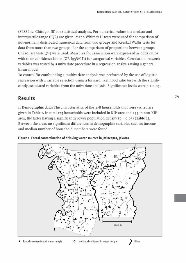
(SPSS Inc, Chicago, Ill) for statistical analysis. For numerical values the median and
interquartile range (IQR) are given. Mann Whitney U-tests were used for comparison of
not-normally distributed numerical data from two groups and Kruskal Wallis tests for
data from more than two groups. For the comparison of proportions between groups
Chi square tests (χ2) were used. Measures for association were expressed as odds ratios
with their confidence limits (OR [95%CI ]) for categorical variables. Correlation between
variables was tested by a univariate procedure in a regression analysis using a general
linear model.
To control for confounding a multivariate analysis was performed by the use of logistic
regression with a variable selection using a forward likelihood ratio test with the signifi-
cantly associated variables from the univariate analysis. Significance levels were p < 0.05.
Results1. Demographic data: The characteristics of the 378 households that were visited are
given in Table 1. In total 123 households were included in KIP-area and 255 in non-KIP-
area, the latter having a significantly lower population density (p = 0.05) (Table 2).
Between the areas no significant differences in demographic variables such as income
and median number of household members were found.
79
Drinking water, sanitation and diarrhoea
Figure 1. Faecal contamination of drinking water sources in Jatinegara, Jakarta
1000 m
Faecally contaminated water sample No faecal coliforms in water sample River
18439 Vollaard 14-12-2004 08:24 Pagina 79
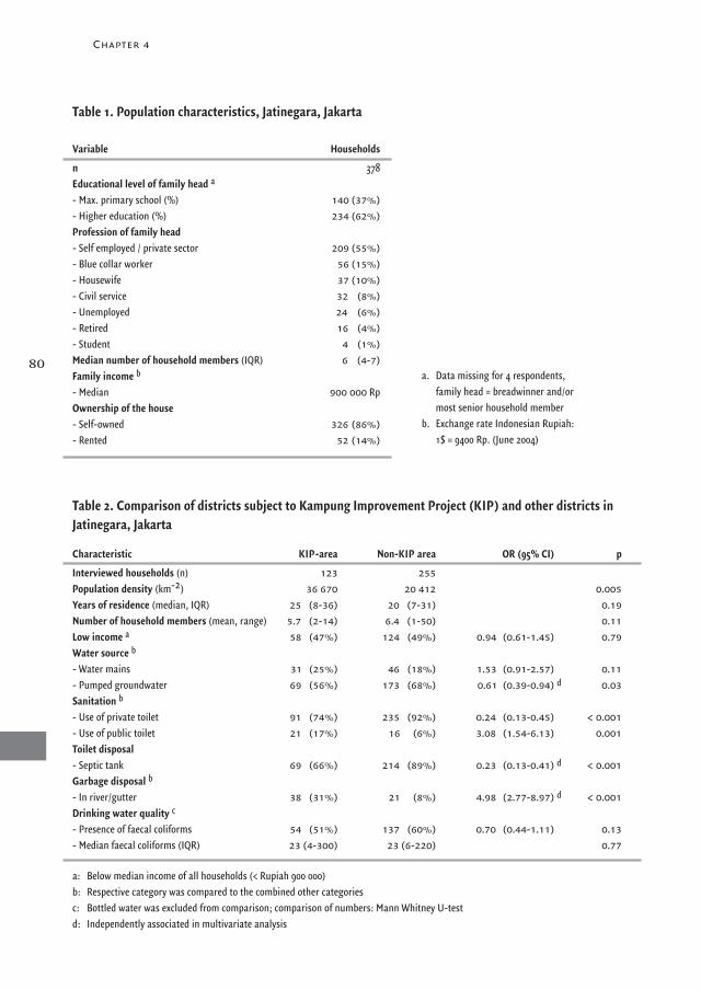
Chapter 4
80
Table 1. Population characteristics, Jatinegara, Jakarta
Table 2. Comparison of districts subject to Kampung Improvement Project (KIP) and other districts inJatinegara, Jakarta
Variable Households
n 378Educational level of family head a
- Max. primary school (%) 140 (37%)- Higher education (%) 234 (62%)Profession of family head- Self employed / private sector 209 (55%)- Blue collar worker 56 (15%)- Housewife 37 (10%)- Civil service 32 (8%)- Unemployed 24 (6%)- Retired 16 (4%)- Student 4 (1%)Median number of household members (IQR) 6 (4-7)Family income b
- Median 900 000 RpOwnership of the house- Self-owned 326 (86%)- Rented 52 (14%)
Characteristic KIP-area Non-KIP area OR (95% CI) p
Interviewed households (n) 123 255Population density (km-2) 36 670 20 412 0.005Years of residence (median, IQR) 25 (8-36) 20 (7-31) 0.19Number of household members (mean, range) 5.7 (2-14) 6.4 (1-50) 0.11Low income a 58 (47%) 124 (49%) 0.94 (0.61-1.45) 0.79Water source b
- Water mains 31 (25%) 46 (18%) 1.53 (0.91-2.57) 0.11- Pumped groundwater 69 (56%) 173 (68%) 0.61 (0.39-0.94) 0.03Sanitation b
- Use of private toilet 91 (74%) 235 (92%) 0.24 (0.13-0.45) < 0.001- Use of public toilet 21 (17%) 16 (6%) 3.08 (1.54-6.13) 0.001Toilet disposal- Septic tank 69 (66%) 214 (89%) 0.23 (0.13-0.41) < 0.001Garbage disposal b
- In river/gutter 38 (31%) 21 (8%) 4.98 (2.77-8.97) < 0.001Drinking water quality c
- Presence of faecal coliforms 54 (51%) 137 (60%) 0.70 (0.44-1.11) 0.13- Median faecal coliforms (IQR) 23 (4-300) 23 (6-220) 0.77
a: Below median income of all households (< Rupiah 900 000)b: Respective category was compared to the combined other categoriesc: Bottled water was excluded from comparison; comparison of numbers: Mann Whitney U-test d: Independently associated in multivariate analysis
a. Data missing for 4 respondents, family head = breadwinner and/or most senior household member
b. Exchange rate Indonesian Rupiah: 1$ = 9400 Rp. (June 2004)
d
d
d
18439 Vollaard 14-12-2004 08:24 Pagina 80
2. Water source: Most residents (64%) used groundwater extracted by electrically or
hand-powered pumps from boreholes (Table 3). The reported depth ranged from 3 to
35 meter (mean 16 meter). Only 5 households used public pumps (2%), the others had
access to privately constructed boreholes in their houses. Twenty percent of the house-
holds used the water mains as the source of drinking water and 12% only drank bottled
mineral water (Table 3). The latter households also had connections to the water mains
(30%) or boreholes (71%) from which water was used for personal hygiene and/or food
preparation. Only very small proportions used water from unprotected dug wells (2%)
or water from vendors (2%) as a source of drinking water.
The high-income group had more often private connections to the water mains than the
low-income group: 55 vs. 44%, but this difference was not significant (p = 0.09).
Consumers of bottled water were left out from this comparison. This group had a signifi-
cantly higher income than the users of all other sources: 84% vs. 47% were from the
high-income group, respectively (p < 0.001).
Fewer pumps were present in households in KIP-area compared to non-KIP area (56% vs.
68%, p = 0.03), but the expansion of the water mains starts to compensate for this deficit
in water access, because more residents in KIP-area had private connections to the water
mains than in non-KIP area (25% vs. 18%, respectively, p = 0.11) (Table 2).
3. Bacteriologic quality of drinking water: In 33 water samples only total coliforms but
no faecal coliforms were detected (Table 4). Piped water was less often faecally contami-
nated than the combined other sources (OR 0.10 [0.05-0.19]). In contrast to 57 (24%)
of the pumped groundwater samples only 2 (3%) of the piped water samples contained
more than 100 faecal coliforms/100 mL (p < 0.001), a level of contamination classified as
high risk. 11 However, the comparison of the median numbers of faecal coliforms in
contaminated piped water and those from pumped groundwater: 4 (IQR 2-50) versus
23 (IQR 6-280) faecal coliforms/100 mL, showed that the difference with groundwater
was non-significant (p = 0.07, Mann Whitney U-test).
Three mineral water bottles contained total coliforms including one bottle with faecal
coliforms. These bottles had been refilled in local refill outlets after prior use.
Water from dug wells and water vendors had particularly high rates and levels of
contamination (Table 4).
4. Sanitation: Most residents (86%) used private toilets in their houses and smaller pro-
portions used public toilets (10%) or the riverbanks (4%) (Table 5). In 86% of the private
toilets excreta were collected in on-site septic tanks. Human excreta and wastewater were
directly disposed into rivers or gutters by 98 households (26%) including the 46 house-
holds with a private toilet but without an on-site septic tank.
81
Drinking water, sanitation and diarrhoea
18439 Vollaard 14-12-2004 08:24 Pagina 81
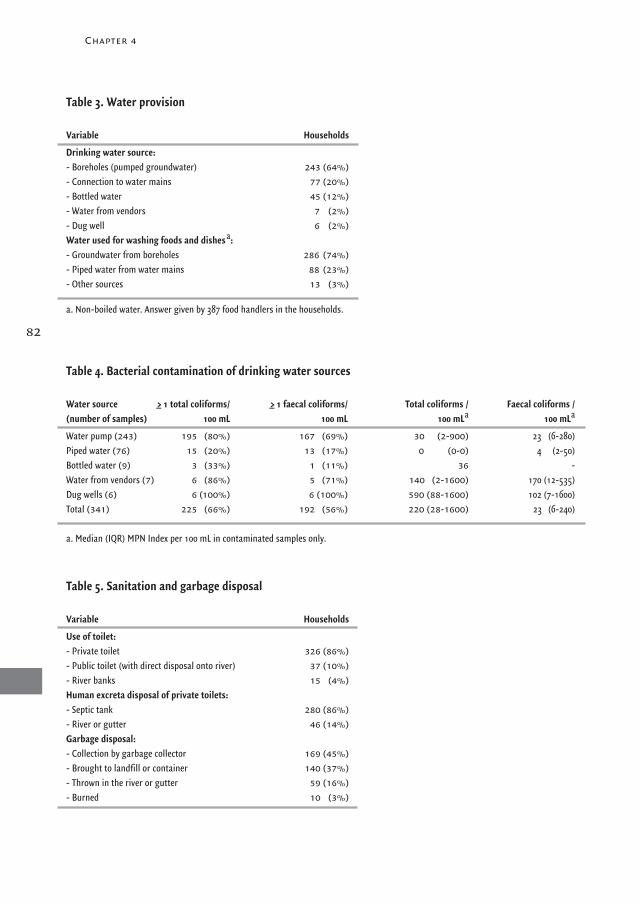
Chapter 4
82
Variable Households
Drinking water source:- Boreholes (pumped groundwater) 243 (64%)- Connection to water mains 77 (20%)- Bottled water 45 (12%)- Water from vendors 7 (2%)- Dug well 6 (2%)Water used for washing foods and dishes a:- Groundwater from boreholes 286 (74%)- Piped water from water mains 88 (23%)- Other sources 13 (3%)
a. Non-boiled water. Answer given by 387 food handlers in the households.
Table 3. Water provision
Table 4. Bacterial contamination of drinking water sources
Water source >_ 1 total coliforms/ >_ 1 faecal coliforms/ Total coliforms / Faecal coliforms / (number of samples) 100 mL 100 mL 100 mLa 100 mLa
Water pump (243) 195 (80%) 167 (69%) 30 (2-900) 23 (6-280)Piped water (76) 15 (20%) 13 (17%) 0 (0-0) 4 (2-50)Bottled water (9) 3 (33%) 1 (11%) 36 -Water from vendors (7) 6 (86%) 5 (71%) 140 (2-1600) 170 (12-535)Dug wells (6) 6 (100%) 6 (100%) 590 (88-1600) 102 (7-1600)Total (341) 225 (66%) 192 (56%) 220 (28-1600) 23 (6-240)
a. Median (IQR) MPN Index per 100 mL in contaminated samples only.
Table 5. Sanitation and garbage disposal
Variable Households
Use of toilet:- Private toilet 326 (86%)- Public toilet (with direct disposal onto river) 37 (10%)- River banks 15 (4%)Human excreta disposal of private toilets:- Septic tank 280 (86%)- River or gutter 46 (14%)Garbage disposal:- Collection by garbage collector 169 (45%)- Brought to landfill or container 140 (37%)- Thrown in the river or gutter 59 (16%)- Burned 10 (3%)
18439 Vollaard 14-12-2004 08:24 Pagina 82

We calculated the hypothetical distance between all septic tanks in Jatinegara study area
using the data from our study group. Assuming a median number of household members
of 6 and that 86% of the households had a private toilet of which 86% had a septic tank,
we found a radius of 10.2 m around each septic tank in the total study area of 10.6 km2
with 263 000 inhabitants.
Next to human excreta also garbage was directly disposed into rivers or gutters by 16%
of the households.
In KIP-area sanitation was still lagging behind, because significantly fewer households
used private toilets (74 vs. 92% in non-KIP-area) and consequently more often public
toilets were used (17 vs. 6%, respectively) (Table 2). The disposal of the private toilets in
KIP-area was less frequently collected into septic tanks than in non-KIP area (66 vs. 89%,
respectively, p < 0.001). Garbage was more frequently thrown into gutters or rivers in
KIP-area than in the other area (31 vs. 8%, p < 0.001).
5. Factors influencing the contamination rate and level
Distance to the river: Faecal contamination was not confined to areas immediately borde-
ring the rivers (Figure 1) nor to one of the 8 sub-districts (p = 0.38, χ2), also not if pumped
groundwater samples were analysed separately (p = 0.65). The number of piped water
samples in some sub-districts was too small to allow statistical comparison of contami-
nation rates and levels in piped water between the distinct sub-districts. The number of
faecal coliform in all samples was not significantly different for the 8 distinct sub-
districts (p = 0.40, Kruskal Wallis test), also not when analyzed for pumped groundwater
separately (p = 0.68).
Seasonality: The faecal contamination rate of all water samples did not vary significantly
per month (p = 0.13, χ2) (Figure 2), also not when piped water was analysed separately
(p = 0.63) or for the pump water samples only (p = 0.64). Also, no significant differences
in the numbers of faecal coliform per month were demonstrated for piped water samples
(p = 0.46, Kruskal Wallis test) or pumped water (p = 0.53).
83
Drinking water, sanitation and diarrhoea
Fig. 2. Proportional contamination of samples from groundwater and water mains (n=324) and rainfall per month
number of samples
month of sampling
18439 Vollaard 14-12-2004 08:24 Pagina 83
Chapter 4
KIP-area: Contamination rates (51% vs. 60%, p = 0.13) and levels (p = 0.77) in the KIP
and non-KIP-area were not significantly different (Table 2).
Depth of boreholes: From 168 pumps both the depth of the borehole and contamination
level was known. In a linear regression model for depth of the borehole and numbers
of faecal coliforms significantly lower numbers were found in deeper boreholes (p=0.017,
R = 0.2), but high bacterial counts were found over the full range of depths. Thirteen
outliers were not confined to one sub-district in the study area and were mostly sampled in
dry season (11 from 13). The mean depth of boreholes containing less than 100 faecal coli-
forms/100 mL was 17.0 m compared to 14.2 m in those with > 100 bacteria/100 mL(p = 0.02).
Chlorination: Thames PAM Jaya monthly measured end-point free chlorine residuals
(ppm) at 17 locations in the study area during 2002, during the study period. An end-
point level of 0.3 ppm free chlorine residual is the minimum bactericidal level the water
company applies which was met in 74% of the measurements. In regression analysis
month and location were both independently (p = 0.03 and p < 0.001, respectively) of
influence on the chlorine level, but rainfall was not independently associated (p = 0.53).
6. Diarrhoeal illness
In 34% of the households diarrhoeal episodes were reported for at least one of the house-
hold members in the 3 months preceding the interview. The occurrence of diarrhoea was
not associated with the use of other sources than piped water for consumption (OR 1.03
[0.61-1.75]) or for washing food (OR 1.21 [0.74-1.97]). Also non-associated with diarrhoea
were flooding (OR 1.40 [0.83-2.33]), the use of private toilets (OR 0.95 [0.52-1.77]),
residence in a KIP-area (OR 1.11 [0.70-1.75]) or faecal contamination of the drinking
water source (OR 1.09 [0.70-1.72]). However, in households with water contamination
> 100 faecal coliforms/100 mL diarrhoea was reported more frequently (51 vs. 30%)
(OR 2.40 [1.38-4.16]).
All respondents reported to boil their drinking water before consumption with the
exception of the 45 consumers of bottled water and 2 subjects who drank non-boiled
water from the water mains. Non-boiled groundwater from boreholes was also used in
74% for food preparation.
7. Kampung Improvement Program
The significant differences in water supply and sanitation in the areas that had been
subject to KIP compared to non-KIP area from univariate analysis include: less access to
pumped water, less use of private toilets and, consequently, more use of public toilets,
fewer septic tanks and inadequate garbage disposal (table 2). In a multivariate analysis by
logistic regression independent characteristics of the KIP-area were: less access of house-
holds to groundwater from pumps as a source of drinking water (OR 0.54 [0.33-0.89]);
84
18439 Vollaard 14-12-2004 08:24 Pagina 84
more frequent garbage disposal in rivers and gutters (OR 2.89 [1.33-6.27]); and less
presence of septic tanks for collection of human excreta (OR 0.33 [0.17-0.65]).
DiscussionThe main finding of the present study in East-Jakarta is that although the supply and
quality of drinking water – though frequently faecally contaminated with low bacterial
numbers – are satisfactory, human waste disposal and environmental hygiene are poor.
Areas subject to the prior Kampung Improvement Program especially were still lagging
behind with respect to provisions of sanitation and environmental hygiene. However, the
health hazard resulting from the latter situation, as assessed by frequency of diarrhoeal
illness, apparently is countered effectively by the generally endorsed practice of boiling
drinking water before consumption.
With respect to the study design the following should be considered. We examined the
drinking water quality at the source and not in containers after storage or boiling. We
choose this method to compare the drinking water quality from different water sources,
since comparative data are scarce in Jakarta. The only samples that had been stored prior
to sampling were those from the water vendors and high numbers of coliforms were
demonstrated in these samples. Contamination was most likely caused by unhygienic
handling or infrequent cleaning of the water containers. Similarly, if boiled water is stored
in unhygienic conditions, the initial gains of microbiologically safe water supply would
be nullified. Second, we only examined the microbiological quality of drinking water and
therefore nothing can be said about the chemical pollution of groundwater. However,
considering the lack of wastewater-treatment in Jakarta and uncontrolled garbage disposal
this problem must be considerable. Finally, the occurrence of diarrhoea in household
members as reported by the food handlers in the households might be an under-estima-
tion of the true incidence.
The access to water sources in the study area was adequate, because most households had
private pumps or connections to the water mains. Freely available groundwater extracted
by pumps was the predominant source of drinking water and water used for food prepa-
ration. Although low income could prevent residents from buying bottled water or acqui-
ring a direct connection to the water mains, the projected future scarcity of groundwater
in Jakarta may eventually limit its availability. The industrial and domestic over-exploita-
tion of groundwater caused a drop in the groundwater table and consequent salination by
intrusion of seawater (Indonesia Water Resources and Irrigation Reform Implementation
Project, Strategic Management Plan Ciliwung-Cisadane, 2003). In the first 4 years of
operation (1998-2002) the number of connections to the water system network of Thames
PAM Jaya increased with 30%. Further expansion and continuation of the existing
85
Drinking water, sanitation and diarrhoea
18439 Vollaard 14-12-2004 08:24 Pagina 85
Chapter 4
cross-subsidy system are therefore necessary to compensate for the expected increase
in demand for piped water.
Chlorinated piped water was least frequently contaminated. Although no significant dif-
ference in the number of faecal coliforms in the contaminated samples from piped water
and groundwater was found, groundwater contained more often contamination levels of
more than 100 faecal coliforms per 100 mL. That contamination level was associated with
the occurrence of diarrhoea in households making water treatment at home (i.e., boiling
or addition of hypochlorite) and hygienic storage crucial for prevention of diarrhoeal
illness. Below this threshold level most likely other transmission routes of diarrhoeal
disease (e.g., food and personal hygiene) contribute equally or more to transmission of
diarrhoeal disease, similar to findings of Moe et al. 12 The association may be explained
by the frequent use of non-boiled groundwater for food preparation, but other factors,
such as unhygienic storage of boiled drinking water, might have contributed as well.
We assume that the inadequate sanitation and human waste disposal in our study area
and the subsequent intrusion of wastewater into water pipes and boreholes are responsible
for the contamination of piped water and groundwater, respectively. Four observations
supported this hypothesis.
First, in our study area with a high population density the septic tanks are located in close
proximity to each other and the minimum recommended distance of 10 m between septic
tanks and water sources is therefore infrequently met. 10,12 The septic tanks require ade-
quate outflow of the liquid effluent and repeated emptying of faecal sludge. Both require-
ments are hard to enforce, since Jakarta lacks good sewage systems to dispose the liquid
effluent and the impaired infrastructure in overcrowded slum-areas makes emptying dif-
ficult or impossible.
Second, contamination of groundwater samples was evenly spread over the study
area. Other factors than proximity to the river (i.e., nearby septic tanks and open
gutters) must therefore be associated with faecal contamination as well. Also the fact
that high numbers of faecal coliforms were found in shallow and even in some of the
deep boreholes demonstrated that groundwater is likely contaminated by intrusion of
wastewater from superficial ground layers into boreholes (and evidently also into dug
wells). Also poor construction or maintenance of boreholes might have contributed in
this respect.
Third, uncontrolled garbage disposal in open sewers is complicating the drainage of
wastewater. Overflow or leakage from clogged gutters could result in intrusion of faecal
bacteria into adjacent boreholes or water pipes. The Kampung Improvement Project as
initiated by the city authorities did provide for concrete gutters, but participation of
residents in maintenance is essential. Garbage collection in Jakarta is a logistic challenge
but necessary to prevent the obstruction of water flows by direct disposal of garbage
86
18439 Vollaard 14-12-2004 08:24 Pagina 86

into gutters and rivers.
Fourth, fluctuating levels of chlorination were demonstrated in samples from the water
mains. Intrusion of wastewater into the water mains decreases the chlorination level and
consequently bacterial numbers rise. Intrusion is certainly possible, because administra-
tive and technical leakage of the water mains is calculated to cause a loss of 50% of the
initial water supply in Jakarta (Case studies from Dhaka, Jakarta and Manila, Malou
Mangahas, Philippine Center for Investigative Journalism, www.adb.org, 2002). As a
consequence, water pressure in the water mains is low and supply is not continuous.
Residents store water at home to bridge intervals in supply. This could give rise to conta-
mination of stored water by immersion of soiled hands or utensils into water reservoirs
or by the use of dirty storage vessels. 14-16 Negative pressures during interruptions in
supply and back-siphonage can facilitate intrusion of surrounding wastewater into
leaking water pipes resulting in post-treatment contamination of the water mains. This
mechanism could cause outbreaks of disease as was illustrated by a massive typhoid fever
epidemic in Tajikistan in 1997 that was associated with the use of municipal water. 17
Rehabilitation of the water network, as was already initiated by the water company, is
consequently a priority in water management to guarantee the distribution of high quality
water and to anticipate the increasing demand for piped water.
In conclusion, the adequate availability of water in most households, the low contamina-
tion levels in most-used drinking water sources and the entrenched habit of boiling piped
and groundwater before consumption reduce the risk of diarrhoeal illness in East Jakarta.
However, the inadequate human excreta disposal is a threat to the quality of both piped
water and groundwater. With respect to hardware interventions in water supply and sani-
tation the rehabilitation and expansion of the existing water network combined with a
24-hour water supply (i.e., permanent pressure in the water mains) are needed to prevent
intrusion of wastewater into pipes and to allow supply of high quality water from the
water network. The separation of human excreta and water provisions is essential to pre-
vent that intrusion. Future Kampung Improvement Programs should specifically focus on
human waste disposal, since water supply in slum-areas is increasingly covered by the
expansion of the water network in recent years. Specific software interventions by the
health sector in Jakarta should address the following issues in public health campaigns:
the continuation of boiling groundwater and piped water before consumption; the use
of piped water or bottled water for food preparation; the warning for the high contamina-
tion rates and levels in water from dug wells and water vendors; the construction of bore-
holes of sufficient depth; the monitoring of mineral water refill outlets; the promotion of
cheap access to the water mains for the urban poor; adequate garbage disposal; and the
construction in safe distance from water sources and frequent emptying of septic tanks.
87
Drinking water, sanitation and diarrhoea
18439 Vollaard 14-12-2004 08:24 Pagina 87

Financial support was provided by the Royal Netherlands Academy of Arts and Sciences
(KNAW). We thank the technicians of the Nusantara Water Centre in West-Jakarta for the
examination of the water samples, Thames PAM Jaya for the provision of data on free
chlorine residuals in Jatinegara and Halim Meteorology Station East Jakarta for provision
of data on rainfall. We also thank Ron Wolterbeek of the Dept. Medical Statistics of the
LUMC for his advice on statistical matters. This study could not have been done without
our research-assistants: Billy Hunsinger, Ferry Kandaw, Rinny Listyani, Meily, Vea
Noveria, Carmelita Ridwan, Min Ali Sugiharto, Lidwina Sutikno, Mariana Tasman and
Lily Yaputra.
References88
Chapter 4
1. Murray CJ, Lopez AD. Global mortality, disability, and the contribution of risk factors: Global Burden of Disease Study. Lancet1997; 349:1436-1442.
2. WHO. Global water supply and sanitation assessment. Geneva: World Health Organization, 2000.3. Esrey SA. Water, waste, and well-being: a multicountry study. Am J Epidem 1996; 143:608-623.4. VanDerslice J, Briscoe J. Environmental interventions in developing countries: interactions and their implications. Am J Epidem
1995; 141:135-144.5. Esrey SA, Potash JB, Roberts L, Shiff C. Effects of improved water supply and sanitation on ascariasis, diarrhoea, dracunculiasis,
hookworm infection, schistosomiasis, and trachoma. Bull World Health Organ 1991; 69:609-621.6. Checkley W, Gilman RH, Black RE, Epstein LD, Cabrera L, Sterling CR et al. Effect of water and sanitation on childhood health in a
poor Peruvian peri-urban community. Lancet 2004; 363:112-118.7. Varley RC, Tarvid J, Chao DN. A reassessment of the cost-effectiveness of water and sanitation interventions in programmes for
controlling childhood diarrhoea. Bull World Health Organ 1998; 76:617-631.8. Oyofo BA, Subekti D, Tjaniadi P, Machpud N, Komalarini S, Setiawan B et al. Enteropathogens associated with acute diarrhea in
community and hospital patients in Jakarta, Indonesia. FEMS Immunol Med Microbiol 2002; 34:139-146.9. Subekti D, Lesmana M, Tjaniadi P, Safari N, Frazier E, Simanjuntak C et al. Incidence of Norwalk-like viruses, rotavirus and
adenovirus infection in patients with acute gastroenteritis in Jakarta, Indonesia. FEMS Immunol Med Microbiol 2002; 33:27-33.10. Vollaard AM, Ali S, van Asten HAGH et al., Risk factors for typhoid and paratyphoid fever in Jakarta, Indonesia. JAMA 2004; 291:
2607-2615.11. WHO. Guidelines for drinking-water quality. Second Edition. Geneva: World Health Organization, 1997.12. Moe CL, Sobsey MD, Samsa GP, Mesolo V. Bacterial indicators of risk of diarrhoeal disease from drinking-water in the
Philippines. Bull World Health Organ1991; 69:305-317.13. Borchardt MA, Chyou PH, DeVries EO, Belongia EA. Septic system density and infectious diarrhea in a defined population of
children. Environm Health Perspect 2003; 111:742-748.14. Jensen PK, Ensink JH, Jayasinghe G, van der HW, Cairncross S, Dalsgaard A. Domestic transmission routes of pathogens: the
problem of in-house contamination of drinking water during storage in developing countries. Trop Med Int Health 2002; 7:604-609.15. Mintz ED, Reiff FM, Tauxe RV. Safe water treatment and storage in the home. A practical new strategy to prevent waterborne
disease. J Am Med Assoc 1995; 273:948-953.16. Wright J, Gundry S, Conroy R. Household drinking water in developing countries: a systematic review of microbiological
contamination between source and point-of-use. Trop Med Int Health 2004; 9:106-117.17. Mermin JH, Villar R, Carpenter J, Roberts L, Samaridden A, Gasanova L et al. A massive epidemic of multidrug-resistant typhoid
fever in Tajikistan associated with consumption of municipal water. J Infect Dis 1999; 179:1416-1422.
18439 Vollaard 14-12-2004 08:24 Pagina 88
Acknowledgements

5
Helicobacter pylori infection and typhoid fever
in Jakarta, Indonesia
Albert M. Vollaard 1 Hein W. Verspaget 2 Soegianto Ali 3
Leo G. Visser 1 Roeland A. Veenendaal 2 Henri A.G.H. van Asten 4
Suwandhi Widjaja 5 Charles Surjadi 6 Jaap T. van Dissel 1
1 Dept. Infectious Diseases, and 2 Dept. Gastroenterology-Hepatology,
Leiden University Medical Center, The Netherlands3 Dept. Biology, 5 Dept. Internal Medicine, and 6 Center for Health Research,
Medical Faculty Atma Jaya Catholic University, Jakarta, Indonesia4 Institute for International Health, Nijmegen University Medical Center, The Netherlands
Submitted for publication
18439 Vollaard 14-12-2004 08:24 Pagina 89
Abstract
We evaluated the association between typhoid fever and Helicobacter pylori infection, as
the latter microorganism may influence gastric acid secretion and consequently increase
susceptibility to Salmonella typhi infection. Anti-H. pylori IgG and IgA antibody titres
(ELISA) and gastrin concentration (RIA) were determined in plasma of 87 blood culture-
confirmed typhoid fever cases (collected after clinical recovery) and 232 random healthy
controls without a history of typhoid fever, in the Jatinegara district, Jakarta. Patients with
typhoid fever more often than controls were seropositive for H. pylori IgG (67 vs 50%,
p<0.008), when antibody titres were dichotomized around median titres observed in con-
trols. H. pylori IgA-seropositivity was not associated with typhoid fever. Plasma gastrin
concentrations indicative of hypochlorhydria (i.e., gastrin >_ 25 or >_ 100 ng/L) were not
significantly elevated in typhoid fever cases as compared to controls (p=0.54 and p=0.27,
respectively). In a multivariate analysis, typhoid fever was independently associated with
young age (< 27 years, median age of all subjects): Odds Ratio (OR) 4.53 [95%-confidence
interval (CI): 2.53-8.10], and H. pylori IgG-seropositivity: OR 1.78 [95%-CI: 1.02-3.09].
Typhoid fever was independently associated with H. pylori IgG-seropositivity, but not
with elevated gastrin concentration. Therefore, the association suggests a common risk
of environmental exposure to both bacteria, e.g., poor hygiene, rather than a causal
relationship via a reduced gastric acid production.
Chapter 5
18439 Vollaard 14-12-2004 08:24 Pagina 90
Introduction
Typhoid fever is a serious systemic illness that each year affects over 20 million people,
predominantly in developing countries. 1 Infection with Salmonella typhi is transmitted by
faecal-oral route and in several epidemiological studies risk factors were identified that
suggested either waterborne transmission 2-6 or foodborne transmission. 2,7-9 The deter-
mination of the relative contribution of distinct environmental risk factors for transmis-
sion of disease is essential to focus local control strategies. Also host-related risk factors
for infection have been examined, identifying both genetic factors 10,11 as well as concur-
rent Helicobacter pylori infection, that was interpreted as a cause of a reduced gastric-acid
barrier. 8
A high incidence of salmonellosis has been observed in individuals with surgically induced
or other types of achlorhydria (pernicious anemia and chronic atrophic gastritis). 12-14
Also H. pylori infection may exert an effect on the secretion of gastric acid. Approximately
50 percent of the world population is infected with H. pylori 15, and even higher prevalences
have been reported in developing countries 16, where acquisition occurs at a younger age
than in the developed world. 17,18 Active infection with H. pylori is associated with a tran-
sient hypochlorhydria that may be present for several months. 19 Furthermore, H. pylori-
induced chronic gastritis of the body of the stomach reduces acid secretion and the per-
sistent hypochlorhydria constitutes a risk for the development of gastric cancer. 20,21
In absence of the acid-mediated inhibition of gastric gastrin release, the serum gastrin
concentration increases. In contrast, antral-predominant, body-sparing gastritis due to
H. pylori increases gastric acid secretion, resulting in duodenal ulcer disease. 21-23 The
localization of gastritis depends on the pre-morbid acid secretory status of a subject, die-
tary, genetic and bacterial factors 22,24, and the age of acquisition of H. pylori infection. 18
Consequently, the association between H. pylori infection as an indicator of hypochlorhy-
dria and the susceptibility to other gastro-intestinal infections is ambiguous. An increa-
sed susceptibility for enteric infections in H. pylori-infected individuals, as measured by
anti-H. pylori IgG-response, was documented for cholera 25,26 and typhoid fever. 8
However, the evidence for the association of H. pylori infection and diarrhea is
conflicting 27-32 and even a protective effect of H. pylori infection was demonstrated. 33,34
To examine the association of H. pylori infection and typhoid fever, we determined the
anti-H. pylori antibody titres and plasma gastrin in patients who had just recovered from
typhoid fever and healthy controls in a case-control study in Jakarta, Indonesia.
91
Helicobacter pylori infection and typhoid fever
18439 Vollaard 14-12-2004 08:24 Pagina 91

Chapter 5
Material and Methods
Study design: From June 2001-October 2003 we identified patients with typhoid fever
(Salmonella typhi infections) in a passive community-based surveillance study among
individuals with 3 or more days of fever in the Jatinegara district, Jakarta, Indonesia, as
described. 35 In addition, 6 patients with typhoid fever were enrolled in the Medistra
Hospital in Central Jakarta. Patients were interviewed using a standardized questionnaire
and blood samples were collected during a household visit, conducted within one month
after making the diagnosis by blood culture, at a time when the patient had recovered
from typhoid fever.
During the study period healthy controls were randomly selected in a random household
in every third rukun tetangga (RT) of a total of 1140 RT’s in Jatinegara; RT being the smal-
lest administrative unit of 40-60 households. Controls were enrolled in a ratio of cases to
controls of 1 : 4 to maximize statistical power of the analysis. The selection of controls
was non-matched for age or sex to prevent over-matching. When a control reported any
type of fever in the 30 days preceding the interview or refused participation, the house on
alternating sides of the initially selected household was approached. From 378 healthy
controls 291 (77%) allowed collection of a blood sample. Fifty-nine (20%) of these 291
controls were excluded from the analysis because they reported a history of typhoid fever
(defined as an episode of 3 or more days of fever, requiring antibiotics prescribed by a
physician who had made a diagnosis of typhoid fever). If cases or controls were less than
13 years of age, the mother or guardian was interviewed.
This study was approved by the Indonesian National Institute of Health Research and
Development (Litbangkes) and provincial authorities. From all participants a written
informed consent was obtained at the household visit or during hospitalization.
Laboratory diagnosis of typhoid fever: The diagnosis of typhoid fever required confirma-
tion by blood culture. To this end, blood was collected of the febrile patients (dependent
on the age, 5-10 mL), inoculated in aerobic bottles (Bactec, Becton Dickinson, Franklin
Lakes, NJ) and incubated. The identification of cultured micro-organisms was done by
biochemical tests and agglutination, as described. 35
Samples in patients and controls: In the household visits blood samples were collected
from non-fasting cases and controls in EDTA-tubes. Following centrifugation, plasma
was separated and frozen at –70˚C.
Measurement of anti-H. pylori IgA and IgG antibodies by ELISA: The ELISA technique
for determination of IgG and IgA antibodies against H. pylori as well as information on
the intra- and interassay variability of the results obtained by this ELISA have been
described in detail. 36,37 The antigen is a mixture of sonicates of six different H. pylori
strains; these had been obtained by sonicating whole bacteria for 6 minutes on a Branson
92
18439 Vollaard 14-12-2004 08:24 Pagina 92
sonifier (stage 4; 30 000 cycles/sec), and adjusting the suspension to a protein concentra-
tion of 3 mg/mL. Optimum concentrations of reagents was determined by checker board
titration as described. 36 ELISA results are expressed as absorbance index (AI):
AI = [Patient’s OD – OD of blank reading] / [Reference OD – OD of blank reading],
where OD is the optical density. Samples were assayed in blinded fashion in one run.
Measurements of gastrin concentrations: Plasma gastrin concentration was measured
using a previously validated radioimmune assay (RIA) as described. 38 Samples were
assayed in blinded fashion in one run.
Statistical methods: Data from the questionnaires were entered twice using EpiInfo 6.04b
software (CDC, Atlanta, USA), validated and imported in SPSS version 11.5 (SPSS Inc,
Chicago, Ill.) for further analysis. Unpaired t-tests for normally distributed variables or
Mann Whitney U-tests for not-normally distributed variables were used to test numerical
variables. Correlation between numerical variables was tested by linear regression and the
Pearson correlation coefficient (R) was provided. For the comparison of proportions the
Chi-square test or Fisher’s Exact test was used. To correct for age strata Mantel-Haenszel
statistic was applied. Measures of association were expressed as odds ratios with their
respective confidence limits (OR [95%-CI]) when categorical exposures were evaluated.
To control for confounding a multivariate analysis was performed by the use of logistic
regression.
ResultsDemographic characteristics: Plasma samples were available of 87 typhoid fever cases,
and were collected at home following clinical recovery of the patient 22 days [median,
IQR: 20-27] after the confirmatory blood culture. Thirteen patients had been hospitalized
and 74 patients were included in local health outpatient facilities. During the same period
blood samples were obtained from 232 healthy controls. The median age of all subjects
was 27 years [IQR: 17-45]. Patients with typhoid fever had a median age of 20 year [IQR:
13-27], and were significantly younger than the controls (median age 33 [IQR: 19-50]
years; p<0.001 by Mann Whitney U-test). The sex ratio did not differ between the typhoid
fever group and controls, i.e., 53 and 59 percent were female individuals, respectively
(p = 0.29, χ2).
Concentration of IgG and IgA antibodies against H.pylori: Taking all subjects together,
the median titre of anti-H. pylori IgG amounted to AI 0.126 (IQR: 0.084-0.186) and for
IgA to AI 0.094 (IQR: 0.057-0.170). In a linear regression model IgG and IgA were
significantly correlated, though not strongly considering the correlation coefficient
(p < 0.001, R = 0.23).
93
Helicobacter pylori infection and typhoid fever
18439 Vollaard 14-12-2004 08:24 Pagina 93

Chapter 5
94
Table 1: H. pylori-serology of typhoid fever cases and controls
Table 2: Univariate analysis: associations with typhoid fever
Age Cases Controls Cases IgG (AI) a Controls IgG (AI) a IgG >_ 0.119 (AI) b p c
Cases Controls0-14 28 23 0.132 (0.068-0.179) 0.114 (0.069-0.155) 17 (61%) 10 (43%) 0.22
15-29 43 82 0.151 (0.097-0.190) 0.118 (0.084-0.162) 30 (70%) 39 (48%) 0.0230-44 12 48 0.143 (0.098-0.192) 0.128 (0.093-0.206) 8 (67%) 26 (54%) 0.43
>_ 45 4 79 0.236 (0.090-0.261) 0.128 (0.080-0.230) 3 (75%) 41 (52%) 0.62
Total: 87 232 0.146 (0.092-0.190) 0.119 (0.082-0.185) 58 (67%) 116 (50%) 0.008
IgA (AI) a IgA (AI) a IgA >_ 0.092 (AI) b p c
0-14 28 23 0.073 (0.046-0.118) 0.038 (0.016-0.102) 12 (43%) 6 (26%) 0.2115-29 43 82 0.100 (0.060-0.164) 0.069 (0.042-0.126) 25 (58%) 29 (35%) 0.0230-44 12 48 0.105 (0.072-0.231) 0.119 (0.073-0.221) 7 (58%) 30 (63%) 0.79
>_ 45 4 79 0.105 (0.074-0.226) 0.134 (0.078-0.296) 2 (50%) 51 (65%) 0.62
Total: 87 232 0.098 (0.060-0.156) 0.092 (0.054-0.178) 46 (53%) 116 (50%) 0.65
a: Median absorbance index (25-75% range)b: Median antibody AI in total number of healthy controls c: Chi square test (Fisher’s Exact test if cells contained less than 5 subjects)
Risk factor Cases (n=87) Controls (n=232) OR (95% CI) p
Age a
- < median (i.e., 27 years) 64 (74%) 94 (41%) 4.09 (2.37-7.04) < 0.001Sex- Male 41 (47%) 94 (41%) 1.31 (0.80-2.15) 0.29Anti-H.pylori antibodies- IgG positive (AI >_ 0.119) b 58 (67%) 116 (50%) 2.00 (1.20-3.35) 0.008- IgA positive (AI >_ 0.092) 46 (53%) 116 (50%) 1.12 (0.69-1.84) 0.65Plasma gastrin- >_ 25 ng/L 80 (92%) 208 (90%) 1.32 (0.55-3.18) 0.54- >_ 100 ng/L 18 (21%) 36 (16%) 1.42 (0.76-2.66) 0.27
a. Independently associated in multivariate analysis: OR 4.53 (95% CI: 2.53-8.10), p < 0.001b. Independently associated in multivariate analysis: OR 1.78 (95% CI: 1.02-3.09), p = 0.04
18439 Vollaard 14-12-2004 08:24 Pagina 94
In individuals who had suffered from typhoid fever in the preceding month the anti-
H. pylori IgG-titres (median 0.146 [IQR: 0.092-0.190]) were higher than those in controls
(median 0.119 [ IQR: 0.082-0.185]), but the difference did not reach a level of significance
(p = 0.24, by Mann Whitney U-test). Also the IgA titres did not differ significantly (p > 0.50)
between typhoid fever cases (median 0.098 [IQR: 0.060-0.156]) and controls (median
0.092 [IQR: 0.054-0.178]).
In typhoid fever patients and controls alike, IgG and IgA against H. pylori increased with
age (Table 1), and in a linear regression model IgG titres and age were significantly corre-
lated (p = 0.005, R = 0.16) as were IgA AI and age (p < 0.001, R = 0.30). In all age cohorts
higher rates of IgG-seropositivity were observed in the group of individuals with typhoid
fever, and in the age cohort of 15-29 years this difference reached a level of significance
(p = 0.02).
Next, the median AI of IgG and IgA in the healthy controls were used to dichotomize the
data to obtain a cutoff-level of H. pylori seropositivity (IgG >_ 0.119 and IgA >_ 0.092, res-
pectively) comparable to study procedures of Bhan et al. 8 The percentage of individuals
with typhoid fever who had an IgG titre above this level (n = 58; 67%) was significantly
(p = 0.008) higher than that in the controls (n = 116; 50%) (Table 1). No significant diffe-
rence of IgA-seropositivity between individuals with typhoid fever and controls was
observed: i.e, 46 (53%) versus 116 (50%), respectively (p = 0.65). In a stratified analysis
for all individuals correcting for age group by use of Mantel-Haenszel statistic, IgG-sero-
positivity remained significantly associated with typhoid fever (p = 0.007), whereas
IgA-seropositivity reached borderline significance (p = 0.05).
Gastrin concentration: Taken all subjects together, plasma gastrin concentration (ng/L)
ranged from 6 to 1253 ng/L, with a median concentration of 54 (IQR 36-85) ng/L. On
average, gastrin concentrations did not change with age (p > 0.50, R = 0.004). The
gastrin concentration in individuals with typhoid fever (median 53 ng/L [IQR: 37-85])
did not differ significantly from that in controls (median 54 ng/L [IQR: 36-85])
(p = 0.74, Mann Whitney U-test).
Gastrin concentrations of >_ 25 ng/L and >_ 100 ng/L have been evaluated as markers of
hypochlorhydria as a condition of chronic atrophic gastritis. 39 In the analysis of the total
group, i.e., both cases and controls, anti-H. pylori antibody indices were higher in the
group of individuals with gastrin >_ 25 ng/L than in those with a lower plasma gastrin,
reaching significance for IgA AI (p = 0.02, Mann Whitney U test) and borderline signifi-
cance for IgG AI (p = 0.06). The IgG and IgA AI of individuals with gastrin < 100 ng/L did
not differ significantly from those with gastrin >_ 100 ng/L (p = 0.44 and p > 0.50, respec-
tively). However, the proportions of typhoid fever cases and controls that had gastrin
>_ 25 ng/L did not differ significantly (p = 0.54) (Table 2). Similarly, in the group of
typhoid fever patients, 18 (21%) individuals had a gastrin concentration >_ 100 ng/L, as
95
Helicobacter pylori infection and typhoid fever
18439 Vollaard 14-12-2004 08:24 Pagina 95
Chapter 5
compared to 36 (16%) in the control group (p = 0.27).
Univariate and multivariate analysis
H. pylori IgG- and IgA-seropositivity and plasma gastrin concentrations in cases and
controls were evaluated by univariate analysis (Table 2). Significantly associated with
typhoid fever were: young age (dichotomized at 27 years, the median age of all subjects):
OR 4.09 (95%-CI: 2.37-7.04), and H. pylori IgG-seropositivity: OR 2.00 (95%-CI:
1.20-3.35). H. pylori IgA-seropositivity nor elevated plasma gastrin concentrations
were significantly associated with typhoid fever.
In a multivariate analysis using multiple logistic regression to control for confounding
the occurrence of typhoid fever was independently associated with H. pylori IgG-
seropositivity: OR 1.78 (95%-CI: 1.02-3.09), and young age (< 27 years): OR 4.53
(95%-CI: 2.53-8.10).
DiscussionThe main finding of this case-control study in Jakarta is that the prevalence of H. pylori
IgG-seropositivity is higher in individuals who recently had recovered from blood culture-
confirmed typhoid fever than in healthy controls without a history of typhoid fever.
However, we did not find an association between typhoid fever and an elevated plasma
gastrin concentration indicative of hypo- or achlorhydria. Furthermore, typhoid fever was
independently associated with young age, which suggests that either exposure to S. typhi
or the proportion of those exposed that develop symptomatic infection (e.g., due to
absence of a prior acquired immune response) is greater among young people.
A reduced secretion of bactericidal gastric acid due to acute or chronic H. pylori infection 22
may increase susceptibility to infection with enteric pathogens. This hypothesis was sup-
ported by case-control studies that examined anti-H. pylori IgG antibody concentrations
in patients with typhoid fever and cholera, and observed a higher prevalence of H. pylori
seropositivity among the cases than controls. 8,25,26 To this end, seropositivity was
defined as an antibody concentration above the median antibody level of the controls 8,
a method we applied as well to be able to compare our results and also to confirm the
demonstrated association of typhoid fever and H. pylori infection.
Although the anti-H. pylori IgG-response reliably indicates prior infection with H. pylori,
H. pylori colonization in the mostly adult typhoid fever cases and controls most likely
occurred at a much younger age, i.e., in early childhood 17, than the acquisition of
typhoid fever in our study. Moreover, the infection itself may exert different effects on the
gastric acid secretion and is therefore not one-dimensionally associated with hypochlor-
hydria as a predisposing condition for enteric infections. 21 In addition, the evidence for
a correlation between anti-H. pylori IgG-antibody levels and the severity of gastritis or
96
18439 Vollaard 14-12-2004 08:24 Pagina 96

H. pylori density remains conflicting 40-43, thereby diminishing the likelihood of a more
severe gastritis in typhoid fever cases compared to controls. Also, anti-H. pylori anti-
bodies may be a sensitive marker of chronic atrophic gastritis as a cause of hypochlorhy-
dria, although not being a very specific marker 39, which can explain the higher antibody
titres in the individuals with gastrin >_ 25 ng/L in our study. However, we did not find
evidence for a higher prevalence of chronic atrophic gastritis, as determined by elevated
concentrations of plasma gastrin, among cases of typhoid fever compared with healthy
controls.
A more likely explanation of the association of increased anti-H. pylori IgG antibodies
and typhoid fever might be a higher susceptibility to colonization by and re-exposure to
H. pylori as a consequence of low hygienic standards and shared risk factors for both
infections, than a supposedly decreased gastric acid secretion in typhoid fever cases.
Since re-infection with H. pylori after eradication is common in developing countries 44
signifying continuous exposure, the association of typhoid fever and H. pylori IgG-
seropositivity may be confounded by poor hygiene.
Some issues related to the outcome of our study need consideration. We did not find an
association between H. pylori IgA-seropositivity and typhoid fever, most likely due to the
fact that the anti-H. pylori IgA serological response is a less sensitive marker for H. pylori
infection than anti-H. pylori IgG. 45-47 We assume that H. pylori infection as determined
by anti-H. pylori antibody response preceded typhoid fever infection, because it takes 1-3
months after H. pylori infection to develop anti-H. pylori IgG antibodies and typhoid fever
cases were visited within 1 month after blood culture-confirmation of S. typhi infection.48
Also, in some cases the antibiotic treatment of typhoid fever might have interfered with
the quantitative anti-H. pylori IgA antibody response, because in H. pylori eradication stu-
dies IgA-titres decreased faster than IgG six weeks after antibiotic treatment, but even
then IgA and IgG titres will remain elevated during 6-12 months after eradication. 37,44,47,49
Furthermore, samples for determination of plasma gastrin were obtained from non-
fasting subjects, but the gastrin concentrations in our study showed little elevation com-
pared to concentrations in a population sample in Mexico, where samples were taking in
a fasting state and median serum gastrin amounted to 35 ng/L [IQR 26-52]. 39 Finally, we
assume that the association of H. pylori infection as defined by anti-H. pylori IgG-response
and typhoid fever may be the result from shared risk factors for exposure to both bacteria.
Infection with H. pylori occurs in early childhood within families. 17,20 Several risk factors
for H. pylori infection have been identified without determination of the exact source of
infection, but most are associated with poor hygiene and sanitation within households,
and could coincide with the intra-household risk factors for typhoid fever in Jakarta. 17,35
The improvement of hygiene within households might therefore reduce the risk of
exposure to both bacteria. Continuous exposure may also explain the correlation of age
97
Helicobacter pylori infection and typhoid fever
18439 Vollaard 14-12-2004 08:24 Pagina 97
Chapter 5
and the anti-H. pylori antibody absorbance index in our study, similar to findings of
Chen et al. 40
In conclusion, our findings demonstrated that typhoid fever is associated with H. pylori
IgG seropositivity, whereas the gastrin concentrations were not significantly higher in
typhoid fever cases compared with healthy controls. The findings do not support the
hypothesis that the association points to H. pylori-induced hypochorhydria as risk factor
for typhoid fever, but suggest that the epidemiological association is likely confounded
by shared risk factors for infection with both bacteria.
AcknowledgementsFinancial support was provided by the Royal Netherlands Academy of Arts and Sciences
(KNAW). We thank the physicians, nurses and technicians of the participating health
centres in Jakarta for their cooperation: Mitra Internasional hospital and microbiology
lab, Budhi Asih, St. Carolus, Medistra and Persahabatan hospital, all puskesmas’ in
Jatinegara and the local private practitioners. We thank Wim van Duijn, Izäk Biemond
and Johan van der Reijden, and Adriette de Visser from the LUMC Laboratory of
Gastroenterology-Hepatology, and Infectious Diseases, respectively. This study could
not have been conducted without our research-assistants in Jakarta.
References
1. Crump JA, Luby SP, Mintz ED. The global burden of typhoid fever. Bull World Health Organ 2004; 82: 346-353.
2. Luby SP, Faizan MK, Fisher-Hoch SP, et al. Risk factors for typhoid fever in an endemic setting, Karachi, Pakistan.
Epidemiol Infect 1998; 120: 129-138.
3. Swaddiwudhipong W, Kanlayanaphotporn J. A common-source water-borne outbreak of multidrug-resistant typhoid fever in a
rural Thai community. J Med Assoc Thai 2001; 84: 1513-1517.
4. King CC, Chen CJ, You SL, Chuang YC, Huang HH, Tsai WC. Community-wide epidemiological investigation of a typhoid
outbreak in a rural township in Taiwan, Republic of China. Int J Epidemiol 1989; 18: 254-260.
5. Gasem MH, Dolmans WM, Keuter MM, Djokomoeljanto RR. Poor food hygiene and housing as risk factors for typhoid fever in
Semarang, Indonesia. Trop Med Int Health 2001; 6: 484-490.
6. Mermin JH, Villar R, Carpenter J, et al. A massive epidemic of multidrug-resistant typhoid fever in Tajikistan associated with
consumption of municipal water. J Infect Dis 1999; 179: 1416-1422.
7. Velema JP, van Wijnen G, Bult P, van Naerssen T, Jota S. Typhoid fever in Ujung Pandang, Indonesia--high-risk groups and
high-risk behaviours. Trop Med Int Health 1997; 2: 1088-1094.
8. Bhan MK, Bahl R, Sazawal S, et al. Association between Helicobacter pylori infection and increased risk of typhoid fever.
J Infect Dis 2002; 186: 1857-1860.
9. Black RE, Cisneros L, Levine MM, Banfi A, Lobos H, Rodriguez H. Case-control study to identify risk factors for paediatric
endemic typhoid fever in Santiago, Chile. Bull World Health Organ 1985; 63: 899-904.
10. Dunstan SJ, Stephens HA, Blackwell JM, et al. Genes of the class II and class III major histocompatibility complex are associated
with typhoid fever in Vietnam. J Infect Dis 2001;183: 261-268.
98
18439 Vollaard 14-12-2004 08:24 Pagina 98
11. Dharmana E, Joosten I, Tijssen HJ, et al. HLA-DRB1*12 is associated with protection against complicated typhoid fever,
independent of tumour necrosis factor alpha. Eur J Immunogenet 2002; 29: 297-300.
12. Kunz LJ, Waddell WR. Association of Salmonella enteritis with operations on the stomach. N Engl J Med 1956; 255: 555-559.
13. Gianella RA, Broitman SA, Zamcheck N. Influence of gastric acidity on bacterial and parasitic enteric infections: a perspective.
Ann Intern Med 1973; 78: 271-276.
14. Gianella RA, Broitman SA, Zamcheck N. The gastric barrier to microorganisms in man: in vivo and in vitro studies. Gut 1972; 13:
251-256.
15. Torres J, Perez-Perez G, Goodman KJ, et al. A comprehensive review of the natural history of Helicobacter pylori infection in
children. Arch Med Res 2000; 31: 431-469.
16. Bardhan PK. Epidemiological features of Helicobacter pylori infection in developing countries. Clin Infect Dis 1997; 25: 973-978.
17. Frenck RW, Jr., Clemens J. Helicobacter in the developing world. Microbes Infect 2003; 5: 705-713.
18. Blaser MJ. Hypothesis: the changing relationships of Helicobacter pylori and humans: implications for health and disease.
J Infect Dis 1999; 179:1523-1530.
19. Harford WV, Barnett C, Lee E, Perez-Perez G, Blaser MJ, Peterson WL. Acute gastritis with hypochlorhydria: report of 35 cases
with long term follow up. Gut 2000; 47: 467-472.
20. Suerbaum S, Michetti P. Helicobacter pylori infection. N Engl J Med 2002; 347: 1175-1186.
21. El Omar EM, Carrington M, Chow WH, et al. Interleukin-1 polymorphisms associated with increased risk of gastric cancer.
Nature 2000; 404: 398-402.
22. El Omar EM, Oien K, El Nujumi A, et al. Helicobacter pylori infection and chronic gastric acid hyposecretion. Gastroenterology
1997; 113: 15-24.
23. Gillen D, El Omar EM, Wirz AA, Ardill JE, McColl KE. The acid response to gastrin distinguishes duodenal ulcer patients from
Helicobacter pylori-infected healthy subjects. Gastroenterology 1998; 114: 50-57.
24. McColl KE, Gillen D, el Omar E. The role of gastrin in ulcer pathogenesis. Baillieres Best Pract Res Clin Gastroenterol 2000; 14: 13-26.
25. Shahinian ML, Passaro DJ, Swerdlow DL, Mintz ED, Rodriguez M, Parsonnel J. Helicobacter pylori and epidemic Vibrio cholerae
O1 infection in Peru. Lancet 2000; 355: 377-378.
26. Clemens J, Albert MJ, Rao M, et al. Impact of infection by Helicobacter pylori on the risk and severity of endemic cholera.
J Infect Dis 1995; 171: 1653-1656.
27. Sullivan PB, Thomas JE, Wight DG, et al. Helicobacter pylori in Gambian children with chronic diarrhoea and malnutrition.
Arch Dis Child 1990; 65: 189-191.
28. Rahman MM, Mahalanabis D, Sarker SA, et al. Helicobacter pylori colonization in infants and young children is not necessarily
associated with diarrhoea. J Trop Pediatr 1998; 44: 283-287.
29. Kehrt R, Becker M, Brosicke H, Kruger N, Helge H. Prevalence of Helicobacter pylori infection in Nicaraguan children with
persistent diarrhea, diagnosed by the 13C-urea breath test. J Pediatr Gastroenterol Nutr 1997; 25:84-88.
30. Passaro DJ, Taylor DN, Meza R, Cabrera L, Gilman RH, Parsonnet J. Acute Helicobacter pylori infection is followed by an
increase in diarrheal disease among Peruvian children. Pediatrics 2001; 108: E87.
31. Weaver LT. Royal Society of Tropical Medicine and Hygiene Meeting at Manson House, London, 16 February 1995. Aspects of
Helicobacter pylori infection in the developing and developed world. Helicobacter pylori infection, nutrition and growth of West
African infants. Trans R Soc Trop Med Hyg 1995; 89: 347-350.
32. Dale A, Thomas JE, Darboe MK, Coward WA, Harding M, Weaver LT. Helicobacter pylori infection, gastric acid secretion, and
infant growth. J Pediatr Gastroenterol Nutr 1998; 26: 393-397.
33. Rothenbacher D, Blaser MJ, Bode G, Brenner H. Inverse relationship between gastric colonization of Helicobacter pylori and
diarrheal illnesses in children: results of a population-based cross-sectional study. J Infect Dis 2000; 182: 1446-1449.
34. Perry S, Sanchez L, Yang S, Haggerty TD, Hurst P, Parsonnet J. Helicobacter pylori and risk of gastroenteritis.
J Infect Dis 2004; 190: 303-310.
99
Helicobacter pylori infection and typhoid fever
18439 Vollaard 14-12-2004 08:24 Pagina 99
35. Vollaard AM, Ali S., van Asten HAGH, et al. Risk factors for typhoid and paratyphoid fever in Jakarta, Indonesia.
JAMA 2004; 291: 2607-2615.
36. Pena AS, Endtz HP, Offerhaus GJ, et al. Value of serology (ELISA and immunoblotting) for the diagnosis of Campylobacter pylori
infection. Digestion 1989; 44:131-141.
37. Veenendaal RA, Pena AS, Meijer JL, et al. Long term serological surveillance after treatment of Helicobacter pylori infection.
Gut 1991; 32: 1291-1294.
38. Jansen JB, Lamers CB. Effect of changes in serum calcium on secretin-stimulated serum gastrin in patients with Zollinger-
Ellison syndrome. Gastroenterology 1982; 83: 173-178.
39. Ley C, Mohar A, Guarner J, et al. Screening markers for chronic atrophic gastritis in Chiapas, Mexico. Cancer Epidemiol Biomarkers
Prev 2001; 10: 107-112.
40. Chen TS, Li FY, Chang FY, Lee SD. Immunoglobulin G antibody against Helicobacter pylori: clinical implications of levels found
in serum. Clin Diagn Lab Immunol 2002; 9: 1044-1048.
41. Hsu PI, Lai KH, Tseng HH, et al. Correlation of serum immunoglobulin G Helicobacter pylori antibody titers with histologic and
endoscopic findings in patients with dyspepsia. J Clin Gastroenterol 1997; 25: 587-591.
42. Kreuning J, Lindeman J, Biemond I, Lamers CB. Relation between IgG and IgA antibody titres against Helicobacter pylori in
serum and severity of gastritis in asymptomatic subjects. J Clin Pathol 1994; 47: 227-231.
43. Yamamoto I, Fukuda Y, Mizuta T, Fukada M, Nishigami T, Shimoyama T. Serum anti-Helicobacter pylori antibodies and
gastritis. J Clin Gastroenterol 1995; 21 Suppl 1: S164-S168.
44. Soto G, Bautista CT, Roth DE, et al. Helicobacter pylori reinfection is common in Peruvian adults after antibiotic eradication
therapy. J Infect Dis 2003; 188: 1263-1275.
45. Karvar S, Karch H, Frosch M, Burghardt W, Gross U. Use of serum-specific immunoglobulins A and G for detection of
Helicobacter pylori infection in patients with chronic gastritis by immunoblot analysis. J Clin Microbiol 1997; 35: 3058-3061.
46. Kolho KL, Korhonen J, Verkasalo M, Lindahl H, Savilahti E, Rautelin H. Helicobacter pylori serology at diagnosis and follow-up
of biopsy-verified infection in children. Scand J Infect Dis 2002; 34: 177-182.
47. Kosunen TU, Seppala K, Sarna S, Sipponen P. Diagnostic value of decreasing IgG, IgA, and IgM antibody titres after eradication
of Helicobacter pylori. Lancet 1992; 339: 893-895.
48. Sobala GM, Crabtree JE, Dixon MF, et al. Acute Helicobacter pylori infection: clinical features, local and systemic immune
response, gastric mucosal histology, and gastric juice ascorbic acid concentrations. Gut 1991; 32: 1415-1418.
49. Cutler AF, Prasad VM, Santogade P. Four-year trends in Helicobacter pylori IgG serology following successful eradication.
Am J Med 1998; 105: 18-20.
100
Chapter 5
18439 Vollaard 14-12-2004 08:24 Pagina 100

General discussion
18439 Vollaard 14-12-2004 08:24 Pagina 101

This thesis describes the epidemiology and clinical presentation of, and risk factors for
Salmonella typhi and S. paratyphi infection, two endemic diseases in Jakarta. Since both
bacteria are strictly human pathogens, eradication of (para)typhoid fever is possible if
human exposure would end. Next to improvements in water supply, human excreta
disposal and general hygienic measures, control of disease requires the development
of a system for disease surveillance through public health agencies and the effective use
of antibiotic agents in patients and chronic carriers.
The above-mentioned perspectives on the control of typhoid and paratyphoid fever in
Jakarta were kept in mind in the present thesis that provided empirical evidence on the
risk factors of disease in Jakarta. The presented articles also suggested initiatives for the
control of disease at the level of residents, health care providers, food vendors, public
health planners and municipal authorities.
Health care providersAt the level of health care providers suggestions were provided for the identification of
(para)typhoid fever cases among patients with fever, in order to target antibiotic treat-
ment and to give advice on specific measures for the prevention of further spread of the
disease in households or professions of patients (chapter 1). In a passive surveillance
study during 20 months involving 4 hospitals and 20 outpatient centres, 88 blood culture-
confirmed typhoid and 26 paratyphoid fever patients were identified among 1019 conse-
cutive fever patients. A conservative estimate of the incidence of enteric fever using our
surveillance data was 0.03% in the study area, lower than the previously reported rate in
another part in Indonesia (1%). Some factors might have influenced the inclusion rate of
patients in the surveillance study: self-treatment through the use of over-the-counter anti-
biotics, the consultation of other private health care providers than the participating
health centres, and a possible under-representation in our study of young children who
may present in an atypical way. However, the demonstrated incidence rate was much
lower than anticipated, which may be due to an improved water supply and consequent
boiling of drinking water in Jakarta (chapter 4).
Also was shown that paratyphoid fever was the cause of disease in one quarter of the
enteric fever patients. This finding contrasted with other surveillance studies that showed
percentages of five to twenty percent of paratyphoid fever in enteric fever. One reason
could be, apart from a relative decrease of the incidence of typhoid fever, that the expan-
ding urban population in post-Asian crisis Jakarta has become increasingly dependent on
street food for nutritional needs. As was shown, hygiene in food stalls and pushcarts is
poorer than in restaurants (chapter 3). Consequently, the hands of recent paratyphoid
fever patients among street vendors might contaminate street foods in which paratyphoid
102
18439 Vollaard 14-12-2004 08:24 Pagina 102
bacteria can multiply to reach a sufficiently high dose for disease in customers (chapter 3).
Health care providers in outpatient clinics, being the physicians dealing most frequently
with (para)typhoid fever patients in Jakarta, should bear in mind that the clinical presen-
tation of (para)typhoid fever is relatively similar to other febrile illnesses (chapter 1).
Paratyphoid fever could not be distinguished clinically from typhoid fever and should
therefore be considered in the differential diagnosis of any febrile illness as well. When
culture facilities are absent, suggestions were provided to increase the index-of-suspicion
for (para)typhoid fever in patients presenting with fever. Decisions on empiric treatment
and advice on hygiene measures should be influenced by the duration of fever of patients,
absence of cough with or without chills in the first week of fever, and delirium in the
second week, to increase the likelihood of (para)typhoid fever in a febrile outpatient from
1 : 10 in the first week to at most 2 : 3 for the latter combination. However, no conclusive
picture could be distilled from our data that could help physicians in the outpatient clinic
to help differentiate enteric fever from other causes of fever. Therefore, blood cultures are
necessary for the diagnosis, for the determination of antibiotic susceptibility of Salmonella
strains, and for disease surveillance and monitoring of public health interventions.
Fortunately, in Jakarta multi-drug resistance of S. typhi is rare, and expensive fluoro-
quinolones or hospitalization to administer intravenous antibiotics are currently not
required for the treatment of both typhoid and paratyphoid fever. Ironically, the empiric
treatment strategy in many outpatient clinics to treat any patient with prolonged fever as
a suspected typhoid fever patient might have helped to limit the spread of (para)typhoid
fever. Notwithstanding the short-term benefits of this rigorous strategy for the treatment
of ten percent of the fever patients with the actual disease, long-term consequences
might be the appearance of multi-drug resistant bacteria. Also unwanted and serious
side-effects, e.g., aplastic anemia due to chloramphenicol, are the price that non-enteric
fever patients pay for the overexposure to antibiotic agents. However, considering the
lack of culture facilities in most centres, the absence of sensitive serological tests, and
the high costs of cultures for most residents, disease surveillance and identification of
chronic carriers by use of cultures will currently remain infeasible instruments in public
health practice in Jakarta.
Health care providers should emphasize adequate hand washing hygiene of (suspected)
typhoid fever patients, because typhoid fever appeared to be spread predominantly within
households by cases. In our study (para)typhoid fever patients continued to excrete bacte-
ria in their stools for several weeks after the acute infection. The combination of bacterial
excretion and both inadequate hand washing hygiene and sharing food with other house-
hold members, could facilitate intra-household transmission (chapter 2). In the chapter
describing risk factors for transmission of food borne illness, hand washing hygiene in
commercial food handling was poor, especially in the food stalls and pushcarts, which
103
General discussion
18439 Vollaard 14-12-2004 08:24 Pagina 103
could explain the association of paratyphoid fever and consumption of street food
(chapter 3). Apart from the use of soap for hand washing, also boiling of drinking water
appeared to be an effective preventive measure to counteract the spread of disease at
household-level, and this practice should be stimulated explicitly in health campaigns
(chapter 2, 4).
In conclusion, in Jakarta (para)typhoid fever is an illness predominantly treated in out-
patient setting. Health care providers in Jakarta should consider (para)typhoid fever in
about 10% of the presenting fever patients, especially in those with prolonged fever and
delirium. The differentiation of (para)typhoid fever from other febrile illnesses is difficult
on clinical grounds only, as is the differentiation of paratyphoid from typhoid fever.
Standard first-line antibiotics suffice for the treatment of (para)typhoid fever, and treat-
ment should be accompanied with advice on adequate hand-washing hygiene in the
weeks during and following treatment .
Public health agenciesThe initiation of mass immunization campaigns does not seem to be a cost-effective
public health intervention in Jakarta. The relatively low-endemicity level of typhoid fever,
the high proportion of paratyphoid fever in enteric fever (that is not covered in current
typhoid vaccines), the cheap and adequate antibiotic treatment options, and the more
effective intervention method of the propagation of the use of soap (which would also
help reduce the overall incidence of febrile illnesses) provide little substantial support for
the initiation of costly mass typhoid fever immunization programmes in Jakarta. Scarce
health resources might better be allocated to the development of culture facilities, the
distribution of soap to suspected typhoid fever patients and to educational programs in
the puskesmas on personal hygiene, safe human waste disposal and domestic treatment
of drinking water.
Empirical evidence on the risk of transmission of waterborne disease was provided in
chapter 4. The assessment was based on the examination of water supply, water quality
and sanitation in 378 households in Jakarta. Piped water was much less often contaminated
and, when contaminated, contained less frequently high numbers of faecal coliforms that
were associated with diarrheal illness in households. These two factors and the expected
reduction of the availability of groundwater due to its overexploitation in Jakarta, should
encourage city administrators and water companies to expand the water network to
supply the remaining 80% of the households in Jatinegara that not are connected to the
water mains.
Also was shown that expansion alone, without the rehabilitation of the existing water
network to allow permanent pressure in water pipes, could limit the health gains of the
104
18439 Vollaard 14-12-2004 08:24 Pagina 104

provision of good quality drinking water due to the poor environmental hygiene and
sanitation in especially the slum-areas that have been subject to Kampung Improvement
Programs. Future improvement programs should specifically focus on human excreta
disposal in slums, because the lack of a sewage system in Jakarta resulted in frequent
contamination of drinking water by intrusion of wastewater into the water mains and
boreholes of pumps. This potential health hazard appeared to be countered effectively
by the entrenched habit of boiling drinking water, but failure to do so could result in
outbreaks of typhoid fever if transient or chronic carriers contaminate central sources.
In conclusion, drinking water supply and quality are not the major contributing factors in
the transmission of typhoid fever, but potential outbreaks of disease should be prevented
by the provision of drinking water of good quality, i.e., water from the water mains, and
adequate human waste disposal. Scarce health care resources might better be allocated
to educational programs on hand-washing hygiene and domestic treatment of drinking
water, than to mass vaccination campaigns.
Food vendorsBecause of the association of paratyphoid fever and street food, a cross-sectional study
was carried out in the same study area that compared hygiene practices, water quality
and faecal carrier rates of Salmonella bacteria of 128 street vendors with that of 74 vendors
in restaurants (chapter 3). The main finding was that one in every twenty-five vendors
excreted Salmonella bacteria, including one case of S. typhi in an asymptomatic individual
among the food vendors. In addition, street vendors observed poorer hand washing
hygiene compared to the other vendors and in food stalls and pushcarts direct hand
contact with foods occurred more frequently. Consequently, since faecal carrier rates of
enteric pathogens did not differ between the two groups of commercial food handlers,
hygienic behavioral components in the street food trade appeared to be contributing
most to the transmission of food borne illness in Jakarta.
Dishwashing standards were inadequate in the street food trade, with high rates of faecal
contamination of dishwater. Samples of drinking water and ice cubes in all food units
were frequently faecally contaminated and might additionally contribute to the transmis-
sion of waterborne diseases such as typhoid fever. The lower hygiene standards of street
vendors compared to those of food handlers in restaurants could therefore explain the
association of (para)typhoid fever and street food, because of the higher probability of
contamination of street food through hands, dishes and water. In the warm climate of
Jakarta bacteria can multiply rapidly in foods and reach sufficiently high doses for
disease in customers.
In conclusion, relatively simple interventions could reduce the role of food vendors in
105
General discussion
18439 Vollaard 14-12-2004 08:24 Pagina 105
the transmission of food borne illness in Jakarta: instruction on hand washing hygiene
and food handling hygiene, frequent renewal of dishwater and the use of soap in dishwater.
To reduce the risk of transmission of waterborne illness in outlets of commercial food
handlers, attention should be given to the quality of drinking water and ice cubes.
Host factorsA reduced gastric acid barrier could predispose to enteric infections. Therefore, the con-
tribution of concurrent Helicobacter pylori infection - that can cause hypochlorhydria and
chronic atrophic gastritis - was determined in typhoid fever patients (chapter 5). H. pylori
infection exerts diverse effects on the gastric acid secretion, depending on the pattern of
gastritis, bacterial factors, nutritional status, pre-morbid acid status and genetic factors
of the human host and the age of acquisition. An independent association of increased
anti-H. pylori IgG-titres and typhoid fever was demonstrated in the comparison of
typhoid fever cases and healthy controls. However, since an association with increased
gastrin and typhoid was not found in the typhoid fever cases, the association might as
well be confounded by poor hygiene that predisposes subjects to both infections.
In conclusion, H. pylori infection and typhoid fever share intra-household risk factors
for disease, and associations between both diseases more likely reflect the shared envi-
ronmental exposure to both pathogens than causative relations within the host.
Methodological aspectsThe risk factor study in chapter 2 demonstrated how the outcome of a case-control study
may be influenced by the selection of the control-group used for comparison. In this
study typhoid and paratyphoid fever patients were compared with two groups of controls.
The selection of random controls from the community was essential to obtain the
prevalence of risk factors in the population.
Compared to the community controls typhoid cases observed poorer hygiene, and there-
fore a predominant intra-household transmission of typhoid fever in Jakarta was postulated.
Several factors consistently hinted at this likely transmission route: non-use of soap,
other household contacts with recent typhoid fever, and sharing of food. However, other
risk factors for typhoid fever (use of ice cubes and female sex) were demonstrated in the
comparison with the fever control-group, because the hygienic standards of the fever
controls were relatively similar to those of typhoid cases. Considering the importance of
personal hygiene for the reduction of the transmission of many endemic febrile illnesses
in Jakarta, e.g., diarrhoeal illness and viral respiratory tract infections, this outcome from
the comparison of typhoid cases and fever controls is not surprising.
106
18439 Vollaard 14-12-2004 08:24 Pagina 106
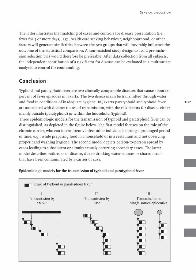
107
General discussion
The latter illustrates that matching of cases and controls for disease presentation (i.e.,
fever for 3 or more days), age, health care seeking behaviour, neighbourhood, or other
factors will generate similarities between the two groups that will inevitably influence the
outcome of the statistical comparison. A non-matched study design to avoid pre-inclu-
sion selection bias would therefore be preferable. After data collection from all subjects,
the independent contribution of a risk factor for disease can be evaluated in a multivariate
analysis to control for confounding.
ConclusionTyphoid and paratyphoid fever are two clinically comparable diseases that cause about ten
percent of fever episodes in Jakarta. The two diseases can be transmitted through water
and food in conditions of inadequate hygiene. In Jakarta paratyphoid and typhoid fever
are associated with distinct routes of transmission, with the risk factors for disease either
mainly outside (paratyphoid) or within the household (typhoid).
Three epidemiologic models for the transmission of typhoid and paratyphoid fever can be
distinguished, as depicted in the figure below. The first model focuses on the role of the
chronic carrier, who can intermittently infect other individuals during a prolonged period
of time, e.g., while preparing food in a household or in a restaurant and not observing
proper hand washing hygiene. The second model depicts person-to-person spread by
cases leading to subsequent or simultaneously occurring secondary cases. The latter
model describes outbreaks of disease, due to drinking water sources or shared meals
that have been contaminated by a carrier or case.
Epidemiologic models for the transmission of typhoid and paratyphoid fever
18439 Vollaard 14-12-2004 08:24 Pagina 107

These simplified transmission models can overlap and intermingle in endemic regions,
but they constitute a useful take-off point for the identification of the weakest link in the
transmission chain. In consequence, the number of reported cases within a household or
a community and the timeframe of occurrence of cases could help to determine the most
likely source or vehicle of transmission of disease. Our surveillance study did not detect
any outbreaks of disease (i.e., model 3) nor intra-household carriers among the primary
food handlers (i.e., model 1).
The first finding can be explained by the entrenched habit of boiling drinking water
before consumption in the study area, and by the fact that most households had private
access to drinking water sources, which decreases the likelihood of waterborne transmis-
sion on a large scale in the community. However, drinking water was frequently faecally
contaminated due to inadequate human waste disposal, which potentially could result in
outbreaks of disease, if domestic treatment of drinking water were neglected. Also
paratyphoid fever patients among food vendors could initiate micro-epidemics among
customers, if inoculation size or the number of replicated bacteria on their foodstuff
exceeds the minimal infective dose.
The second finding increases the likelihood of transmission of disease by (convalescent)
cases instead of chronic carriers (model 2). This was particularly true for typhoid trans-
mission within households, where recent typhoid cases observed poor personal hygiene.
We identified shared food as a likely vehicle for transmission of typhoid fever. The most
likely explanation of the finding that transmission of paratyphoid fever within house-
holds was not likely to occur, is that the relatively higher infective dose for paratyphoid
fever compared to that for typhoid fever, is only infrequently met in food that is shared
with household contacts. Food can be implicated as a vehicle in the transmission of
paratyphoid fever as well, but our study showed that food purchased from street vendors
was a more likely vehicle than shared food items within households. This finding could
be explained by the presence of chronic carriers or paratyphoid cases among street ven-
dors (model 1 or 2), who observed inadequate hand washing hygiene and touched food
items with bare hands frequently. The contamination of foods with paratyphoid (or
typhoid) bacteria and favorable conditions for bacterial growth during storage could
result in sufficiently high doses for illness in customers.
In Jakarta containment strategies should focus on the prevention of intra-household
spread of typhoid fever by instruction on adequate hygiene of (recent) cases and, to a
lesser extent, immunization of household contacts, especially those without a history of
typhoid fever. The control of paratyphoid fever requires monitoring of food vendors
(e.g., by repeated stool samples) and the detection of convalescent cases and carriers
among vendors operating in a district where recent paratyphoid cases have been reported.
Simple interventions such as hand washing using soap at home and in commercial food
108
18439 Vollaard 14-12-2004 08:24 Pagina 108
handling could reduce the food borne transmission of typhoid and paratyphoid fever.
The growing global urbanization has important consequences for the epidemiology of
infectious diseases, such as typhoid fever. In the first half of the 20th century the incidence
of typhoid fever in developed countries rapidly declined due to the improved water supply
in urban areas and chlorination of drinking water. However, other members of the
Salmonella family continue to circulate and infect people, because of their host-specificity
(i.e., not confined to humans alone) and because they effectively infiltrated in the
industrialized food-processing in these countries. Although water supply and sanitation
are far from ideal in many developing countries, improvements are underway, such as in
the Kampung Improvement Program in Jakarta and through the expansion of the water
network. A future prediction would therefore be a reduced global importance of typhoid
fever as we already signaled in our study, and a predominantly food borne transmission
of S. typhi. Consequently, an improved food processing hygiene and personal hygiene
would be detrimental for the survival of all Salmonella family members, including its
relatively fragile (i.e., most host-specific) members S. typhi and S. paratyphi.
109
General discussion
18439 Vollaard 14-12-2004 08:24 Pagina 109
In dit proefschrift is geëvalueerd hoe buiktyfus zich presenteert in Jakarta en welke risico-
factoren een rol spelen bij de transmissie van deze infectie. Buiktyfus wordt veroorzaakt
door een infectie met de Salmonella typhi bacterie, welke alleen de mens treft. Infectie
met Salmonella paratyphi, vaak met de term paratyfus aangeduid, geeft een vergelijkbaar
ziektebeeld, maar komt ongeveer tien keer minder vaak voor dan buiktyfus.
Buiktyfus en paratyfusBuiktyfus is een verwarrende naam, want de ziekte geeft –behalve in ernstige gevallen–
in het algemeen weinig klachten van de buik. De naam is terug te voeren op de tijd dat
koortsende ziekten niet nader konden worden gedetermineerd, en buiktyfus zich alleen
van die bij tyfus leek te onderscheiden door het optreden van zweren in de darm.
Inmiddels weten we dat tyfus een koortsende ziekte is die wordt overgedragen door beten
door luizen, vlooien of mijten en veroorzaakt wordt door infectie met Rickettsiae. Deze
infectieziekte heeft met een Salmonella infectie dus niks te maken.
Buiktyfus was een gevreesde ziekte die onbehandeld tien tot twintig procent van de
patiënten deed overlijden. In Europa en de VS kwamen vroeger massale epidemieën voor,
maar daar is sinds de aanleg van waterleidingen en chlorering van het drinkwater een
drastische daling in opgetreden. Tegenwoordig zijn de meeste buiktyfus-patiënten in
Nederland toeristen die uit het buitenland terugkomen. In ontwikkelingslanden komen
buiktyfus en ook paratyfus nog frequent voor.
S. typhi en S. paratyphi hebben geen dierlijk reservoir en veroorzaken uitsluitend ziekte bij
mensen. De overdracht van infectie is faeco-oraal, dat wil zeggen via ontlasting dat water
of voedsel heeft verontreinigd. Er zijn waarschijnlijk minimaal 100 tot 1000 bacteriën
nodig om ziek te worden van buiktyfus. In één gram ontlasting van een patiënt met buik-
tyfus kunnen vele miljoenen Salmonella bacteriën worden aangetroffen, en deze veroor-
zaken door verontreiniging van drinkwater of voedsel overdracht van de infectie. In voedsel,
zoals melk, roomijs, kokosmelk en vleesprodukten, kan de Salmonella bacterie zich
vermenigvuldigen - de bacterie deelt dan elk half uur - waardoor het aantal micro-
organismen in het voedsel toeneemt en de kans op overdracht van ziekte groter wordt.
Paratyfus zou met name via voedsel worden overgedragen, omdat er meer bacteriën nodig
zijn voor een infectie dan voor buiktyfus.
De lichamelijke gevolgen van het inslikken van een voldoende dosis bacteriën manifesteren
zich pas na ruim één week. Er ontstaat oplopende lichaamstemperatuur, soms met
rillingen en ijlen, en daarbij: misselijkheid, apathie, gebrek aan eetlust, vage buikpijn,
soms diarree of juist obstipatie en soms hoest. Met antibiotica is de infectie goed te
110
Nederlandse samenvatting
18439 Vollaard 14-12-2004 08:24 Pagina 110
111
behandelen en na drie tot vijf dagen zakt dan de koorts, waarna de patiënt verder kan her-
stellen. Voordat antibiotica geïntroduceerd werden, kreeg tien procent van de patiënten
bloedingen uit de darmwand door erosie van het laatste stuk van de dunne darm. Soms
traden daarbij perforaties van ernstig ontstoken darmwandgedeelten op. Die perforaties
behoeven altijd operatief ingrijpen om het gat in de darm af te dichten, want zonder
behandeling leidt dit tot een fataal verlopende buikvliesontsteking.
Behalve dat patiënten tijdens een acute infectie bacteriën uitscheiden in de ontlasting
gedurende drie tot vier weken, blijft een kleine groep patiënten (4%) gedurende jaren
na de infectie grote aantallen bacteriën uitscheiden, overigens zonder zelf klachten te
hebben. Deze bacterie-strooiers worden chronische dragers (‘carriers’) genoemd. Zo kan
de bacterie ondanks zijn kieskeurigheid voor de menselijke gastheer zich toch langdurig
handhaven in een populatie, mits de hygiëne, sanitaire voorzieningen en drinkwatervoor-
ziening de verspreiding van bacteriën uit ontlasting van acute patiënten of chronische
dragers via drinkwater of voedsel veilig stellen. En daarmee blijft de mens opgezadeld
met deze infectieziekte, tenzij ze erin slaagt verbeteringen door te voeren in de drie
bovengenoemde risicofactoren voor buiktyfus en paratyfus.
Onderzoek in JakartaIn dit proefschrift wordt beschreven hoe buiktyfus- en paratyfus-patiënten werden
geïdentificeerd in Jakarta. In Jatinegara, Oost-Jakarta, werkten 4 ziekenhuizen, 12
gezondheidscentra van de overheid (puskesmas) en 8 privé-kliniekjes mee gedurende 20
maanden aan een surveillance studie. Bij alle patiënten met drie of meer dagen koorts
werd 5-10 mL bloed afgenomen en in een speciale kweekfles geïnjecteerd. Die kweekflesjes
waren door de studiegroep zonder kosten voor patiënten en dokters gedistribueerd en
werden in een laboratorium op kweek gezet. In de onderzoeksperiode voldeden er 1019
patiënten aan de onderzoekscriteria, waarvan de meeste (89%) buiten de ziekenhuizen
werden geïncludeerd. In het bloed van 88 patiënten werd een S. typhi en bij 26 een S.
paratyphi A bacterie geïsoleerd in het centrale laboratorium van een privé-ziekenhuis. De
buiktyfus- en paratyfus-patiënten werden thuis bezocht, evenals de helft van de mensen
met koorts bij wie in het bloed geen Salmonella bacteriën waren geïsoleerd. In totaal wer-
den 69 mensen met buiktyfus geïnterviewd, 24 paratyfus-patiënten en 289 patiënten met
een koortsende ziekte van andere origine (de zogenaamde koorts-controles). Daarnaast
werden nog 378 huishoudens in het studiegebied bezocht, die volstrekt willekeurig van
een lijst werden gekozen. Dit was de gemeenschaps-controlegroep, die een afspiegeling
gaf hoe het er in de bevolking aan toeging.
In al deze huishoudens werd er drinkwater afgetapt dat in het laboratorium op bacteriën
werd onderzocht, dat wil zeggen op bacteriën die normaal in de ontlasting voorkomen
(en dus niet speciaal S. typhi of S. paratyphi). Hierdoor kan nagegaan worden of besmet-
Nederlandse samenvatting
18439 Vollaard 14-12-2004 08:24 Pagina 111
ting van de waterleiding met fecaliën is opgetreden (en daarmee in potentie ook de moge-
lijkheid om S. typhi over te dragen). Bovendien werd er bij alle bezochte mensen en van
de personen in het huishouden die het eten klaarmaakten, ontlasting onderzocht om na
te gaan of er carriers in het huishouden voorkwamen. Naast het interview, het monster
van het drinkwater en ontlasting, werd er ook van iedereen bloed afgenomen voor onder-
zoek naar gastheer-factoren.
Tenslotte werden in het onderzoeksgebied 202 voedselverkopers geïnterviewd, met als
doel inzicht te krijgen in de hygiëne in restaurants, en in stalletjes en karren. Daarbij
werd ook ontlasting van deze verkopers onderzocht op pathogene bacteriën en parasieten
en werd drinkwater en afwaswater onderzocht op fecale verontreiniging.
Hoofdstuk 1: ZiektepresentatieOm voor artsen in de ambulante gezondheidszorg een richtlijn te ontwerpen die zou kun-
nen helpen buiktyfus en paratyfus in een vroeg stadium te kunnen herkennen, werden de
symptomen van 59 buiktyfus-, 23 paratyfus- en 259 andere koorts-patiënten vergeleken.
De meeste patiënten waren tieners of jong-volwassenen. Er was geen significant verschil
tussen symptomen bij buiktyfus en die bij paratyfus. Wel waren er verschillen met de
andere koortsende patiënten: langere duur van koorts voor er een bloedkweek werd afge-
nomen, vaker last van koude rillingen en ijlen, vaker buikpijn en minder vaak klachten
van hoest. Met de symptomen zoals die door de patiënten waren gerapporteerd in de
interviews, kon er een richtlijn gegeven worden aan de artsen. Bij patiënten met minder
dan 7 dagen koorts is er een kans van 10% dat een patiënt buiktyfus of paratyfus heeft.
Als de patiënt niet hoest of daarnaast ook nog rillingen heeft, wordt die kans twee keer zo
groot. Voor patiënten met een week koorts of meer, is de aanwezigheid van ijlen (‘delier’)
belangrijk in de diagnostiek, want daardoor stijgt de kans op buiktyfus of paratyfus voor
een patiënt van 20% naar maximaal 67%. Maar ondanks het feit dat sommige symptomen
suggestief zijn voor buiktyfus of paratyfus, is de klinische presentatie van beide ziekten in
hoge mate overeenkomstig met die van andere infectieziekten in Jakarta, zoals dengue of
darminfecties. Bloedkweken of serologische testen blijven daarom noodzakelijk voor
het definitief vaststellen van een infectie met S. typhi of S. paratyphi, omdat de klinische
presentatie alleen te weinig onderscheidend vermogen biedt. Wel bleek dat de behande-
lingsopties in Jakarta goed zijn, want de S. typhi bacteriën waren maar in een gering
percentage (3%) ongevoelig voor de meest gebruikte, goedkope antibiotica.
Hoofdstuk 2: RisicofactorenOm te onderzoeken welke risicofactoren een rol spelen in de overdracht van buiktyfus
en paratyfus in Jakarta, werden beide patiëntengroepen vergeleken met de beide controle-
groepen. Paratyfus blijkt met name buiten het huishouden opgelopen te worden, vooral
112
18439 Vollaard 14-12-2004 08:24 Pagina 112
113
Nederlandse samenvatting
door het eten van straatvoedsel. Ook bleek overstroming van het huishouden een risico-
factor voor ziekte te zijn. Deze beide risicofactoren kwamen naar voren in vergelijking
met beide controlegroepen. Voor buiktyfus lag dat anders. In de vergelijking met de
gemeenschapsgroep waren het vooral factoren die binnen het huishouden lagen: een eer-
dere buiktyfus episode van een huisgenoot in het laatste jaar, het niet wassen van handen
met zeep, delen van voedsel van hetzelfde bord en de afwezigheid van een toilet. Ook
jonge leeftijd was een risicofactor voor buiktyfus. De factoren die met onvoldoende hygiëne
te maken hadden, speelden niet mee toen buiktyfus-patiënten werden vergeleken met de
andere patiënten-controlegroep. Alleen het gebruik van ijsklontjes was gerelateerd aan
buiktyfus en bovendien waren buiktyfus-patiënten vaker van het vrouwelijk geslacht dan
de andere patiënten. Het was wel te verklaren dat in de vergelijking met deze controle-
groep de factoren die wezen op een slechtere hygiëne van deze patiënten niet naar voren
kwamen, omdat onvoldoende hygiëne de overdracht van andere ziekten die gepaard gaan
met koorts, zoals sommige vormen van diarree of virale luchtweginfecties, eveneens
bevordert.
Hoofdstuk 3: VoedselverkopersIn hetzelfde gebied werden verkopers van etenswaren geïnterviewd: 11 uit restaurants, 63
uit kleine restaurantjes (warung), 110 uit stalletjes en 18 mobiele verkopers. Omdat in het
risicofactoronderzoek gebleken was dat het eten bij de laatste twee groepen van straatver-
kopers een risico inhield op het oplopen van paratyfus, werden deze twee groepen geza-
menlijk vergeleken met de eerste twee groepen. Naast de interviews, werden ontlasting,
drinkwater en afwaswater onderzocht. Bij één op de vijfentwintig verkopers werden
Salmonella bacteriën gekweekt in de ontlasting, waarvan bij één (circa 0.6%) S. typhi werd
geïsoleerd. De hygiëne van de straat-verkopers was minder dan die van de verkopers in
restaurants. Niet alleen werd er minder vaak handen gewassen, maar ook werd minder
vaak zeep gebruikt en vaker etenswaren met blote handen aangeraakt. Afwaswater bleek
in het algemeen besmet met fecale coliforme bacteriën, waarbij de hoogste aantallen
bacteriën werden aangetroffen bij straatverkopers, die hun water zelden verversten.
Bovendien was drinkwater vaak verontreinigd, zonder dat er verschillen waren tussen
beide groepen van verkopers. In 23 monsters van ijsklontjes bleken alle monsters veront-
reinigd. Op basis van deze gegevens kan worden begrepen dat straatvoedsel een risico-
factor is voor de overdracht van paratyfus en buiktyfus. Slechte handwashygiëne na ont-
lasting en voor voedselbereiding, en direct contact van handen met voedsel in de stalletjes
en karren leidt tot contaminatie van de etenswaren, waarna de bacteriën verder kunnen
groeien, zodat in de etenswaren het noodzakelijke aantal bacteriën voor ontwikkeling
van ziekte wordt bereikt.
18439 Vollaard 14-12-2004 08:24 Pagina 113
Hoofdstuk 4: Drinkwater en sanitaire voorzieningenDe gegevens over drinkwatervoorziening en –kwaliteit en over sanitaire voorzieningen
van de willekeurig gekozen controlegroep uit het onderzoeksgebied werden afzonderlijk
nader uitgediept. Van de 378 huishouden had 20% een aansluiting op de waterleiding,
64% gebruikte grondwater uit pompen en het restant gebruikte of mineraalwater, of
water van waterventers of uit putten. Het water van de waterleiding was in een gering
percentage besmet met faecale bacteriën (17%) en bovendien waren de aantallen bacteriën
laag. Ondanks het feit dat grondwater vaker verontreinigd was (69%), lag het aantal
bacteriën in het grondwater van pompen daar niet significant boven, maar wel werden
vaker hogere aantallen micro-organismen aangetroffen. In ruim een kwart van de huis-
houdens loost het toilet rechtstreeks in een open goot of de rivier. De bron van drinkwater
of het bezitten van een privé toilet had geen invloed op het vóórkomen van diarree. Wel
werd er vaak diarree gerapporteerd in huishoudens met drinkwater dat zwaar verontreinigd
was (> 100 fecale coliforme bacteriën per 100 mL). Van belang is verder te melden dat alle
inwoners vertelden dat ze hun drinkwater langdurig kookten voor gebruik, waarmee ze
het risico op een infectieuze diarree natuurlijk verminderen, ondanks de gebrekkige sani-
taire voorzieningen. Behalve verdere uitbreiding van het waterleidingnetwerk is ook ver-
vanging van lekkende pijpen, waarin verontreinigd omgevingswater kan binnendringen,
van belang om hoge kwaliteit drinkwater aan de inwoners van Jakarta te kunnen leveren.
Hoofdstuk 5: Helicobacter pylori infectie en buiktyfusDe maaginfectie met Helicobacter pylori komt geregeld voor over de hele wereld, vooral
in ontwikkelingslanden (tot bij 80% van de volwassenen). Afhankelijk van de plek in de
maag waar de infectie een ontstekingsreactie genereert, kan er een afname van maag-
zuurproduktie optreden. Als gevolg van verminderde zuurproductie zou de kans om bij
geringe aantallen S. typhi in het voedsel toch buiktyfus op te lopen toenemen. Om dit na
te gaan werd bij patiënten met buiktyfus en controle-personen de concentratie van anti-
lichamen tegen H. pylori bepaald in het bloed (zowel IgG als IgA). Ook werd de gastrine
concentratie in het bloed bepaald, als maat voor een lage maagzuurproduktie, zoals die
bijvoorbeeld wordt gemeten voor de bepaling van atrofie (slijmvliesverlies) van de maag.
Er werd een associatie van optreden van buiktyfus en anti-Helicobacter IgG-spiegels vast-
gesteld, maar de gastrine concentraties waren niet verschillend tussen patiënten met
buiktyfus en controles. Het lijkt er dus op dat de associatie tussen Helicobacter infecties
(afgemeten aan IgG) en buiktyfus niet op een causaal verband berust. Deze associatie
werd in een ander onderzoek gedacht te berusten op een verlaagde zuurproductie bij
mensen met een gelijktijdige Helicobacter infectie. Maar het is eerder waarschijnlijk dat de
gebeurtenissen die leiden tot een Helicobacter infectie en/of buiktyfus geassocieerd zijn,
bijvoorbeeld door een gebrek aan (voedsel-)hygiëne in het huishouden.
114
18439 Vollaard 14-12-2004 08:24 Pagina 114

Nederlandse samenvatting
ConclusieBuiktyfus en paratyfus zijn twee endemische ziekten in Jakarta, die klinisch niet van
elkaar te onderscheiden zijn en gezamenlijk circa 10% van de episodes met 3 of meer
dagen koorts veroorzaken. Bloedkweken of gevoelige serologische bepalingen zijn
noodzakelijk om de infectie met zekerheid vast te stellen, want de klinische presentatie
van beide ziekten biedt te weinig houvast voor artsen om een eenduidig onderscheid te
maken met andere koortsende ziekten. Zowel buiktyfus als paratyfus worden overgedragen
via drinkwater of voedsel, als de toilet- en voedselhygiëne onvoldoende in acht genomen
wordt door patiënten die de bacteriën in hun ontlasting uitscheiden. Uit ons onderzoek
in Jakarta werd duidelijk dat paratyfus met name buiten het huishouden overgedragen
wordt (via straatvoedsel), maar dat de meeste risicofactoren voor buiktyfus binnen het
huishouden lagen (personen die eerder een buiktyfus opgelopen hadden).
Weinig bewijs werd gevonden voor overdracht van beide ziekten via drinkwater, omdat
de mensen in Jakarta hun water consequent koken voorafgaande aan consumptie, en ook
omdat in de laatste jaren de drinkwatervoorziening verbeterd is door uitbreiding van de
centrale waterleiding. Toch bevatte het drinkwater vaak fecale coliforme bacteriën, een
besmetting die erop wijst dat er contact is van menselijke ontlasting met de drinkwater-
voorziening. Verbetering van sanitaire voorzieningen zou dus moeten nagestreefd
worden, door het aanleggen van goede riolering en ‘septic’ opvangtanks. Immers, door
slechte omgevingshygiëne zouden er epidemieën kunnen ontstaan als een buiktyfus
patiënt een centrale waterleiding besmet met S. typhi, en er tegelijkertijd minder zorgvuldig
met het drinkwater wordt omgegaan (als bijvoorbeeld het koken van drinkwater wordt
veronachtzaamd).
Het was duidelijk dat beide ziektes overgedragen worden via voedsel. Voor buiktyfus:
gedeeld voedsel binnen het huishouden, en voor paratyfus: straatvoedsel. Voor beide
ziekten bleek een belangrijke rol weggelegd voor voedselhygiëne, of beter gezegd een
gebrek daaraan: bij het handenwassen wordt niet consequent zeep gebruikt en daarmee
wordt in veel gevallen voorbijgegaan aan een eenvoudige doch efficiënte manier om
overdracht van buiktyfus en paratyfus te voorkómen.
18439 Vollaard 14-12-2004 08:24 Pagina 115

18439 Vollaard 14-12-2004 08:24 Pagina 116
117
Acknowledgements
Soegianto Ali has been most essential for the success of this PhD-period, because he was
my committed local partner during the surveillance study in Jakarta. He was a tireless
companion and willing sparring-partner in epidemiological aspects and in the explana-
tion of many facets of the Indonesian culture.
I am very grateful to Prof. Dr. Jaap van Dissel for his unconditional support during the
project.
Our work in the study area, laboratory and Center for Health Research of Medical Faculty
of the Atma Jaya University was only possible with the help of our committed research-
assistants: Ferry Kandaw, Rinny Listyani, Meily, Vea Noveria, Carmelita Ridwan, Min Ali
Sugiharto, Lidwina Sutikno, Mariana Tasman and Lily Yaputra.
In Jakarta the staff of the Center of Health Research (Puslitkes) of the Atma Jaya Catholic
University helped in unravelling causes of stagnation and signalling moments of reorien-
tation. The Suwandhi Widjaja family always showed their interest and hospitality.
Also other members of the Typhoid Team have been very helpful. Especially Henri van
Asten from the International Institute of Health, Nijmegen Medical Faculty, has been a
great counterpart and his methodological input was significant. Leo Visser from the
LUMC department of Infectious Diseases has been a very supportive colleague and critic
during our discussions. Dr. Wil Dolmans from the Nijmegen Medical Faculty gave his
important contribution in the initial stage of our project.
The physicians and nurses in the participating health centers in Jatinegara have been very
helpful and they were essential for the fine outcome of our study. Patients and controls
should be explicitly thanked for their participation and open doors.
The staff of our central reference lab in RS Mitra Internasional: dr. Sian, Chandra, Atiek,
Sri, Meira and Diyah were very helpful, diligent and friendly.
During my stay in Jakarta several people have been very supportive. Dr. Narain Punjabi
from the NAMRU Jakarta has always been an interested counterpart. Dr. Is Suharia Ismid
from the department of Parasitology of the Universitas Indonesia and her staff have been
very helpful during the food vendor study. In Jakarta Manoe Mesters was a dedicated sup-
porter. Billy Hunsinger, his wife Heni and their children Kevin, Boudewijn and Albert
Junior have contributed to a sort of homecoming and I hope we continue our friendship
in the future. Jeroen Alberts, Robert Reid, Maaike Schellekens and Lene Nielsen have
been wonderful mates who shared their friendship and houses.
In the Netherlands my loving parents Rien and Greetje Vollaard took care of the ongoing
affairs and have managed my administration meticulously. I am very grateful to Nettie
Kaat of the department of Infectious Diseases LUMC for her quick responses to all my
trivial requests. My colleagues Emile Schippers, Margje Haverkamp, Offra Duerink and
18439 Vollaard 14-12-2004 08:24 Pagina 117
118
Eliane Leijten in the LUMC have been great sparring-partners on methodological aspects.
I thank Thijs Kolhoff and Juliet Wermenbol, and Gert de Visser and Julian Veenker for
providing a roof during my visits to the Netherlands.
Jo Smet of the IRC in Delft was very helpful in the evaluation of our study on the water
supply and sanitation in Jakarta. The study on the association of Helicobacter pylori
infection and typhoid fever was impossible without the input of Hein Verspaget and Izäk
Biemond of the department of Gastroenterology-Hepatology LUMC and the lab-
assistants Wim van Duyn and Johan van der Reijden. The contribution of Adriette de
Visser of the Lab of Infectious Diseases LUMC was crucial in the serology study and DNA
analysis. I am thankful to Ron Wolterbeek of the Department of Medical Statistics of the
LUMC for his suggestions.
I thank Ben Vollaard and Liesbeth van Rijn for critically reviewing parts of the manu-
script. Eliene Maria Soares da Silva, and Ken Sykes and family have continuously radiated
love and interest since ages.
During the period in Jakarta I very much appreciated the visits of my brothers Harrie and
Hans Vollaard, and friends: Albert van IJzeren, Onno Hamburger, Sweder van de Poll,
Jan Willem Mazel, and last but not least Jurgen Huiskes.
18439 Vollaard 14-12-2004 08:24 Pagina 118
About the author
Albert Meint Vollaard was born on March 23rd, 1970 in Veenendaal, the Netherlands.
Undergraduate education was received at the Guido de Brès Scholengemeenschap (VWO)
in Amersfoort from 1982 till 1988. After one year of traveling he studied Latin American
Languages and Cultures from 1989 till 1991 in Leiden and obtained his propaedeusis in
1990.
In 1991 he could enter the Medical Faculty of Leiden University and obtained his Medical
Degree in 1998. During this period he participated in 1995-96 in a field study of the
Fundação Oswaldo Cruz on the “Socio-economic risk factors for Schistosoma mansoni
infection” in Belo Horizonte, Brazil.
After graduation in 1998 he worked in Delft, Reinier de Graaf Gasthuis, Department of
Cardiology, and from 1999 till 2000 in Den Haag, Bronovo Ziekenhuis, Department of
Internal Medicine.
In 2000 his PhD-research started in the Department of Infectious Diseases, Leiden
University Medical Center (Prof. Dr. J.T. van Dissel) and he joined the staff of the Center
for Health Research, Atma Jaya University, Jakarta, Indonesia, for the Typhoid Fever
Research Project from 2001 till 2003.
In 2004 he began his residency in Internal Medicine in Medisch Centrum Haaglanden,
Den Haag (head: Dr. P.H.L.M. Geelhoed-Duijvestein).
18439 Vollaard 14-12-2004 08:24 Pagina 119

18439 Vollaard 14-12-2004 08:24 Pagina 120


