http://jdr.sagepub.com/ Journal of Dental Research http://jdr.sagepub.com/content/66/3/784 The online version of this article can be found at: DOI: 10.1177/00220345870660031601 1987 66: 784 J DENT RES I. Gorter de Vries, E. Quartier, P. Boute, E. Wisse and D. Coomans Immunocytochemical Localization of Osteocalcin in Developing Rat Teeth Published by: http://www.sagepublications.com On behalf of: International and American Associations for Dental Research can be found at: Journal of Dental Research Additional services and information for http://jdr.sagepub.com/cgi/alerts Email Alerts: http://jdr.sagepub.com/subscriptions Subscriptions: http://www.sagepub.com/journalsReprints.nav Reprints: http://www.sagepub.com/journalsPermissions.nav Permissions: http://jdr.sagepub.com/content/66/3/784.refs.html Citations: by guest on July 24, 2011 For personal use only. No other uses without permission. jdr.sagepub.com Downloaded from
Transcript
http://jdr.sagepub.com/Journal of Dental Research
http://jdr.sagepub.com/content/66/3/784The online version of this article can be found at:
DOI: 10.1177/00220345870660031601
1987 66: 784J DENT RESI. Gorter de Vries, E. Quartier, P. Boute, E. Wisse and D. Coomans
Immunocytochemical Localization of Osteocalcin in Developing Rat Teeth
Published by:
http://www.sagepublications.com
On behalf of:
International and American Associations for Dental Research
can be found at:Journal of Dental ResearchAdditional services and information for
Immunocytochemical Localization of Osteocalcin in Developing Rat Teeth§
I. GORTER DE VRIES, E. QUARTIER, P. BOUTE, E. WISSE1, and D. COOMANS2
Institute of Dentistry and 'Laboratory for Cell Biology and Histology, Vrije Universiteit Brussel, Laarbeeklaan 103, 1090 Brussels, Belgium
Osteocalcin was purified by gel chromatography from a crude extractobtained after decalcification of rat incisors. The apparent molecularweight, as determined by 5-15% SDS-polyacrylamide gel electropho-resis, was 18,000, and amino acid analysis revealed 60 y-carboxy-glutamic acid residues per 1000. Antisera against osteocalcin, raisedin rabbits, reacted specifically with osteocalcin when investigated byimmuno-electroblotting of dentin crude extract. 4-pim cryosections offormaldehyde-fixed tooth germs showed positive immunocytochemicalstaining for osteocalcin in dentin and odontoblasts. The staining ofthe mantle dentin at the coronal sides of the tooth germs was moreintense than that of the adjacent circumpulpal dentin, while the odon-toblasts involved in the formation of mantle dentin showed strongerimmunoreactivitv than did odontoblasts involved in circumpulpal den-tin formation. This marked difference was not observed on the rootsides of the tooth germs. In 1h-pm cryosections, osteocalcin immu-noreactivity was found evenly distributed throughout the entire cellbody, with the exception of the Golgi region, which was less intenselystained, while the nucleus and the cell process were negative. Thepositive staining reaction with anti-osteocalcin antiserum was foundin dentin from the very onset of its formation in the fetus. In conclu-sion, our results demonstrate the presence of osteocalcin in odonto-blasts and dentin. Its immunocytochemical localization may becompatible with a distinct role in early dentinogenesis.
J Dent Res 66(3):784-790, March, 1987
Introduction.
Osteocalcin or bone Gla protein (BGP) is a major protein ofbone (Hauschka et al., 1975; Price et al., 1976) and dentin(Linde et al., 1980), constituting from 5 to 10% of the non-collagenous proteins. In most species, it contains three y-car-boxyglutamic acid (Gla) residues per molecule of approximately50 amino acids. Gla is formed by the carboxylation of glutamicacid, a reaction which is dependent on vitamin K. The Glaresidues bind calcium ions and are responsible for the highaffinity of osteocalcin for hydroxyapatite (Price et al., 1976;Poser and Price, 1979; Hauschka and Carr, 1982). The abun-dance of osteocalcin among the non-collagenous proteins ofbone and dentin, and the strong correlation of its serum con-centration with the level of bone turnover (Price et al., 1980b)indicate an important function of osteocalcin in the metabolismof mineralized tissues.
Osteocalcin in rat dentin consists of four closely relatedproteins (Linde et al., 1982). It was found to be synthesizedin organ culture of dentin-odontoblast fragments (Dimuzio etal., 1983). Odontoblasts and dentin of rat molar tooth germsin vivo (Bronckers et al., 1985) and cultured in vitro (Finkel-man and Butler, 1985) stain positive after incubation with anti-dentin osteocalcin antiserum. In this study, we have furtherinvestigated the immunocytochemical localization of osteocal-cin, especially in incisor tooth germs, where root-side dentinis found in addition to crown-side dentin. We were also inter-ested in the developmental appearance of osteocalcin in odon-
Received for publication April 16, 1986Accepted for publication October 17, 1986§This investigation was supported by the Belgian "Fonds voor Ge-
neeskundig Wetenschappelijk Onderzoek", grant number 3.0038.81.2To whom correspondence and reprint requests should be addressed
784
toblasts and dentin during fetal life, since it has been reportedthat newly formed rat bone is devoid of the protein (Price etal., 1980a; Price et al., 1981).
Materials and methods.Purification of osteocalcin.-Incisors were excised from male
Wistar rats (weighing 200 g each), cleaned of adhering tissue,and stored frozen. All manipulations were carried out at 4TC.The incisors were broken and extracted with a solution con-taining 4 mol/L guanidine-HC1, 50 mmol/L Na-acetate, 10mmol/L EDTA, and several protease inhibitors (Gorter de Vrieset al., 1986) at pH 6.5. The tooth fragments were furtherextracted with 0.25 mol/L EDTA, 20 mmol/L Tris at pH 7.4,also containing protease inhibitors, resulting in a crude dentinextract. Dentin phosphoprotein was precipitated from the de-salted extract with CaCl2. Osteocalcin was purified from thesupernatant as described (Butler et al., 1981) and eluted as asingle peak in repeated chromatography over a Sephadex G-50 fine column (Pharmacia, Uppsala, Sweden) in 100 mmol/L NH4HCO3 (Price et al., 1976).
Characterization of osteocalcin.-The peak fractions of theSephadex G-50 column were pooled and further analyzed byDEAE-cellulose chromatography, by amino acid analysis, andby polyacrylamide gel electrophoresis. The material was chro-matographed at 4TC on a I x 6 cm column of DEAE-cellulose(DE-52, Whatman, Maidstone, England) with 50 mmol/L Tris-HCl at pH 8.0. Elution was performed with a linear gradientof 0-0.6 mol/L NaCI over a total volume of 500 mL. Foramino acid analysis, samples were hydrolyzed in 6 mol/L HC1at 1 10C for 22 hours or, for the Gla determinations, in 2 mol/L KOH at 1 0IC for 22 hours (Hauschka, 1977), since Gladecarboxylates to form glutamic acid on acid hydrolysis. Aminoacid analysis was performed with a Beckman type 119 CLamino acid analyzer. Glutamic acid was used as internal ref-erence for calculation of the final amino acid composition fromthe data obtained from the alkaline and acid hydrolysates, sinceGla is converted quantitatively into Glu on acid hydrolysis(Lian et al., 1979). Polyacrylamide gel electrophoresis in thepresence of sodium dodecyl sulphate (SDS) was carried out in5-15% or 20% gels (Laemmli, 1970). Gels were stained withCoomassie Brilliant Blue or with Silver Stain (Biorad Labo-ratories, Utrecht, The Netherlands) and scanned with a VernonPhotometer.
Preparation and characterization of antisera.-White NewZealand rabbits were immunized with 1 mg of osteocalcin,prepared as described, dissolved in 0.5 mL phosphate-bufferedsaline (PBS), and emulsified with 0.5 mL Freund's completeadjuvant (Difco, Detroit, MI) through multiple intracutaneousinjections on the back and two intramuscular injections in thehind legs. After 80 days, booster injections were given sub-cutaneously with 100 pLg osteocalcin dissolved in 0.5 mL PBSand emulsified with an equal volume of Freund's incompleteadjuvant. Animals were bled eight days later from the centralear vein.
Sera were tested for the presence of anti-osteocalcin anti-bodies by enzyme-linked immunosorbent assay (ELISA) onmicrotiter plates (Dynatech, Greiner, Nurtingen, West Ger-
by guest on July 24, 2011 For personal use only. No other uses without permission.jdr.sagepub.comDownloaded from
Fig. 1 DEAE-cellulose chromatography of 5.3 mg osteocalcin purifiedby G-50 gel filtration. No chromatographic peaks were observed at highersalt concentrations.
TABLE 1AMINO-ACID COMPOSITION OF RAT DENTIN OSTEOCALCIN*
*Mean value of two independently hydrolyzed samples is given.
many). Binding was visualized with horseradish peroxidase(HRP) conjugated to sheep anti-rabbit IgG (Institut Pasteur,Paris, France), with 5-aminosalicylic acid used as a substrate.The reaction was quantified by measurement of absorbance at450 nm on a multiscan Titertek system (Flow Lab., Asnieres,France). Cross-reactivity with rat collagen type I (tail tendon)and with rat dentin phosphoprotein (Gorter de Vries et al.,1986) was also investigated by ELISA. The specificity of theantisera was further determined by immuno-electroblotting.Proteins were transferred from 5-15% SDS polyacrylamidegels to nitrocellulose (NC) sheets (Biorad Lab., Utrecht, TheNetherlands), essentially as described by Towbin et al. (1979)in a gel-destaining apparatus (Pharmacia, Uppsala, Sweden)for 15 or 30 min at 36 V. The transfer buffer contained 25mmol/L Tris, 192 mmol/L glycine, and 20% (v/v) methanolat pH 8.3. Protein transfer was checked by staining of narrow
strips of the NC sheets with Auro Dye (Janssen Pharmaceutica,Beerse, Belgium). The rest of the sheet was used for the im-munochemical reactions. Protein-binding sites were blockedby incubation with 3% bovine serum albumin (BSA) in PBSfor three hours at 37°C. Next, NC strips were incubated withanti-osteocalcin antiserum or with pre-immune serum dilutedin PBS containing 1% BSA for 16 hours at 4°C. After being
E 0.8 -
CD(0
. 0.2 - _
0 5 10MIGRATION DISTANCE (cm)
Fig. 2 5-15% SDS polyacrylamide gel electrophoresis. The gel wasstained with Coomassie Brilliant Blue and scanned with a Vernon pho-tometer. Lane a, low-molecular-weight standards (Pharmacia Fine Chem-icals, Uppsala, Sweden) expressed in kiloDaltons; lane b, dentin osteocalcin(30 keg); lane c, crude dentin extract (200 ,ug).
extensively washed in PBS containing 0.1% Tween 20, thestrips were incubated with protein A conjugated to 5 nm goldparticles (prot A-Au 5 nm; Janssen Pharmaceutica, Beerse,Belgium) diluted 100 X in PBS-BSA, for five hours at roomtemperature. The binding was visualized, after extensive wash-ing with PBS-0. 1% Tween 20, with a silver enhancement pro-cedure (Janssen Pharmaceutica, Beerse, Belgium), as describedby the manufacturer.
Tissue processing.-Rat pups (each from zero to six daysold) were perfused via the left ventricle with PBS containingheparin, quickly followed by a fixative containing 3% for-maldehyde and 0.08 mol/L sodium phosphate at pH 7.3 for10 min. Some animals were perfused with Zamboni's fixative(Stefanini et al., 1967). Molar and incisor tooth germs wereexcised and fixed further in the same fixative for several hours.Upper and lower jaws were excised from 18-, 19-, 20-, and21-day-old fetuses (birth was on the 22nd day) and fixed byimmersion. Part of the tissue was decalcified at 4°C in for-maldehyde fixative containing 0.1 mol/L EDTA. The tissuewas frozen on stubs in liquid nitrogen just prior to being sec-tioned. Unfixed tissue, which was quickly excised from ratpups and fetuses, was frozen in freon 22 cooled in liquid ni-trogen. 4-,um frozen sections were made on an American Op-tical cryotome and 1-p>m frozen sections on an LKB Vultracryotome.Immunocytochemistry.-Osteocalcin immunoreactivity was
demonstrated by means of a two-step immunocytochemical
Vol. 66 No. 3 785
by guest on July 24, 2011 For personal use only. No other uses without permission.jdr.sagepub.comDownloaded from
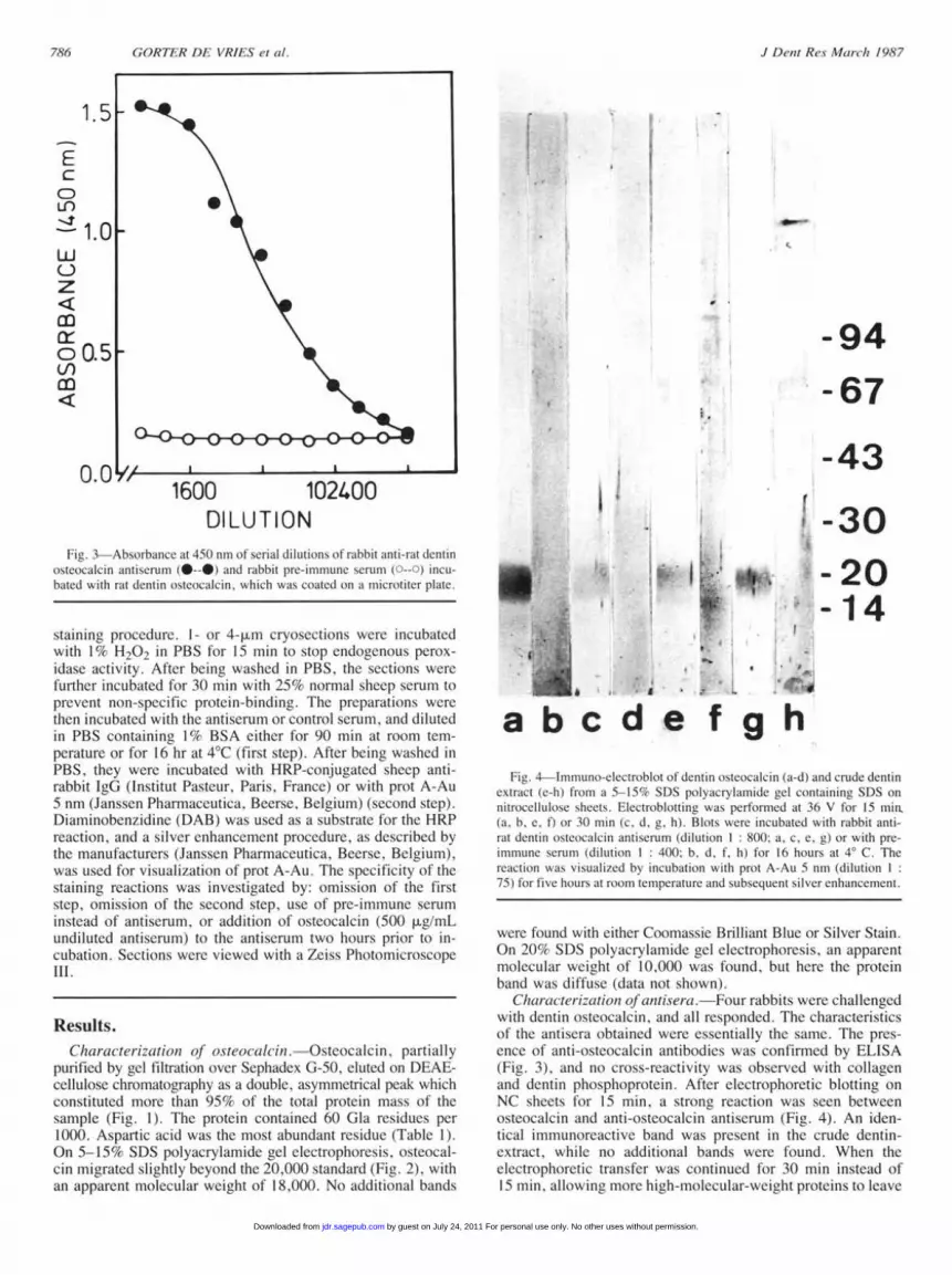
DILUTION -30Fi. 3 Absorbance at 450 nm of serial dilutions of rabbit anti-rat dentin
ostcocalcin antiserum (0--0) and rabbit pic-1immun111e scrum ( --o) incu-bated with rat dentin osteocalcin, which was coated on a microtiter plate.
staining procedure. I- or 4-pm cryosections were incubatedwith 1% H207 in PBS for 15 min to stop endogenous perox-
idase activity. After being washed in PBS, the sections were
further incubated for 30 min with 25% normal sheep serum toprevent non-specific protein-binding. The preparations were
then incubated with the antiserum or control serum, and dilutedin PBS containing 1% BSA either for 90 min at room tem-perature or for 16 hr at 4°C (first step). After being washed in
PBS, they were incubated with HRP-conjugated sheep anti-rabbit IgG (Institut Pasteur, Paris, France) or with prot A-Au5 nm (Janssen Pharmaccutica, Beerse. Belgium) (second step).Diaminobenzidine (DAB) was used as a substrate for the HRPreaction, and a silver enhancement procedure, as described bythe manufacturers (Janssen Pharmaccutica, Beerse, Belgium),was used for visualization of prot A-Au. The specificity of thestaining reactions was investigated by: omission of the firststep, omission of the second step, use of pre-immune serum
instead of antiserum, or addition of osteocalcin (500 pg/mLundiluted antiserum) to the antiscrum two hours prior to in-
cubation. Sections were viewed with a Zeiss PhotomicroscopeIll.
Results.Chairac'terizationj of osteocailcin. Osteocalcin. partially
purified by gel filtration over Sephadex G-50. cluted on DEAE-cellulose chromatography as a double, asymmetrical peak whichconstituted more than 95% of the total protein mass of thesample (Fig. 1). The protein contained 60 Gla residues per1000. Aspartic acid was the most abundant residue (Table 1).On 5 15% SDS polyacrylamide gel clectrophoresis, osteocal-cin migrated slightly beyond the 20,000 standard (Fig. 2). withan apparent molecular weight of 18,000. No additional bands
abcde fghFig-. 4-Immuno-cletroblot of dentin osteocalcin (a-d) and crude dentin
extract (e h) from a 5-15%/1 SDS polyacrylamide gel containin S1)S onnitiocellulose sheets. Flectroblotting was performed at 36 V for 15 minn.(a. b. e, f) or 30 min (c. d. g. hW. Blots were incubated with rabbit anti-rat dentin osteecalcin antiserum (dilution 1: 800; a. .c ge or with pre-immune serum (dilution 1: 400( b, d. f, h) for 16 hours at 4° C. Thereaction was visualized by incubation with prot A-Aui 5 nm (dilution75) for five hours at room temperature and subsequent silver enhanccment.
were found with either Coomassic Brilliant Blue or Silver Stain.On 20% SDS polyacrylamide gel electrophoresis, an apparentmolecular weight of 10,000 was found, but here the proteinband was diffuse (data not shown).
Chara-cteriz-cation of antiser-a. Four rabbits were challengedwith dentin osteocalcin, and all responded. The characteristicsof the antisera obtained were essentially the same. The pres-
ence of anti-osteocalcin antibodies was confirmed by ELISA(Fig. 3), and no cross-rcactivity was observed with collagenand dentin phosphoprotein. After electrophoretic blotting on
NC sheets for 15 min, a strong reaction was seen betweenosteocalcin and anti-osteocalcin antiserum (Fig. 4). An iden-tical immunoreactive band was present in the crude dentin-extract, while no additional bands were found. When theclectrophoretic transfer was continued for 30 min instead of15 min, allowing more high-molecular-weight proteins to leave
1.5F
0
F-
EcCDU-)No-I1.0.
hLJC-)z
cr5n0.5cn
o.0 V,
-20- 14
A I I A
J Dentl Res. March, 1987
i I a
I
I 4I * -,
it '.1 I ...:, W
by guest on July 24, 2011 For personal use only. No other uses without permission.jdr.sagepub.comDownloaded from
B 0.3 mm_______________________S___a________________________________________________________
7
_
** ~~~pA
150 jam
0
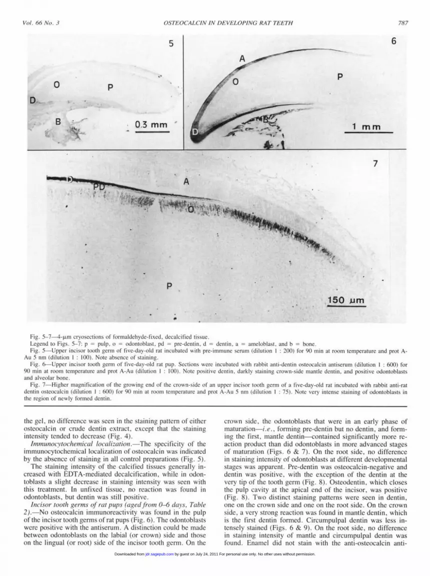
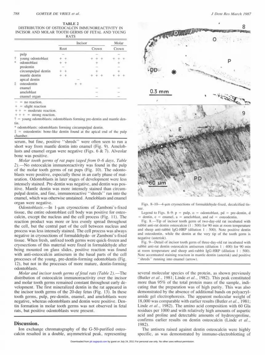
-iz. 5-7-4 pLm cryoscctions of torildalechyde-fixed. decalcified tissue.cge'end to Figs. 57: p = pulp, o = odontoblast. pd = pre-dentin. d ddentin, a = ameloblast, and b bone.
Fig. 5 Upper incisor tooth germ of five-day-old rat incubated with pre-immune serum (dilution 1: 200) for 90 min at room temperature and prot A-Au 5 nm (dilution 1 : 100). Note absence of staining.
Fig. (--Uppcr incisor tooth germ of five-day-old rat pp. Sections were incubated with rabbit anti-dentin osteocalcin antiserum (dilution 1: 600) for90 nin at roomn temperature and prot A-AI (dilution 1: 10)(. Note positive dentin, darkly staining crown-sidc mantle dentin, and positive odontoblastsand alveolar bone.
Fig. 7-Higher magnification of the growing end of the crown-side of an Lupper incisor tooth cerm of a five-day-old rat incubated with rabbit anti-ratdentin osteocalCin (dilution : 6)0() for 90 min at room temperatuie and prot A-AU 5 nri (dilution 1 : 75). Note very intense staining of odontoblasts inthe region of newly formed dentin.::7,
the gel, no difference was seen in the staining pattern of eitherosteocalcin or crude dentin extract, except that the stainingintensity tended to decrease (Fig. 4).IhnmunocN'tochemnical local cation. The specificity of the
immunocytochetntical localization of' osteocalcin was indicatedby the absence of staining in all control preparations (Fig. 5).The staining intensity of the calcified tissues generally in-
creased with EDTA-mediated decalcification, while in odon-toblasts a slight decrease in staining intensity was seen withthis treatment. In unfixed tissue, no reaction was found inodontoblasts, but dentin was still positive.
Incisor tooth germ.s t-at p)ups (aged/rf-in 0-6 daCxs, Table2). No osteocalcin immunoreactivity was found in the pulpof the incisor tooth germs of rat pups (Fig. 6). The odontoblastswere positive with the antiserum. A distinction could be madebetween odontoblasts on the labial (or crown) side and thoseon the lingual (or root) side of the incisor tooth germ. On the
crown side, the odontoblasts that were in an early phase ofmaturation i.e., forming pre-dcntin but no dentin, and form-
ing the first, mantle dentin contained significantly more rc-action product than dtd odontoblasts in more advanced stagesof maturation (Figs. 6 & 7). On the root side, no differencein staining intensity of odontoblasts at different developmentalstates was apparent. Pre-dentin was osteocalcin-negative anddentin was positive, with the exception of the dentin at thevery tip of the tooth germ (Fit. 8). Osteodentin, which closesthe pulp cavity at the apical end of the incisor, was positive(Fig. 8). Two distinct staining patterns were seen in dentin,one on the crown side and one on the root side. On the crownside, a very strong reaction was found in mantle dentin, whichis the first dentin formed. Circumpulpal dentin was less in-tensely stained (Figs. 6 & 9). On the root side, no differencein staining intensity of mantle and circumpulpal dentin was
found. Enamel did not stain with the anti-osteocalcin anti-
6
p
1mm
Vol. 06~e NoJ. 3 787
by guest on July 24, 2011 For personal use only. No other uses without permission.jdr.sagepub.comDownloaded from
TABLE 2DISTRIBUTION OF OSTEOCALCIN IMMUNORELAC TIVITY IN
INCISOR AND MN0 AR r[00 H GLRMS OF FETAL AND YOUNGRATS
Incisor
Root
pulpyount odontoblastodontoblastpredentincircumpulpal dentinmantle dentinapical dentinosteodentinenamelamceloblastenamel organ
+
+±+
Molai-Crown Cl-own
±±+t +-+-+
+-- +
+--I H+ -H-
+±+
0.3 mm
no reaction.shligt reactionl
+ ± -modetate reactionI+ + + = stiron reaction.
young odontoblasts: odontoblasts forming pie-dentin and mtatle dell-tin.odontoblasts: odontobl asts FOrminng cirrc1lUmlpu pal (lentin.
=ste)dentin. hone-likc dentin found at the apical end of the pulpchamber.
serum, but fine, positive 'shreds"' were oftcn seen to run ashort way from mantle dentin into enamel (Fig. 9). Amnelob-lasts and enamel organ were negative (Flis. 6 & 7). Alveolarbone was positive.Molar toot/i germns o/ rat ptUfL (agedr front 0-6 dla s, Table
2). -No osteocalcin immunoreactivity was tound in the pulpof the molar tooth germs of' rat pups (Fig. 10). The odonto-blasts were positive, especially those in an early phase of mat-uration. Odontoblasts in later stages of development were lessintensely stained. Pre-dentin was ncativc and dentin was pos-itive. Mantle dentin was more intensely stained than circum-pulpal dentin, and fine. immunorcactive ''shreds'' ran into theenamel, which was otherwise unstained. Ameloblasts and cnamclorgan were negative.
Odotooblasts. In I -m cryoscctions of Zamboni's-fixedtissue, the entire odontoblast cell body was positive for ostce-calcin, except the nucleus and the cell process (Fig. I 1 ). Thereaction product was more or less evenly spread throughoutthe cell, but the central part of the cell between nucleus andprocess was less intensely stained. The cell process was alwaysnegative in cryosections of formaldehyde- or Zamboni's-fixedtissue. When fresh, unfixed tooth germs were quick-frozen andcryosections of this material were fixed in formaldehyde afterbeing mounted on glass slides, positive reaction was foundwith anti-osteocalcin antiscrum in the basal parts of the cellprocesses of the young, pre-dentin-forming odontoblasts (Fig.12), but not in the processes of more mature, dentin-formingodontoblasts.Molar and iwnisor tooth germ.'.s oJctal rats (Table 2).- The
distribution of osteocalcin immunoreactivity over the incisorand molar tooth germs remained constant throughout early de-velopment. The first mineralized dentin in the rat appeared inthe incisor tooth germs of the 20-day fetus (Fig. 13). In thesetooth germs, pulp, pre-dentin, enamel, and ameloblasts werenegative, whereas odontoblasts and dentin were positive. Den-tin formation in molar tooth germs was not observed in fetalrats, but positive odontoblasts were present.
I"N-
S
4 I 9
A6OjimI- s" PD
Figs. 8- 1-4 cryosections otf fra nilellbsdc- ixcld d1ecaicific tis-sue.
Leielnd to lis. S-9: p puIp. o = odon11toblast. pC - d-C-letin. ddenitnl c = clniamc, at amieloblast. and od = osteodentin.lie. 8 Tip of Incisor tooth eerni of two-ldav-old rat incubated with
rabbit anti-rit dentin ostcocalcin : 51)0)) for 91) nui ait room tempeiaCI-<toi-cand sheep anti-iabbit leG LHRI (dilution 1: 500). Note positive dcntilland ostcodcntin. while the dentin at the very tip of the tooth gctr01 isnet-lativc asteriskk).
Li(e. 9-Detail oftiicisoi tooth cerm of three-day-old rat incLbatCd Withrabbit anti-rait dentin osteocalcin aIntiSCeiuni (dhilItion1 1- 41)0)) loti 90 nlunat room tcin]pctatLIrc and sheep anti-rabbit lgGIG-RP (dilution : 50I}).Note accentuLIted stainlin reaction in mantle dentin (astciisk) and positiveshicils riinnrn" iat enaniel (iarrw).
several molecular species of the protein, as shown previously(Butler et al., 1981; Linde et al., 1982). This peak constitutedmore than 95% of the total protein mass of the sample, indi-cating that the preparation was of high purity. This wa Isalodemonstrated by the absence of additional brands on polyacryl-a'ide gel electrophoresis. The apparent molecular weight of18.000 was comparable with earlier results (Butler et al., 198 1;Linde et al., 1982). The amino acid composition with 60 Glaresidues per 1000 and with relatively high amounts of asparticacid and proline and detectable amounts of hydroxyprolinceconfirms earlier results on dentin osteocalcin (Linde et Cl.,1982).The antisera raised against dentin osteocalcin were highly
specific, as was demonstrated by immunoeclectroblotting of
8
0
OD *i-
p
Discussion.Ion exchange chromatography of the G-50-purified osteo
calcin resulted in a double, asymmetrical peak, representing
J Detil Res. March8 19)8'7
by guest on July 24, 2011 For personal use only. No other uses without permission.jdr.sagepub.comDownloaded from
Fig. lO-Upper second molar tooth germ of five-day-old rat pup. Scc-tions were incubated with rabbit anti-dentin osteocalcin antiserum (dilution1:600) for 90 min at roonm temperature and prot A-Au (dilution 1:100).Note positive dentin and odontoblasts.
the dentin crude extract. No components of this crudc extractother than osteocalcin reacted with the antiserum after transferon NC sheets. No cross-reactivity was found in ELISA on
microtiter plates with collagen type 1, which is absent fromthe crude extract (Linde et al., 1980)) nor with dentin phos-phoprotein, which is present in crude extract but could not bedetected on the NC blots with anti-dcntin phosphoprotein anti-serum (Gorter de Vries et al., unpublished).The specificity of the immunocytochemical reactions with
anti-osteocalctn antiserum was demonstrated by four controls.Staining was absent after incubation with pre-immune serumor antiserum adsorbed with osteocalcin, and when the first or
second incubation step was omitted.The absence of staining with anti-osteocalcin antiserum in
odontoblasts in unfixed cryosections indicated that osteocalcin,which is a small protein, is easily washed from the tissue byincubation fluids. The absence of staining in odontoblastprocesses, however, is not yet understood. The positive reac-
tion seen in the basal parts of the processes of young odon-toblasts in quick-frozen tissue, which was fixed after beingcryosectioned, might indicate that osteocalcin is lost from thesecell regions during the time needed for the fixative to penetratethe tissue, even in the case of perfusion fixation.The presence of osteocalcin immunoreactivity in dentin and
odontoblasts confirms earlier results (Bronckers et al., 1985;Finkelman and Butler, 1985) and suggests that osteocalcin is
synthesized by the odontoblasts and secreted into dentin.The localization of osteocalcin immunoreactivity observed
in molar tooth germs was similar to the localization on thecrown sides of the incisor tooth germs. Since in molar toothgerms only the crown is forming at the developmental stagesstudied, it can be concluded that the crown side immunoreac-tivity in the tooth germs differs from the root side immuno-
reactivity. Mantle dentin and circumpulpal dentin on the rootsides of the incisor tooth germs were equally positive. Thisseems to agree with the uniform staining intensity of all odon-toblasts those forming mantle dentin as well as those formingcircumpulpal dentin. On the crown side, on the contrary, theintense staining of the mantle dentin seems to be compatiblewith the relatively high immunoreactivity of young odonto-
.P
13
ODp
0
B"
A.
0.5 mm
Legend to Figs. 11 13: p = pulp. ) = odontoblast, n - nucleus, pdpre-dentin. d = dentin, od = osteodentin, and b = bone.Fig. 11 1 -pm cryosection of Zamboni s-fixed, decalcified incisor tooth
germ of thrce-day-old rat incubated with rabbit anti-rat dentin osteocalcinantiserum (dilution 1: 6000) for 16 hrs at 4° C and sheep anti-rabbit IgG-HRP (dilution 1: 500). Note positive staining of odontoblasts, while thenucleus remains unstained. Arrowhead indicates Golgi region. which stains
less intensely. Dentin is positive, and pre-dentin and odontoblast processes
are negative.Fig. 12-4-pim cryosection of unfixed incisor tooth germ of three-day-
old rat which was fixed with formaldehyde after being sectioned, andincubated with rabbit anti-rat dentin osteocalcin antiserum (dilution 1:400) for 90 min at room temperature and sheep anti rabbit IgG-HRP (di-lution 1: 500). Note positive reaction in odontoblasts and in the bases oftheir cellular processes (arrow). No dentin has been formed yet.
Fig. 13-4 pLm cryosection of fixed. undecalcified lower incisor toothgerm of a 90-day fetus incubated with rabbit anti-rat dentin osteocalcinantiserum (dilution 1: 500) for 90 min at room temperature and sheepanti-rabbit IgG-HRP (dilution I: 500). Note positive reaction in newlyformed dentin, in osteodentin, odontoblasts, and alveolar bone.
20 umP
PD12
Vol. 66 Nod. 3 789
by guest on July 24, 2011 For personal use only. No other uses without permission.jdr.sagepub.comDownloaded from
blasts involved in the formation of pre-dentin and mantle den-tin. This staining pattern suggests that osteocalcin is stored inyoung odontoblasts. At the start of mineralization, osteocalcincould be rapidly secreted, resulting in highly immunoreactivemantle dentin and slightly immunoreactive odontoblasts. Therestriction of the high immunoreactivity of mantle dentin andyoung odontoblasts to the crown sides of the tooth germs sug-gests some involvement of ameloblasts in osteocalcin forma-tion. This involvement might be the stimulation of youngodontoblasts to synthesize osteocalcin. The fine, positive"shreds'" running into enamel might also be compatible withameloblast involvement.The distribution of osteocalcin immunoreactivity in the
odontoblast cell body, as seen in 1- pm cryosections of Zam-boni's-fixed tooth germs, suggests that osteocalcin is diffuselypresent in the cytoplasm and is absent from the cell process.The fainter staining of the Golgi region (Takuma and Nagai,1971) suggests that less osteocalcin is present here, but mightalso be due to antigenic masking. Further ultrastructural in-vestigations of the immunocytochemical localization of osteo-calcin are required to reveal the biosynthetic pathway of thisprotein.The distribution of osteocalcin immunoreactivity in incisor
and molar tooth germs in various stages of development wasconstant from the very first appearance of dentin, suggestingan important function in initial as well as in continued dentinformation. The situation in newly formed rat bone seems tobe very different, where osteocalcin is reported to appear wellafter the first deposition of mineral (Price et al., 1980a; Priceet al., 1981), indicating that osteocalcin does not play a majorrole in initial bone formation.The suggested function of osteocalcin as a chemotactic agent
for cells involved in bone turnover (Malone et al., 1982; Mundyand Poser, 1983; Lian et al., 1984, 1986) is difficult to un-derstand for the dental situation, since dentin does not have alevel of turnover comparable with that of bone. The normaldevelopment of osteocalcin-deficient rats (Price and William-son, 1981), which were not reported to have tooth pathology,seems to be contradictory to the impression of the importanceof osteocalcin in dentin formation.
In conclusion, our results demonstrate the presence of os-teocalcin in early dentin formation, but the precise nature ofits role remains to be elucidated.
Acknowledgments.We sincerely thank Dr. C.F.H. Van Schravendijk for helpful
discussion and careful reading of the manuscript. We thankDr. L. Schimpfessel, who performed the amino acid analyses,Dr. A. Geerts for providing rat-tail tendon collagen, and Dr.E. Wijffels and Ms. M. Cresens for their help with the animals.Ms. C. Derom gave excellent phototechnical assistance. Ms.J. Rembaut typed the manuscript.
REFERENCES
BRONCKERS, A.L.J.J.; GAY, S.; DiMUZIO, M.T.; and BUTLER, W.T.(1985): Immunolocalization of y-Carboxyglutamic Acid-containingProteins in Developing Molar Tooth Germs of the Rat, Coll Rel Res5:17-22.
BUTLER, W.T.; BHOWN, M.; DiMUZIO, M.T.; and LINDE, A. (1981):Noncollagenous Proteins of Dentin. Isolation and Partial Characteri-zation of Rat Dentin Proteins and Proteoglycans using a Three StepPreparative Method, Coll Rel Res 1: 187- 199.
DiMUZIO, M.T.; BHOWN, M.; and BUTLER, W.T. (1983): The Bio-synthesis of Dentine -yCarboxyglutamic Acid-containing Proteins byRat Incisor Odontoblasts in Organ Culture, Biochem J 216:249-257.
FINKELMAN, R.D. and BUTLER, W.T. (1985): Appearance of Dentiny-Carboxyglutamic Acid-containing Proteins in Developing Rat Mo-lars in vitro, J Dent Res 64:1008-1015.
GORTER DE VRIES, I.; QUARTIER, E.; VAN STEIRTEGHEM, A.;BOUTE, P.; COOMANS, D.; and WISSE, E. (1986): Characteriza-tion and Immunocytochemical Localization of Dentine Phosphoproteinin Rat and Bovine Teeth, Arch Oral Biol 31:57-66.
HAUSCHKA, P.V.; LIAN, J.B.; and GALLOP, P.M. (1975): DirectIdentification of the Calcium-binding Amino Acid, y-Carboxygluta-mate, in Mineralized Tissue, Proc Natl Acad Sci USA 72:3925-3929.
HAUSCHKA, P.V. (1977): Quantitative Determination of -y-Carboxyglu-tamic Acid in Proteins, Anal Biochem 80:212-223.
HAUSCHKA, P.V. and CARR, S.A. (1982): Calcium-dependent (x-Hel-ical Structure in Osteocalcin, Biochemistry 21:2538-2547.
LAEMMLI, U.K. (1970): Cleavage of Structural Proteins during the As-sembly of the Head of Bacteriophage T4, Nature 227:680-685.
LIAN, J.B. and HEROUX, K.M. (1979): In vitro Studies of OsteocalcinBiosynthesis in Embryonic Chick Bone Cultures. In: Vitamin K Me-tabolism and Vitamin K-Dependent Proteins, J.W. Suttie, Ed., Bal-timore: Univ. Park Press, pp. 245-254.
LIAN, J.B.; TASSINARI, M.; and GLOWACKI, J. (1984): Resorptionof Implanted Bone Prepared from Normal and Warfarin-treated Rats,J Clin Invest 73:1223-1226.
LIAN, J.B.; DUNN, K.; and KEY, L.L., Jr. (1986): In vitro Degradationof Bone Particles by Human Monocytes is Decreased with the Deple-tion of the Vitamin K-Dependent Bone Protein from the Matrix, En-docrinology 118:1636-1642.
LINDE, A.; BHOWN, M.; and BUTLER, W.T. (1980): NoncollagenousProteins of Dentin. A Reexamination of Proteins from Rat IncisorDentin Utilizing Techniques to Avoid Artefacts, J Biol Chem 225:593 1-5942.
LINDE, A.; BHOWN, M.; COTHRAN, W.C.; HOGLUND, A.; andBUTLER, W.T. (1982): Evidence for Several -y-Carboxyglutamic Acid-containing Proteins in Dentin, Biochim Biophys Acta 704:235-239.
MUNDY, G.R. and POSER, J.W. (1983): Chemotactic Activity of the-y-Carboxyglutamic Acid-containing Protein in Bone, Calcif Tissue Int35:164-168.
POSER, J.W. and PRICE, P.A. (1979): A Method for Decarboxylationof y-Carboxyglutamic Acid in Proteins, J Biol Chem 254:431-436.
PRICE, P.A.; LOTHRINGER, J.W.; BAUKOL, S.A.; and REDDI, A.H.(1981): Developmental Appearance of the Vitamin K-dependent Pro-tein of Bone During Calcification, J Biol Chem 256:3781-3784.
PRICE, P.A.; LOTHRINGER, J.W.; and NISHIMOTO, S.K. (1980a):Absence of the Vitamin K-dependent Protein in Fetal Rat Mineral, JBiol Chem 225:2938-2942.
PRICE, P.A.; OTSUKA, A.S.; POSER, J.W.; KRISTAPONIS, J.; andRAMAN, N. (1976): Characterization of a -y-Carboxyglutamic Acid-containing Protein from Bone, Proc Natl Acad Sci USA 73:1447-145 1.
PRICE, P.A.; PARTHEMORE, J.G.; and DEFTOS, L.J. (1980b): NewBiochemical Marker for Bone Metabolism. Measurement by Radioim-munoassay of Bone Gla-protein in the Plasma of Normal Subjects andPatients with Bone Disease, J Clin Invest 66:878-883.
PRICE, P.A. and WILLIAMSON, M.K. (1981): Effects of Warfarin onBone. Studies on the Vitamin K-dependent Protein of Rat Bone, JBiol Chem 256:12754-12759.
STEFANINI, M.; de MARTINO, C.; and ZAMBONI, L. (1967): Fixationof Ejaculated Spermatozoa for Electron Microscopy, Nature 216:173-174.
TAKUMA, S. and NAGAI, N. (1971): Ultrastructure of Rat Odontoblastsin Various Stages of their Development and Maturation, Arch OralBiol 16:993-1011.
TOWBIN, H.; STAEHELIN, T.; and GORDON, J. (1979): Electropho-retic Transfer of Proteins from Polyacrylamide Gels to NitrocelluloseSheets: Procedure and some Applications, Proc Natl Acad Sci USA76:4350-4354.
J Dent Res March 1987
by guest on July 24, 2011 For personal use only. No other uses without permission.jdr.sagepub.comDownloaded from