y 185 (2007) 123–129www.elsevier.com/locate/jneuroim
Journal of Neuroimmunolog
Inhibition of naive Th1 CD4+ T cells by glatiramer acetate inmultiple sclerosis
S. Kantengwa a, M.S. Weber b, C. Juillard a, M. Benkhoucha a,c, B. Fellay d, S.S. Zamvil b,M.-L. Gougeon e, M. Chofflon a, P.H. Lalive a,c,⁎
a Department of Neurology, Neuroimmunology Laboratories, University Hospital of Geneva, Micheli-du-Crest 24, 1211 Geneva 14, Switzerlandb Department of Neurology, University of California, San Francisco, USA
c Department of Pathology and Immunology, University of Geneva, 1211, Geneva 4, Switzerlandd Department of Chemistry and Haematology, University Hospital of Fribourg, Switzerland
e Antiviral Immunity, Biotherapy and Vaccine Unit, Molecular Medicine Department, Institut Pasteur, Paris, France
Received 25 September 2006; received in revised form 12 December 2006; accepted 22 December 2006
Abstract
We investigated whether glatiramer acetate (GA) treatment may affect Th1 differentiation at various T-cell maturation stages. Specifically,we analyzed the effect of in vivo GA treatment on intracellular synthesis of IL-2 and TNF-α by naive, memory and effector CD4+ and CD8+
Keywords: Multiple sclerosis; Glatiramer acetate; T-cell differentiation; CD4+ T cell; CD8+ T cell
1. Introduction
Multiple sclerosis (MS) is characterized by the develop-ment of multifocal demyelinating lesions disseminated intime and space throughout the central nervous system(CNS). Relapsing–remitting multiple sclerosis (RRMS) isthe most common MS subtype that includes about 85% ofpatients at initial presentation and is characterized byalternating worsening (relapse) and recovery (remission).MS is a chronic inflammatory disease thought to be mediatedby CD4+ myelin-reactive Th1 cells although other immunefactors may contribute to the pathogenesis (Steinman, 1996;Hemmer et al., 2002, 2003; O'Connor et al., 2001; Sospedraand Martin, 2005). Glatiramer acetate (GA) (Copaxone®) is
⁎ Corresponding author. Department of Neurology Laboratories, Univer-sity Hospital of Geneva, Micheli-du-Crest 24, 1211 Geneva 14, Switzerland.Tel.: +41 22 372 83 18; fax: +41 22 372 83 82.
one approved therapy for RRMS and has shown to decreasethe number of CNS lesions and the frequency of relapses.GA is a copolymer of 40–100 residues which is randomlycomposed of the four amino acids glutamate, lysine, alanineand tyrosine in a defined molar ratio. The mechanism ofaction of GA is thought to be mediated mainly by GA-specific Th2 cells, producers of IL-4, which dampen theactivity of autoreactive T cells (Duda et al., 2000; Neuhauset al., 2001, 2000; Farina et al., 2005). Further, GA might actas an altered peptide ligand modifying the T-cell response toseveral myelin antigens. Other mechanisms of GA include adecreased activation of antigen presenting cells secreting ananti-inflammatory cytokine profile (Karandikar et al., 2002;Weber et al., 2004; Vieira et al., 2003). In addition, a possibleneuroprotective effect of GA has been suggested by the factthat GA-activated T cells can produce brain-derivedneurotrophic factor (BDNF), a key regulator of neuronaldevelopment that may promote axonal regeneration (Ahar-oni et al., 2003; Ziemssen et al., 2002; Azoulay et al., 2005).
124 S. Kantengwa et al. / Journal of Neuroimmunology 185 (2007) 123–129
We previously reported that the frequency of CD4+ Tcellsprimed for TNF-α synthesis is increased in all stages of MS,including RRMS, and normalized to values of HC in GA-treated patients (Fellay et al., 2001). In addition, a significantdecrease in the frequency of CD4+ T cells primed forsecretion of IL-2 and TNF-α was shown in GA-treatedpatients, reaching values of HC (Fellay et al., 2001). In thepresent study, we investigated the impact of GA on cytokineresponses by naive, memory and effector CD4+ T cells. Weanalysed CD4+ T-cell subsets according to van Lier andcolleagues (Hamann et al., 1997; Baars et al., 1995) whodemonstrated the presence of phenotypically and function-ally distinct subsets of both CD8+ and CD4+ primed T cellsby analyzing the cell surface co-expression of CD27 andCD45RA molecules.
In this report, we demonstrate that GA treatment decreasesthe priming of memory and effector CD4+ T cells for pro-
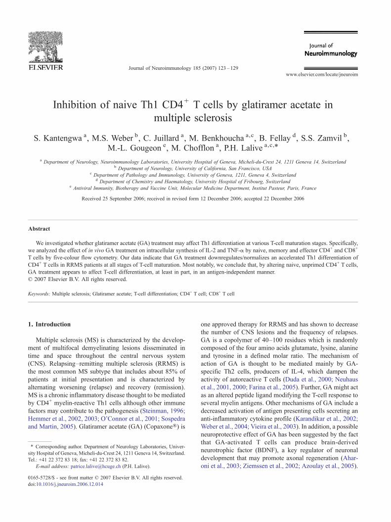
Fig. 1. Representative FACS analysis of CD4+ T-cell subsets. Mononuclear cells wetreated) or a RRMS patient receiving GA as immunotherapy (GA-treated). PlottingB). On the basis of expression of CD45RA and CD27 markers, the CD4+ T cells(naive), R4 = CD45RA−CD27+ (memory) and R5 = CD45RA−CD27− (effector). PCD8+ T cells were gated in R6 (B).
inflammatory cytokine synthesis. Interestingly, GA treatmentwas found to inhibit the priming of the naive CD4+ T cells forTNF-α and IL-2 synthesis which suggests that GAmay act, atleast in part, in an antigen-independent manner.
2. Materials and methods
2.1. Patients and healthy donors
Eighteen MS patients fulfilling Poser criteria (Poser et al.,1983) as well as 13 age- and gender-matched healthyindividuals were recruited at the University Hospital ofGeneva in accordance with institutional guidelines. Allpatients were classified as RRMS in remission according toclinical history. Seven patients treated by GA were undertherapy for at least 1 year, whereas the 11 non-treatedpatients had not received any immunomodulatory therapyfor at least 1 year. Characteristics of patients and HC aredescribed in Table 1. All individuals had peripheral bloodcounts within the reference range.
2.2. Antibodies and reagents
Labelled monoclonal antibodies and reagents used inthis study were: human CD3 biotinylated (Diaclone,Besançon, France); CD8-APC-Cy7, CD45RA-APC andStreptavidin-PE-TR (Caltag, Burlingame, CA, USA);CD27-FITC, TNFα-PE and IL-2-PE (BD PharMingen,
re isolated from healthy control (HC), RRMS patient without treatment (non-CD3 vs CD8+ allowed to gate on CD4+ cells in region R2 as CD3+CD8− (A,were further distributed into the different subsets: R3 = CD45RA+CD27+
ercentage of each subset is displayed in the corresponding quadrant (C–E).
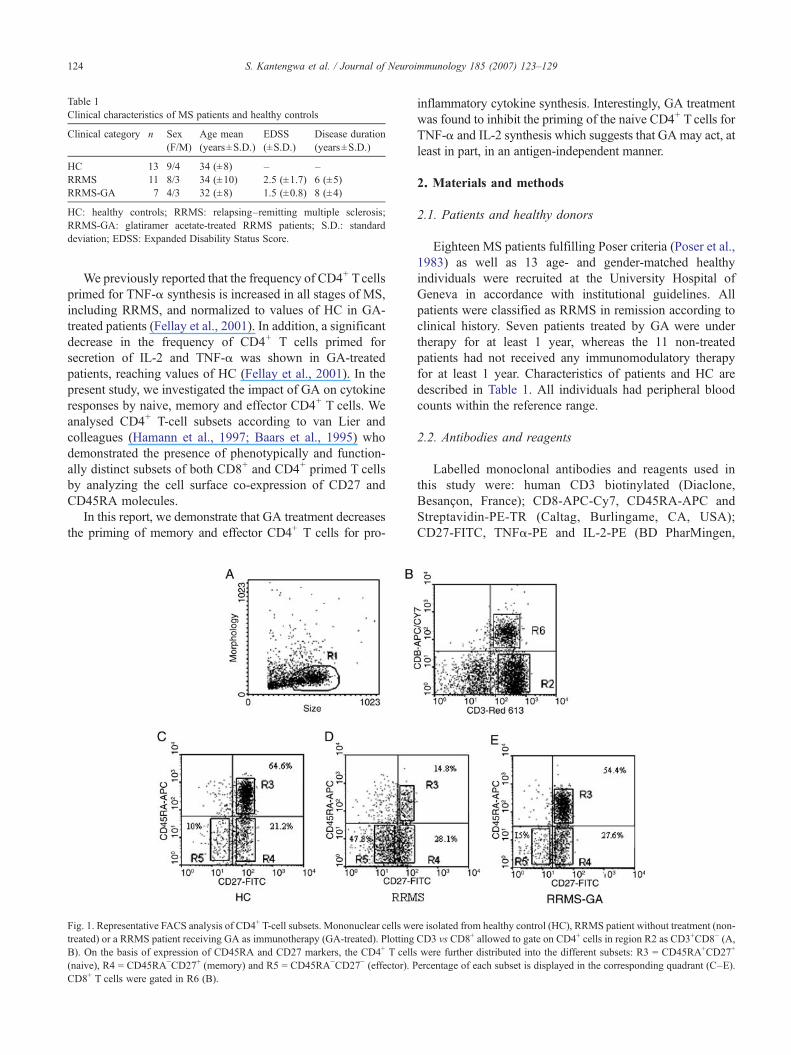
Fig. 2. Frequencies of naive, effector and memory CD4+ Tcells from healthycontrols (HC), non-treated RRMS (RRMS) and GA-treated (RRMS-GA)patients. Data are means±SEM from HC (n=13), RRMS (n=11) andRRMS-GA (n=7). The number of naive cells was significantly decreased inRRMS ( p=0.013) whereas effector cells were increased ( p=0.048) inRRMS, when compared to HC. No difference was found between RRMS-GA and HC in the memory T-cell subset ( p=ns).
125S. Kantengwa et al. / Journal of Neuroimmunology 185 (2007) 123–129
San Diego, CA, USA); ionomycin (Alexis Biochemicals,San Diego, CA, USA); phorbol 12-myristate 13-acetate(PMA) and brefeldin A (Sigma, St. Louis, MI, USA);RPMI 1640 medium, L-glutamine and penicillin–strepto-mycin (Invitrogen, Praisley, Scotland); fetal calf serum(FCS) (Sera-Tech, Aidenbach).
PBMCs were isolated from ethylenediaminetetraacetic(EDTA)-anticoagulated blood by standard Ficoll–Hypaque(Amersham Pharmacia Biotech, Uppsala, Sweden) densitygradient centrifugation. After 3 washes with Hanks’balanced salt solution (HBSS), PBMCs (106 cells/ml)were resuspended in RPMI medium (Invitrogen LifeTechnologies) containing 10% heat-inactivated fetal calfserum (FCS), 1% penicillin–streptomycin and 2% gluta-mine (Invitrogen Life Technologies). The cells werestimulated for 16 h with 20 ng/ml phorbol 12-myristate13-acetate (PMA) (Sigma) and 1 μg/ml ionomycin (P/I)(Sigma). The protein transport inhibitor brefeldin A (10 μg/ml) was added for the last 14 h of incubation (O'Neil-Andersen and Lawrence 2002).
2.4. Five-colour staining and flow cytometry analysis
PBMCs from HC, non-treated or GA-treated RRMSpatients were analysed by fluorescence-activated cell sorter(FACS). PBMCs (2.5×10⁵/well) were first labelled forsurface markers: CD3, CD8, CD45RA and CD27 for 30 minin phosphate-buffered saline (PBS) containing 1% bovineserum albumin (BSA) and 0.1% azide (NaN3) (labellingbuffer). Cells were fixed in 1% paraformaldehyde (PFA) inthe labelling buffer for 15 min at room temperature (RT). Thecells were further permeabilized with 0.05% saponin in thelabelling buffer for 15 min at RT and stained for intracellularcytokine IL-2 or TNF-α. Five-colour-stained CD4+ andCD8+ T cells were gated and analysed in a FACSvantage SE(BD Biosciences). FACS analysis was performed within 24 hafter blood sampling. For each sample, 5×10⁴ cells wereacquired and results were analysed with WinMDI 2.8software.
A representative FACS analysis of CD4+ T-cell subsets isshown in Fig. 1. PBMCs were gated according to forwardscatter/side scatter (FSC/SSC) criteria (R1) and analyzed forCD3/CD8 co-staining. The stimulation of PBMC withphorbol 12-myristate 13-acetate (PMA) downregulates thesurface membrane CD4 by internalisation (Moller et al.,1990; Petersen et al., 1992). Therefore, in accordance withseveral previous studies (Picker et al., 1995; Ledru et al.,1998; Fellay et al., 2001), the percentage of CD4+ T cellswas determined as the difference between the percentage ofCD3+ T cells and the percentage of CD8+ T cells (in R2region). Co-expression of CD45RA and CD27 markers wasthen analyzed on R3-gated cells. The following CD4+ T-cell
subsets were identified: CD45RA+CD27+ (naive) in R3,CD45RA−CD27+ (memory) in R4 and CD45RA−CD27−
(effectors) in R5. The percentage of each population isdisplayed in the corresponding areas.
2.5. Statistical analyses
Statistical analyses were performed using PRISM soft-ware (GraphPad, San Diego, CA). The statistical significance
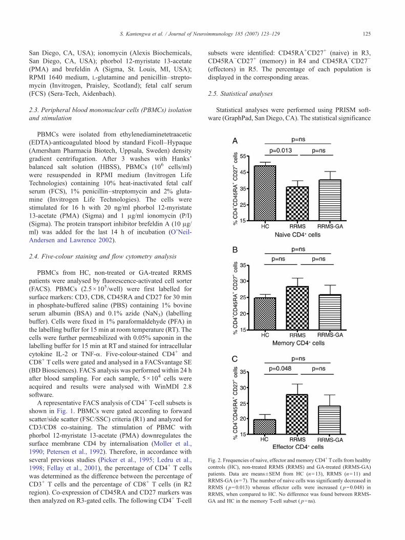
Fig. 3. Intracellular TNF-α and IL-2 production in naive, memory and effector CD4+ T cells. PBMCs derived from HC (n=13), RRMS (n=11) and RRMS-GA (n=7) were stimulated with PMA/ionomycin in the presence of brefeldin A and intracellular cytokine was analysed with specific antibody according tothe five-colour technique (see details in the Materials and methods section). Compared to RRMS, RRMS-GA patients showed a significant decrease ofTNF-α in naive ( p=0.033) (A) and effector ( p=0.006) (C) subsets while IL-2 was lowered by GA in the three subpopulations: naive ( p=0.046) (D),memory ( p=0.007) (E) and effector ( p=0.05) (F). No cytokine difference was found when HC were compared to RRMS except for IL-2 in the effectorsubset ( p=0.007) (F).
126 S. Kantengwa et al. / Journal of Neuroimmunology 185 (2007) 123–129
( p value of b0.05) was determined by the non-parametricMann Whitney test.
3. Results
3.1. CD4+ T-cell subsets in HC, untreated RRMS and GA-treated RRMS patients
The percentages of naive, memory and effector CD4+ T-cell subsets were determined in HC, RRMS and RRMS-GApatients (Fig. 2). A significant decrease of naive CD4+ Tcells (36±3.8% (mean±SEM) vs 48.9±2.5%; p=0.013) as
Table 2Frequencies of TNF-α and IL-2 expressing cells in naive, memory and effector C
% TNF-α+ cells
Naive Memory Effector
HC 8.4±3.9 45.2±8.2 47.8±16RRMS 13.1±7.3 47.1±9 55.1±11RRMS-GA 7.2±2.6 37.9±9.9 39.6±13
PBMCs were analyzed by flow cytometry as indicated in Fig. 1, after gating on CDn=7). Statistical differences are described in Fig. 3. HC: healthy controls; RRMS: rRRMS patients.
well as an increase of effector CD4+ T cell (27.7±3.5% vs19.7±1.8%; p=0.048) was observed in non-treated RRMSpatients compared to HC (Fig. 2A, C). In contrast, comparedto HC GA-treated RRMS patients showed similar propor-tions of naive (40.4±5.3% vs 48.9±2.5%; p=ns), memory(25.8±3.1% vs 24.8±1.1%; p=ns) and effector (24±3.7%vs 19.7±1.8%; p=ns) CD4+ T cells. Interestingly, GA-treated RRMS patients showed an increase in naive CD4+ Tcells (40.4±5.3% vs 36±3.8%) and a decrease in effectorCD4+ T cells (24±3.7% vs 27.7±3.5%) compared to RRMSpatients, although the difference was not statisticallysignificant ( p=ns).
D4+ T cells
% IL-2+ cells
Naive Memory Effector
.4 58.5±16.6 59.5±16.9 29.5±12
.1 65.8±10.5 71.6±6.2 38.5±9.1
.4 48.9±23 59.5±16.9 27.0±7.6
3+CD8− cells. Data are means±S.D. (HC, n=13; RRMS, n=11; RRMS-GA,elapsing–remitting multiple sclerosis; RRMS-GA: glatiramer acetate-treated
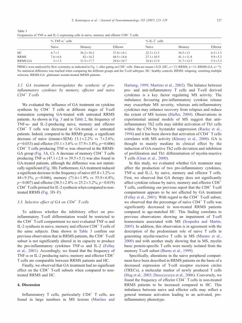
Table 3Frequencies of TNF-α and IL-2 expressing cells in naive, memory and effector CD8+ T cells
PBMCs were analyzed by flow cytometry as indicated in Fig. 1, after gating on CD8+ cells. Data are means±S.D. (HC, n=13; RRMS, n=11; RRMS-GA, n=7).No statistical difference was reached when comparing the different groups and the T-cell subtypes. HC: healthy controls; RRMS: relapsing–remitting multiplesclerosis; RRMS-GA: glatiramer acetate-treated RRMS patients.
127S. Kantengwa et al. / Journal of Neuroimmunology 185 (2007) 123–129
3.2. GA treatment downregulates the synthesis of pro-inflammatory cytokines by memory, effector and naiveCD4+ T cells
We evaluated the influence of GA treatment on cytokinesynthesis by CD4+ T cells at different stages of T-cellmaturation comparing GA-treated with untreated RRMSpatients. As shown in Fig. 3 and in Table 2, the frequency ofTNF-α- and IL-2-producing naive, memory and effectorCD4+ T cells was decreased in GA-treated vs untreatedpatients. Indeed, compared to the RRMS group, a significantdecrease of naive ((mean±SEM) 13.1±2.2% vs 7±2.6%;p=0.033) and effector (55.1±3.6% vs 37.9±3.8%; p=0.006)CD4+ T cells producing TNF-α was observed in the RRMS-GA group (Fig. 3A, C). A decrease of memory CD4+ T cellsproducing TNF-α (47.1±2.8 vs 39.5±5.1) was also found inGA-treated patients, although the difference was not statisti-cally significant (Fig. 3B). Furthermore, GA treatment induceda significant decrease in the frequency of naive (65.8±3.2% vs44±9.3%; p=0.046), memory (71.6±1.9% vs 55.9±6.8%;p=0.007) and effector (38.5±2.8% vs 25.2±3.2%; p=0.019)CD4+ Tcells primed for IL-2 synthesiswhen compared to non-treated RRMS (Fig. 3D–F).
3.3. Selective effect of GA on CD4+ T cells
To address whether the inhibitory effect on pro-inflammatory T-cell differentiation would be restricted tothe CD4+ T-cell compartment we next evaluated TNF-α andIL-2 synthesis in naive, memory and effector CD8+ T cells ofthe same subjects. Data shown in Table 3 confirm ourprevious observation that in RRMS patients, the CD8+ T-cellsubset is not significantly altered in its capacity to producethe pro-inflammatory cytokines TNF-α and IL-2 (Fellayet al., 2001). Accordingly, we found that the frequency ofTNF-α or IL-2 producing naive, memory and effector CD8+
T cells are comparable between RRMS patients and HC.Finally, we observed that GA treatment had no significant
effect on the CD8+ T-cell subsets when compared to non-treated RRMS and HC.
4. Discussion
Inflammatory T cells, particularly CD4+ T cells, arefound in large numbers in MS lesions (Martino and
Hartung, 1999; Martino et al., 2002). The balance betweenpro- and anti-inflammatory T cells and T-cell derivedcytokines is a key factor regulating MS activity. Theimbalance favouring pro-inflammatory cytokine releasemay exacerbate MS severity, whereas anti-inflammatorycytokines may enhance recovery from relapses and reducethe extent of MS lesions (Hafler, 2004). Observations inexperimental animal models of MS suggest that anti-inflammatory Th2 cells may inhibit activation of Th1 cellswithin the CNS by bystander suppression (Racke et al.,1994) and it has been shown that activation of CD4+ T cellscorrelates with MS activity (Khoury et al., 2000). GA isthought to mainly mediate its clinical effect by theinduction of GA-reactive Th2 cells deviation and inhibitionof proliferation and Th1 differentiation of myelin-reactiveT cells (Gran et al., 2000).
In this study, we evaluated whether GA treatment mayaffect the production of two pro-inflammatory cytokines,TNF-α and IL-2, by naive, memory and effector T cells.First, we observed that GA therapy does not significantlyaffect cytokine release by naive, memory and effector CD8+
T cells, confirming our previous report that the CD8+ T-cellcompartment appears to be not affected by GA treatment(Fellay et al., 2001). With regard to the CD4+ T-cell subset,we observed that the percentage of naive CD4+ T cells wassignificantly decreased in non-treated RRMS patientscompared to age-matched HC. This finding correlates toprevious observations showing an impairment of T-cellhomeostasis associated with MS (Sospedra and Martin,2005). In addition, this observation is in agreement with thedescription of the predominant role of naive T cells ingenerating myelin-reactive T cells in MS (Muraro et al.,2000) and with another study showing that in MS, myelinbasic protein-specific T cells were mostly isolated from thememory T-cell subset (Burns et al., 1999).
Specifically, alterations in the naive peripheral compart-ment have been described in RRMS patients on the basis of adecreased expression of T-cell receptor excision circles(TRECs), a molecular marker of newly produced T cells(Hug et al., 2003; Duszczyszyn et al., 2006). Conversely, wefound the frequency of effector CD4+ T cells in non-treatedRRMS patients to be increased compared to HC. Thisimbalance between naive and effector cells may reflect ageneral immune activation leading to an activated, pro-inflammatory phenotype.
128 S. Kantengwa et al. / Journal of Neuroimmunology 185 (2007) 123–129
Pro-inflammatory cytokines produced by activated T cellshave been suggested to be involved in the pathogenesis ofMS (Clerici et al., 2001). In particular, TNF-α has beenassociated with enhanced MS activity and demyelination(Navikas and Link, 1996; Matusevicius et al., 1996; Bitschet al., 2000; Chofflon and Fellay, 1998), although somehomeostatic role has also been suggested (Martino et al.,2002; Liu et al., 1998). TNF-α is predominantly released byT cells but also by macrophages and microglia (Bitsch et al.,2000; Hartung et al., 2004). In comparison with non-treatedRRMS patients, we found that GA-treated patients revealed asignificant decrease in the frequency of effector and memoryCD4+ T cells primed for TNF-α synthesis. Interestingly, thiswas also observed for the naive CD4+ T-cell subset,suggesting that the effect of GA on T-cell priming is notdependent on prior T-cell antigen recognition.
IL-2 is a growth factor required for T-cell activation,proliferation, and survival. High affinity IL-2 receptor (IL-2R) was found to be upregulated in MS patients and anincreased frequency of IL-2-responsive T cells specific formyelin antigens has been reported (Zhang et al., 1994). TheIL-2 receptor is likely to be an important component ofinflammation in MS and has accordingly been targeted innovel MS therapies (Rose et al., 2004). Correlation of IL-2and soluble IL-2R with activity of MS has been established(Sharief and Thompson, 1993). Our study shows that IL-2was mostly expressed by naive and memory CD4+ T cells inboth MS patients and healthy donors. In all three CD4+ T-cell subsets RRMS patients showed a significantly enhancedsynthesis of IL-2 when compared to HC. Strikingly, GAtreatment again decreased the frequency of IL-2-synthesiz-ing CD4+ T cells, whether expressing markers of naive,memory or effectors. Thus, GA treatment appears todownregulate the priming of all three CD4+ T-cell subsetsfor the pro-inflammatory cytokines TNF-α and IL-2.Furthermore, the comparison to corresponding maturationstages of CD8+ T cells confirms the selective functionalalteration of CD4+ T cells in MS and the beneficialimmunomodulatory effects of GA on this subset.
GA treatment is thought to primarily induce GA-reactiveTh2-like regulatory cells (Neuhaus et al., 2001, 2000). Ourstudy indicates that GA treatment exerts an immunomodu-latory effect on both activated/differentiated and naive CD4+
T cells independent of their antigen-specificity. Thus, thebeneficial effect of GA is detected not only on GA-specificT-cell clones but also on polyclonal CD4+ T cells at differentlevels of maturation and on non-primed Tcells, as it has beensuggested by in vitro studies on PBMC of non-treated MSpatients (Wiesemann et al., 2001).
Interestingly, the immunomodulatory effect of GA wasrecently shown to influence the innate immune system(Weber et al., 2004; Vieira et al., 2003). The fact that naiveT cells are influenced by GA treatment could be also seen asa secondary effect of GA on APC. Indeed, following GAtreatment, APC could directly influence effector andmemory T cells but could also have an effect on the
priming of naive T cells. It thus can be hypothesized that theinhibitory impact of GA on Th1 priming of CD4+ T cellsmay occur secondary to APC immunomodulation by GA.
In conclusion, our results give new insights on themechanism of action of GA in vivo and confirm recentstudies demonstrating that GA has a broad immunologicaleffect not restricted to an antigen-specific immunomodula-tion of the acquired immune system. In addition, this reportunderscores the therapeutic potential of GA for treatment ofautoimmune diseases other than MS.
Acknowledgements
We thank Anne-Marie Paunier-Doret for technical support.This study was supported by the Swiss National Foundation(grant # 310000-113653 and # 31-63192.00), the Swiss Mul-tiple Sclerosis Society and the Alliance SEP foundation.
Reference
Aharoni, R., Kayhan, B., Eilam, R., Sela, M., Arnon, R., 2003. Glatirameracetate-specific T cells in the brain express T helper 2/3 cytokines andbrain-derived neurotrophic factor in situ. Proc. Natl. Acad. Sci. U. S. A.100, 14157–14162.
Azoulay, D., Vachapova, V., Shihman, B., Miler, A., Karni, A., 2005. Lowerbrain-derived neurotrophic factor in serum of relapsing remitting MS:reversal by glatiramer acetate. J Neuroimmunol. 167, 215–218.
Baars, P.A., Maurice, M.M., Repe, M., Hooibrink, B., van Lier, R.A., 1995.Heterogeneity of the circulating human CD4+ T cell population. Furtherevidence that the CD4+CD45RA−CD27− T cell subset containsspecialized primed T cells. J. Immunol. 154, 17–25.
Bitsch, A., Kuhlmann, T., Da Costa, C., Bunkowski, S., Polak, T., Bruck,W., 2000. Tumour necrosis factor alpha mRNA expression in earlymultiple sclerosis lesions: correlation with demyelinating activity andoligodendrocyte pathology. Glia 29, 366–375.
Burns, J., Bartholomew, B., Lobo, S., 1999. Isolation of myelin basicprotein-specific T cells predominantly from the memory T-cellcompartment in multiple sclerosis. Ann. Neurol. 45, 33–39.
Chofflon, M., Fellay, B., 1998. Monitoring multiple sclerosis course andactivity with TNF-alpha. Mult. Scler. 4, 188–192.
Clerici, M., Saresella, M., Trabattoni, D., Speciale, L., Fossati, S., Ruzzante, S.,Cavaretta, R., Filippi, M., Caputo, D., Ferrante, P., 2001. Single-cellanalysis of cytokine production shows different immune profiles in mul-tiple sclerosis patients with active or quiescent disease. J. Neuroimmunol.121, 88–101.
Duda, P.W., Schmied, M.C., Cook, S.L., Krieger, J.I., Hafler, D.A., 2000.Glatiramer acetate (Copaxone) induces degenerate, Th2-polarizedimmune responses in patients with multiple sclerosis. J. Clin. Invest.105, 967–976.
Duszczyszyn, D.A., Beck, J.D., Antel, J., Bar-Or, A., Lapierre, Y., Gadag, V.,Haegert, D.G., 2006. Altered naive CD4 and CD8 T cell homeostasis inpatients with relapsing–remitting multiple sclerosis: thymic versusperipheral (non-thymic) mechanisms. Clin. Exp. Immunol. 143, 305–313.
Farina, C., Weber, M.S., Meinl, E., Wekerle, H., Hohlfeld, R., 2005.Glatiramer acetate in multiple sclerosis: update on potential mechanismsof action. Lancet Neurol. 4, 567–575.
Fellay, B., Chofflon, M., Juillard, C., Paunier, A.M., Landis, T., Roth, S.,Gougeon, M.L., 2001. Beneficial effect of co-polymer 1 on cytokine pro-duction by CD4 T cells in multiple sclerosis. Immunology 104, 383–391.
Gran, B., Tranquill, L.R., Chen, M., Bielekova, B., Zhou, W., Dhib-Jalbut,S., Martin, R., 2000. Mechanisms of immunomodulation by glatirameracetate. Neurology 55, 1704–1714.
Hafler, D.A., 2004. Multiple sclerosis. J. Clin. Invest. 113, 788–794.
129S. Kantengwa et al. / Journal of Neuroimmunology 185 (2007) 123–129
Hamann, D., Baars, P.A., Rep, M.H., Hooibrink, B., Kerkhof-Garde, S.R.,Klein, M.R., van Lier, R.A., 1997. Phenotypic and functional separationof memory and effector human CD8+ T cells. J. Exp. Med. 186,1407–1418.
Hartung, H.P., Bar-Or, A., Zoukos, Y., 2004. What do we know about themechanism of action of disease-modifying treatments in MS? J. Neurol251 (Suppl. 5), v12–v29.
Hemmer, B., Archelos, J.J., Hartung, H.P., 2002. New concepts in theimmunopathogenesis of multiple sclerosis. Nat. Rev. Neurosci. 3,291–301.
Hemmer, B., Kieseier, B., Cepok, S., Hartung, H.P., 2003. New immuno-pathologic insights into multiple sclerosis. Curr. Neurol. Neurosci. Rep. 3,246–255.
Hug, A., Korporal, M., Schroder, I., Haas, J., Glatz, K., Storch-Hagenlocher,B., Wildemann, B., 2003. Thymic export function and T cellhomeostasis in patients with relapsing remitting multiple sclerosis.J. Immunol. 171, 432–437.
Khoury, S.J., Guttmann, C.R., Orav, E.J., Kikinis, R., Jolesz, F.A., Weiner,H.L., 2000. Changes in activated T cells in the blood correlate withdisease activity in multiple sclerosis. Arch. Neurol. 57, 1183–1189.
Ledru, E., Lecoeur, H., Garcia, S., Debord, T., Gougeon, M.L., 1998.Differential susceptibility to activation-induced apoptosis amongperipheral Th1 subsets: correlation with Bcl-2 expression and con-sequences for AIDS pathogenesis. J. Immunol. 160, 3194–3206.
Liu, J., Marino, M.W., Wong, G., Grail, D., Dunn, A., Bettadapura, J.,Slavin, A.J., Old, L., Bernard, C.C., 1998. TNF is a potent anti-inflammatory cytokine in autoimmune-mediated demyelination. Nat.Med. 4, 78–83.
Martino, G., Hartung, H.P., 1999. Immunopathogenesis of multiplesclerosis: the role of T cells. Curr. Opin. Neurol. 12, 309–321.
Martino, G., Adorini, L., Rieckmann, P., Hillert, J., Kallmann, B., Comi, G.,Filippi, M., 2002. Inflammation in multiple sclerosis: the good, the bad,and the complex. Lancet Neurol. 1, 499–509.
Matusevicius, D., Navikas, V., Soderstrom, M., Xiao, B.G., Haglund, M.,Fredrikson, S., Link, H., 1996. Multiple sclerosis: the proinflammatorycytokines lymphotoxin-alpha and tumour necrosis factor-alpha areupregulated in cerebrospinal fluid mononuclear cells. J. Neuroimmunol.66, 115–123.
Moller, B.K., Andresen, B.S., Christensen, E.I., Petersen, C.M., 1990. Surfacemembrane CD4 turnover in phorbol ester stimulated T-lymphocytes.Evidence of degradation and increased synthesis. FEBS Lett. 276, 59–62.
Muraro, P.A., Pette, M., Bielekova, B., McFarland, H.F., Martin, R., 2000.Human autoreactive CD4+ T cells from naive CD45RA+ and memoryCD45RO+ subsets differ with respect to epitope specificity andfunctional antigen avidity. J. Immunol. 164, 5474–5481.
Navikas, V., Link, H., 1996. Review: cytokines and the pathogenesis ofmultiple sclerosis. J. Neurosci. Res. 45, 322–333.
Neuhaus, O., Farina, C., Yassouridis, A., Wiendl, H., Then, B.F., Dose, T.,Wekerle, H., Hohlfeld, R., 2000. Multiple sclerosis: comparison ofcopolymer-1-reactive T cell lines from treated and untreated subjectsreveals cytokine shift from T helper 1 to T helper 2 cells. Proc. Natl.Acad. Sci. U. S. A. 97, 7452–7457.
Neuhaus, O., Farina, C., Wekerle, H., Hohlfeld, R., 2001. Mechanisms ofaction of glatiramer acetate in multiple sclerosis. Neurology 56,702–708.
O'Connor, K.C., Bar-Or, A., Hafler, D.A., 2001. The neuroimmunology ofmultiple sclerosis: possible roles of T and B lymphocytes inimmunopathogenesis. J. Clin. Immunol. 21, 81–92.
O'Neil-Andersen, N.J., Lawrence, D.A., 2002. Differential modulation ofsurface and intracellular protein expression by T cells after stimulation inthe presence of monensin or brefeldin A. Clin. Diagn. Lab. Immunol. 9,243–250.
Petersen, C.M., Christensen, E.I., Andresen, B.S., Moller, B.K., 1992.Internalization, lysosomal degradation and new synthesis of surfacemembrane CD4 in phorbol ester-activated T-lymphocytes and U-937cells. Exp. Cell Res. 201, 160–173.
Picker, L.J., Singh, M.K., Zdraveski, Z., Treer, J.R., Waldrop, S.L.,Bergstresser, P.R., Maino, V.C., 1995. Direct demonstration of cytokinesynthesis heterogeneity among human memory/effector T cells by flowcytometry. Blood 86, 1408–1419.
Racke, M.K., Bonomo, A., Scott, D.E., Cannella, B., Levine, A., Raine, C.S.,Shevach, E.M., Rocken, M., 1994. Cytokine-induced immune deviationas a therapy for inflammatory autoimmune disease. J Exp. Med. 180,1961–1966.
Rose, J.W., Watt, H.E., White, A.T., Carlson, N.G., 2004. Treatment ofmultiple sclerosis with an anti-interleukin-2 receptor monoclonalantibody. Ann. Neurol. 56, 864–867.
Sharief, M.K., Thompson, E.J., 1993. Correlation of interleukin-2 andsoluble interleukin-2 receptor with clinical activity of multiple sclerosis.J. Neurol. Neurosurg. Psychiatry 56, 169–174.
Sospedra, M., Martin, R., 2005. Immunology of multiple sclerosis ⁎. Annu.Rev. Immunol. 23, 683–747.
Steinman, L., 1996. Multiple sclerosis: a coordinated immunological attackagainst myelin in the central nervous system. Cell 85, 299–302.
Vieira, P.L., Heystek, H.C., Wormmeester, J., Wierenga, E.A., Kapsenberg,M.L., 2003. Glatiramer acetate (copolymer-1, Copaxone) promotes Th2cell development and increased IL-10 production through modulation ofdendritic cells. J. Immunol. 170, 4483–4488.
Weber, M.S., Starck, M., Wagenpfeil, S., Meinl, E., Hohlfeld, R., Farina, C.,2004. Multiple sclerosis: glatiramer acetate inhibits monocyte reactivityin vitro and in vivo. Brain 127, 1370–1378.
Wiesemann, E., Klatt, J., Sonmez, D., Blasczyk, R., Heidenreich, F.,Windhagen, A., 2001. Glatiramer acetate (GA) induces IL-13/IL-5secretion in naive T cells. J. Neuroimmunol. 119, 137–144.
Zhang, J., Markovic-Plese, S., Lacet, B., Raus, J., Weiner, H.L., Hafler, D.A.,1994. Increased frequency of interleukin 2-responsive T cells specific formyelin basic protein and proteolipid protein in peripheral blood andcerebrospinal fluid of patients with multiple sclerosis. J. Exp. Med. 179,973–984.
Ziemssen, T., Kumpfel, T., Klinkert, W.E., Neuhaus, O., Hohlfeld, R., 2002.Glatiramer acetate-specific T-helper 1- and 2-type cell lines produceBDNF: implications for multiple sclerosis therapy. Brain-derivedneurotrophic factor. Brain 125, 2381–2391.


![[(Methyl)1-11C]-Acetate Metabolism in Hepatocellular Carcinoma](https://static.documents.pub/doc/80x56/6348dbfb7442d262850f3c5e/methyl1-11c-acetate-metabolism-in-hepatocellular-carcinoma.jpg)


