Interaction between Coat Morphogenetic Proteins SafA and SpoVID�
Teresa Costa,1† Anabela L. Isidro,1 Charles P. Moran, Jr.,2 and Adriano O. Henriques1*Instituto de Tecnologia Quımica e Biologica, Universidade Nova de Lisboa, Avenida da Republica, EAN, 2781-157 Oeiras,
Portugal,1 and Emory University School of Medicine, Department of Microbiology and Immunology,3010 Rollins Research Center, Atlanta, Georgia 303222
Received 26 May 2006/Accepted 23 August 2006
Morphogenetic proteins such as SpoVID and SafA govern assembly of the Bacillus subtilis endospore coat byguiding the various protein structural components to the surface of the developing spore. Previously, a screenfor peptides able to interact with SpoVID led to the identification of a PYYH motif present in the C-terminalhalf of the SafA protein and to the subsequent demonstration that SpoVID and SafA directly interact. spoVIDand safA spores show deficiencies in coat assembly and are lysozyme susceptible. Both proteins, orthologs ofwhich are found in all Bacillus species, have LysM domains for peptidoglycan binding and localize to thecortex-coat interface. Here, we show that the interaction between SafA and SpoVID involves the PYYH motif(region B) but also a 13-amino-acid region (region A) just downstream of the N-terminal LysM domain of SafA.We show that deletion of region B does not block the interaction of SafA with SpoVID, nor does it bring aboutspore susceptibility to lysozyme. Nevertheless, it appears to reduce the interaction and affects the complex. Incontrast, lesions in region A impaired the interaction of SafA with SpoVID in vitro and, while not affecting theaccumulation of SafA in vivo, interfered with the localization of SafA around the developing spore, causingaberrant assembly of the coat and lysozyme sensitivity. A peptide corresponding to region A interacts withSpoVID, suggesting that residues within this region directly contact SpoVID. Since region A is highly conservedamong SafA orthologs, this motif may be an important determinant of coat assembly in the group of Bacillusspore formers.
A large number of proteins produced in the mother cell ofsporulating cells of Bacillus subtilis are targeted to the surfaceof the developing spore to form a structure known as the sporecoat (11, 16, 18). Assembly of the coat confers protectionagainst lytic enzymes and small noxious molecules and allowsefficient interaction of the spore with compounds able to trig-ger germination (11, 16, 18). Because of the resistance it im-parts to the action of the lytic enzymes, the coat also protectsphagocytosed spores from digestion by predatory microorgan-isms (22). Structurally, the coat consists of an amorphous un-dercoat that contacts the underlying cortex peptidoglycan, alamellar inner layer, and a thick electron-dense outer layer (11,16, 18). The genes encoding the various coat components areexpressed at different times during the deployment of a mothercell cascade of gene expression, and this temporal control is animportant factor contributing to the ordered assembly of thecoat (16). However, assembly of the coat relies to a large extenton the action of a class of so-called morphogenetic proteinsthat guide the assembly of the structural components (11, 16,18, 29). One, called SpoIVA, localizes early to the mother cellside of the asymmetric septum and is responsible for recruitinganother morphogenetic protein, SpoVID, to the developingspore (11, 12, 33). A third morphogenetic protein, CotE, lo-calizes in a SpoIVA-dependent manner close to the developing
spore, around a region of unknown composition called thematrix (11, 12). The localizations of SpoIVA, SpoVID, andCotE take place prior to the complete engulfment of the pre-spore by the mother cell when �E and the ancillary transcrip-tion factors SpoIIID and GerR govern gene expression in themother cell (13, 41). Following engulfment completion, andthe activation of the late mother cell-specific regulators �K andlater of GerE, the coat layers start gaining their final appear-ance (11, 13, 16, 18, 41). The prior localization of CotE at theedge of the matrix region is thought to be required to nucleateassembly of the outer coat, whereas the matrix region developsinto the inner coat (11, 12). CotE appears to have a modulardesign, specifying information for its targeting to the surface ofthe developing spore, for oligomerization, and also for inter-action with other coat proteins (3, 28). Presumably, CotE con-trols outer coat assembly by means of complex network ofdirect or indirect interactions leading to the recruitment ofmany proteins to this coat layer (21, 28).
SpoVID does not influence the localization of CotE at anearly stage, but following engulfment completion in cells mu-tant for spoVID, CotE and the rest of the coat detaches fromthe surface of the prespore and forms swirls of partially struc-tured material dispersed throughout the mother cell cytoplasm(4, 12). Left with an exposed cortex, spoVID spores are sensi-tive to lysozyme (12). To study the function of SpoVID in theassembly process, Ozin et al. (32) used a phage display screen-ing to identify peptides able to interact with SpoVID. Theyfound a peptide motif, PYYH, occurring in the C-terminal halfof another coat protein, SafA (387 residues long), the absenceof which causes deficient formation of the coat and sporesusceptibility to lysozyme (32, 40). At least three forms of SafAaccumulate in sporulating cells: a full-length form (SafAFL) (45
* Corresponding author. Mailing address: Instituto de TecnologiaQuımica e Biologica, Universidade Nova de Lisboa, Avenida da Re-publica, EAN, 2781-157 Oeiras, Portugal. Phone: 351-21-4469522. Fax:351-21-4411277. E-mail: [email protected].
† Present address: Department of Molecular, Cellular, and Devel-opmental Biology, Yale University, New Haven, CT 06520.
kDa), a C-terminal form derived from internal translation ofthe safA mRNA at methionine 164 (SafAC30, 30 kDa) and anN-terminal form (SafAN21, 21 kDa) (31). Assays in vitro and invivo showed that SafA and SpoVID directly interact, that SafAinteracts with itself, and that each of the C- and N-terminalforms are capable of interacting with SafAFL, with themselves,and, for at least SafAN21, with SpoVID (33). Both SpoVID (atits C terminus) and SafA (at its N terminus) contain a LysMdomain, which has a general peptidoglycan-binding function(2, 5, 20, 27, 35, 39), and both proteins localize to the cortex/coat interface (12, 32). Moreover, the localization of SafAdepends on SpoVID (33). These results have led to a modelaccording to which the interaction between SafA and SpoVIDrecruits the former to the coat region in close proximity to thecortex layer, and the two proteins function in this region bypromoting attachment of the nascent coat to the underlyingcortex layer (33). However, in spite of the fact that it containsthe PYYH motif, the SafAC30 form is not recruited to theprespore in the absence of SafAFL, suggesting that a secondregion of interaction could exist in the N-terminal half ofSafA (33). This second region of interaction between SafAand SpoVID was attributed to the LysM domain of both pro-teins, which is presumed to be involved in multimerization (2,5, 20, 27, 35).
Here, we have analyzed the interaction between SafA andSpoVID. We present biochemical, cytological, and genetic evi-
dence that strongly suggests that the interaction of SafA withSpoVID via a region just downstream of the LysM domain isresponsible for the targeting of SafA to the surface of thedeveloping spore and for the proper assembly and function ofthe coat structure.
MATERIALS AND METHODS
Strains and general techniques. All of the B. subtilis strains used in this studyare congenic derivatives of the wild-type strain MB24 (trpC2 metC3) (Table 1).The Escherichia coli strain DH5� (Bethesda Research Laboratories) was usedfor molecular cloning, and CC118(DE3)/pLysS was used for the overproductionof native or glutathione S-transferase (GST) fusion proteins (Table 1). Luria-Bertani medium was used for maintenance and growth of E. coli and B. subtilisstrains, with appropriate antibiotic selection when needed. Sporulation was in-duced by growth and nutrient exhaustion in Difco sporulation medium (DSM)(30). Genetic manipulations of B. subtilis and spore resistance or germinationproperties were assessed as previously described (9).
GST-SafA fusions. Primer safA204D and primer safA-STOP51R, safA-STOP64R, safA-STOP78R, safA-STOP91R, or safA-STOP99R (the sequencesof all primers are available upon request) were used to PCR amplify the codingregions of safA from residues 1 to 50, 1 to 63, 1 to 77, 1 to 90, and 1 to 98,respectively. The PCR fragments were digested with BamHI and XhoI andcloned between the same sites of pGex4T-3 (Pharmacia) to form pTC138 (GST-SafAE50), pTC159 (GST-SafAS63), pTC160 (GST-SafAK77), pTC161 (GST-SafAK90), and pTC139 (GST-SafAP98), respectively. The region encompassingcodons 1 to 162 of safA was amplified with primers safA204D and safA693R,digested with BamHI, and cloned between the BamHI site and a filled-in XhoIsite of pGex4T-3 (Pharmacia) to yield pTC141 (GST-SafAS162). pTC165 (GST-SafA�G51-S63) was obtained after digestion with HincII and autoligation of a
TABLE 1. Bacterial strains used in this study
Strain Genotype/phenotypea Origin or reference
E. coli strainsCC118 (DE3)/pLysS Colin ManoilAH2687 CC118(DE3)/pLysS/pTC55 Cmr Ampr This workAH2688 CC118(DE3)/pLysS/pTC49 Cmr Ampr This workAH2689 CC118(DE3)/pLysS/pTC50 Cmr Ampr This workAH2690 CC118(DE3)/pLysS/pTC51 Cmr Ampr This workAH2691 CC118(DE3)/pLysS/pTC52 Cmr Ampr This workAH2718 CC118(DE3)/pLysS/pTC61 Cmr Ampr This workAH2719 CC118(DE3)/pLysS/pTC62 Cmr Ampr This workAH2692 CC118(DE3)/pLysS/pOZ169 Cmr Ampr This workAH2934 CC118(DE3)/pLysS/pTC138 Cmr Ampr This workAH2935 CC118(DE3)/pLysS/pTC139 Cmr Ampr This workAH2937 CC118(DE3)/pLysS/pTC141 Cmr Ampr This workAH2978 CC118(DE3)/pLysS/pTC159 Cmr Ampr This workAH2979 CC118(DE3)/pLysS/pTC160 Cmr Ampr This workAH2980 CC118(DE3)/pLysS/pTC161 Cmr Ampr This workAH2985 CC118(DE3)/pLysS/pTC165 Cmr Ampr This workAH4086 CC118(DE3)/pLysS/pTC196 Cmr Ampr This workAH4087 CC118(DE3)/pLysS/pTC197 Cmr Ampr This work
B. subtilis strainsAOB68 trpC2 metC3 �safA::sp Spr 32AOB90 trpC2 metC3 safA-flag Cmr 33AH2781 trpC2 metC3 �safA::safA�PYYH Spr Cmr This workAH2787 trpC2 metC3 �safA::safAN1–162 Spr Cmr This workAH2788 trpC2 metC3 �safA::safAC164–387 Spr Cmr This workAH4006 trpC2 metC3 �safA::safA�G51-S63 Spr Cmr This workAH4090 trpC2 metC3 �safA::safA�G51-E57 Spr Cmr This workAH4091 trpC2 metC3 �safA::safA�P58-S63 Spr Cmr This workAH4007 trpC2 metC3 �safA::safA4�Ala Spr Cmr This workAH4102 trpC2 metC3 �safA::safA�-gfp Spr Nmr This workAH4103 trpC2 metC3 �safA::safA�G51-S63-gfp Spr Nmr This workAH4107 trpC2 metC3 �spoVID::cm �safA::safA�-gfp Spr Cmr Nmr This workAH4108 trpC2 metC3 �spoVID::cm �safA::safA�G51-S63-gfp Spr Cmr Nmr This work
a Spr, spectinomycin resistant; Cmr, chloramphenicol resistant; Nmr, neomycin resistant.
PCR obtained from pTC139 (see above) with primers safA50-64D and safA50-64R. pTC196 (GST-SafA�G51-E57) and pTC197 (GST-SafA�P58-S63) were con-structed as follows. First, the safA coding regions from residues 1 to 50 and 1 to57 were PCR amplified with primer sets safA204D/safA51-57R and safA204D/safA58-63R, respectively. Second, the regions corresponding to residues 58 to 98and 64 to 98 of safA were PCR amplified with primer sets safA51-57D/safA-STOP99R and safA58-63D/safA-STOP99R, respectively. The fragments (codons1 to 50 and 58 to 98 or 1 to 57 and 64 to 98) were mixed and subjected to PCRusing primers safA204D and safA-STOP99R to obtain safA fragments comprisingthe region coding for the first 98 residues but excluding residues 51 to 57 or 58to 63, respectively. The fragments were digested with BamHI and XhoI andintroduced between identical sites of pGex4T-3 (Pharmacia) to create pTC196and pTC197. All PCR products were sequenced. Plasmids pTC138, pTC159,pTC160, pTC161, pTC139, pTC141, pTC165, pTC196, and pTC197 were used totransform E. coli CC118(DE3)/pLysS to obtain strains AH2934, AH2978,AH2979, AH2980, AH2935, AH2937, AH2985, AH4086, and AH4087, respec-tively (Table 1).
GST-SpoVID fusions. Strain AH2692 (Table 1) was obtained by transforma-tion of E. coli CC118(DE3)/pLysS cells with pOZ169 (GST-SpoVIDFL) in whichthe entire spoVID coding sequence was translationally fused to the gst gene (33).We used pOZ169 and primer pairs spoVIDSTOP1498D/spoVID-STOP1498R,spoVID-STOP1198D/spoVID-STOP1198R, spoVIDSTOP907D/spoVID-STOP907R,and spoVID-STOP607D/spoVID-STOP607R to replace codons 500, 400, 303, and203 in spoVID with nonsense codons by using the QuickChange site-directedmutagenesis system (Stratagene) to obtain plasmids pTC52 (GST-SpoVID499),pTC51 (GST-SpoVID399), pTC50 (GST-SpoVID302), and pTC49 (GST-SpoVID202),respectively. Plasmids pTC52, pTC51, pTC50, and pTC49 were used to trans-form E. coli CC118(DE3)/pLysS, yielding strains AH2691, AH2690, AH2689,and AH2688, respectively (Table 1). Fusion of GST to residues 201 to 575 and201 to 399 of SpoVID was done as follows. We PCR amplified DNA fragmentsof spoVID (codons 201 to 575 and 201 to 399) with primers spoVID�800D andspoVID1935R and the pOZ169 and pTC51 templates, respectively. The productswere digested with BamHI and XhoI and cloned between the same sites ofpOZ169, creating pTC61 (GST-SpoVID201–575) and pTC62 (GST-SpoVID201–399),which were then introduced into CC118(DE3)/pLysS, yielding AH2718 andAH2719, respectively (Table 1). In order to obtain strain AH2687, producingnative GST (Table 1), we excised the spoVID coding region from pOZ169 (33)by digestion with BamHI and XhoI and filled-in the ends before autoligation.The resulting plasmid, pTC55, was used to transform CC118(DE3)/pLysS, cre-ating AH2687 (Table 1).
Mutations in regions A and B of SafA. A PCR fragment comprising the entiresafA coding region and 595 bp upstream of its start codon, generated withprimers safA-595D and safA�1176R, was introduced in pCR2.1TOPO (Invitro-gen) to create pTC75. Primers safA791D and safA834R were used to deletecodons 203 to 206 (the PYYH motif) of safA in pTC75 by the QuickChangemethod, yielding pTC78. A chloramphenicol resistance (Cmr) cassette was re-leased from pMS38 (43) with XbaI and XhoI and inserted between the same sitesof pTC78, generating pTC90. A PCR fragment encompassing codons 1 to 162 ofsafA, including 595 bp upstream of the safA start codon, was amplified withprimers safA-595D and safA693R and inserted into pCR2.1TOPO (Invitrogene)to obtain pTC93 (SafAN1–162). pTC93 was then digested with XbaI and XhoI,and a Cmr cassette released from pMS38 (43) with the same enzymes wasinserted to form pTC95. To fuse the safA promoter directly to codons 164 to 387,codons 1 to 163 were eliminated by amplification of pTC88 (obtained by cloningof the Cmr cassette from pMS38 between the XbaI and XhoI sites of pTC75)with primers safA203R and safA677D, digestion of the resulting product withHincII, and religation to form pTC97 (SafAC164–387). To delete residues 51 to 63of SafA, a product generated from pTC88 with primers safA50-64D and safA50-64R was cut with SacII and religated, to yield pTC170 (SafA�G51-S63). pTC198(SafA�G51-E57) and pTC199 (SafA�P58-S63) were constructed by cloning 143- and146-bp fragments released from pTC196 and pTC197 (see above) with BbvII andEspI, respectively, between the same sites of pTC88 (see above). Alanine sub-stitutions in SafA (R55A, K56A, K59A, and K62A) were obtained by the Quick-Change method (Stratagene) by using pTC75 and primers safAAlamut50-64Dand safAAlamut50-64R. The result was pTC172 (SafA4�Ala), from whichpTC173 was made by introducing a Cmr cassette (see above) between its XbaIand XhoI sites. All PCR products were sequenced. pTC90, pTC95, pTC97,pTC170, pTC190, pTC191, and pTC173 were transformed into B. subtilis AOB68to form AH2781, AH2787, AH2788, AH4006, AH4090, AH4091, and AH4007,respectively (Table 1).
Green fluorescent protein (GFP) fusions. pTC204 (SafA�-GFP) and pTC205(SafA�G51-S63-GFP) were constructed as follows. First, the safA 3� coding region(684 bp) was PCR amplified with primers safA677D and safA-gfpR. Second, a
714-bp fragment comprising the coding region of the gfp gene was PCR amplifiedwith pEA18 (a gift from Alan Grossman) and primers gfp-30D and gfpmut2/749R. The resulting fragments were mixed and subjected to PCR with primerssafA677D and gfpmut2/749R. The resulting 1,398-bp �safA-gfp fragment wascleaved with SmaI and EcoRI, and the 1,110-bp fragment was gel purified. DNAfragments corresponding to the 5� regions of safA� or safA�G51-S63 were ob-tained by digestion of pTC75 and pTC170 (see above), respectively, with SmaIand NotI. The safA� and safA�G51-S63 fragments were cloned together with the�safA-gfp fragment between the NotI and EcoRI sites of pBEST501 (19) to yieldpTC204 and pTC205, respectively. All PCR products were verified by sequenc-ing. Strains AH4102 and AH4103 resulted from the integration of pTC204 orpTC205, respectively, at the safA locus of AOB68 via a single reciprocal (Camp-bell-type) crossover (Table 1). The spoVID null mutant AH1910 was transformedwith chromosomal DNA of strains AH4102 and AH4103 to yield AH4107 andAH4108, respectively (Table 1).
Pull-down assays. In vitro pull-down assays were performed as describedpreviously (33), except that 60 �l of a 50% slurry of glutathione-Sepharose beads(Amersham Biosciences) were used and the interaction mixtures were resus-pended in a final volume of 30 �l of loading buffer, before analysis by sodiumdodecyl sulfate (SDS)–12.5% polyacrylamide gel electrophoresis (PAGE) andimmunoblotting.
Purification of spores and analysis of the spore coat. Spores were harvested bycentrifugation of DSM cultures 24 h after the onset of sporulation, washed, andpurified on step gradients of 20% to 50% Gastrografin (Schering) (8, 42). Coatproteins were extracted from purified spores at an optical density at 580 nm(OD580) of about 2 and resolved by SDS–15% PAGE (14, 15). The gels werestained with Coomassie brilliant blue R-250 or transferred to nitrocellulose forimmunoblot analysis as described below.
Western blot analysis. Pull-down samples and coat protein extracts wereresolved by SDS-PAGE (12.5 and 15% gels, respectively) and immunoblot anal-ysis conducted as previously described (8). Anti-SafA (32), anti-CotE (our un-published work), and anti-CotG (43) antibodies were used at dilutions of1:15,000, 1:1,000, and 1:7,500, respectively, whereas a horseradish peroxidase-conjugated secondary antibody (Sigma) was used at a dilution of 1:6,000. Theimmunoblots were developed with chemiluminescence reagents (Amersham Bio-sciences).
Microscopy. Samples (0.6 ml) of DSM cultures were collected about 2.5 and4 h after the initiation of sporulation, resuspended in 0.2 ml of phosphate-buffered saline supplemented with 2 �l of a 1-mg ml�1 solution of 4,6-diamidino-2-phenylindole dihydrochloride (DAPI) for visualization of DNA. Microscopywas carried out as described previously (8, 36). DAPI staining allowed theidentification of the prespore region and the ability to distinguish between cellsthat had just completed asymmetric division (with highly condensed presporechromosomes) and those that had completed the engulfment process (diffuseDAPI signal in the prespore) (8, 36).
SPR experiments. Surface plasmon resonance (SPR) experiments were con-ducted on a BIACORE 2000 instrument with a CM5 sensor chip (BiacoreInternational AB). For all measurements and immobilization procedures, thetemperature was set to 25°C. The running buffer was HEPES-buffered saline(150 mM NaCl, 10 mM HEPES [pH 7.2], 3 mM EDTA, and 0.005% surfactantP20). Two flow cells (Fc) of the CM5 chip were prepared for the immobilizationof GST (Fc3) and GST-SpoVID202 (Fc4) by the standard amine coupling methodprovided by the manufacturer. Ligand immobilization was done at a flow rate of10 �l/min with a buffer containing 10 mM sodium acetate, pH 3.9. The immo-bilization levels were established from 0.1 to 0.23 pmol/mm2 (about 3,000 to13,500 resonance units [RUs]). Two peptides were used as analytes: SafA-regionA(GVPVRKEPKAGKS) and SafA-Ala (GVPVAAEPAAGAS) (GenScript Cor-poration). Binding assays were performed in triplicate in HEPES-buffered salinewith a 0.8 mM concentration of each analyte and a flow rate of 10 �l/min. Theflow cells were regenerated with 10 mM glycine (pH 2.0) after each cycle.Injection times were 3 min for binding and 30 s for regeneration.
Protein identification by peptide mass fingerprinting. Proteins were excisedfrom Coomassie brilliant blue R-250-stained gels and sent to the MicrochemicalFacility at Emory University for matrix-assisted laser desorption ionization–time-of-flight analysis.
RESULTS
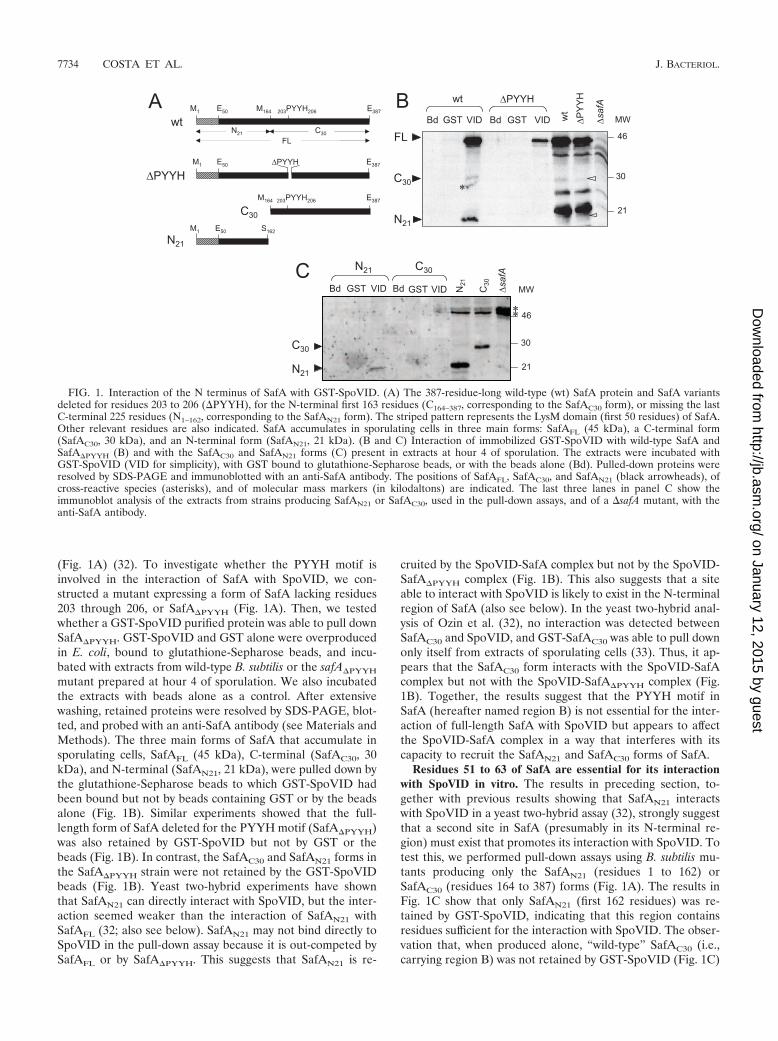
The PYYH motif is involved in formation of the SafA-SpoVID complex. The PYYH motif, found in a phage displayscreen for peptides able to interact with SpoVID, is present inthe C-terminal half of the SafA protein (residues 203 to 206)
(Fig. 1A) (32). To investigate whether the PYYH motif isinvolved in the interaction of SafA with SpoVID, we con-structed a mutant expressing a form of SafA lacking residues203 through 206, or SafA�PYYH (Fig. 1A). Then, we testedwhether a GST-SpoVID purified protein was able to pull downSafA�PYYH. GST-SpoVID and GST alone were overproducedin E. coli, bound to glutathione-Sepharose beads, and incu-bated with extracts from wild-type B. subtilis or the safA�PYYH
mutant prepared at hour 4 of sporulation. We also incubatedthe extracts with beads alone as a control. After extensivewashing, retained proteins were resolved by SDS-PAGE, blot-ted, and probed with an anti-SafA antibody (see Materials andMethods). The three main forms of SafA that accumulate insporulating cells, SafAFL (45 kDa), C-terminal (SafAC30, 30kDa), and N-terminal (SafAN21, 21 kDa), were pulled down bythe glutathione-Sepharose beads to which GST-SpoVID hadbeen bound but not by beads containing GST or by the beadsalone (Fig. 1B). Similar experiments showed that the full-length form of SafA deleted for the PYYH motif (SafA�PYYH)was also retained by GST-SpoVID but not by GST or thebeads (Fig. 1B). In contrast, the SafAC30 and SafAN21 forms inthe SafA�PYYH strain were not retained by the GST-SpoVIDbeads (Fig. 1B). Yeast two-hybrid experiments have shownthat SafAN21 can directly interact with SpoVID, but the inter-action seemed weaker than the interaction of SafAN21 withSafAFL (32; also see below). SafAN21 may not bind directly toSpoVID in the pull-down assay because it is out-competed bySafAFL or by SafA�PYYH. This suggests that SafAN21 is re-
cruited by the SpoVID-SafA complex but not by the SpoVID-SafA�PYYH complex (Fig. 1B). This also suggests that a siteable to interact with SpoVID is likely to exist in the N-terminalregion of SafA (also see below). In the yeast two-hybrid anal-ysis of Ozin et al. (32), no interaction was detected betweenSafAC30 and SpoVID, and GST-SafAC30 was able to pull downonly itself from extracts of sporulating cells (33). Thus, it ap-pears that the SafAC30 form interacts with the SpoVID-SafAcomplex but not with the SpoVID-SafA�PYYH complex (Fig.1B). Together, the results suggest that the PYYH motif inSafA (hereafter named region B) is not essential for the inter-action of full-length SafA with SpoVID but appears to affectthe SpoVID-SafA complex in a way that interferes with itscapacity to recruit the SafAN21 and SafAC30 forms of SafA.
Residues 51 to 63 of SafA are essential for its interactionwith SpoVID in vitro. The results in preceding section, to-gether with previous results showing that SafAN21 interactswith SpoVID in a yeast two-hybrid assay (32), strongly suggestthat a second site in SafA (presumably in its N-terminal re-gion) must exist that promotes its interaction with SpoVID. Totest this, we performed pull-down assays using B. subtilis mu-tants producing only the SafAN21 (residues 1 to 162) orSafAC30 (residues 164 to 387) forms (Fig. 1A). The results inFig. 1C show that only SafAN21 (first 162 residues) was re-tained by GST-SpoVID, indicating that this region containsresidues sufficient for the interaction with SpoVID. The obser-vation that, when produced alone, “wild-type” SafAC30 (i.e.,carrying region B) was not retained by GST-SpoVID (Fig. 1C)
FIG. 1. Interaction of the N terminus of SafA with GST-SpoVID. (A) The 387-residue-long wild-type (wt) SafA protein and SafA variantsdeleted for residues 203 to 206 (�PYYH), for the N-terminal first 163 residues (C164–387, corresponding to the SafAC30 form), or missing the lastC-terminal 225 residues (N1–162, corresponding to the SafAN21 form). The striped pattern represents the LysM domain (first 50 residues) of SafA.Other relevant residues are also indicated. SafA accumulates in sporulating cells in three main forms: SafAFL (45 kDa), a C-terminal form(SafAC30, 30 kDa), and an N-terminal form (SafAN21, 21 kDa). (B and C) Interaction of immobilized GST-SpoVID with wild-type SafA andSafA�PYYH (B) and with the SafAC30 and SafAN21 forms (C) present in extracts at hour 4 of sporulation. The extracts were incubated withGST-SpoVID (VID for simplicity), with GST bound to glutathione-Sepharose beads, or with the beads alone (Bd). Pulled-down proteins wereresolved by SDS-PAGE and immunoblotted with an anti-SafA antibody. The positions of SafAFL, SafAC30, and SafAN21 (black arrowheads), ofcross-reactive species (asterisks), and of molecular mass markers (in kilodaltons) are indicated. The last three lanes in panel C show theimmunoblot analysis of the extracts from strains producing SafAN21 or SafAC30, used in the pull-down assays, and of a �safA mutant, with theanti-SafA antibody.
is consistent with results of previous studies in which SafAC30
and SpoVID did not interact (32, 33).To determine whether a specific region within the first 162
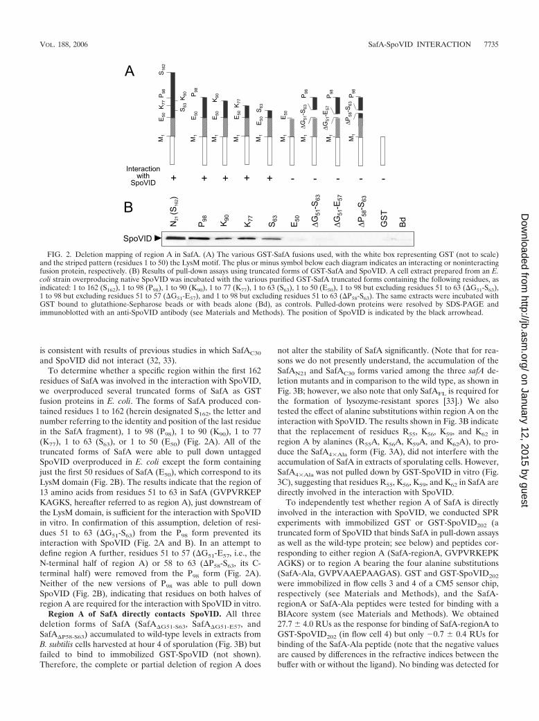
residues of SafA was involved in the interaction with SpoVID,we overproduced several truncated forms of SafA as GSTfusion proteins in E. coli. The forms of SafA produced con-tained residues 1 to 162 (herein designated S162, the letter andnumber referring to the identity and position of the last residuein the SafA fragment), 1 to 98 (P98), 1 to 90 (K90), 1 to 77(K77), 1 to 63 (S63), or 1 to 50 (E50) (Fig. 2A). All of thetruncated forms of SafA were able to pull down untaggedSpoVID overproduced in E. coli except the form containingjust the first 50 residues of SafA (E50), which correspond to itsLysM domain (Fig. 2B). The results indicate that the region of13 amino acids from residues 51 to 63 in SafA (GVPVRKEPKAGKS, hereafter referred to as region A), just downstream ofthe LysM domain, is sufficient for the interaction with SpoVIDin vitro. In confirmation of this assumption, deletion of resi-dues 51 to 63 (�G51-S63) from the P98 form prevented itsinteraction with SpoVID (Fig. 2A and B). In an attempt todefine region A further, residues 51 to 57 (�G51-E57, i.e., theN-terminal half of region A) or 58 to 63 (�P58-S63, its C-terminal half) were removed from the P98 form (Fig. 2A).Neither of the new versions of P98 was able to pull downSpoVID (Fig. 2B), indicating that residues on both halves ofregion A are required for the interaction with SpoVID in vitro.
Region A of SafA directly contacts SpoVID. All threedeletion forms of SafA (SafA�G51-S63, SafA�G51-E57, andSafA�P58-S63) accumulated to wild-type levels in extracts fromB. subtilis cells harvested at hour 4 of sporulation (Fig. 3B) butfailed to bind to immobilized GST-SpoVID (not shown).Therefore, the complete or partial deletion of region A does
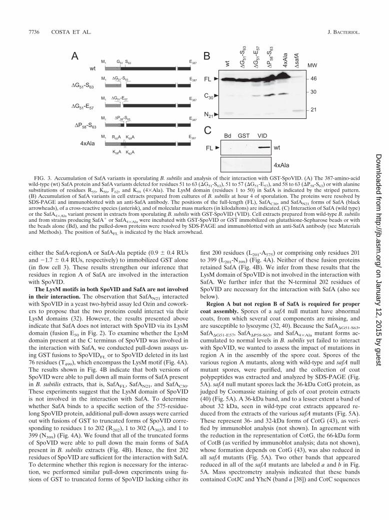
not alter the stability of SafA significantly. (Note that for rea-sons we do not presently understand, the accumulation of theSafAN21 and SafAC30 forms varied among the three safA de-letion mutants and in comparison to the wild type, as shown inFig. 3B; however, we also note that only SafAFL is required forthe formation of lysozyme-resistant spores [33].) We alsotested the effect of alanine substitutions within region A on theinteraction with SpoVID. The results shown in Fig. 3B indicatethat the replacement of residues R55, K56, K59, and K62 inregion A by alanines (R55A, K56A, K59A, and K62A), to pro-duce the SafA4�Ala form (Fig. 3A), did not interfere with theaccumulation of SafA in extracts of sporulating cells. However,SafA4�Ala was not pulled down by GST-SpoVID in vitro (Fig.3C), suggesting that residues R55, K56, K59, and K62 in SafA aredirectly involved in the interaction with SpoVID.
To independently test whether region A of SafA is directlyinvolved in the interaction with SpoVID, we conducted SPRexperiments with immobilized GST or GST-SpoVID202 (atruncated form of SpoVID that binds SafA in pull-down assaysas well as the wild-type protein; see below) and peptides cor-responding to either region A (SafA-regionA, GVPVRKEPKAGKS) or to region A bearing the four alanine substitutions(SafA-Ala, GVPVAAEPAAGAS). GST and GST-SpoVID202
were immobilized in flow cells 3 and 4 of a CM5 sensor chip,respectively (see Materials and Methods), and the SafA-regionA or SafA-Ala peptides were tested for binding with aBIAcore system (see Materials and Methods). We obtained27.7 4.0 RUs as the response for binding of SafA-regionA toGST-SpoVID202 (in flow cell 4) but only �0.7 0.4 RUs forbinding of the SafA-Ala peptide (note that the negative valuesare caused by differences in the refractive indices between thebuffer with or without the ligand). No binding was detected for
FIG. 2. Deletion mapping of region A in SafA. (A) The various GST-SafA fusions used, with the white box representing GST (not to scale)and the striped pattern (residues 1 to 50) the LysM motif. The plus or minus symbol below each diagram indicates an interacting or noninteractingfusion protein, respectively. (B) Results of pull-down assays using truncated forms of GST-SafA and SpoVID. A cell extract prepared from an E.coli strain overproducing native SpoVID was incubated with the various purified GST-SafA truncated forms containing the following residues, asindicated: 1 to 162 (S162), 1 to 98 (P98), 1 to 90 (K90), 1 to 77 (K77), 1 to 63 (S63), 1 to 50 (E50), 1 to 98 but excluding residues 51 to 63 (�G51-S63),1 to 98 but excluding residues 51 to 57 (�G51-E57), and 1 to 98 but excluding residues 51 to 63 (�P58-S63). The same extracts were incubated withGST bound to glutathione-Sepharose beads or with beads alone (Bd), as controls. Pulled-down proteins were resolved by SDS-PAGE andimmunoblotted with an anti-SpoVID antibody (see Materials and Methods). The position of SpoVID is indicated by the black arrowhead.
either the SafA-regionA or SafA-Ala peptide (0.9 0.4 RUsand �1.7 0.4 RUs, respectively) to immobilized GST alone(in flow cell 3). These results strengthen our inference thatresidues in region A of SafA are involved in the interactionwith SpoVID.
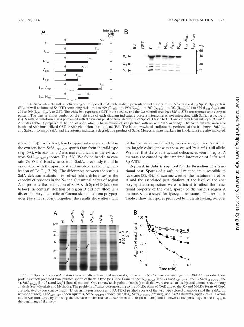
The LysM motifs in both SpoVID and SafA are not involvedin their interaction. The observation that SafAN21 interactedwith SpoVID in a yeast two-hybrid assay led Ozin and cowork-ers to propose that the two proteins could interact via theirLysM domains (32). However, the results presented aboveindicate that SafA does not interact with SpoVID via its LysMdomain (fusion E50 in Fig. 2). To examine whether the LysMdomain present at the C terminus of SpoVID was involved inthe interaction with SafA, we conducted pull-down assays us-ing GST fusions to SpoVIDFL or to SpoVID deleted in its last76 residues (T499), which encompass the LysM motif (Fig. 4A).The results shown in Fig. 4B indicate that both versions ofSpoVID were able to pull down all main forms of SafA presentin B. subtilis extracts, that is, SafAFL, SafAN21, and SafAC30.These experiments suggest that the LysM domain of SpoVIDis not involved in the interaction with SafA. To determinewhether SafA binds to a specific section of the 575-residue-long SpoVID protein, additional pull-down assays were carriedout with fusions of GST to truncated forms of SpoVID corre-sponding to residues 1 to 202 (R202), 1 to 302 (A302), and 1 to399 (N399) (Fig. 4A). We found that all of the truncated formsof SpoVID were able to pull down the main forms of SafApresent in B. subtilis extracts (Fig. 4B). Hence, the first 202residues of SpoVID are sufficient for the interaction with SafA.To determine whether this region is necessary for the interac-tion, we performed similar pull-down experiments using fu-sions of GST to truncated forms of SpoVID lacking either its
first 200 residues (L201-A575) or comprising only residues 201to 399 (L201-N399) (Fig. 4A). Neither of these fusion proteinsretained SafA (Fig. 4B). We infer from these results that theLysM domain of SpoVID is not involved in the interaction withSafA. We further infer that the N-terminal 202 residues ofSpoVID are necessary for the interaction with SafA (also seebelow).
Region A but not region B of SafA is required for propercoat assembly. Spores of a safA null mutant have abnormalcoats, from which several coat components are missing, andare susceptible to lysozyme (32, 40). Because the SafA�G51-S63,SafA�G51-E57, SafA�P58-S63, and SafA4�Ala mutant forms ac-cumulated to normal levels in B. subtilis yet failed to interactwith SpoVID, we wanted to assess the impact of mutations inregion A in the assembly of the spore coat. Spores of thevarious region A mutants, along with wild-type and safA nullmutant spores, were purified, and the collection of coatpolypeptides was extracted and analyzed by SDS-PAGE (Fig.5A). safA null mutant spores lack the 36-kDa CotG protein, asjudged by Coomassie staining of gels of coat protein extracts(40) (Fig. 5A). A 36-kDa band, and to a lesser extent a band ofabout 32 kDa, seen in wild-type coat extracts appeared re-duced from the extracts of the various safA mutants (Fig. 5A).These represent 36- and 32-kDa forms of CotG (43), as veri-fied by immunoblot analysis (not shown). In agreement withthe reduction in the representation of CotG, the 66-kDa formof CotB (as verified by immunoblot analysis; data not shown),whose formation depends on CotG (43), was also reduced inall safA mutants (Fig. 5A). Two other bands that appearedreduced in all of the safA mutants are labeled a and b in Fig.5A. Mass spectrometry analysis indicated that these bandscontained CotJC and YhcN (band a [38]) and CotC sequences
FIG. 3. Accumulation of SafA variants in sporulating B. subtilis and analysis of their interaction with GST-SpoVID. (A) The 387-amino-acidwild-type (wt) SafA protein and SafA variants deleted for residues 51 to 63 (�G51-S63), 51 to 57 (�G51-E57), and 58 to 63 (�P58-S63) or with alaninesubstitutions of residues R55, K56, F62, and K59 (4�Ala). The LysM domain (residues 1 to 50) in SafA is indicated by the striped pattern.(B) Accumulation of SafA variants in cell extracts prepared from cultures of B. subtilis at hour 4 of sporulation. The proteins were resolved bySDS-PAGE and immunoblotted with an anti-SafA antibody. The positions of the full-length (FL), SafAC30, and SafAN21 forms of SafA (blackarrowheads), of a cross-reactive species (asterisk), and of molecular mass markers (in kilodaltons) are indicated. (C) Interaction of SafA (wild type)or the SafA4�Ala variant present in extracts from sporulating B. subtilis with GST-SpoVID (VID). Cell extracts prepared from wild-type B. subtilisand from strains producing SafA� or SafA4�Ala were incubated with GST-SpoVID or GST immobilized on glutathione-Sepharose beads or withthe beads alone (Bd), and the pulled-down proteins were resolved by SDS-PAGE and immunoblotted with an anti-SafA antibody (see Materialsand Methods). The position of SafAFL is indicated by the black arrowhead.
(band b [10]). In contrast, band c appeared more abundant inthe extracts from SafA�G51-S63 spores than from the wild type(Fig. 5A), whereas band d was more abundant in the extractsfrom SafA�G51-E57 spores (Fig. 5A). We found band c to con-tain GerQ and band d to contain SodA, previously found inassociation with the spore coat and involved in the oligomer-ization of CotG (17, 25). The differences between the variousSafA deletion mutants may reflect subtle differences in thecapacity of residues in the N- and C-terminal halves of regionA to promote the interaction of SafA with SpoVID (also seebelow). In contrast, deletion of region B did not affect in adiscernible way the profile of Coomassie-stained coat polypep-tides (data not shown). Together, the results show alterations
of the coat structure caused by lesions in region A of SafA thatare largely coincident with those caused by a safA null allele.We infer that the coat structural deficiencies seen in region Amutants are caused by the impaired interaction of SafA withSpoVID.
Region A in SafA is required for the formation of a func-tional coat. Spores of a safA null mutant are susceptible tolysozyme (32, 40). To examine whether the mutations in regionA and the associated perturbations at the level of the coatpolypeptide composition were sufficient to affect this func-tional property of the coat, spores of the various region Amutants were assayed for lysozyme resistance. The results inTable 2 show that spores produced by mutants lacking residues
∗
FIG. 4. SafA interacts with a defined region of SpoVID. (A) Schematic representation of fusions of the 575-residue-long SpoVIDFL protein(FL), as well as forms of SpoVID containing residues 1 to 499 (T499), 1 to 399 (N399), 1 to 302 (A302), 1 to 202 (R201), 201 to 575 (L201-A575), and201 to 399 (L201- N399), to GST. The white box represents GST (not to scale), and the LysM motif (residues 525 to 575) corresponds to the stripedpattern. The plus or minus symbol on the right side of each diagram indicates a protein interacting or not interacting with SafA, respectively.(B) Results of pull-down assays performed with the various purified truncated forms of SpoVID fused to GST and extracts from wild-type B. subtilisAOB90 (Table 1) prepared at hour 4 of sporulation. The immunoblot was probed with an anti-SafA antibody. The same extracts were alsoincubated with immobilized GST or with glutathione beads alone (Bd). The black arrowheads indicate the positions of the full-length, SafAC30,and SafAN21 forms of SafA, and the asterisk indicates a degradation product of SafA. Molecular mass markers (in kilodaltons) are also indicated.
a
b
cd
FIG. 5. Spores of region A mutants have an altered coat and impaired germination. (A) Coomassie-stained gel of SDS-PAGE-resolved coatprotein extracts prepared from purified spores of the wild type (wt) (lane 1) and the SafA�G51-S63 (lane 2), SafA�G51-E57 (lane 3), SafA�P58-S63 (lane4), SafA4�Ala (lane 5), and �safA (lane 6) mutants. Open arrowheads point to bands (a to d) that were excised and subjected to mass spectrometryanalysis (see Materials and Methods). The positions of bands corresponding to the 66-kDa form of CotB and to the 32- and 36-kDa forms of CotGare indicated by black arrowheads. (B) Germination responses to AGFK of purified spores of the wild type (closed diamonds) and the SafA4�Ala(closed squares), SafA�G51-S63 (open squares), SafA�G51-E57 (closed triangles), SafA�P58-S63 (crosses), and �safA mutants (open circles). Germi-nation was monitored by following the decrease in absorbance at 580 nm over time (in minutes) and is shown as the percentage of the OD580 atthe beginning of the assay.
51 to 63 (region A), 51 to 57, or 58 to 63 of SafA have a degreeof susceptibility to lysozyme treatment in the range of thatobserved for spores of a safA null mutant (about 107 lysozyme-resistant spores/ml of culture, compared to 108 for wild-typespores). None of the mutations in region A affected spore heatresistance (Table 2), a property that relies on proper cortexformation, and hence the effects herein described are specifi-cally related to the assembly of the spore coat. We have alsofound that spores of the SafA�PYYH mutant are fully resistantto lysozyme (Table 2), an observation that is in agreement withabsence of any discernible alteration in the profile of Coomas-sie-stained coat polypeptides (see above). This reinforces thesuggestion that the PYYH motif is not a critical determinantfor the SafA-SpoVID interaction (see above). Surprisinglyhowever, spores of the SafA4�Ala mutant were also found to beresistant to lysozyme (Table 2). Presumably, the alanine sub-stitutions within region A still permit some degree of interac-tion of SafA with SpoVID in vivo, sufficient to support sporelysozyme resistance. In support of this interpretation, we foundthat when examined for their germination in response to amixture of asparagine, glucose, fructose, and KCl (AGFK), aproperty that is dependent on safA (40), spores of theSafA4�Ala mutant showed an intermediate phenotype betweenthe wild type and the safA null mutant (Fig. 5B). In contrast,the results in Fig. 5B show that spores of the SafA�G51-E57 andSafA�G51-S63 mutants were as impaired in AGFK-induced ger-mination as were spores of the safA null mutant. Spores of theSafA�P58-E63 mutant however, showed a response to AGFKthat closely paralleled that of the SafA4�Ala mutant (Fig. 5B),suggesting that residues in the N-terminal half of region Ahave a greater impact on the SafA-SpoVID interaction, at leastas assessed by this spore property. Overall, the finding thatmutations in region A led to a germination phenotype thatlargely mimics that of a safA null mutant strengthens the viewthat the function of SafA critically depends on its interactionwith SpoVID.
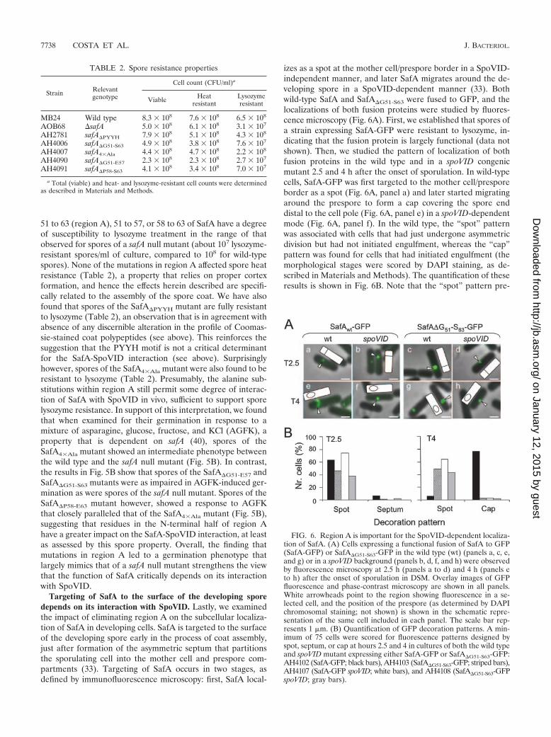
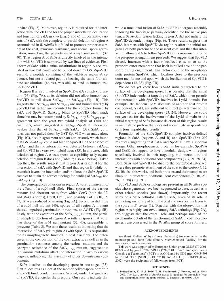
Targeting of SafA to the surface of the developing sporedepends on its interaction with SpoVID. Lastly, we examinedthe impact of eliminating region A on the subcellular localiza-tion of SafA in developing cells. SafA is targeted to the surfaceof the developing spore early in the process of coat assembly,just after formation of the asymmetric septum that partitionsthe sporulating cell into the mother cell and prespore com-partments (33). Targeting of SafA occurs in two stages, asdefined by immunofluorescence microscopy: first, SafA local-
izes as a spot at the mother cell/prespore border in a SpoVID-independent manner, and later SafA migrates around the de-veloping spore in a SpoVID-dependent manner (33). Bothwild-type SafA and SafA�G51-S63 were fused to GFP, and thelocalizations of both fusion proteins were studied by fluores-cence microscopy (Fig. 6A). First, we established that spores ofa strain expressing SafA-GFP were resistant to lysozyme, in-dicating that the fusion protein is largely functional (data notshown). Then, we studied the pattern of localization of bothfusion proteins in the wild type and in a spoVID congenicmutant 2.5 and 4 h after the onset of sporulation. In wild-typecells, SafA-GFP was first targeted to the mother cell/presporeborder as a spot (Fig. 6A, panel a) and later started migratingaround the prespore to form a cap covering the spore enddistal to the cell pole (Fig. 6A, panel e) in a spoVID-dependentmode (Fig. 6A, panel f). In the wild type, the “spot” patternwas associated with cells that had just undergone asymmetricdivision but had not initiated engulfment, whereas the “cap”pattern was found for cells that had initiated engulfment (themorphological stages were scored by DAPI staining, as de-scribed in Materials and Methods). The quantification of theseresults is shown in Fig. 6B. Note that the “spot” pattern pre-
FIG. 6. Region A is important for the SpoVID-dependent localiza-tion of SafA. (A) Cells expressing a functional fusion of SafA to GFP(SafA-GFP) or SafA�G51-S63-GFP in the wild type (wt) (panels a, c, e,and g) or in a spoVID background (panels b, d, f, and h) were observedby fluorescence microscopy at 2.5 h (panels a to d) and 4 h (panels eto h) after the onset of sporulation in DSM. Overlay images of GFPfluorescence and phase-contrast microscopy are shown in all panels.White arrowheads point to the region showing fluorescence in a se-lected cell, and the position of the prespore (as determined by DAPIchromosomal staining; not shown) is shown in the schematic repre-sentation of the same cell included in each panel. The scale bar rep-resents 1 �m. (B) Quantification of GFP decoration patterns. A min-imum of 75 cells were scored for fluorescence patterns designed byspot, septum, or cap at hours 2.5 and 4 in cultures of both the wild typeand spoVID mutant expressing either SafA-GFP or SafA�G51-S63-GFP:AH4102 (SafA-GFP; black bars), AH4103 (SafA�G51-S63-GFP; striped bars),AH4107 (SafA-GFP spoVID; white bars), and AH4108 (SafA�G51-S63-GFPspoVID; gray bars).
TABLE 2. Spore resistance properties
Strain Relevantgenotype
Cell count (CFU/ml)a
Viable Heatresistant
Lysozymeresistant
MB24 Wild type 8.3 � 108 7.6 � 108 6.5 � 108
AOB68 �safA 5.0 � 108 6.1 � 108 3.1 � 107
AH2781 safA�PYYH 7.9 � 108 5.1 � 108 4.3 � 108
AH4006 safA�G51-S63 4.9 � 108 3.8 � 108 7.6 � 107
AH4007 safA4�Ala 4.4 � 108 4.7 � 108 2.2 � 108
AH4090 safA�G51-E57 2.3 � 108 2.3 � 108 2.7 � 107
AH4091 safA�P58-S63 4.1 � 108 3.4 � 108 7.0 � 107
a Total (viable) and heat- and lysozyme-resistant cell counts were determinedas described in Materials and Methods.
dominated in samples at hour 2.5 of sporulation and was notaffected by mutation of spoVID, whereas accumulation of the“cap” pattern, which predominated at hour 4, required expres-sion of spoVID (Fig. 6B). These results agree with those ofOzin and coworkers (33), except that in their experiments SafAwas found as caps covering both spore ends. However, thesignal at the pole-proximal spore end was always weaker thanthe signal at the spore end away from the cell pole (33). Weemphasize that our SafA-GFP fusion is functional, and wepresume that the fluorescence at the pole-proximal spore endis not detected under our conditions. In any event, a strikinglydifferent pattern was found for the SafA�G51-S63-GFP fusionprotein. SafA�G51-S63-GFP was still targeted at an early time(hour 2.5) to the asymmetric septum, where it accumulated asa spot (Fig. 6A, panels c and d). However, for this protein theincidence of the “spot” pattern persisted at hour 4, at theexpense of the “cap” pattern, whose representation was severelycurtailed regardless of the presence of a functional spoVIDlocus (Fig. 6A, panels g and h, and Fig. 6B). We conclude
that deletion of region A in SafA strongly interferes with theSpoVID-dependent stage of SafA localization, and we inferthat the interaction of SafA with SpoVID via region A isrequired for the correct localization of SafA to the devel-oping coat.
DISCUSSION
Previous work has suggested that the PYYH motif (regionB) in the C-terminal half of SafA, found in a phage displayscreen for peptides able to interact with SpoVID, could medi-ate the interaction of SafA with SpoVID in vivo (32, 33) (Fig.7A). However, a second region of interaction was postulated toexist in the N-terminal half of SafA (33). We now show thatsuch a region indeed exists in the N-terminal half of SafA (Fig.7A). SafAN21 (but not SafAC30) could be pulled out by immo-bilized GST-SpoVID, and deletion mapping indicated that aregion of 13 amino acids encompassing residues 51 to 63 ofSafA, or region A, is sufficient for the interaction with SpoVID
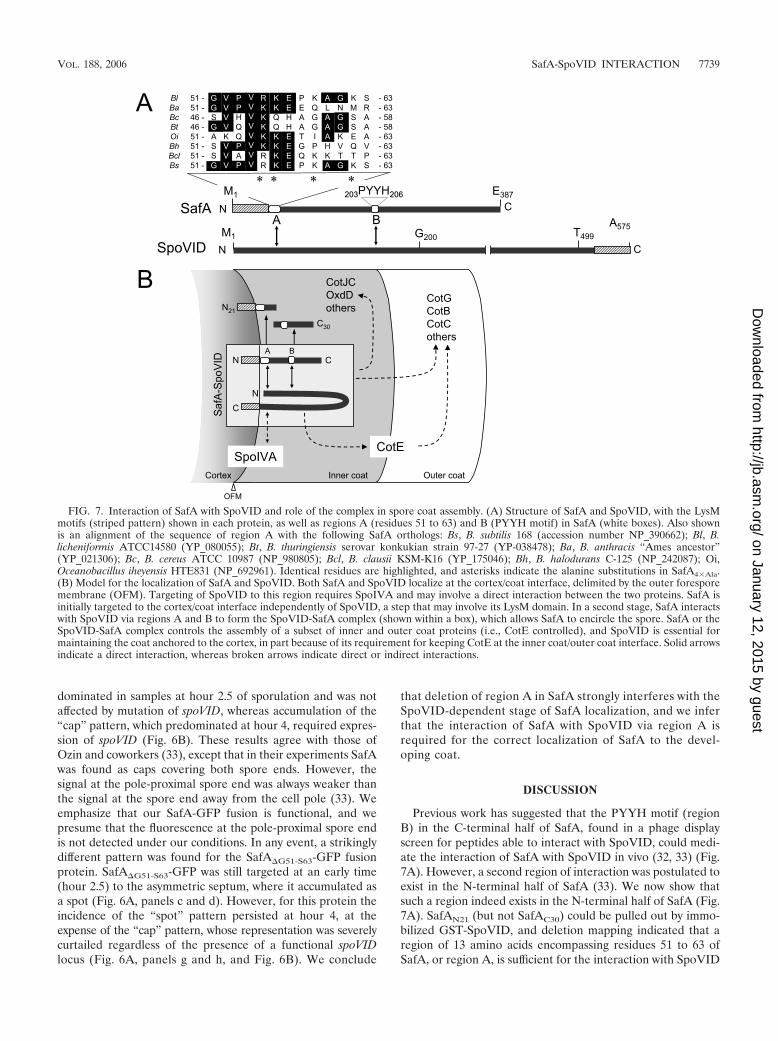
FIG. 7. Interaction of SafA with SpoVID and role of the complex in spore coat assembly. (A) Structure of SafA and SpoVID, with the LysMmotifs (striped pattern) shown in each protein, as well as regions A (residues 51 to 63) and B (PYYH motif) in SafA (white boxes). Also shownis an alignment of the sequence of region A with the following SafA orthologs: Bs, B. subtilis 168 (accession number NP_390662); Bl, B.licheniformis ATCC14580 (YP_080055); Bt, B. thuringiensis serovar konkukian strain 97-27 (YP-038478); Ba, B. anthracis “Ames ancestor”(YP_021306); Bc, B. cereus ATCC 10987 (NP_980805); Bcl, B. clausii KSM-K16 (YP_175046); Bh, B. halodurans C-125 (NP_242087); Oi,Oceanobacillus iheyensis HTE831 (NP_692961). Identical residues are highlighted, and asterisks indicate the alanine substitutions in SafA4�Ala.(B) Model for the localization of SafA and SpoVID. Both SafA and SpoVID localize at the cortex/coat interface, delimited by the outer foresporemembrane (OFM). Targeting of SpoVID to this region requires SpoIVA and may involve a direct interaction between the two proteins. SafA isinitially targeted to the cortex/coat interface independently of SpoVID, a step that may involve its LysM domain. In a second stage, SafA interactswith SpoVID via regions A and B to form the SpoVID-SafA complex (shown within a box), which allows SafA to encircle the spore. SafA or theSpoVID-SafA complex controls the assembly of a subset of inner and outer coat proteins (i.e., CotE controlled), and SpoVID is essential formaintaining the coat anchored to the cortex, in part because of its requirement for keeping CotE at the inner coat/outer coat interface. Solid arrowsindicate a direct interaction, whereas broken arrows indicate direct or indirect interactions.
in vitro (Fig. 2). Moreover, region A is required for the inter-action with SpoVID and for the proper subcellular localizationand function of SafA in vivo (Fig. 5 and 6). Importantly, vari-ants of SafA with the complete or partial deletion of region Aaccumulated in B. subtilis but failed to promote proper assem-bly of the coat, lysozyme resistance, and normal spore germi-nation, mimicking the phenotypes of a safA null mutant (32,40). That region A of SafA is directly involved in the interac-tion with SpoVID is supported by two lines of evidence. First,a form of SafA with alanine substitutions in region A accumu-lated in vivo but could not be pulled down by GST-SpoVID.Second, a peptide consisting of the wild-type region A se-quence, but not a related peptide bearing the same four ala-nine substitutions (above), was able to bind to immobilizedGST-SpoVID.
Region B is also involved in SpoVID-SafA complex forma-tion (33) (Fig. 7A), as its deletion did not allow immobilizedSpoVID to pull down SafAN21 or SafAC30 (Fig. 1B). Thissuggests that SafAN21 and SafAC30 are not bound directly bySpoVID but rather are recruited by the complex formed bySafA and SpoVID. SafAN21 binds to SpoVID when madealone but may be outcompeted by SafAFL or by SafA�PYYH, inagreement with the yeast two-hybrid analysis of Ozin andcoauthors, which suggested a SafAN21-SpoVID interactionweaker than that of SafAN21 with SafAFL (33). SafAC30, inturn, was not pulled down by GST-SpoVID when made alone(Fig. 1C), also in agreement with an earlier study which foundthat GST-SafAC30 could not bind to SpoVID in the absence ofSafAFL and that no interaction was detected between SafAC30
and SpoVID in a yeast two-hybrid assay (33). In any case, whiledeletion of region A causes spore susceptibility to lysozyme,deletion of region B does not (Table 2; also see below). Takentogether, the results suggest that region A is essential for theinteraction of SafA with SpoVID and that region B (while notessential) favors the interaction and/or allows the SafA-SpoVIDcomplex to attain the correct topology for binding of SafAN21 andSafAC30 (Fig. 7B).
The consequences of lesions in region A were reminiscent ofthe effects of a safA null allele. First, spores of the variousmutants had aberrant coats, from which CotG (both the 32-and 36-kDa forms), CotB, CotC, and possibly CotJC (10, 15,37, 38) were reduced or missing (Fig. 5A). Second, as did thoseof a safA null mutant (40), spores of all region A mutantsshowed impaired germination in response to AGFK (Fig. 5B).Lastly, with the exception of the SafA4�Ala mutant, the partialor complete deletion of region A results in spores that were,like those of the safA null mutant (32, 40), susceptible tolysozyme (Table 2). We take these results as indicating that theinteraction of SafA (via region A) with SpoVID is responsiblefor its morphogenetic function in coat assembly. The differ-ences in the composition of the coat extracts, as well as in thegermination responses among the various mutants and thelysozyme resistance of the SafA4�Ala mutant, suggest thatthe various mutations affect the interaction in vivo to variousdegrees, influencing the assembly of other downstream com-ponents.
SafA localizes to the developing spore in two stages (33).First it localizes as a dot at the mother cell/prespore border ina SpoVID-independent manner. Second, under the guidanceof SpoVID, it encircles the prespore. Our results indicate that
while a functional fusion of SafA to GFP undergoes assemblyfollowing the two-stage pathway described for the native pro-tein, a SafA-GFP fusion lacking region A did not initiate theSpoVID-dependent stage (Fig. 6). These results suggest thatSafA interacts with SpoVID via region A after the initial tar-geting of both proteins to the nascent coat and that this inter-action allows SafA to follow SpoVID in its movement aroundthe prespore as engulfment proceeds. We suggest that SpoVIDdirectly interacts with a factor localized close to or at theprespore outer membrane that itself is pulled around the pre-spore during engulfment. This factor could be the morphoge-netic protein SpoIVA, which localizes close to the presporeouter membrane and upon which the localization of SpoVID isdependent (12, 33) (Fig. 7B).
We do not yet know how is SafA initially targeted to thesurface of the developing spore. It is possible that the initial(SpoVID-independent) targeting of SafA, which then permitsthe interaction with SpoVID, involves its LysM domain. Forexample, the tandem LysM domains of another coat proteincomponent, YaaH, are sufficient to direct -lactamase to thesurface of the developing spore (23, 24). However, we couldnot yet test for the involvement of the LysM domain in theinitial targeting of SafA because deletion of this region resultsin an unstable protein that does not accumulate in sporulatingcells (our unpublished results).
Formation of the SafA-SpoVID complex involves definedregions in SafA (regions A and B) and SpoVID (first 202residues), suggesting that SafA and SpoVID have a modulardesign. Other morphogenetic proteins, for example, SpoIVAand CotE, also appear to have a modular design, with regionsspecifically involved in their targeting, multimerization, andinteractions with additional coat components (3, 7, 21, 28, 34).Both SafA and SpoVID localize to the cortex/coat interface,where they promote attachment of the coat to the cortex (4, 12,32, 40; also this work), and both proteins and their complex arelikely to interact with additional coat components (6, 10, 23–26, 33, 38) (Fig. 7B).
SpoVID and SafA orthologs are present in all Bacillus spe-cies whose genomes have been sequenced to date, as well as inother related species (not shown). Importantly, the recentstudy of a SafA ortholog, called ExsA, revealed its role inpromoting anchoring of both the coat and exosporium layers tothe spore in B. cereus (1). Together with the observation thatregion A is highly conserved among SafA orthologs (Fig. 7A),this suggests that the overall role and perhaps some of themechanistic details of the functioning of SafA in coat morpho-genesis are conserved in the Bacillus group of spore formers.
ACKNOWLEDGMENTS
We thank Melissa Wilby (Emory University) for comments on themanuscript and John Pohl (Emory Microchemical Facility) for themass spectrometry analysis.
This work was supported by European Union grant QLK5-CT-2001-01729 and by grant CONC-REEQ/692/2001 from “Fundacao para aCiencia e a Tecnologia” (FCT) to A.O.H. and by NIH grant GM54395to C.P.M. T.C. (SFRH/BD/1167/00) and A.L.I. (SFRH/BPD/8967/2002) were the recipients of fellowships from FCT.
REFERENCES
1. Bailey-Smith, K., S. J. Todd, T. W. Southworth, J. Proctor, and A. Moir.2005. The ExsA protein of Bacillus cereus is required for assembly of coatand exosporium onto the spore surface. J. Bacteriol. 187:3800–3806.
2. Bateman, A., and M. Bycroft. 2000. The structure of a LysM domain from E.coli membrane-bound lytic murein transglycosylase D (MltD). J. Mol. Biol.299:1113–1119.
3. Bauer, T., S. Little, A. G. Stover, and A. Driks. 1999. Functional regions ofthe Bacillus subtilis spore coat morphogenetic protein CotE. J. Bacteriol.181:7043–7051.
4. Beall, B., A. Driks, R. Losick, and C. P. Moran, Jr. 1993. Cloning andcharacterization of a gene required for assembly of the Bacillus subtilis sporecoat. J. Bacteriol. 175:1705–1716.
5. Birkeland, N. K. 1994. Cloning, molecular characterization, and expressionof the genes encoding the lytic functions of lactococcal bacteriophage phiLC3: a dual lysis system of modular design. Can. J. Microbiol. 40:658–665.
6. Bourne, N., P. C. FitzJames, and A. I. Aronson. 1991. Structural and germi-nation defects of Bacillus subtilis spores with altered contents of a spore coatprotein. J. Bacteriol. 173:6618–6625.
7. Catalano, F. A., J. Meador-Parton, D. L. Popham, and A. Driks. 2001.Amino acids in the Bacillus subtilis morphogenetic protein SpoIVA withroles in spore coat and cortex formation. J. Bacteriol. 183:1645–1654.
8. Costa, T., L. Steil, L. O. Martins, U. Volker, and A. O. Henriques. 2004.Assembly of an oxalate decarboxylase produced under �K control into theBacillus subtilis spore coat. J. Bacteriol. 186:1462–1474.
9. Cutting, S. M., and P. B. V. Horn. 1990. Genetic analysis, p. 27–74. In C. R.Harwood and S. M. Cutting (ed.), Molecular biological methods for Bacillus.John Wiley & Sons Ltd., Chichester, England.
10. Donovan, W., L. B. Zheng, K. Sandman, and R. Losick. 1987. Genes encod-ing spore coat polypeptides from Bacillus subtilis. J. Mol. Biol. 196:1–10.
11. Driks, A. 1999. Bacillus subtilis spore coat. Microbiol. Mol. Biol. Rev. 63:1–20.12. Driks, A., S. Roels, B. Beall, C. P. Moran, Jr., and R. Losick. 1994. Subcel-
lular localization of proteins involved in the assembly of the spore coat ofBacillus subtilis. Genes Dev. 8:234–244.
13. Eichenberger, P., M. Fujita, S. T. Jensen, E. M. Conlon, D. Z. Rudner, S. T.Wang, C. Ferguson, K. Haga, T. Sato, J. S. Liu, and R. Losick. 2004. Theprogram of gene transcription for a single differentiating cell type duringsporulation in Bacillus subtilis. PLoS Biol. 2:e328.
14. Henriques, A. O., B. W. Beall, and C. P. Moran, Jr. 1997. CotM of Bacillussubtilis, a member of the alpha-crystallin family of stress proteins, is inducedduring development and participates in spore outer coat formation. J. Bac-teriol. 179:1887–1897.
15. Henriques, A. O., B. W. Beall, K. Roland, and C. P. Moran, Jr. 1995.Characterization of cotJ, a �E-controlled operon affecting the polypeptidecomposition of the coat of Bacillus subtilis spores. J. Bacteriol. 177:3394–3406.
16. Henriques, A. O., T. Costa, L. O. Martins, and R. Zilhao. 2004. Functionalarchitecture and assembly of the spore coat, p. 34–52. In E. Ricca, A. O.Henriques, and S. M. Cutting (ed.), Bacterial spores: probiotics and emerg-ing applications. Horizon Scientific Press, London, United Kingdom.
17. Henriques, A. O., L. R. Melsen, and C. P. Moran, Jr. 1998. Involvement ofsuperoxide dismutase in spore coat assembly in Bacillus subtilis. J. Bacteriol.180:2285–2291.
18. Henriques, A. O., and C. P. Moran, Jr. 2000. Structure and assembly of thebacterial endospore coat. Methods 20:95–110.
19. Itaya, M., K. Kondo, and T. Tanaka. 1989. A neomycin resistance genecassette selectable in a single copy state in the Bacillus subtilis chromosome.Nucleic Acids Res. 17:4410.
20. Joris, B., S. Englebert, C. P. Chu, R. Kariyama, L. Daneo-Moore, G. D.Shockman, and J. M. Ghuysen. 1992. Modular design of the Enterococcushirae muramidase-2 and Streptococcus faecalis autolysin. FEMS Microbiol.Lett. 70:257–264.
21. Kim, H., M. Hahn, P. Grabowski, D. C. McPherson, M. M. Otte, R. Wang,C. C. Ferguson, P. Eichenberger, and A. Driks. 2006. The Bacillus subtilisspore coat protein interaction network. Mol. Microbiol. 59:487–502.
22. Klobutcher, L. A., K. Ragkousi, and P. Setlow. 2006. The Bacillus subtilisspore coat provides “eat resistance” during phagocytic predation by theprotozoan Tetrahymena thermophila. Proc. Natl. Acad. Sci. USA 103:165–170.
23. Kodama, T., H. Takamatsu, K. Asai, K. Kobayashi, N. Ogasawara, and K.
Watabe. 1999. The Bacillus subtilis yaaH gene is transcribed by SigE RNApolymerase during sporulation, and its product is involved in germination ofspores. J. Bacteriol. 181:4584–4591.
24. Kodama, T., H. Takamatsu, K. Asai, N. Ogasawara, Y. Sadaie, and K.Watabe. 2000. Synthesis and characterization of the spore proteins of Bacil-lus subtilis, YdhD, YkuD, and YkvP, which carry a motif conserved amongcell wall binding proteins. J. Biochem. (Tokyo) 128:655–663.
25. Kuwana, R., Y. Kasahara, M. Fujibayashi, H. Takamatsu, N. Ogasawara,and K. Watabe. 2002. Proteomics characterization of novel spore proteins ofBacillus subtilis. Microbiology 148:3971–3982.
26. Lai, E. M., N. D. Phadke, M. T. Kachman, R. Giorno, S. Vazquez, J. A.Vazquez, J. R. Maddock, and A. Driks. 2003. Proteomic analysis of the sporecoats of Bacillus subtilis and Bacillus anthracis. J. Bacteriol. 185:1443–1454.
27. Limpens, E., C. Franken, P. Smit, J. Willemse, T. Bisseling, and R. Geurts.2003. LysM domain receptor kinases regulating rhizobial Nod factor-inducedinfection. Science 302:630–633.
28. Little, S., and A. Driks. 2001. Functional analysis of the Bacillus subtilismorphogenetic spore coat protein CotE. Mol. Microbiol. 42:1107–1120.
29. McPherson, D. C., H. Kim, M. Hahn, R. Wang, P. Grabowski, P. Eichen-berger, and A. Driks. 2005. Characterization of the Bacillus subtilis sporemorphogenetic coat protein CotO. J. Bacteriol. 187:8278–8290.
30. Nicholson, W. L., and P. Setlow. 1990. Sporulation, germination and out-growth, p. 391–450. In C. R. Harwood and S. M. Cutting (ed.), Molecularbiology methods for Bacillus. John Wiley & Sons Ltd., Chichester, England.
31. Ozin, A. J., T. Costa, A. O. Henriques, and C. P. Moran, Jr. 2001. Alternativetranslation initiation produces a short form of a spore coat protein in Bacillussubtilis. J. Bacteriol. 183:2032–2040.
32. Ozin, A. J., A. O. Henriques, H. Yi, and C. P. Moran, Jr. 2000. Morphoge-netic proteins SpoVID and SafA form a complex during assembly of theBacillus subtilis spore coat. J. Bacteriol. 182:1828–1833.
33. Ozin, A. J., C. S. Samford, A. O. Henriques, and C. P. Moran, Jr. 2001.SpoVID guides SafA to the spore coat in Bacillus subtilis. J. Bacteriol.183:3041–3049.
34. Price, K. D., and R. Losick. 1999. A four-dimensional view of assembly of amorphogenetic protein during sporulation in Bacillus subtilis. J. Bacteriol.181:781–790.
35. Radutoiu, S., L. H. Madsen, E. B. Madsen, H. H. Felle, Y. Umehara, M.Gronlund, S. Sato, Y. Nakamura, S. Tabata, N. Sandal, and J. Stougaard.2003. Plant recognition of symbiotic bacteria requires two LysM receptor-like kinases. Nature 425:585–592.
36. Real, G., S. Autret, E. J. Harry, J. Errington, and A. O. Henriques. 2005. Celldivision protein DivIB influences the Spo0J/Soj system of chromosome seg-regation in Bacillus subtilis. Mol. Microbiol. 55:349–367.
37. Sacco, M., E. Ricca, R. Losick, and S. Cutting. 1995. An additional GerE-controlled gene encoding an abundant spore coat protein from Bacillussubtilis. J. Bacteriol. 177:372–377.
38. Seyler, R. W., Jr., A. O. Henriques, A. J. Ozin, and C. P. Moran, Jr. 1997.Assembly and interactions of cotJ-encoded proteins, constituents of the innerlayers of the Bacillus subtilis spore coat. Mol. Microbiol. 25:955–966.
39. Steen, A., G. Buist, K. J. Leenhouts, M. El Khattabi, F. Grijpstra, A. L.Zomer, G. Venema, O. P. Kuipers, and J. Kok. 2003. Cell wall attachment ofa widely distributed peptidoglycan binding domain is hindered by cell wallconstituents. J. Biol. Chem. 278:23874–23881.
40. Takamatsu, H., T. Kodama, T. Nakayama, and K. Watabe. 1999. Charac-terization of the yrbA gene of Bacillus subtilis, involved in resistance andgermination of spores. J. Bacteriol. 181:4986–4994.
41. Zheng, L. B., and R. Losick. 1990. Cascade regulation of spore coat geneexpression in Bacillus subtilis. J. Mol. Biol. 212:645–660.
42. Zilhao, R., R. Isticato, L. O. Martins, L. Steil, U. Volker, E. Ricca, C. P.Moran, Jr., and A. O. Henriques. 2005. Assembly and function of a sporecoat-associated transglutaminase of Bacillus subtilis. J. Bacteriol. 187:7753–7764.
43. Zilhao, R., M. Serrano, R. Isticato, E. Ricca, C. P. Moran, Jr., and A. O.Henriques. 2004. Interactions among CotB, CotG, and CotH during assem-bly of the Bacillus subtilis spore coat. J. Bacteriol. 186:1110–1119.