(3) Ear II Leader: Maha Allhaidan Done by: Shaikha Aldossari Revised by: Hind Almuhaya 431 teamwork Important Not important Team's note note Doctor's (431 teamwork do not highlight it in yellow, but put it in a yellow “box”)
Transcript
(3) Ear I I
Leader: Maha Allhaidan
Done by: Shaikha Aldossari
Revised by: Hind Almuhaya
431 teamwork Important Not important Team's note note Doctor's (431 teamwork do not highlight it in yellow, but put it in a yellow “box”)
Objectives:
• Congenital anomalies of auricle
• Traumatic injury and its complications
• Perichondritis
• Otitis externa, classifications, presentation and treatment
• Acute otitis media
• Recurrent otitis media
External Ear
Congenital malformations
Otits Externa
Otitis Media
Disaeses
• We’d like to thank 431 team for their notes • Please refer to the original presentation from group A for more pictures • The doctor didn’t gave us the presentation, but we typed everything in
the slides in the last two pages (before the summary)
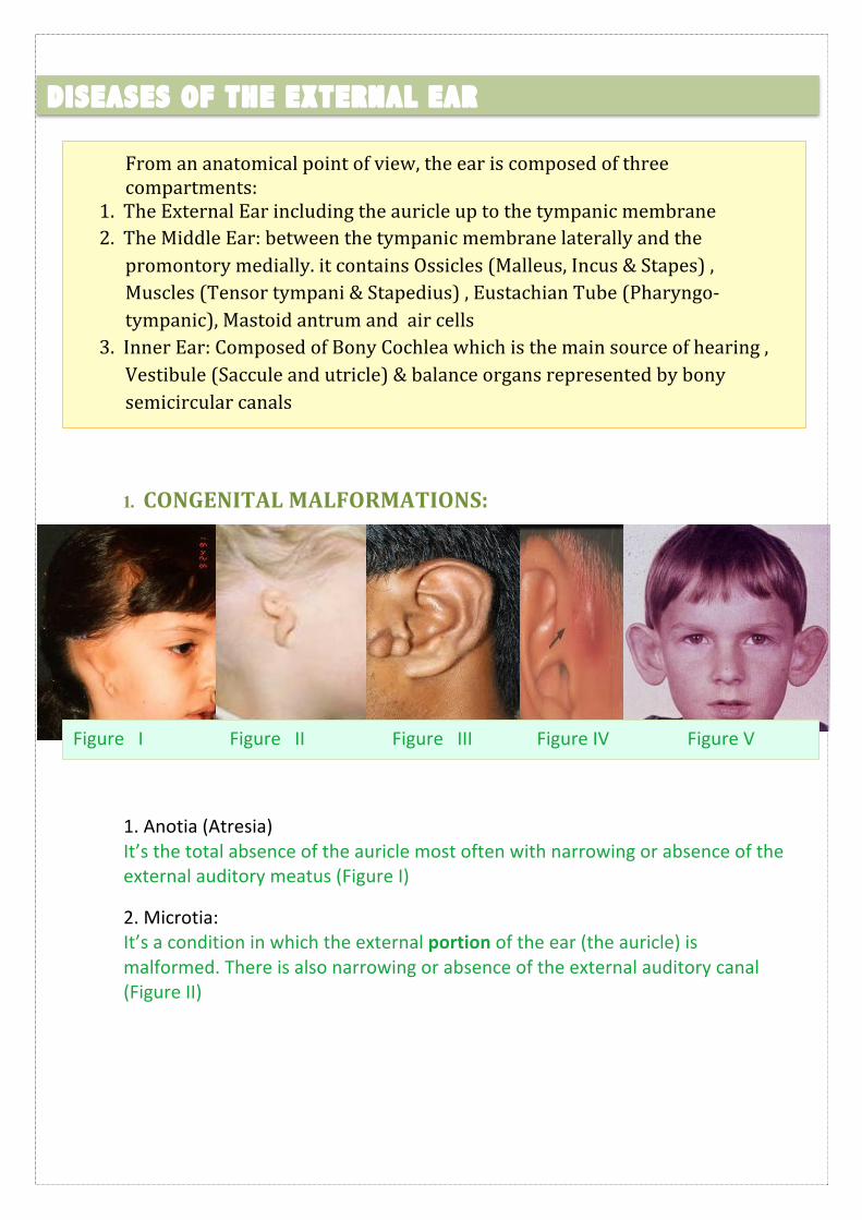
1. CONGENITAL MALFORMATIONS:
1. Anotia (Atresia) It’s the total absence of the auricle most often with narrowing or absence of the external auditory meatus (Figure I)
2. Microtia: It’s a condition in which the external portion of the ear (the auricle) is malformed. There is also narrowing or absence of the external auditory canal (Figure II)
DISEASES OF THE EXTERNAL EAR
From an anatomical point of view, the ear is composed of three compartments:
1. The External Ear including the auricle up to the tympanic membrane 2. The Middle Ear: between the tympanic membrane laterally and the promontory medially. it contains Ossicles (Malleus, Incus & Stapes) , Muscles (Tensor tympani & Stapedius) , Eustachian Tube (Pharyngo-‐ tympanic), Mastoid antrum and air cells
3. Inner Ear: Composed of Bony Cochlea which is the main source of hearing , Vestibule (Saccule and utricle) & balance organs represented by bony semicircular canals
Figure I Figure II Figure III Figure IV Figure V
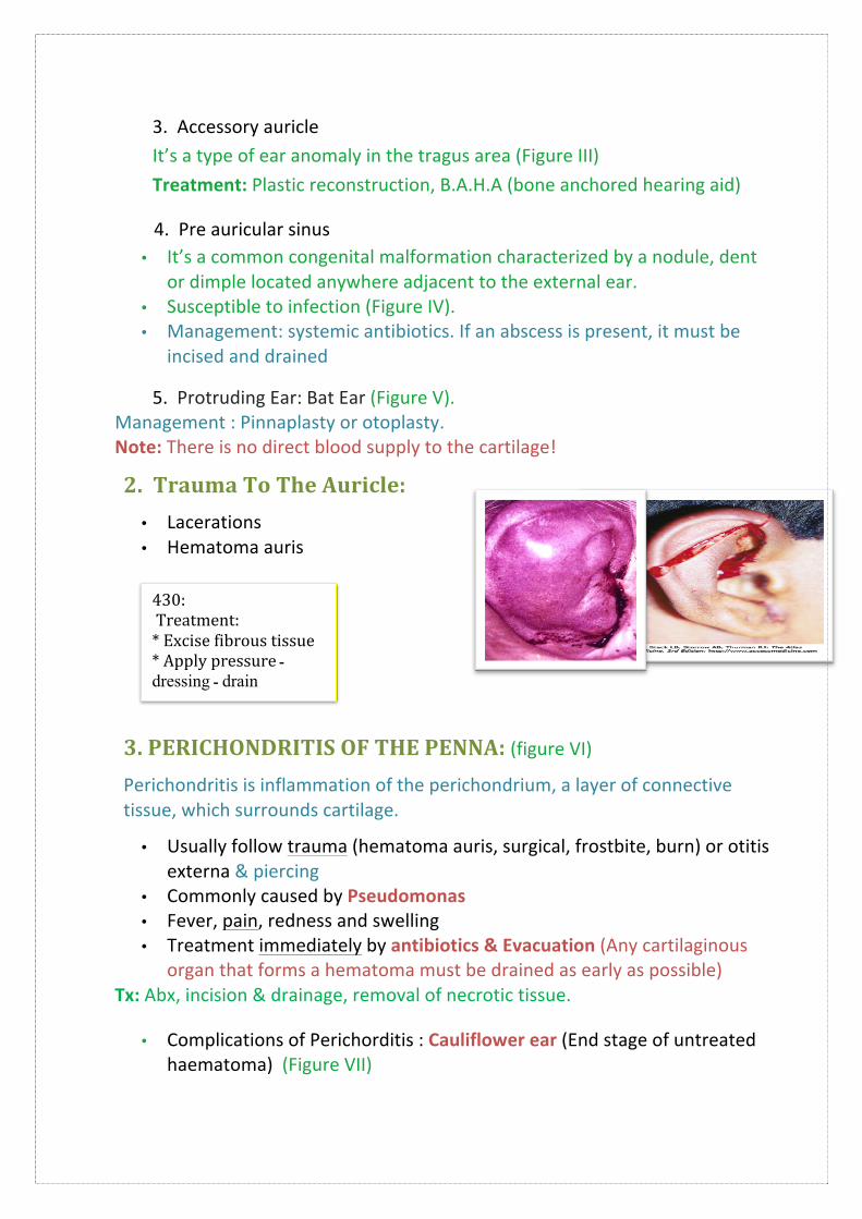
3. Accessory auricle It’s a type of ear anomaly in the tragus area (Figure III) Treatment: Plastic reconstruction, B.A.H.A (bone anchored hearing aid)
4. Pre auricular sinus
• It’s a common congenital malformation characterized by a nodule, dent or dimple located anywhere adjacent to the external ear.
• Susceptible to infection (Figure IV). • Management: systemic antibiotics. If an abscess is present, it must be
incised and drained
5. Protruding Ear: Bat Ear (Figure V). Management : Pinnaplasty or otoplasty. Note: There is no direct blood supply to the cartilage!
2. Trauma To The Auricle: • Lacerations • Hematoma auris
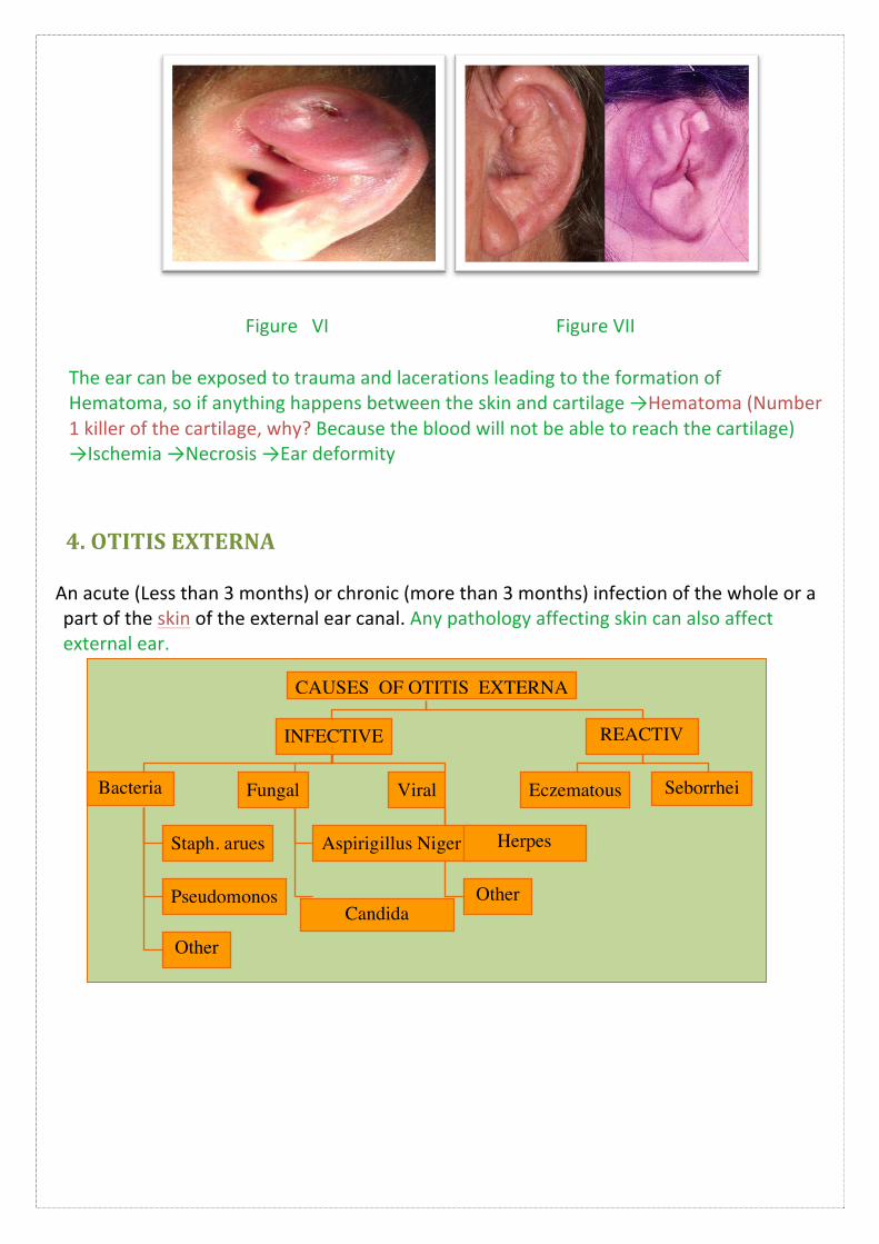
3. PERICHONDRITIS OF THE PENNA: (figure VI) Perichondritis is inflammation of the perichondrium, a layer of connective tissue, which surrounds cartilage.
• Usually follow trauma (hematoma auris, surgical, frostbite, burn) or otitis externa & piercing
• Commonly caused by Pseudomonas • Fever, pain, redness and swelling • Treatment immediately by antibiotics & Evacuation (Any cartilaginous
organ that forms a hematoma must be drained as early as possible) Tx: Abx, incision & drainage, removal of necrotic tissue.
• Complications of Perichorditis : Cauliflower ear (End stage of untreated haematoma) (Figure VII)
430: Treatment: * Excise fibrous tissue * Apply pressure ـ dressing ـ drain
Figure VI Figure VII
The ear can be exposed to trauma and lacerations leading to the formation of Hematoma, so if anything happens between the skin and cartilage →Hematoma (Number 1 killer of the cartilage, why? Because the blood will not be able to reach the cartilage) →Ischemia →Necrosis →Ear deformity
4. OTITIS EXTERNA
An acute (Less than 3 months) or chronic (more than 3 months) infection of the whole or a part of the skin of the external ear canal. Any pathology affecting skin can also affect external ear.
·∙ Seborrhea: A disease of the sebaceous glands characterized by excessive secretion of sebum or an alteration in its quality, resulting in an oily coating, crusts, or scales on the skin. It’s usually painless
·∙ Eczema or Dermatitis: A noncontagious inflammation of the skin, characterized chiefly by redness, itching
Clinical features of Otitis Externa
1. Itching 2. Pain: could be very severe because of underlying cartilage, evoked by movement of the
jaw, because the ear auricle and external canal is attached to the TMJ (temporomandibular joint) pain can radiate to the throat!
3. Tenderness and swelling, absent in otitis media. 4. Otorrhea: No discharge or very little and scanty, not muciod. Large discharge in otitis
media. (Not mucus discharge because the skin does not contain mucus-‐secreting cells. If the discharge doesn’t contain mucus then it is from the External ear however if it contains mucus it is originating from the middle ear)
5. Deafness: deafness caused by external ear needs to be completely obstructed, which is rare in otitis externa.
6. Changes in the lumen and skin of EAM (external auditory meatus)
Clinical Types of Otitis Externa:
Pathophysiology: -‐ Aggressive washing of wax or retention water -‐ Microtrauma (cotton swabs, fingernails)
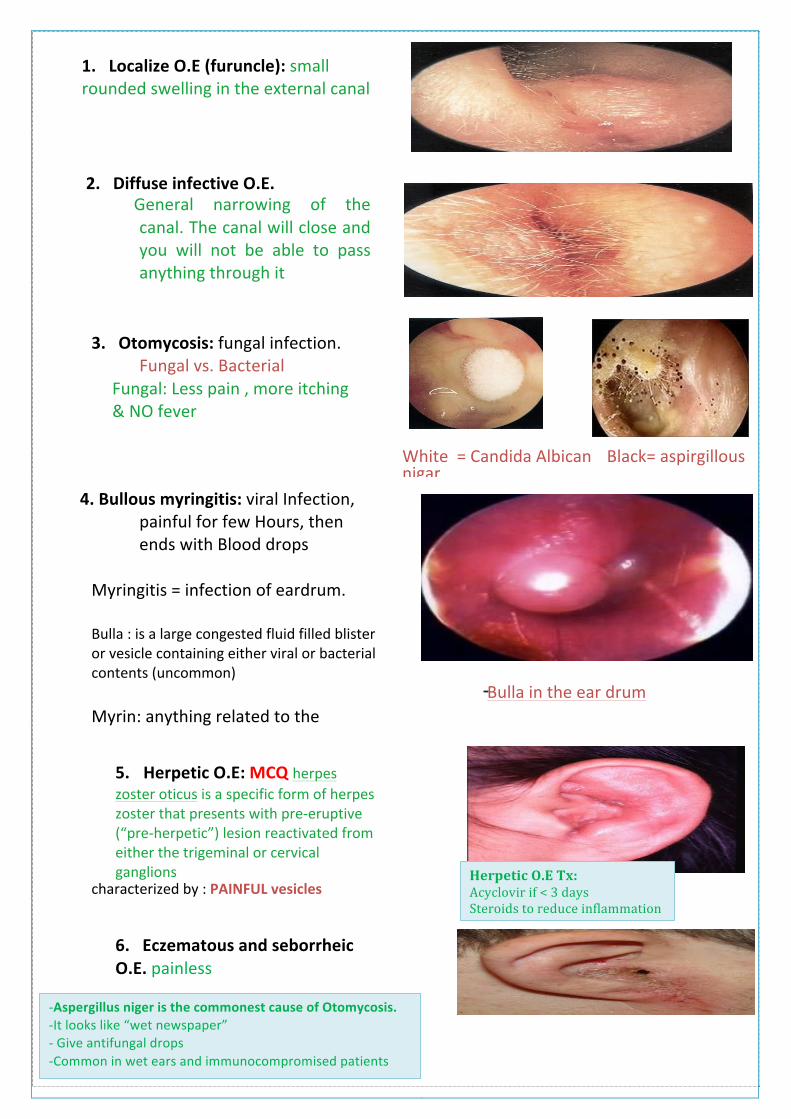
1. Localize O.E (furuncle): small rounded swelling in the external canal
2. Diffuse infective O.E. General narrowing of the canal. The canal will close and you will not be able to pass anything through it
3. Otomycosis: fungal infection. Fungal vs. Bacterial
Fungal: Less pain , more itching & NO fever
Bacterial: Fever , More acute , Sever pain
White = Candida Albican Black= aspirgillous nigar
4. Bullous myringitis: viral Infection, painful for few Hours, then ends with Blood drops
Myringitis = infection of eardrum.
Bulla : is a large congested fluid filled blister or vesicle containing either viral or bacterial contents (uncommon)
Myrin: anything related to the tympanic membrane.
Bulla in the ear drum
5. Herpetic O.E: MCQ herpes zoster oticus is a specific form of herpes zoster that presents with pre-‐eruptive (“pre-‐herpetic”) lesion reactivated from either the trigeminal or cervical ganglions
characterized by : PAINFUL vesicles
6. Eczematous and seborrheic O.E. painless
Herpetic O.E Tx: Acyclovir if < 3 days Steroids to reduce inflammation
-‐Aspergillus niger is the commonest cause of Otomycosis. -‐It looks like “wet newspaper” -‐ Give antifungal drops -‐Common in wet ears and immunocompromised patients
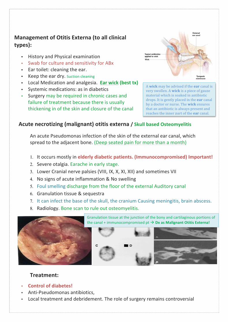
Management of Otitis Externa (to all clinical types):
• History and Physical examination • Swab for culture and sensitivity for ABx • Ear toilet: cleaning the ear. • Keep the ear dry. Suction cleaning • Local Medication and analgesia. Ear wick (best tx) • Systemic medications: as in diabetics • Surgery may be required in chronic cases and
failure of treatment because there is usually thickening in of the skin and closure of the canal
Acute necrotizing (malignant) otitis externa / Skull based Osteomyelitis
An acute Pseudomonas infection of the skin of the external ear canal, which spread to the adjacent bone. (Deep seated pain for more than a month)
1. It occurs mostly in elderly diabetic patients. (Immunocompromised) Important! 2. Severe otalgia. Earache in early stage. 3. Lower Cranial nerve palsies (VIII, IX, X, XI, XII) and sometimes VII 4. No signs of acute inflammation & No swelling 5. Foul smelling discharge from the floor of the external Auditory canal 6. Granulation tissue & sequestra 7. It can infect the base of the skull, the cranium Causing meningitis, brain abscess. 8. Radiology. Bone scan to rule out osteomyelitis.
Treatment:
• Control of diabetes! • Anti-‐Pseudomonas antibiotics, • Local treatment and debridement. The role of surgery remains controversial
A wick may be advised if the ear canal is very swollen. A wick is a piece of gauze material which is soaked in antibiotic drops. It is gently placed in the ear canal by a doctor or nurse. The wick ensures that an antibiotic is always present and reaches the inner part of the ear canal.
Granulation tissue at the junction of the bony and cartilaginous portions of the canal + immunocompromised pt à Dx as Malignant Otitis Externa!
WAX:
·∙ Mixture of ceruminous (in the outer third of the canal) and sebaceous glands secretion
·∙ Normally is expelled from the canal aided by movements of the jaw
·∙ When accumulated it may cause deafness, earache or tinnitus
·∙ Deafness caused by wax accumulation is sudden in onset, as the hearing won’t be affected in 90% obstruction, but once it’s 100% it causes deafness
Treatment: is by removal using syringing, suction or instrumentation “microsuction under vision”
Acute infection of the mucous membrane lining of the middle ear cleft
The definition is specific to infection because in chronic Otitis media it can be due to infection of normal inflammation
Predisposing factors:
• Age: common in children as the Eustachian tube is more horizontal, wider and shorter.
• Males
• Bottle feeding: more likely to have milk regurgitation in meddle ear
• Climate
• Crowded living conditions
• Heredity
• Associated conditions: cleft palate, immunodeficiency, ciliary dyskinesia, Down syndrome, and cystic fibrosis
430: Acute otitis media: -‐ Most common reason for visit to Pediatrician -‐ Tympanostomy tube placement is second most common surgical procedure in children
430: commonest cause of conductive hearing loss
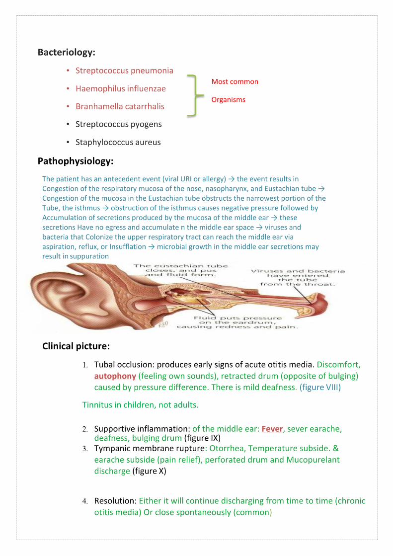
Bacteriology:
• Streptococcus pneumonia
• Haemophilus influenzae
• Branhamella catarrhalis
• Streptococcus pyogens
• Staphylococcus aureus
Pathophysiology:
Most common
Organisms
The patient has an antecedent event (viral URI or allergy) → the event results in Congestion of the respiratory mucosa of the nose, nasopharynx, and Eustachian tube → Congestion of the mucosa in the Eustachian tube obstructs the narrowest portion of the Tube, the isthmus → obstruction of the isthmus causes negative pressure followed by Accumulation of secretions produced by the mucosa of the middle ear → these secretions Have no egress and accumulate n the middle ear space → viruses and bacteria that Colonize the upper respiratory tract can reach the middle ear via aspiration, reflux, or Insufflation → microbial growth in the middle ear secretions may result in suppuration
Clinical picture:
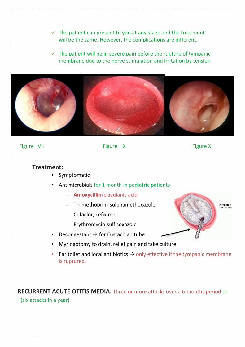
1. Tubal occlusion: produces early signs of acute otitis media. Discomfort, autophony (feeling own sounds), retracted drum (opposite of bulging) caused by pressure difference. There is mild deafness. (figure VIII)
Tinnitus in children, not adults.
2. Supportive inflammation: of the middle ear: Fever, sever earache, deafness, bulging drum (figure IX)
3. Tympanic membrane rupture: Otorrhea, Temperature subside. & earache subside (pain relief), perforated drum and Mucopurelant discharge (figure X)
4. Resolution: Either it will continue discharging from time to time (chronic otitis media) Or close spontaneously (common)
Treatment:
ü The patient can present to you at any stage and the treatment will be the same. However, the complications are different.
ü The patient will be in severe pain before the rupture of tympanic
membrane due to the nerve stimulation and irritation by tension
Figure VII Figure IX Figure X
• Symptomatic
• Antimicrobials for 1 month in pediatric patients
– Amoxycillin/clavulanic acid
– Tri-‐methoprim-‐sulphamethoxazole
– Cefaclor, cefixime
– Erythromycin-‐sulfisoxazole
• Decongestant → for Eustachian tube
• Myringotomy to drain, relief pain and take culture
• Ear toilet and local antibiotics → only effective if the tympanic membrane is ruptured.
RECURRENT ACUTE OTITIS MEDIA: Three or more attacks over a 6-‐months period or (six attacks in a year)
Treatment: • Long-‐term low dose antimicrobials
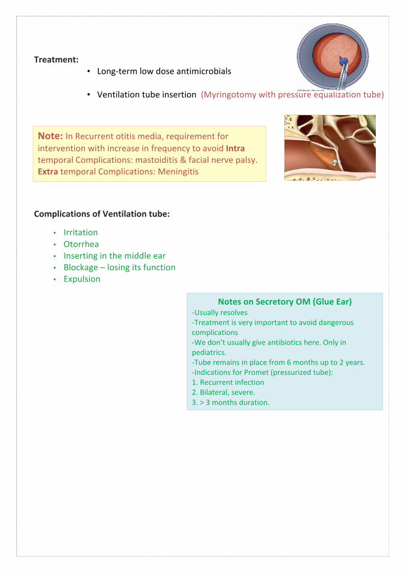
• Ventilation tube insertion (Myringotomy with pressure equalization tube)
Complications of Ventilation tube:
• Irritation • Otorrhea • Inserting in the middle ear • Blockage – losing its function • Expulsion
Note: In Recurrent otitis media, requirement for intervention with increase in frequency to avoid Intra temporal Complications: mastoiditis & facial nerve palsy. Extra temporal Complications: Meningitis
Notes on Secretory OM (Glue Ear) -‐Usually resolves -‐Treatment is very important to avoid dangerous complications -‐We don’t usually give antibiotics here. Only in pediatrics. -‐Tube remains in place from 6 months up to 2 years. -‐Indications for Promet (pressurized tube): 1. Recurrent infection 2. Bilateral, severe. 3. > 3 months duration.
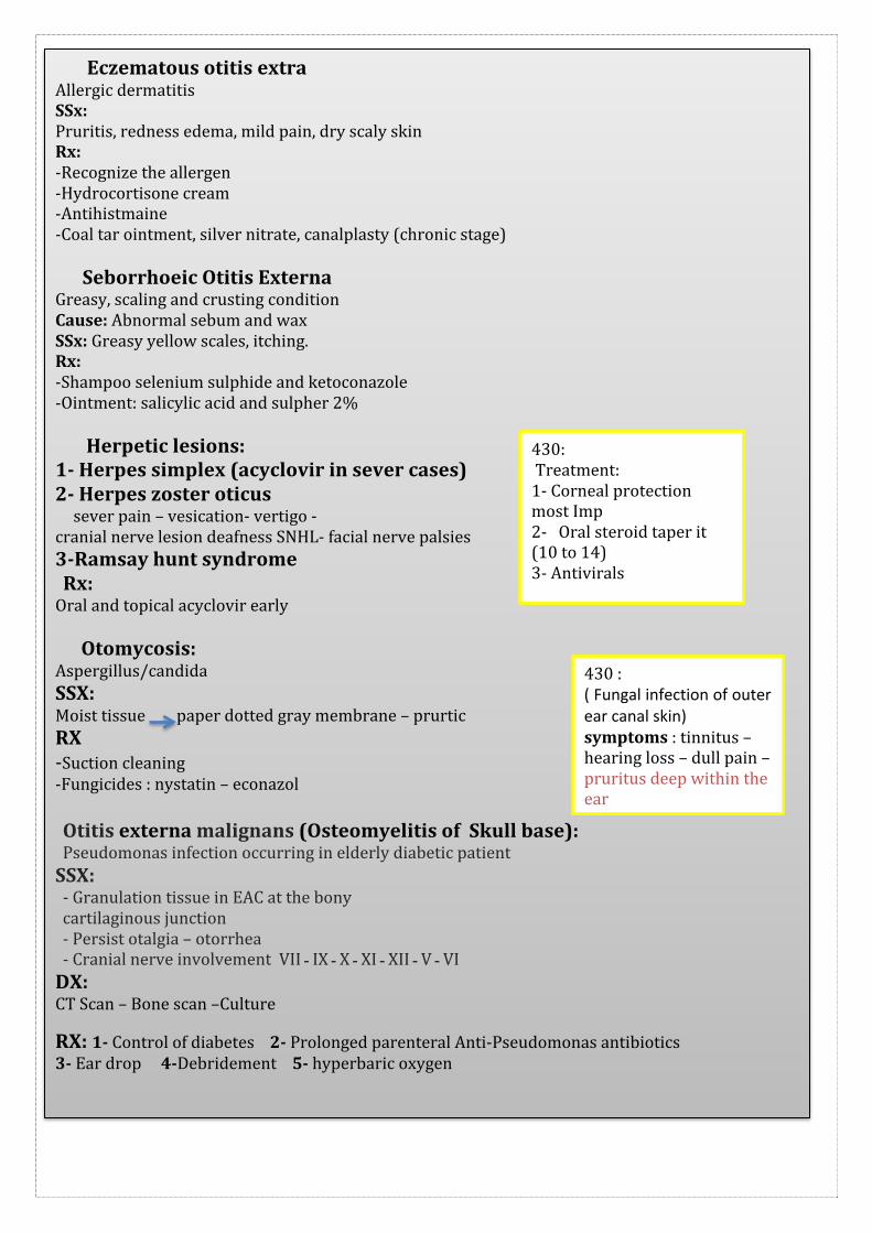
Eczematous otitis extra Allergic dermatitis SSx: Pruritis, redness edema, mild pain, dry scaly skin Rx: -‐Recognize the allergen -‐Hydrocortisone cream -‐Antihistmaine -‐Coal tar ointment, silver nitrate, canalplasty (chronic stage) Seborrhoeic Otitis Externa Greasy, scaling and crusting condition Cause: Abnormal sebum and wax SSx: Greasy yellow scales, itching. Rx: -‐Shampoo selenium sulphide and ketoconazole -‐Ointment: salicylic acid and sulpher 2% Herpetic lesions: 1-‐ Herpes simplex (acyclovir in sever cases) 2-‐ Herpes zoster oticus sever pain – vesication-‐ vertigo -‐ cranial nerve lesion deafness SNHL-‐ facial nerve palsies 3-‐Ramsay hunt syndrome Rx: Oral and topical acyclovir early Otomycosis: Aspergillus/candida SSX: Moist tissue paper dotted gray membrane – prurtic RX -‐Suction cleaning -‐Fungicides : nystatin – econazol Otitis externa malignans (Osteomyelitis of Skull base): Pseudomonas infection occurring in elderly diabetic patient SSX: -‐ Granulation tissue in EAC at the bony cartilaginous junction -‐ Persist otalgia – otorrhea -‐ Cranial nerve involvement VII ـ IX ـ X ـ XI ـ XII ـ V ـ VI DX: CT Scan – Bone scan –Culture
RX: 1-‐ Control of diabetes 2-‐ Prolonged parenteral Anti-‐Pseudomonas antibiotics 3-‐ Ear drop 4-‐Debridement 5-‐ hyperbaric oxygen
430 : ( Fungal infection of outer ear canal skin) symptoms : tinnitus – hearing loss – dull pain – pruritus deep within the ear
430: Treatment: 1-‐ Corneal protection most Imp 2-‐ Oral steroid taper it (10 to 14) 3-‐ Antivirals
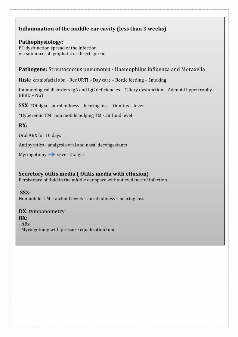
Inflammation of the middle ear cavity (less than 3 weeks) Pathophysiology: ET dysfunction spread of the infection via submucosal lymphatic or direct spread
Pathogens: Streptococcus pneumonia -‐ Haemophilus influenza and Moraxella Risk: craniofacial abn -‐ Rec URTI – Day care – Bottle feeding – Smoking Immunological disorders IgA and IgG deficiencies – Ciliary dysfunction – Adenoid hypertrophy – GERD – NGT
*Hyperemic TM ـ non mobile bulging TM -‐ air fluid level
RX:
Oral ABX for 10 days
Antipyretics -‐ analgesia oral and nasal decongestants
Myringotomy sever Otalgia Secretory otitis media ( Otitis media with effusion) Persistence of fluid in the middle ear space without evidence of infection SSX: Nonmobile TM -‐ airfluid levels – aural fullness – hearing loss DX: tympanometry RX: -‐ ABx -‐ Myringotomy with pressure equalization tube
Lecture Title ENT Teamwork 432
15
Summary
DISEASES OF THE EXTERNAL EAR: 1. Congenital malformations:
4. Treatment immediately by antibiotics & Evacuation (Any cartilaginous organ that forms hematoma must be drained as early as possible)
5. Complications : Cauliflower ear
3. OTITIS EXTERNA: An acute (Less than 3 months) or chronic (more than 3 months) infection of the whole or a part of the skin of the external ear canal
• Acute necrotizing (malignant) otitis externa: Consider malignant otitis externa in any old diabetic patient
• Acute Otitis Media is an acute infection of the mucous membrane lining of the middle ear cleft
Lecture Title ENT Teamwork 432
16
MCQs :
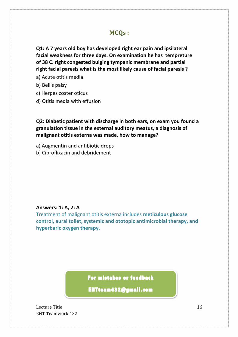
Q1: A 7 years old boy has developed right ear pain and ipsilateral facial weakness for three days. On examination he has tempreture of 38 C. right congested bulging tympanic membrane and partial right facial paresis what is the most likely cause of facial paresis ?
a) Acute otitis media
b) Bell's palsy
c) Herpes zoster oticus
d) Otitis media with effusion
Q2: Diabetic patient with discharge in both ears, on exam you found a granulation tissue in the external auditory meatus, a diagnosis of malignant otitis externa was made, how to manage?
a) Augmentin and antibiotic drops b) Ciproflixacin and debridement Answers: 1: A, 2: A Treatment of malignant otitis externa includes meticulous glucose control, aural toilet, systemic and ototopic antimicrobial therapy, and hyperbaric oxygen therapy.