Molecular Cell Resource Mammalian Mirtron Genes Eugene Berezikov, 1, * Wei-Jen Chung, 2 Jason Willis, 2 Edwin Cuppen, 1 and Eric C. Lai 2, * 1 Hubrecht Institute, Uppsalalaan 8, 3584 CT Utrecht, The Netherlands 2 Sloan-Kettering Institute, 1275 York Avenue, Box 252, New York, NY 10021, USA *Correspondence: [email protected](E.B.), [email protected](E.C.L.) DOI 10.1016/j.molcel.2007.09.028 SUMMARY Mirtrons are alternative precursors for micro- RNA biogenesis that were recently described in invertebrates. These short hairpin introns use splicing to bypass Drosha cleavage, which is otherwise essential for the generation of canonical animal microRNAs. Using computa- tional and experimental strategies, we now es- tablish that mammals have mirtrons as well. We identified 3 mirtrons that are well conserved and expressed in diverse mammals, 16 pri- mate-specific mirtrons, and 46 candidates sup- ported by limited cloning evidence in primates. As with some fly and worm mirtrons, the exis- tence of well-conserved mammalian mirtrons indicates their relatively ancient incorporation into endogenous regulatory pathways. How- ever, as worms, flies, and mammals each have different sets of mirtrons, we hypothesize that different animals may have independently evolved the capacity for this hybrid small RNA pathway. This notion is supported by our obser- vation of several clade-specific features of mammalian and invertebrate mirtrons. INTRODUCTION MicroRNAs (miRNAs) are 22 nucleotide (nt) RNAs that typically repress the activity of complementary messenger RNAs (Lai, 2003). Canonical animal miRNAs derive from longer primary transcripts bearing hairpin structures, which are processed in a stepwise fashion by the RNase III enzymes Drosha and Dicer. In the nucleus, Drosha cleaves near the hairpin base to release the pre-miRNA hairpin (Lee et al., 2003). Following its export to the cyto- plasm, Dicer cleaves on the loop side of the hairpin to gen- erate an miRNA:miRNA* duplex, one strand of which is preferentially incorporated into a silencing complex (Du and Zamore, 2005). An alternative nuclear pathway for miRNA biogenesis was recently described in invertebrates (Okamura et al., 2007; Ruby et al., 2007a). Short introns with hairpin poten- tial, termed mirtrons, can be spliced and debranched into pre-miRNA hairpin mimics that appear to bypass Drosha cleavage. Debranched mirtrons access the canonical miRNA pathway during nuclear export, and are then cleaved by Dicer and incorporated into silencing com- plexes (Okamura et al., 2007; Ruby et al., 2007a). Mirtrons were found only in nematodes and flies thus far. It was suggested that the evolutionary emergence of invertebrate mirtrons was aided by the sheer number of short introns whose length is typical of pre-miRNA hair- pins (Ruby et al., 2007a). The relative proportion of such introns in different species is flies > worms > mammals (Lim and Burge, 2001; Yandell et al., 2006). However, be- cause mammals have many more introns than do worms and flies, the difference in absolute numbers of short in- trons among these species is less substantial. In this study, we addressed the possibility that mirtrons might exist in mammals. Using computational methods, we identified a small set of mammalian short hairpin in- trons as possible well-conserved mirtron candidates. Cloned 22 nt RNA products from the ends of three of these candidates were present in multiple small RNA li- braries from human, macaque, chimpanzee, rat, and/or mouse, validating the existence of conserved mammalian mirtrons. Emboldened by these findings, we analyzed whether more ‘‘newly evolved’’ mirtrons could be de- tected, as these comprise the majority of identified fly and worm mirtrons. Indeed, by analyzing large-scale pri- mate small RNA data sets, we could confidently classify 16 additional primate-specific mirtrons from human and macaque brain; nearly 50 additional candidates were sup- ported by more tentative evidence (one to two clones). These findings indicate that mirtrons constitute a substan- tial and highly dynamic class of regulatory RNA in both in- vertebrates and vertebrates. Curiously, we identified sev- eral basic distinctions between mirtrons from these different clades, suggesting that this alternative strategy to generate microRNAs may have arisen more than once during animal evolution. RESULTS AND DISCUSSION Computational Survey for Well-Conserved Mammalian Mirtrons At least some invertebrate mirtrons have been well conserved during fly or worm evolution. These exhibit characteristic features that reflect their status as micro- RNA-class genes (Lai et al., 2003), namely that they are short, straight, hairpin introns that exhibit preferential 328 Molecular Cell 28, 328–336, October 26, 2007 ª2007 Elsevier Inc.
Transcript
Molecular Cell
Resource
Mammalian Mirtron GenesEugene Berezikov,1,* Wei-Jen Chung,2 Jason Willis,2 Edwin Cuppen,1 and Eric C. Lai2,*1Hubrecht Institute, Uppsalalaan 8, 3584 CT Utrecht, The Netherlands2Sloan-Kettering Institute, 1275 York Avenue, Box 252, New York, NY 10021, USA
Mirtrons are alternative precursors for micro-RNA biogenesis that were recently describedin invertebrates. These short hairpin intronsuse splicing to bypass Drosha cleavage, whichis otherwise essential for the generation ofcanonical animal microRNAs. Using computa-tional and experimental strategies, we now es-tablish that mammals have mirtrons as well.We identified 3 mirtrons that are well conservedand expressed in diverse mammals, 16 pri-mate-specific mirtrons, and 46 candidates sup-ported by limited cloning evidence in primates.As with some fly and worm mirtrons, the exis-tence of well-conserved mammalian mirtronsindicates their relatively ancient incorporationinto endogenous regulatory pathways. How-ever, as worms, flies, and mammals each havedifferent sets of mirtrons, we hypothesize thatdifferent animals may have independentlyevolved the capacity for this hybrid small RNApathway. This notion is supported by our obser-vation of several clade-specific features ofmammalian and invertebrate mirtrons.
INTRODUCTION
MicroRNAs (miRNAs) are �22 nucleotide (nt) RNAs that
typically repress the activity of complementary messenger
RNAs (Lai, 2003). Canonical animal miRNAs derive from
to both the 50 and 30 ends of host introns (i.e., miRNA/
miRNA*) were found in human, chimpanzee, rat, and/or
mouse small RNA data sets for three loci (mir-877, mir-
1224, and mir-1225, Figures 1A and 2 and Figures S1
and S4). As with invertebrate mirtrons, mammalian mir-
trons generally lacked the pairing between their flanking
exons needed for recognition by the Drosha/DGCR8 com-
plex (Figure 1 and Figure S1); where pairing was found, it
was typically not conserved and followed codon wobble
rules.
The mirtrons mir-877, mir-1224, and mir-1225 were
clearly maintained as hairpins in mammals as diverse as
rodents, dog, and horse, indicating their persistence over
at least �80 million years of eutherian evolution (Figures
S1 and S2). We note that small RNAs from the mir-877 lo-
cus were recently cloned independently by Tuschl and col-
leagues, who annotated it as a canonical miRNA gene
(Landgraf et al., 2007). Its reclassification as a mirtron is
akin to that of nematode mir-62, which was only recently
recognized as a mirtron gene (Ruby et al., 2007a). We
also note that two of the most abundantly cloned mirtron
products were derived from mir-877 and mir-1224
(Figure S4), which were also two of the most perfectly con-
served predicted mirtrons. This parallels the finding that the
most highly expressed invertebrate mirtrons are also the
most highly conserved ones (Okamura et al., 2007; Ruby
et al., 2007a), as is also generally the case for canonical an-
imal miRNAs (Berezikov et al., 2006b; Ruby et al., 2007b).
Molecul
A Plethora of Primate-Specific MirtronsAlthough some are well conserved, most invertebrate mir-
trons arose quite recently during Drosophilid and nema-
tode radiation (Okamura et al., 2007; Ruby et al., 2007a);
thus, the consideration of evolutionary conservation
does not aid their computational identification. However,
newly evolved miRNAs have emerged through high-
throughput small RNA sequencing efforts. In D. mela-
nogaster, adult heads expressed a high diversity of mir-
trons and canonical miRNAs (Ruby et al., 2007b). This is
consistent with the fact that brains harbor an exceptional
diversity of neurons, a cell type that intrinsically has ex-
ceptional needs for translational regulation. We therefore
mined a data set of 30 additional small RNA libraries
from 15 matched anatomical regions of human and rhesus
macaque brains (Figure S3), represented by 18,000–
45,000 sequences each (E.B. and E.C., unpublished data).
In addition to revealing cloned evidence for mirtrons
mir-877, mir-1224, and mir-1225 in macaque, analysis of
these small RNA data sets yielded another 16 mirtrons ex-
pressed in primate brains with evidence justifying official
nomenclature (Figure 2 and Figure S4). We considered
minimum evidence to be the recovery of clones from inde-
pendent libraries, or at least three clones from any individ-
ual library. In several cases, higher levels of evidence were
attained, including their cloning from multiple species (i.e.,
mir-1226 and mir-1227 both from human and macaque),
the isolation of many clones (i.e., mir-1229, 16 clones
from 12 different libraries), and/or the isolation of both
miRNA and miRNA* species (i.e., mir-1227 and mir-
1228). These mirtrons appeared to be phylogenetically re-
stricted to primates, with some presenting conserved
hairpin structures in human/rhesus/chimp, and others
that were restricted to a primate subset. We have summa-
rized the sequences and secondary structures of the or-
thologous primate mirtronic introns in Figure S5.
Finally, we classified 46 additional hairpin introns from
human (23 loci), macaque (16 loci), chimpanzee (3 loci),
or mouse (4 loci) as mirtron candidates (Figure S6). The
greater number of human and macaque candidates was
due in part to the deeper sampling of human and macaque
brains. A few of these candidates were cloned three or
more times, but we considered their candidacy tentative
because of an atypical intronic extension of 8–10 nt
on one side of the hairpin (i.e., macaque_block210826
[3 reads/2 libs], and human_block172399 [3 reads/1 lib]).
In Drosophila, at least one conserved mirtron-like locus
(mir-1017) exhibits a long intronic extension on one side
of the hairpin (Ruby et al., 2007a), suggesting that
such ‘‘half-mirtron’’ loci might have one side defined by
splicing and the other by exonucleolytic digestion. Of
the remaining candidates, five (human_block107544,
chimp_block23965, macaque_block550558, macaque_
block137121, and mouse_block283) were sequenced
twice while the rest were defined by single reads. Many
of these candidate mirtrons exhibit compelling extended
hairpin structures; thus, we anticipate that at least some
of them (along with some of the uncloned, conserved,
ar Cell 28, 328–336, October 26, 2007 ª2007 Elsevier Inc. 329
Molecular Cell
Mammalian Mirtron Genes
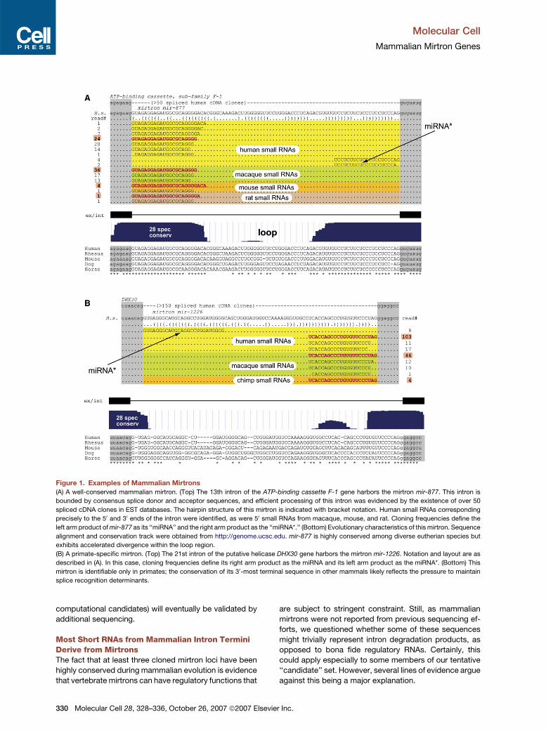
Figure 1. Examples of Mammalian Mirtrons
(A) A well-conserved mammalian mirtron. (Top) The 13th intron of the ATP-binding cassette F-1 gene harbors the mirtron mir-877. This intron is
bounded by consensus splice donor and acceptor sequences, and efficient processing of this intron was evidenced by the existence of over 50
spliced cDNA clones in EST databases. The hairpin structure of this mirtron is indicated with bracket notation. Human small RNAs corresponding
precisely to the 50 and 30 ends of the intron were identified, as were 50 small RNAs from macaque, mouse, and rat. Cloning frequencies define the
left arm product of mir-877 as its ‘‘miRNA’’ and the right arm product as the ‘‘miRNA*.’’ (Bottom) Evolutionary characteristics of this mirtron. Sequence
alignment and conservation track were obtained from http://genome.ucsc.edu. mir-877 is highly conserved among diverse eutherian species but
exhibits accelerated divergence within the loop region.
(B) A primate-specific mirtron. (Top) The 21st intron of the putative helicase DHX30 gene harbors the mirtron mir-1226. Notation and layout are as
described in (A). In this case, cloning frequencies define its right arm product as the miRNA and its left arm product as the miRNA*. (Bottom) This
mirtron is identifiable only in primates; the conservation of its 30-most terminal sequence in other mammals likely reflects the pressure to maintain
splice recognition determinants.
computational candidates) will eventually be validated by
additional sequencing.
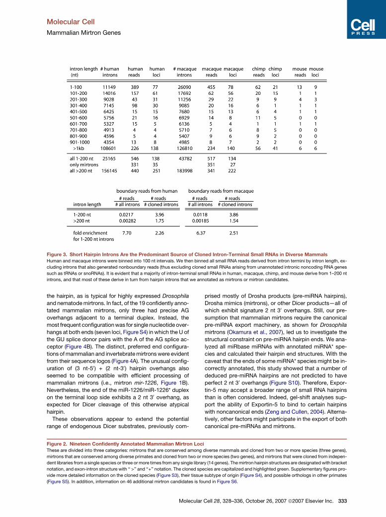
Most Short RNAs from Mammalian Intron TerminiDerive from MirtronsThe fact that at least three cloned mirtron loci have been
highly conserved during mammalian evolution is evidence
that vertebrate mirtrons can have regulatory functions that
330 Molecular Cell 28, 328–336, October 26, 2007 ª2007 Elsevi
are subject to stringent constraint. Still, as mammalian
mirtrons were not reported from previous sequencing ef-
forts, we questioned whether some of these sequences
might trivially represent intron degradation products, as
opposed to bona fide regulatory RNAs. Certainly, this
could apply especially to some members of our tentative
‘‘candidate’’ set. However, several lines of evidence argue
Nevertheless, the end of the miR-1226/miR-1226* duplex
on the terminal loop side exhibits a 2 nt 30 overhang, as
expected for Dicer cleavage of this otherwise atypical
hairpin.
These observations appear to extend the potential
range of endogenous Dicer substrates, previously com-
Molecula
prised mostly of Drosha products (pre-miRNA hairpins),
Drosha mimics (mirtrons), or other Dicer products—all of
which exhibit signature 2 nt 30 overhangs. Still, our pre-
sumption that mammalian mirtrons require the canonical
pre-miRNA export machinery, as shown for Drosophila
mirtrons (Okamura et al., 2007), led us to investigate the
structural constraint on pre-miRNA hairpin ends. We ana-
lyzed all miRbase miRNAs with annotated miRNA* spe-
cies and calculated their hairpin end structures. With the
caveat that the ends of some miRNA* species might be in-
correctly annotated, this study showed that a number of
deduced pre-miRNA hairpins are not predicted to have
perfect 2 nt 30 overhangs (Figure S10). Therefore, Expor-
tin-5 may accept a broader range of small RNA hairpins
than is often considered. Indeed, gel-shift analyses sup-
port the ability of Exportin-5 to bind to certain hairpins
with noncanonical ends (Zeng and Cullen, 2004). Alterna-
tively, other factors might participate in the export of both
canonical pre-miRNAs and mirtrons.
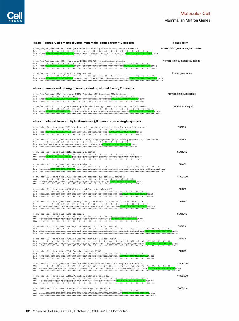
Figure 2. Nineteen Confidently Annotated Mammalian Mirtron Loci
These are divided into three categories: mirtrons that are conserved among diverse mammals and cloned from two or more species (three genes),
mirtrons that are conserved among diverse primates and cloned from two or more species (two genes), and mirtrons that were cloned from indepen-
dent libraries from a single species or three or more times from any single library (14 genes). The mirtron hairpin structures are designated with bracket
notation, and exon-intron structure with ‘‘ >’’ and ‘‘+’’ notation. The cloned species are capitalized and highlighted green. Supplementary figures pro-
vide more detailed information on the cloned species (Figure S3), their tissue subtype of origin (Figure S4), and possible orthologs in other primates
(Figure S5). In addition, information on 46 additional mirtron candidates is found in Figure S6.
r Cell 28, 328–336, October 26, 2007 ª2007 Elsevier Inc. 333
Molecular Cell
Mammalian Mirtron Genes
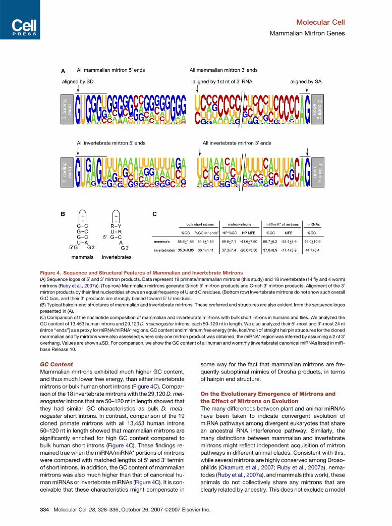
Figure 4. Sequence and Structural Features of Mammalian and Invertebrate Mirtrons
(A) Sequence logos of 50 and 30 mirtron products. Data represent 19 primate/mammalian mirtrons (this study) and 18 invertebrate (14 fly and 4 worm)
mirtrons (Ruby et al., 2007a). (Top row) Mammalian mirtrons generate G-rich 50 mirtron products and C-rich 30 mirtron products. Alignment of the 30
mirtron products by their first nucleotides shows an equal frequency of U and C residues. (Bottom row) Invertebrate mirtrons do not show such overall
G:C bias, and their 30 products are strongly biased toward 50 U residues.
(B) Typical hairpin-end structures of mammalian and invertebrate mirtrons. These preferred end structures are also evident from the sequence logos
presented in (A).
(C) Comparison of the nucleotide composition of mammalian and invertebrate mirtrons with bulk short introns in humans and flies. We analyzed the
GC content of 13,453 human introns and 29,120 D. melanogaster introns, each 50–120 nt in length. We also analyzed their 50-most and 30-most 24 nt
(intron ‘‘ends’’) as a proxy for miRNA/miRNA* regions. GC content and minimum free energy (mfe, kcal/mol) of straight hairpin structures for the cloned
mammalian and fly mirtrons were also assessed; where only one mirtron product was obtained, the miRNA* region was inferred by assuming a 2 nt 30
overhang. Values are shown ±SD. For comparison, we show the GC content of all human and worm/fly (invertebrate) canonical miRNAs listed in miR-
base Release 10.
GC Content
Mammalian mirtrons exhibited much higher GC content,
and thus much lower free energy, than either invertebrate
mirtrons or bulk human short introns (Figure 4C). Compar-
ison of the 18 invertebrate mirtrons with the 29,120 D. mel-
anogaster introns that are 50–120 nt in length showed that
they had similar GC characteristics as bulk D. mela-
nogaster short introns. In contrast, comparison of the 19
cloned primate mirtrons with all 13,453 human introns
50–120 nt in length showed that mammalian mirtrons are
significantly enriched for high GC content compared to
bulk human short introns (Figure 4C). These findings re-
mained true when the miRNA/miRNA* portions of mirtrons
were compared with matched lengths of 50 and 30 termini
of short introns. In addition, the GC content of mammalian
mirtrons was also much higher than that of canonical hu-
man miRNAs or invertebrate miRNAs (Figure 4C). It is con-
ceivable that these characteristics might compensate in
334 Molecular Cell 28, 328–336, October 26, 2007 ª2007 Elsevie
some way for the fact that mammalian mirtrons are fre-
quently suboptimal mimics of Drosha products, in terms
of hairpin end structure.
On the Evolutionary Emergence of Mirtrons andthe Effect of Mirtrons on EvolutionThe many differences between plant and animal miRNAs
have been taken to indicate convergent evolution of
miRNA pathways among divergent eukaryotes that share
an ancestral RNA interference pathway. Similarly, the
many distinctions between mammalian and invertebrate
mirtrons might reflect independent acquisition of mirtron
pathways in different animal clades. Consistent with this,
while several mirtrons are highly conserved among Droso-
philids (Okamura et al., 2007; Ruby et al., 2007a), nema-
todes (Ruby et al., 2007a), and mammals (this work), these
animals do not collectively share any mirtrons that are
clearly related by ancestry. This does not exclude a model
r Inc.
Molecular Cell
Mammalian Mirtron Genes
in which mirtrons facilitated the evolution of a canonical
animal miRNA pathway, prior to the evolution of a Dro-
sha-type activity (Ruby et al., 2007a). However, in this sce-
nario, it is necessary to posit that none of these ancient
mirtrons evolved substantial functions and were all lost
through evolution, or that all of them accumulated so
many sequence changes that their ancestry is no longer
apparent from sequence alignment. These scenarios are
not easily reconciled with the fact that highly conserved
mirtrons have subsequently emerged in three different
animal lineages, nor with the fact that many canonical
miRNAs have been retained completely unchanged from
the bilaterian ancestor of invertebrates and vertebrates
(Prochnik et al., 2007).
Our findings also do not clearly support a model in
which mirtrons arise in genomes strictly proportionally to
the fraction of short introns whose size is comparable to
pre-miRNA hairpins (Ruby et al., 2007a). The extant evi-
dence demonstrates that primate brains express a greater
number of mirtrons than do flies and worms put together,
despite the fact that these invertebrates have more short
introns (Lim and Burge, 2001; Yandell et al., 2006). In ad-
dition, because mammalian mirtrons have very high GC
content relative to bulk mammalian short introns, they ev-
idently do not comprise a random sampling of mammalian
short introns (Figure 4C). Indeed, the differences in se-
quence composition and structure between mammalian
mirtron and pre-miRNA hairpins (Figure 4C) further sug-
gest that they are not simply pre-miRNA mimics, as ap-
pears to be the case for their invertebrate counterparts.
Overall, the observation of cloned products from many
newly evolved mirtrons in diverse animal species suggests
that the mirtron might represent an evolutionarily opportu-
nistic and facile strategy for the birth of regulatory RNAs in
animal species with a preexisting canonical miRNA path-
way. This is conceptually similar to the notion that animals
and plants may have evolved miRNA genes indepen-
dently, building their respective pathways via an ancestral
RNA interference pathway. The fact that a majority of
D. melanogaster mirtrons arose quite recently during Dro-
sophilid evolution, combined with the observation that
miRNAs have relatively minimal requirements for target iden-
tification, suggested that mirtrons could have a palpable
effect on insect speciation. Our parallel observation that
primates, and specifically primate brains, express a strong
diversity of processed mirtrons similarly suggests that
they might also contribute to primate evolution and/or pri-
mate-specific behavior.
EXPERIMENTAL PROCEDURES
Computational Screen for Conserved Mammalian Mirtrons
From the UCSC Genome Browser (Kuhn et al., 2007), we extracted
21,883 RefSeq human introns 50–200 nt in length, and supplemented
these with a nonredundant set of 4052 Ensembl-exclusive human in-
trons 50–200 nt in length (many of which might be misannotated cod-
ing exons). We then identified introns for which at least 17 nt in the 50-
most 25 nt and 30-most 25 nt exhibited phastCons score of >0.7 across
17 mammalian species. This yielded 220 and 223 conserved introns
Molecul
from Refseq and Ensembl-only intron data set, respectively. Of these,
89 RefSeq and 34 Ensembl introns exhibited a saddle shape conserva-
tion profile, in which a minimum of five continuous nucleotides ex-
hibited phastCons score < 0.1 within the central region of the intron.
Operationally, we required that the diverged region either overlapped
the midpoint of the intron, or its closest boundary was no more than
5 nt away from the midpoint. In addition to selecting for candidates
with microRNA-like evolutionary properties, saddle selection
proved useful for removing misannotated coding regions from consid-
eration.
The mammalian orthologs of these selected introns were then folded
using RNAfold (Hofacker, 2003) and RNAshapes (Steffen et al., 2006).
We used these algorithms because at least one Drosophila mirtron
(mir-1015) is not predicted to adopt a straight hairpin in any alternative
mfold structure, but is using either RNAshapes or RNAfold. The ability
of RNAshapes to report a diversity of suboptimal minimum free energy
structures proved useful to cull single arm, straight hairpin folds. We
defined a potential mirtron candidate to be a straight arm hairpin in
which at least 16 out of the 50 terminal 30 nt and 17 out of the 30 terminal
nt were base paired to each other (these numbers were not the same
because of the nonsymmetrical nature of many hairpins). Candidates
with an overhang of >8 nt at either end were also excluded. Finally,
we defined a conserved mammalian mirtron candidate as a locus for
which orthologs of at least some primate and nonprimate introns sat-
isfied the minimum hairpin criteria. Note that we did not set a lower limit
on the minimum free energy of conserved hairpin candidates. This