MtbHLH1, a bHLH transcription factor involved in Medicago truncatula nodule vascular patterning and nodule to plant metabolic exchanges Laurence Godiard 1 , Agne `s Lepage 1 , Sandra Moreau 1 , Damien Laporte 2 , Marion Verdenaud 1 , Ton Timmers 1 and Pascal Gamas 1 1 Laboratoire des Interactions Plantes Microorganismes, Unite ´ Mixte de Recherche, Institut National de la Recherche Agronomique – Centre National de la Recherche Scientifique 441 ⁄ 2594, F–31320 Castanet Tolosan, France; 2 Jian-Qiu Wu’s laboratory, Ohio State University, 612 Biosciences Building, 484 W 12th Ave, Columbus, OH 43210, USA Author for correspondence: Laurence Godiard Tel: +33 5 61285490 Email: [email protected]Received: 25 February 2011 Accepted: 1 March 2011 New Phytologist (2011) 191: 391–404 doi: 10.1111/j.1469-8137.2011.03718.x Key words: auxin regulation, bHLH, CRES-T, LOB, nodulation, transcription factor, uninfected cells, vascularization. Summary • This study aimed at defining the role of a basic helix–loop–helix (bHLH) tran- scription factor gene from Medicago truncatula, MtbHLH1, whose expression is upregulated during the development of root nodules produced upon infection by rhizobia bacteria. • We used MtbHLH1 promoter::GUS fusions and quantitative reverse-transcription polymerase chain reaction analyses to finely characterize the MtbHLH1 expression pattern. We altered MtbHLH1 function by expressing a dominantly repressed construct (CRES-T approach) and looked for possible MtbHLH1 target genes by transcriptomics. • We found that MtbHLH1 is expressed in nodule primordia cells derived from pericycle divisions, in nodule vascular bundles (VBs) and in uninfected cells of the nitrogen (N) fixation zone. MtbHLH1 is also expressed in root tips, lateral root primordia cells and root VBs, and induced upon auxin treatment. Altering MtbHLH1 function led to an unusual phenotype, with a modified patterning of nodule VB development and a reduced growth of aerial parts of the plant, even though the nodules were able to fix atmospheric N. Several putative MtbHLH1 regulated genes were identified, including an asparagine synthase and a LOB (lateral organ boundary) transcription factor. • Our results suggest that the MtbHLH1 gene is involved in the control of nodule vasculature patterning and nutrient exchanges between nodules and roots. Introduction Legumes play a crucial role in both ecological and agri- cultural systems by their capacity to establish a symbiosis with nitrogen-fixing bacteria called rhizobia. This process involves the formation of a specific organ, the root nodule and relies on the mutual recognition of both partners via molecular signals and activation of a plant symbiotic program. The root nodule provides rhizobia with a carbon (C) source derived from photosynthesis and an appropriate cellular environment allowing the bacterial nitrogenase to fix atmospheric nitrogen (N). In temperate legumes, repre- sented by the model legume Medicago truncatula, the nodule is a highly structured organ with an indeterminate growth, resulting from the activity of an apical meristematic region (also called zone I) and the differentiation of several peripheral and central tissues. The peripheral tissues include the nodule cortex, the nod- ule endodermis and the nodule parenchyma in which the nodule vascular bundles (VBs) are located; in indeterminate nodules, the VBs are connected at their proximal end to the root vasculature and are open at the distal end. The nodule VBs are composed of xylem and phloem vessels in paren- chyma cells surrounded by a pericycle cell layer and a vascular Re-use of this article is permitted in accordance with the Terms and Conditions set out at http://wileyonlinelibrary.com/onlineopen# OnlineOpen_Terms New Phytologist Research Ó 2011 The Authors New Phytologist Ó 2011 New Phytologist Trust New Phytologist (2011) 191: 391–404 391 www.newphytologist.com
Transcript
MtbHLH1, a bHLH transcription factor involved inMedicago truncatula nodule vascular patterning andnodule to plant metabolic exchanges
Laurence Godiard1, Agnes Lepage1, Sandra Moreau1, Damien Laporte2, Marion Verdenaud1, Ton Timmers1
and Pascal Gamas1
1Laboratoire des Interactions Plantes Microorganismes, Unite Mixte de Recherche, Institut National de la Recherche Agronomique – Centre National de la
Recherche Scientifique 441 ⁄ 2594, F–31320 Castanet Tolosan, France; 2Jian-Qiu Wu’s laboratory, Ohio State University, 612 Biosciences Building, 484 W
• This study aimed at defining the role of a basic helix–loop–helix (bHLH) tran-
scription factor gene from Medicago truncatula, MtbHLH1, whose expression is
upregulated during the development of root nodules produced upon infection by
rhizobia bacteria.
• We used MtbHLH1 promoter::GUS fusions and quantitative reverse-transcription
polymerase chain reaction analyses to finely characterize the MtbHLH1 expression
pattern. We altered MtbHLH1 function by expressing a dominantly repressed
construct (CRES-T approach) and looked for possible MtbHLH1 target genes by
transcriptomics.
• We found that MtbHLH1 is expressed in nodule primordia cells derived from
pericycle divisions, in nodule vascular bundles (VBs) and in uninfected cells of the
nitrogen (N) fixation zone. MtbHLH1 is also expressed in root tips, lateral root
primordia cells and root VBs, and induced upon auxin treatment. Altering
MtbHLH1 function led to an unusual phenotype, with a modified patterning of
nodule VB development and a reduced growth of aerial parts of the plant, even
though the nodules were able to fix atmospheric N. Several putative MtbHLH1
regulated genes were identified, including an asparagine synthase and a LOB
(lateral organ boundary) transcription factor.
• Our results suggest that the MtbHLH1 gene is involved in the control of nodule
vasculature patterning and nutrient exchanges between nodules and roots.
Introduction
Legumes play a crucial role in both ecological and agri-cultural systems by their capacity to establish a symbiosis withnitrogen-fixing bacteria called rhizobia. This process involvesthe formation of a specific organ, the root nodule and relies onthe mutual recognition of both partners via molecular signalsand activation of a plant symbiotic program.
The root nodule provides rhizobia with a carbon (C)source derived from photosynthesis and an appropriate
cellular environment allowing the bacterial nitrogenase tofix atmospheric nitrogen (N). In temperate legumes, repre-sented by the model legume Medicago truncatula, thenodule is a highly structured organ with an indeterminategrowth, resulting from the activity of an apical meristematicregion (also called zone I) and the differentiation of severalperipheral and central tissues.
The peripheral tissues include the nodule cortex, the nod-ule endodermis and the nodule parenchyma in which thenodule vascular bundles (VBs) are located; in indeterminatenodules, the VBs are connected at their proximal end to theroot vasculature and are open at the distal end. The noduleVBs are composed of xylem and phloem vessels in paren-chyma cells surrounded by a pericycle cell layer and a vascular
Re-use of this article is permitted in accordance with the Termsand Conditions set out at http://wileyonlinelibrary.com/onlineopen#OnlineOpen_Terms
NewPhytologist Research
� 2011 The Authors
New Phytologist � 2011 New Phytologist Trust
New Phytologist (2011) 191: 391–404 391www.newphytologist.com
endodermis, which constitutes an apoplastic barrier betweenthe VB and the nodule central tissues (Schubert, 2007).
The central nodule tissues comprise an infection zone IIwhere Sinorhizobium meliloti bacteria are released fromtranscellular infection threads (ITs) and where coordinateddifferentiation of both plant and bacterial cells takes place,accompanied by several cycles of endoreduplication ininfected plant cells. This leads to the formation of thefixation zone III, followed at its proximal part by the senes-cence zone IV where both symbionts degenerate (Vasseet al., 1990). The fixation zone III occupies the largestregion of mature nodules and is composed of two types ofcells: large infected cells (ICs), in which N fixation is carriedout by terminally differentiated bacteroids, and smalleruninfected cells (UCs), which are interspersed between theICs and whose function is still unclear. The nodule is a rootorgan with metabolite fluxes playing an essential role bothinward, with photosynthates brought by the phloemproviding a C source and energy for the N fixation andassimilation processes, and outward, with the ensuing nitro-genous compounds which are transported by the xylem. Indeterminate nodules, UCs are specifically involved in synthe-sis and transport of ureides, the major fixed N producttransported from these nodule types. In indeterminatenodules, no specific role has yet been assigned to UCs in thetransport of asparagine, the main product of N fixation (forreview, Vance, 2002). The universal presence of UCs in theinfected tissue of determinate and indeterminate noduleswith ITs suggests, however, that they have an important rolein nodule functioning (Sprent & James, 2007).
In the past two decades, genetics and molecularapproaches, using the model legumes Lotus japonicus andM. truncatula, have led to the identification of plant tran-scription factor (TF) genes involved in the initial symbioticstages associated with Nod factor perception and signal trans-duction (Stougaard, 2000; Oldroyd & Downie, 2008; Libaultet al., 2009). However, very few regulatory host genes havebeen associated with the structural development of the func-tional nodule, and particularly VB development. One notableexception is the Kruppel-like zinc finger TF gene, Mszpt2-1,in Medicago sativa, which is strongly induced in the noduleVB upon S. meliloti infection. Plants expressing an antisenseMszpt2-1 construct develop nonfixing nodules, in which bac-terial invasion and differentiation of the central fixation zoneis arrested (Frugier et al., 2000). More recently class-IIIhomeodomain-leucine zipper (HD-ZIPIII) genes have beendescribed to be expressed in root and nodule vascular bundlesas well as in the nodule zone I and II (Boualem et al., 2008).Overexpressing MIR166, which targets these genes, reducedthe number of nodules and lateral roots, and strongly modi-fied the vascular bundle development in roots (Boualemet al., 2008), but nodule vascularization was not examined.
Basic helix–loop–helix (bHLH) proteins constitute one ofthe largest TF families, widely distributed in all eukaryotes
(Stevens et al., 2008), and involved in a variety of signallingand developmental processes in plants (Heim et al., 2003;Toledo-Ortiz et al., 2003; Li et al., 2006; Carretero-Pauletet al., 2010). The bHLH signature motif is 60 amino acidslong and composed of a basic region of 15–20 residues,followed by the HLH region composed of two amphipathichelices consisting of hydrophobic residues linked by a moredivergent loop region. The HLH region is a protein–proteininteraction domain and the basic regions of two homodimer-ized or heterodimerized bHLHs are able to bind DNA at aspecific recognition sequence, known as the E-box (5¢-CANNTG-3¢). Among 133 bHLH genes described initiallyin Arabidopsis thaliana, 113 were shown by reverse-transcrip-tion polymerase chain reaction (RT-PCR) to be expressed inat least one out of 12 tissues or conditions tested, most ofthem showing a broad expression pattern, and only twoexhibited a root specific expression (Heim et al., 2003). Inlegumes, no bHLH survey has yet been published, although> 100 bHLH sequences are present in the M. truncatula geneatlas data base (MtGEA) (Benedito et al., 2008). Only twolegume bHLH genes associated with root development ornodulation have been studied so far: GmSAT, originallydescribed as encoding a soybean ammonium transporter(Kaiser et al., 1998; Marini et al., 2000), and LjRHL1involved in L. japonicus root hair development (Karas et al.,2009).
Here we present the characterization of a M. truncatulabHLH gene, MtbHLH1, which is specifically expressed inroots and nodules. Based upon various functional data wepropose that this gene is involved in nodule vasculaturepatterning and in the control of nutrient exchange betweennodules and the rest of the plant.
Materials and Methods
Plant growth, bacterial strains
Medicago truncatula Gaertn. cv Jemalong A17 was used asthe wild-type reference for all the experiments. Surface-sterilized seeds were placed on inverted agar plates in thedark for 3 d at 8�C and 1 d at 20�C. For hormone andNod factor (NF) treatments, germinated seeds were grownon Farhaeus medium agar plates covered with growthpouch paper, at 25�C with a photoperiod of 16 hlight: 8 h dark.
Following transformation, composite plants with trans-genic roots were transferred in growth pouches for rhizobialinoculations (Vernie et al., 2008). For root phenotype stud-ies they were transferred onto 21 cm2 Farhaeus agar platescontaining 1 mM NH4NO3 (to avoid N starvation), coveredwith growth pouch paper. Wild-type S. meliloti RCR2011pXLGD4 (GMI6526) and S. meliloti RCR2011 exoApXLGD4 (GMI3072) were grown as described by Vernieet al. (2008).
10 mM stock solutions were prepared in 0.1 M KOHfor benzyl-amino-purine (BAP), and in 50% water-50%ethanol for IAA, ABA (Sigma-Aldrich) and purified NFfrom S. meliloti. The 10 lM hormone and 1 nM NFworking solutions were then prepared in water. Aliquots of2 ml of these solutions were applied with a pipette ontoroots of 10 5-d-old A17 seedlings, placed on Farhaeus agarplates covered with growth pouch paper. Thirty roots pertime-point (0, 2, 4, 8 and 24 h after treatment) were cutand frozen before RNA extraction. Three biological repe-titions were done for each of these treatments. RNAextraction and quantitative (q)RT-PCR were performed asdescribed in Combier et al., 2006).
Plasmid constructs and A. rhizogenes transformation
The T3 primer and two nested primers 5¢-CAATCTTC-ATAAGTTGTCCTGG-3¢ and 5¢-GGTAATTGTGTT-GTTCCATTGTG-3¢ designed from the initial 601 bpSSH (suppression subtractive hybridization) fragment,MtD19113 (Godiard et al., 2007) were used to amplify thelacking 5¢ cDNA region by primer extension in a lambda-Zap cDNA library of M. truncatula 4-d-old nodules(Gamas et al., 1996). A 921 bp DNA fragment overlapping176 bp of the initial SSH DNA fragment was cloned andsequenced, resulting in a full size 1389 bp MtbHLH1cDNA fragment.
To generate the P35S::MtbHLH1-EAR construct, we firstintroduced the EAR domain in the pPex vector (Combieret al., 2006). The EAR domain was obtained by fusing twooligonucleotides corresponding to the EAR sequence(Hiratsu et al., 2003) carrying a BamHI and a XbaI restric-tion sites at the 5¢ and 3¢ site, respectively: 5¢-GAT-CCCTTGATCTGGACCTAGAATTGAGACTTGGA-TTCGCT-3¢ and 5¢-CTAGAGCGAATCCAAGTCTC-ATTACTAGGTCCAGATCAAGG-3¢ (Invitrogen). Theresulting DNA fragment was introduced in the pPex vectorbetween BamHI and XbaI sites, resulting in the pPex-EARvector. We amplified the complete MtbHLH1 codingsequence from the nodule cDNA library (Gamas et al.,1996) using Pfx polymerase (Invitrogen) and primers 5¢-TACTCGAGATGGCTCTTGAAACTGTGG-3¢ and 5¢-GCGGATCCATTTAGTTGATAAGCCAGTTC-3¢ andinserted it into pPex-EAR between the XhoI and BamHIsites. We checked by sequencing that the MtbHLH1 codingsequence was fused in frame with the 12 amino acidsLDLDLELRLGFA of the EAR sequence, as required forCRES-T. Finally, we added into this plasmid, at its KpnIsite, the DsRED expression cassette from the pRed Rootvector (Limpens et al., 2004). The pPex vector carryingthe same DsRED construct was used as control in all thetransformation experiments.
To generate the PMtbHLH1::GUS construct, we amplifieda 1463-bp fragment (3903-5365) from the MTH2-155M7genomic BAC clone using Pfx polymerase and primers5¢-CGGGGTACCACCGTGTTCACGAACGAGAT-3¢ and5¢-CATGCCATGGTATTATTATTAATTTGTGACT-AATC-3¢ and inserted it between the KpnI and NcoI sites ofthe pPex-GUS vector (Combier et al., 2006).
All the constructs were checked by sequencing, introducedinto A. rhizogenes strain ARqua1 by electroporation and usedfor M. truncatula root transformation (Boisson-Dernieret al., 2001). Transgenic roots were selected on Farhaeus agarplates supplemented with 25 lg ml)1 kanamycin and werechecked for fluorescence resulting from the expression of aDsRed gene present on the T-DNA. The DsRed negativeroots were eliminated as soon as they were detected.
Histochemical staining and microscopy studies
Histochemical glucuronidase (GUS) staining (using X-Gluc, 5-bromo-4-chloro-3-indolyl-b-glucuronic acid; MPBiomedicals, Europe, Illkirch, France), preparation andobservation of nodule or root sections (100 or 50 lm), orthinner sections (10 lm) embedded in Technovit 7100resin, were performed as described in Combier et al., (2007).
Before clearing with Hoyer’s solution (Bougourd et al.,2000), the nodules were detached from roots and fixed in a1.5% glutaraldehyde solution in phosphate buffer 0.1 M, pH7, rinsed three times in the same buffer and briefly (< 1 min)treated with 1% sodium hypochlorite until the nodule cellwalls became translucent, and finally washed three times inwater. They were placed on a glass slide, superficially dried withpure ethanol and rapidly immerged in the Hoyer’s solution.The cleared entire nodule content could be observed 3 d later.
Microarray studies and qRT-PCR analyses
RNA was extracted and amplified from P35S::MtbHLH1-EAR and control nodules as previously described (Vernieet al., 2008). Mt16KOLIPlus microarray hybridizationsand analyses were performed as indicated in Vernie et al.(2008). Validation of microarray results were performed byqRT-PCR on 384-well plates with a Lightcycler LC480(Roche) using first-strand cDNA obtained from 500 ng ofnonamplified total RNA extracted at 24 d post-inoculation(dpi) from either P35S::MtbHLH1-EAR or control pPex-DsRed isolated nodules, from three independent experi-ments. The primers used (Supporting Information Table S1)were designed with PRIMER EXPRESS v2.0 Software (AppliedBiosystems France, Sainte Genevieve des Bois, France).
Accession numbers
All data files for Mt16KOLIPlus microarrays are avail-able through the ArrayExpress database (ArrayExpress;
http://www.ebi.ac.uk/arrayexpress/; array accession numberE-TABM-719). The MtbHLH1 gene name and sequencehave been registered at the Genbank database (accessionnumber FR697055).
Results
MtbHLH1-encoded protein has a typical bHLHtranscription factor domain
MtbHLH1 was initially identified from a SSH cDNA librarymade from whole-root systems of the supernodulating M.truncatula sunn-2 mutant inoculated with S. meliloti. Thecorresponding expressed sequences tag (EST) (termedMtD19113) was found to encode a bHLH transcriptionfactor domain, and shown to be upregulated in M. truncatula4-, 10- and 14-d-old nodules (Godiard et al., 2007). Wefound with the Legoo knowledge data base (http://www.legoo.org) that MtD19113 corresponds to MTGI7-TC84416,described to be upregulated by S. meliloti as early as 12 hpost-inoculation (Lohar et al., 2006), and to Mtr.10993.1.S1_at, that shows a maximal expression in roots and nodulesamong a large range of tested organs and conditions(MtGEA, Benedito et al., 2008).
The 5¢ cDNA region, absent from the MtD19113 ESTclone, was amplified by primer extension from a cDNAlibrary (see the Materials and Methods section). The result-ing 1389 bp cDNA fragment corresponded to the size of theMtbHLH1 mRNA detected by Northern blot (data notshown) and was thus considered to be full size. This tran-script is predicted to encode a 321 amino acid protein,showing the typical basic helix–loop–helix motif found inplant bHLH proteins (Carretero-Paulet et al., 2010) atamino acids 121 to 181 (Fig. S1). In the basic region pre-dicted to bind DNA, MtbHLH1 has five basic amino acidsand His-Glu-Arg-Arg (H-E-R-R) residues at positions 9, 13,16 and 17 shown to be critical for DNA binding in severalbHLH proteins (Brownlie et al., 1997). These conserved res-idues classify MtbHLH1 as a putative G-box (5¢-CACGTG-3¢) binder, which is a specific type of E-box (Brownlie et al.,1997; Atchley et al., 1999; Carretero-Paulet et al., 2010).Highly hydrophobic residues are conserved in helix 1 and 2at every position reported to be involved in protein–proteininteractions, including notably the Leu27 residue in helix 1and the Leu73 in helix 2, which are reported to be required inthe dimerization process and in DNA–protein complex sta-bility (Brownlie et al., 1997; Massari & Murre, 2000).
The MtbHLH1 bHLH domain therefore fulfils the consen-sus sequence criteria and contains all the amino acid residuesdescribed as important for DNA binding or protein–proteininteractions, suggesting that MtbHLH1 is likely to be func-tional as a bHLH TF.
To take advantage of functional information available forsome of the 155 described A. thaliana bHLH genes (Heim
et al., 2003; Toledo-Ortiz et al., 2003), the A. thalianabHLH proteins exhibiting the highest conservation withMtbHLH1 were searched. The best score was obtained withAtbHLH096 bHLH protein (AT1G72210), which presents48% identity, and 61% similarity with MtbHLH1 (expectedvalue = 2e-76) (Fig. S1). An alignment of both proteinsequences shows that the best conserved regions are thebHLH motif and adjacent amino acids as well as a region of c.80 amino acids near the C-terminus. An alignment ofthe MtbHLH1 cDNA and genomic (Medtr3g150170.1)sequences indicated that the MtbHLH1 gene has two intronsof, respectively, 892 bp and 99 bp, the position of which isconserved in AtbHLH096 gene (Fig. S1). The expressiondata available for AtbHLH096 reveal that it is transcribed inmany conditions and organs, including roots (Heim et al.,2003). More detailed functional data are available for anotherclosely related AtbHLH protein, FAMA (=AtbHLH097;43% identity, 54% similarity) which is required in the firstcell divisions establishing the stomatal guard cell lineage(Ohashi-Ito & Bergmann, 2006; MacAlister et al., 2007).
MtbHLH1 gene is expressed in roots and induced byauxin treatment
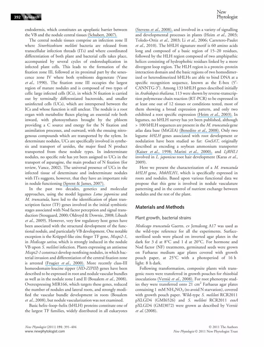
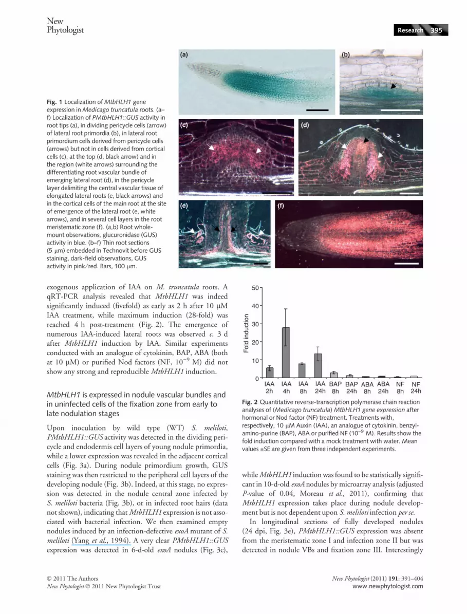
To precisely determine the tissue localization of MtbHLH1transcripts, we generated a transcriptional fusion between a1.44 kbp MtbHLH1 promoter fragment and the GUS repor-ter gene. The expression pattern of this PMtbHLH1::GUSconstruct was examined in A. rhizogenes-transformedM. truncatula roots. The highest nonsymbiotic expressionwas found in the root meristematic region, with a signalstrongly diminishing in the root elongation zone (Fig. 1a).MtbHLH1 expression was also detected in lateral root pri-mordia, where it was first confined to the dividing pericyclecells while it was undetectable in the adjacent endodermis orcortical cell layers (Fig. 1b). At a later stage of development,most internal root primordium cells were intensely stainedfor GUS activity (Fig. 1c). A closer observation revealed thatGUS expression was found in primordium cells derived frompericycle cells but not in cells derived from cortical cells(Fig. 1c). When the lateral root began to emerge, GUS stain-ing was largely restricted to the regions at the top andsurrounding the differentiating root vascular bundle(Fig. 1d). On elongated lateral roots, GUS activity wasobserved in the pericycle layer delimiting the central vasculartissue (Fig. 1e) and in the cortical cells of the main root at thesite of emergence of the lateral root (Fig. 1e, arrowheads).Finally, GUS staining was also detected in several cell layersin the lateral root meristematic zone (Fig. 1f), as in the mainroot (Fig. 1a).
Such an expression pattern is reminiscent of that exhibitedby auxin-induced genes, notably in M. truncatula (vanNoorden et al., 2007; Mathesius, 2008). We thus decided totest whether MtbHLH1 expression could be upregulated by
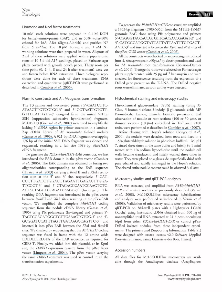
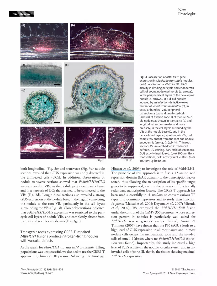
exogenous application of IAA on M. truncatula roots. AqRT-PCR analysis revealed that MtbHLH1 was indeedsignificantly induced (fivefold) as early as 2 h after 10 lMIAA treatment, while maximum induction (28-fold) wasreached 4 h post-treatment (Fig. 2). The emergence ofnumerous IAA-induced lateral roots was observed c. 3 dafter MtbHLH1 induction by IAA. Similar experimentsconducted with an analogue of cytokinin, BAP, ABA (bothat 10 lM) or purified Nod factors (NF, 10)9 M) did notshow any strong and reproducible MtbHLH1 induction.
MtbHLH1 is expressed in nodule vascular bundles andin uninfected cells of the fixation zone from early tolate nodulation stages
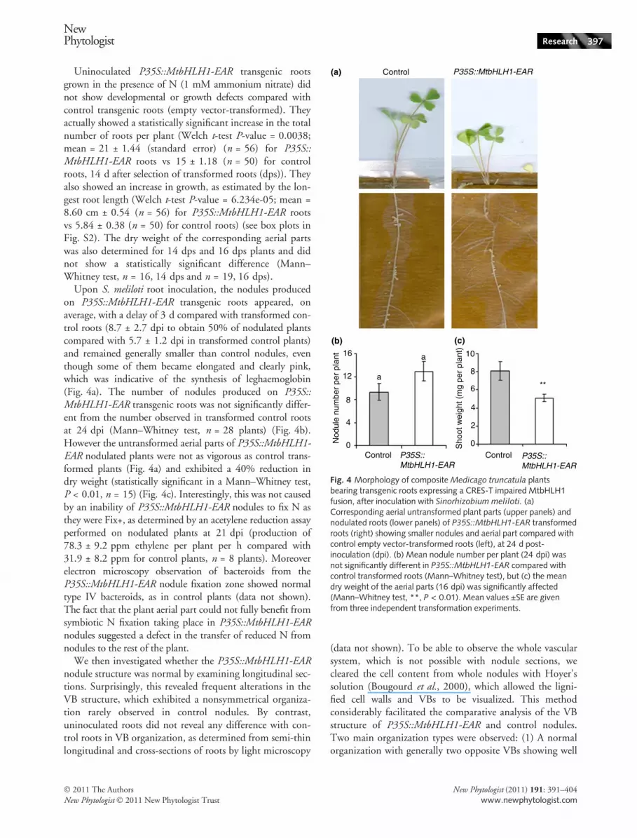
Upon inoculation by wild type (WT) S. meliloti,PMtbHLH1::GUS activity was detected in the dividing peri-cycle and endodermis cell layers of young nodule primordia,while a lower expression was revealed in the adjacent corticalcells (Fig. 3a). During nodule primordium growth, GUSstaining was then restricted to the peripheral cell layers of thedeveloping nodule (Fig. 3b). Indeed, at this stage, no expres-sion was detected in the nodule central zone infected byS. meliloti bacteria (Fig. 3b), or in infected root hairs (datanot shown), indicating that MtbHLH1 expression is not asso-ciated with bacterial infection. We then examined emptynodules induced by an infection-defective exoA mutant of S.meliloti (Yang et al., 1994). A very clear PMtbHLH1::GUSexpression was detected in 6-d-old exoA nodules (Fig. 3c),
while MtbHLH1 induction was found to be statistically signifi-cant in 10-d-old exoA nodules by microarray analysis (adjustedP-value of 0.04, Moreau et al., 2011), confirming thatMtbHLH1 expression takes place during nodule develop-ment but is not dependent upon S. meliloti infection per se.
In longitudinal sections of fully developed nodules(24 dpi, Fig. 3e), PMtbHLH1::GUS expression was absentfrom the meristematic zone I and infection zone II but wasdetected in nodule VBs and fixation zone III. Interestingly
(a) (b)
(c) (d)
(e) (f)
Fig. 1 Localization of MtbHLH1 geneexpression in Medicago truncatula roots. (a–f) Localization of PMtbHLH1::GUS activity inroot tips (a), in dividing pericycle cells (arrow)of lateral root primordia (b), in lateral rootprimordium cells derived from pericycle cells(arrows) but not in cells derived from corticalcells (c), at the top (d, black arrow) and inthe region (white arrows) surrounding thedifferentiating root vascular bundle ofemerging lateral root (d), in the pericyclelayer delimiting the central vascular tissue ofelongated lateral roots (e, black arrows) andin the cortical cells of the main root at the siteof emergence of the lateral root (e, whitearrows), and in several cell layers in the rootmeristematic zone (f). (a,b) Root whole-mount observations, glucuronidase (GUS)activity in blue. (b–f) Thin root sections(5 lm) embedded in Technovit before GUSstaining, dark-field observations, GUSactivity in pink ⁄ red. Bars, 100 lm.
50
40
30
20
10
0
Fol
d in
duct
ion
IAA IAA IAA IAA2h 4h 8h 24h 8h 24h 8h 24h 8h 24h
BAP BAP ABA ABA NF NF
Fig. 2 Quantitative reverse-transcription polymerase chain reactionanalyses of (Medicago truncatula) MtbHLH1 gene expression afterhormonal or Nod factor (NF) treatment. Treatments with,respectively, 10 lM Auxin (IAA), an analogue of cytokinin, benzyl-amino-purine (BAP), ABA or purified NF (10)9 M). Results show thefold induction compared with a mock treatment with water. Meanvalues ±SE are given from three independent experiments.
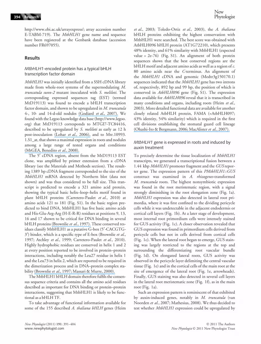
both longitudinal (Fig. 3e) and transverse (Fig. 3d) nodulesections revealed that GUS expression was only detected inthe uninfected cells (UCs). In addition, observations ofnodule transverse sections showed that PMtbHLH1::GUSwas expressed in VBs, in the nodule peripheral parenchymaand in a network of UCs that seemed to be connected to theVBs (Fig. 3d). Longitudinal sections also revealed a strongGUS expression at the nodule base, in the region connectingthe nodule to the root VB, particularly in the cell layerssurrounding the VBs (Fig. 3f). Closer observations indicatedthat PMtbHLH1::GUS expression was restricted to the peri-cycle cell layers of nodule VBs, and completely absent fromthe root and nodule endodermis (Fig. 3g,h).
As the search for MtbHLH1 mutants in M. truncatula Tillingpopulations was unsuccessful, we decided to use the CRES-Tapproach (Chimeric REpressor Silencing Technology,
Hiratsu et al., 2003) to investigate the role of MtbHLH1.The principle of this approach is to fuse a 12 amino acidrepression domain (EAR domain) to the transcription factortested, thus allowing the transcription of its specific targetgenes to be suppressed, even in the presence of functionallyredundant transcription factors. The CRES-T approach hasbeen used successfully in A. thaliana to convert various TFtypes into dominant repressors and to study their functionin planta (Matsui et al., 2005; Koyama et al., 2007; Mitsudaet al., 2007). We expressed the MtbHLH1::EAR fusionunder the control of the CaMV 35S promoter, whose expres-sion pattern in nodules is particularly well suited forMtbHLH1 reverse genetics studies. Indeed, Auriac &Timmers (2007) have shown that the P35S::GUS leads to ahigh level of GUS expression in all root tissues and in mostnodule cells except the meristematic zone and the invadedcells of zone III (tissues where no PMtbHLH1::GUS expres-sion was found). Importantly, this study indicated a highlevel of P35S activity in the nodule vascular system and in un-invaded cells of zone III, that is, the tissues showing maximalMtbHLH1 expression.
(a) (b)
(c) (e)
(d)
(f) (g) (h)
Fig. 3 Localization of MtbHLH1 geneexpression in Medicago truncatula nodules.(a–h) Localization of PMtbHLH1::GUSactivity in dividing pericycle and endodermiscells of young nodule primordia (a, arrows),in the peripheral cell layers of the developingnodule (b, arrows), in 6-d-old nodulesinduced by an infection-defective exoA
mutant of Sinorhizobium meliloti (c), invascular bundles (VB), peripheralparenchyma (pa) and uninfected cells(arrows) of fixation zone III of mature 24-d-old nodules as shown in transverse (d) andlongitudinal sections (e–h), and moreprecisely, in the cell layers surrounding theVBs at the nodule base (f), and in thepericycle cell layers (pe) of nodule VBs, butcompletely absent from the root and noduleendodermis (en) (g,h). (a,b,f–h) Thin rootsections (5 lm) embedded in Technovitbefore GUS staining, dark-field observations,GUS activity in pink ⁄ red. (c–e) 100-lm thickroot sections, GUS activity in blue. Bars: (a–f)100 lm; (g,h) 50 lm.
Uninoculated P35S::MtbHLH1-EAR transgenic rootsgrown in the presence of N (1 mM ammonium nitrate) didnot show developmental or growth defects compared withcontrol transgenic roots (empty vector-transformed). Theyactually showed a statistically significant increase in the totalnumber of roots per plant (Welch t-test P-value = 0.0038;mean = 21 ± 1.44 (standard error) (n = 56) for P35S::MtbHLH1-EAR roots vs 15 ± 1.18 (n = 50) for controlroots, 14 d after selection of transformed roots (dps)). Theyalso showed an increase in growth, as estimated by the lon-gest root length (Welch t-test P-value = 6.234e-05; mean =8.60 cm ± 0.54 (n = 56) for P35S::MtbHLH1-EAR rootsvs 5.84 ± 0.38 (n = 50) for control roots) (see box plots inFig. S2). The dry weight of the corresponding aerial partswas also determined for 14 dps and 16 dps plants and didnot show a statistically significant difference (Mann–Whitney test, n = 16, 14 dps and n = 19, 16 dps).
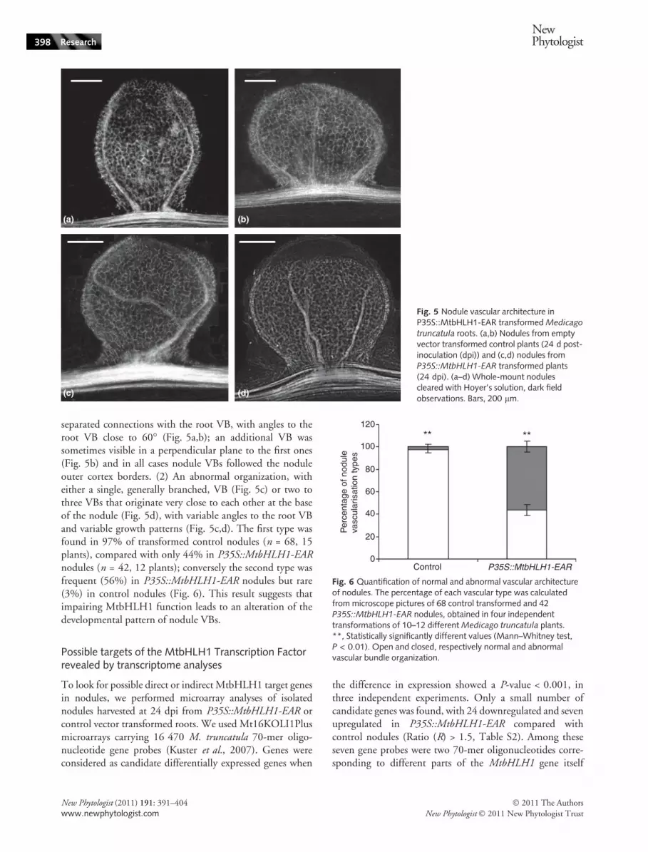
Upon S. meliloti root inoculation, the nodules producedon P35S::MtbHLH1-EAR transgenic roots appeared, onaverage, with a delay of 3 d compared with transformed con-trol roots (8.7 ± 2.7 dpi to obtain 50% of nodulated plantscompared with 5.7 ± 1.2 dpi in transformed control plants)and remained generally smaller than control nodules, eventhough some of them became elongated and clearly pink,which was indicative of the synthesis of leghaemoglobin(Fig. 4a). The number of nodules produced on P35S::MtbHLH1-EAR transgenic roots was not significantly differ-ent from the number observed in transformed control rootsat 24 dpi (Mann–Whitney test, n = 28 plants) (Fig. 4b).However the untransformed aerial parts of P35S::MtbHLH1-EAR nodulated plants were not as vigorous as control trans-formed plants (Fig. 4a) and exhibited a 40% reduction indry weight (statistically significant in a Mann–Whitney test,P < 0.01, n = 15) (Fig. 4c). Interestingly, this was not causedby an inability of P35S::MtbHLH1-EAR nodules to fix N asthey were Fix+, as determined by an acetylene reduction assayperformed on nodulated plants at 21 dpi (production of78.3 ± 9.2 ppm ethylene per plant per h compared with31.9 ± 8.2 ppm for control plants, n = 8 plants). Moreoverelectron microscopy observation of bacteroids from theP35S::MtbHLH1-EAR nodule fixation zone showed normaltype IV bacteroids, as in control plants (data not shown).The fact that the plant aerial part could not fully benefit fromsymbiotic N fixation taking place in P35S::MtbHLH1-EARnodules suggested a defect in the transfer of reduced N fromnodules to the rest of the plant.
We then investigated whether the P35S::MtbHLH1-EARnodule structure was normal by examining longitudinal sec-tions. Surprisingly, this revealed frequent alterations in theVB structure, which exhibited a nonsymmetrical organiza-tion rarely observed in control nodules. By contrast,uninoculated roots did not reveal any difference with con-trol roots in VB organization, as determined from semi-thinlongitudinal and cross-sections of roots by light microscopy
(data not shown). To be able to observe the whole vascularsystem, which is not possible with nodule sections, wecleared the cell content from whole nodules with Hoyer’ssolution (Bougourd et al., 2000), which allowed the ligni-fied cell walls and VBs to be visualized. This methodconsiderably facilitated the comparative analysis of the VBstructure of P35S::MtbHLH1-EAR and control nodules.Two main organization types were observed: (1) A normalorganization with generally two opposite VBs showing well
10
8
6
0Nod
ule
num
ber
per
plan
t
4
2
a
a
**
Control
Control Control
P35S::MtbHLH1-EAR
P35S::MtbHLH1-EAR
P35S::MtbHLH1-EAR
1016
12
8
8
6
4
0 0Sho
ot w
eigh
t (m
g pe
r pl
ant)
4
2
a
a
**
(b)
(a)
(c)
Fig. 4 Morphology of composite Medicago truncatula plantsbearing transgenic roots expressing a CRES-T impaired MtbHLH1fusion, after inoculation with Sinorhizobium meliloti. (a)Corresponding aerial untransformed plant parts (upper panels) andnodulated roots (lower panels) of P35S::MtbHLH1-EAR transformedroots (right) showing smaller nodules and aerial part compared withcontrol empty vector-transformed roots (left), at 24 d post-inoculation (dpi). (b) Mean nodule number per plant (24 dpi) wasnot significantly different in P35S::MtbHLH1-EAR compared withcontrol transformed roots (Mann–Whitney test), but (c) the meandry weight of the aerial parts (16 dpi) was significantly affected(Mann–Whitney test, **, P < 0.01). Mean values ±SE are givenfrom three independent transformation experiments.
separated connections with the root VB, with angles to theroot VB close to 60� (Fig. 5a,b); an additional VB wassometimes visible in a perpendicular plane to the first ones(Fig. 5b) and in all cases nodule VBs followed the noduleouter cortex borders. (2) An abnormal organization, witheither a single, generally branched, VB (Fig. 5c) or two tothree VBs that originate very close to each other at the baseof the nodule (Fig. 5d), with variable angles to the root VBand variable growth patterns (Fig. 5c,d). The first type wasfound in 97% of transformed control nodules (n = 68, 15plants), compared with only 44% in P35S::MtbHLH1-EARnodules (n = 42, 12 plants); conversely the second type wasfrequent (56%) in P35S::MtbHLH1-EAR nodules but rare(3%) in control nodules (Fig. 6). This result suggests thatimpairing MtbHLH1 function leads to an alteration of thedevelopmental pattern of nodule VBs.
Possible targets of the MtbHLH1 Transcription Factorrevealed by transcriptome analyses
To look for possible direct or indirect MtbHLH1 target genesin nodules, we performed microarray analyses of isolatednodules harvested at 24 dpi from P35S::MtbHLH1-EAR orcontrol vector transformed roots. We used Mt16KOLI1Plusmicroarrays carrying 16 470 M. truncatula 70-mer oligo-nucleotide gene probes (Kuster et al., 2007). Genes wereconsidered as candidate differentially expressed genes when
the difference in expression showed a P-value < 0.001, inthree independent experiments. Only a small number ofcandidate genes was found, with 24 downregulated and sevenupregulated in P35S::MtbHLH1-EAR compared withcontrol nodules (Ratio (R) > 1.5, Table S2). Among theseseven gene probes were two 70-mer oligonucleotides corre-sponding to different parts of the MtbHLH1 gene itself
120
100
80
60
40
20
0
Per
cent
age
of n
odul
eva
scul
aris
atio
n ty
pes
Control P35S::MtbHLH1-EAR
** **
Fig. 6 Quantification of normal and abnormal vascular architectureof nodules. The percentage of each vascular type was calculatedfrom microscope pictures of 68 control transformed and 42P35S::MtbHLH1-EAR nodules, obtained in four independenttransformations of 10–12 different Medicago truncatula plants.**, Statistically significantly different values (Mann–Whitney test,P < 0.01). Open and closed, respectively normal and abnormalvascular bundle organization.
truncatula roots. (a,b) Nodules from emptyvector transformed control plants (24 d post-inoculation (dpi)) and (c,d) nodules fromP35S::MtbHLH1-EAR transformed plants(24 dpi). (a–d) Whole-mount nodulescleared with Hoyer’s solution, dark fieldobservations. Bars, 200 lm.
398 Research
NewPhytologist
� 2011 The Authors
New Phytologist � 2011 New Phytologist Trust
New Phytologist (2011) 191: 391–404
www.newphytologist.com
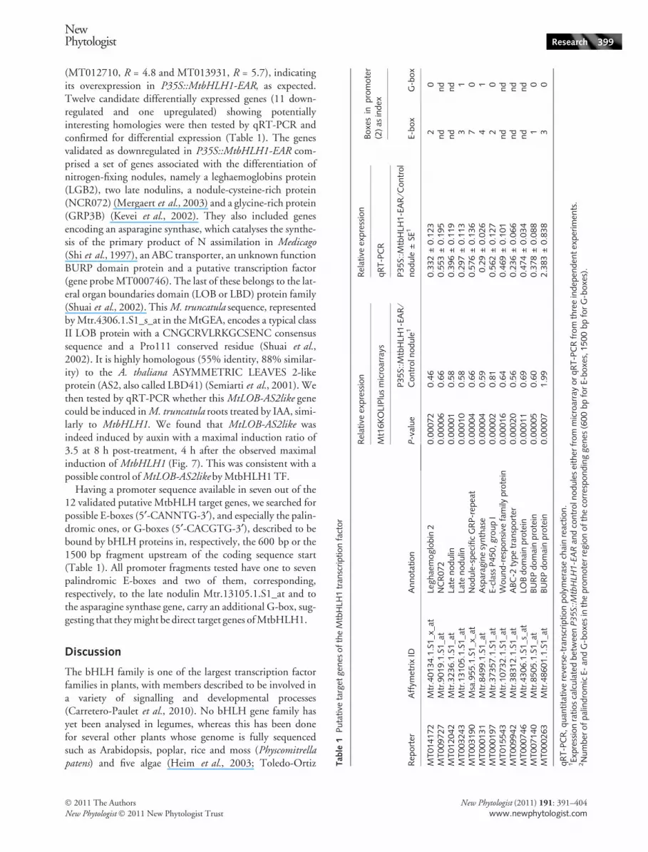
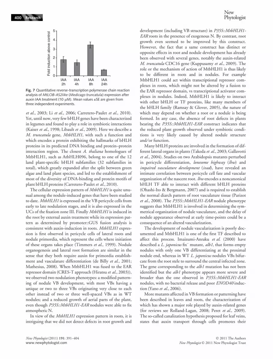
(MT012710, R = 4.8 and MT013931, R = 5.7), indicatingits overexpression in P35S::MtbHLH1-EAR, as expected.Twelve candidate differentially expressed genes (11 down-regulated and one upregulated) showing potentiallyinteresting homologies were then tested by qRT-PCR andconfirmed for differential expression (Table 1). The genesvalidated as downregulated in P35S::MtbHLH1-EAR com-prised a set of genes associated with the differentiation ofnitrogen-fixing nodules, namely a leghaemoglobins protein(LGB2), two late nodulins, a nodule-cysteine-rich protein(NCR072) (Mergaert et al., 2003) and a glycine-rich protein(GRP3B) (Kevei et al., 2002). They also included genesencoding an asparagine synthase, which catalyses the synthe-sis of the primary product of N assimilation in Medicago(Shi et al., 1997), an ABC transporter, an unknown functionBURP domain protein and a putative transcription factor(gene probe MT000746). The last of these belongs to the lat-eral organ boundaries domain (LOB or LBD) protein family(Shuai et al., 2002). This M. truncatula sequence, representedby Mtr.4306.1.S1_s_at in the MtGEA, encodes a typical classII LOB protein with a CNGCRVLRKGCSENC consensussequence and a Pro111 conserved residue (Shuai et al.,2002). It is highly homologous (55% identity, 88% similar-ity) to the A. thaliana ASYMMETRIC LEAVES 2-likeprotein (AS2, also called LBD41) (Semiarti et al., 2001). Wethen tested by qRT-PCR whether this MtLOB-AS2like genecould be induced in M. truncatula roots treated by IAA, simi-larly to MtbHLH1. We found that MtLOB-AS2like wasindeed induced by auxin with a maximal induction ratio of3.5 at 8 h post-treatment, 4 h after the observed maximalinduction of MtbHLH1 (Fig. 7). This was consistent with apossible control of MtLOB-AS2like by MtbHLH1 TF.
Having a promoter sequence available in seven out of the12 validated putative MtbHLH target genes, we searched forpossible E-boxes (5¢-CANNTG-3¢), and especially the palin-dromic ones, or G-boxes (5¢-CACGTG-3¢), described to bebound by bHLH proteins in, respectively, the 600 bp or the1500 bp fragment upstream of the coding sequence start(Table 1). All promoter fragments tested have one to sevenpalindromic E-boxes and two of them, corresponding,respectively, to the late nodulin Mtr.13105.1.S1_at and tothe asparagine synthase gene, carry an additional G-box, sug-gesting that they might be direct target genes of MtbHLH1.
Discussion
The bHLH family is one of the largest transcription factorfamilies in plants, with members described to be involved ina variety of signalling and developmental processes(Carretero-Paulet et al., 2010). No bHLH gene family hasyet been analysed in legumes, whereas this has been donefor several other plants whose genome is fully sequencedsuch as Arabidopsis, poplar, rice and moss (Physcomitrellapatens) and five algae (Heim et al., 2003; Toledo-Ortiz T
et al., 2003; Li et al., 2006; Carretero-Paulet et al., 2010).Yet, until now, very few bHLH genes have been characterizedin legumes and found to play a role in symbiotic interactions(Kaiser et al., 1998; Libault et al., 2009). Here we describe aM. truncatula gene, MtbHLH1, with such a function andwhich encodes a protein exhibiting the hallmarks of bHLHproteins in its predicted DNA binding and protein–proteininteraction region. The closest A. thaliana homologues ofMtbHLH1, such as AtbHLH096, belong to one of the 12land plant-specific bHLH subfamilies (32 subfamilies intotal), which greatly expanded after the split between greenalgae and land plant species, and led to the establishment ofmost of the diversity of DNA-binding and protein motifs ofplant bHLH proteins (Carretero-Paulet et al., 2010).
The cellular expression pattern of MtbHLH1 is quite unu-sual among the nodule-induced genes that have been studiedto date. MtbHLH1 is expressed in the VB pericycle cells fromearly to late nodulation stages, and it is also expressed in theUCs of the fixation zone III. Finally MtbHLH1 is induced inthe root by external auxin treatment while its expression pat-tern as determined by promoter::GUS fusion analysis isconsistent with auxin-induction in roots. MtbHLH1 expres-sion is first observed in pericycle cells of lateral roots andnodule primordia, which represent the cells where initiationof these organs takes place (Timmers et al., 1999). Noduleorganogenesis and lateral root formation are similar in thesense that they both require auxin for primordia establish-ment and vasculature differentiation (de Billy et al., 2001;Mathesius, 2008). When MtbHLH1 was fused to the EARrepressor domain (CRES-T approach (Hiratsu et al., 2003)),we observed two nodulation phenotypes: a modified pattern-ing of nodule VB development, with most VBs having aunique or two to three VBs originating very close to eachother instead of two or three well-spaced VBs as in WTnodules; and a reduced growth of aerial parts of the plant,even though P35S::MtbHLH1-EAR nodules were able to fixatmospheric N.
In view of the MtbHLH1 expression pattern in roots, it isintriguing that we did not detect defects in root growth and
development (including VB structure) in P35S::MtbHLH1-EAR roots in the presence of exogenous N. By contrast, rootgrowth even seemed to be improved by this construct.However, the fact that a same construct has distinct oropposite effects in root and nodule development has alreadybeen observed with several genes, notably the auxin-relatedM. truncatula CDC16 gene (Kuppusamy et al., 2009). Therole or the mechanism of action of MtbHLH1 is thus likelyto be different in roots and in nodules. For exampleMtbHLH1 could act within transcriptional repressor com-plexes in roots, which might not be altered by a fusion tothe EAR repressor domain, vs transcriptional activator com-plexes in nodules. Indeed, MtbHLH1 is likely to interactwith other bHLH or TF proteins, like many members ofthe bHLH family (Ramsay & Glover, 2005), the nature ofwhich may depend on whether a root or a nodule is beingformed. In any case, the absence of root defects in plantsbearing the P35S::MtbHLH1-EAR construct indicates thatthe reduced plant growth observed under symbiotic condi-tions is very likely caused by altered nodule structureand ⁄ or function.
Many bHLH proteins are involved in the formation of dif-ferent lateral organs in plants (Takeda et al., 2003; Gallavottiet al., 2004). Studies on two Arabidopsis mutants perturbedin pericycle differentiation, lonesome highway (lhw) andimpaired vasculature development (ivad), have revealed anintimate correlation between pericycle cell fate and vascularorganization of the nascent root. lhw encodes a noncanonicalbHLH TF able to interact with different bHLH proteins(Ohashi-Ito & Bergmann, 2007) and is required to establishthe normal diarch pattern of root vasculature tissue (Parizotet al., 2008). The P35S::MtbHLH1-EAR nodule phenotypesuggests that MtbHLH1 is involved in determining the sym-metrical organization of nodule vasculature, and the delay ofnodule appearance observed at early time-points could be aconsequence of an altered vascularization.
The development of nodule vascularization is poorly doc-umented and MtbHLH1 is one of the first TF described toaffect this process. Imaizumi-Anraku et al. (2000) havedescribed a L. japonicus fix– mutant, alb1, that forms emptynodules with only one VB differentiating at the proximalnodule end, whereas in WT L. japonicus nodules VBs bifur-cate from the root stele to surround the central infected zone.The gene corresponding to the alb1 mutation has not beenidentified but the alb1 phenotype appears more severe andbroader than the one observed in P35S::MtbHLH1-EARnodules, with no bacterial release and poor ENOD40 induc-tion (Yano et al., 2006).
More mutants affected in VB formation or patterning havebeen described in leaves and roots, the characterization ofwhich has shown a major role played by auxin-related genes(for reviews see Rolland-Lagan, 2008; Peret et al., 2009).The so-called canalization hypothesis proposed for leaf veins,states that auxin transport through cells promotes their
5
4
3
2
Fol
d in
duct
ion
abov
e co
ntro
l
1
0IAA2h
IAA4h
IAA8h
IAA24h
Fig. 7 Quantitative reverse-transcription polymerase chain reactionanalysis of MtLOB-AS2like (Medicago truncatula) expression afterauxin IAA treatment (10 lM). Mean values ±SE are given fromthree independent experiments.
400 Research
NewPhytologist
� 2011 The Authors
New Phytologist � 2011 New Phytologist Trust
New Phytologist (2011) 191: 391–404
www.newphytologist.com
differentiation into veins and thereby increases their capacityto transport auxin. It is very likely that patterning of noduleVBs also involves local auxin fluxes, and it should be recalledthat expression of MtLAX genes encoding auxin influx pro-teins correlates with VB formation in nodule primordia (deBilly et al., 2001). MtbHLH1 may therefore be involved inthe localization of auxin maxima or in auxin-regulatedevents. The position of auxin maxima probably variesdepending on whether a root or a nodule is being formed,leading either to a single central VB (in roots) or severalperipheral VBs (in nodules). MtbHLH1 expression is effi-ciently induced by auxin but we have no evidence thatMtbHLH1 controls auxin transporters and thereby contrib-utes to a positive feedback loop as proposed in thecanalization hypothesis. However, we found that MtbHLH1controls the expression of a gene encoding a protein verysimilar to the LOB transcription factor AS2 described to reg-ulate leaf venation. Thus, in A. thaliana, an as2 mutantexhibits asymmetrical venation and disconnected or insuffi-ciently connected veins (Semiarti et al., 2001), while ectopicAS2 expression leads to an altered vein patterning (Lin et al.,2003). Moreover Zgurski et al. (2005) have shown that theas2 phenotype is correlated with asymmetric auxin response.The MtLOB-AS2like gene thus represents an attractive can-didate to mediate MtbHLH1 role on VB development.Interestingly, the A. thaliana LOB gene AT5G63090, whichis expressed in a band of cells at the base of all lateral organs(Shuai et al., 2002), encodes a protein that has been shownto interact directly with members of the bHLH family(Husbands et al., 2007).
We interpret the reduced growth of the aerial part ofP35S::MtbHLH1-EAR composite plants as an alteration ofthe nodule capacity to deliver products of symbiotic N fixationto the plant, as plant growth was found to be similar to WT inthe presence of ammonium nitrate (without S. meliloti inocu-lation). The reduced growth could be caused by altered VBsor changes in the functioning of the cells where MtbHLH1 isexpressed, that is, VB pericycle cells and ⁄ or zone III UCs. Itshould be recalled that pericycle cells play a critical role fornutrient exchange with nearby tissues, and have been shownin different legume genera to exhibit an intense metabolicactivity (Pate et al., 1969), and that Abd-Alla et al. (2000)have reported that UCs from Vicia faba indeterminatenodules build up a symplasmic network through frequentplasmodesmata. Thus c. 30 times more plasmodesmata werecounted between UCs, and between UCs and infected cells(ICs) than between ICs, suggesting a role for UCs in metabo-lite transport. Such a role has already been established indeterminate nodules where UCs are specifically involved insynthesis and transport of ureides, the major product of Nfixation transported in determinate nodules (Vance, 2002).In Vicia faba indeterminate nodules, uptake experimentswith protoplasts isolated either from UCs or ICs, have led tothe proposition that UCs are involved in bringing sugar from
the phloem sap to the infected cells and transferring aminoacids symbiotically produced by ICs to the peripheral vascu-lar system (Peiter & Schubert, 2003; Peiter et al., 2004). It isthen tempting to propose that MtbHLH1 contributes tocontrolling nutrient exchange between nodule and root cells,that is, between ICs (source) and the rest of the plant (sink).Some of the genes, like the asparagine synthase or the ABCtransporter genes, that showed reduced expression inP35S::MtbHLH1-EAR nodules may be involved in this pro-cess. Only a few differentially expressed genes were found inP35S::MtbHLH1-EAR nodules at 24 dpi compared withcontrol nodules. Several of them were validated by indepen-dent qRT-PCR experiments and could be direct or indirecttarget genes of the MtbHLH1 TF. MtbHLH1 is likely to bea G-box DNA binder, as indicated by the presence of keyamino acids (H-E-R-R) in the basic region of the bHLHdomain, found in 44% of 638 plant bHLH proteins(Carretero-Paulet et al., 2010). The MtbHLH1 protein,once dimerized, could physically bind to the G-box found inthe promoters of some of the putative target genes revealedby transcriptome analyses, such as the asparagine synthaseand a late nodulin genes, and directly induce their transcrip-tion. In the bean legume, a bHLH protein carrying H-E-R-Ramino acids in the basic region has been shown to bind to theG-box motif of a gene encoding a seed-storage protein, the b-phaseolin (Kawagoe & Murai, 1996).
As far as early symbiotic stages are concerned,Complainville et al. (2003) have shown that M. truncatulanodule initiation induces symplasmic continuity betweenthe root phloem and nodule initials such as the pericyclecells and immature sieve elements that will give rise to vas-cularization. The symplasmic field created precedes nodulecell division and allows the transport of macromoleculesbetween root phloem and nodule (phloem unloading).MtbHLH1 might be involved in contributing to such cellto cell communications during early symbiotic stages, whichcould also explain the delay in nodulation observed inP35S::MtbHLH1:EAR roots. It would be very interesting inthe future to analyse more specifically the transcriptome ofnodule primordia cells and nodule UCs, for example bytaking advantage of laser microdissection, to avoid dilutionproblems and thereby have a better understanding of theirrole and the consequences of MtbHLH1 alteration.
Acknowledgements
We acknowledge the contribution of Francoise de Billy forcytological studies, Tatiana Vernie for providing the pPex-EAR vector, Jose Garcia, Anne Turchetti and AudreyLabarde for technical help, Etienne-Pascal Journet for hishelp with acetylene reduction assays, Jerome Gouzy andcollaborators, and Marie-Francoise Jardinaud for her helpwith statistical analyses (LIPM, Toulouse). We are gratefulto Julie Cullimore for critical reading and useful comments
NewPhytologist Research 401
� 2011 The Authors
New Phytologist � 2011 New Phytologist Trust
New Phytologist (2011) 191: 391–404
www.newphytologist.com
on the manuscript and to Patrick Vincourt for the timegiven to L.G. to finish this work on M. truncatula (LIPM).We thank Helge Kuster (Bielefeld University, Germany) forproviding 16K+ microarrays and Jean-Marie Prosperi(INRA, Montpellier) for A17 seeds. Quantitative RT-PCRexperiments were carried out at the Toulouse Genopole‘PLAGE’ platform. This work was funded by an EU grant(FP6 Grain Legume integrated project, GLIP). M.V. wassupported by two grants from the Agence Nationale de laRecherche (LEGoo GPLA06026G and SYMbiMICS PCS-08-GENO-106).
References
Abd-Alla MH, Koyro H-W, Yan F, Schubert S, Peiter E. 2000.
Functional structure of the indeterminate Vicia faba L. root nodule:
implications for metabolite transport. Journal of Plant Physiology 157:
335–343.
Atchley WR, Terhalle W, Dress A. 1999. Positional dependence, cliques,
and predictive motifs in the bHLH protein domain. Journal of MolecularEvolution 48: 501–516.
Auriac MC, Timmers AC. 2007. Nodulation studies in the model legume
Medicago truncatula: advantages of using the constitutive EF1alpha
promoter and limitations in detecting fluorescent reporter proteins in
Vandenbosch KA. 2009. Knockdown of CELL DIVISION CYCLE16
reveals an inverse relationship between lateral root and nodule numbers
and a link to auxin in Medicago truncatula. Plant Physiology 151:
1155–1166.
Kuster H, Becker A, Firnhaber C, Hohnjec N, Manthey K, Perlick AM,
Bekel T, Dondrup M, Henckel K, Goesmann A et al. 2007.
Development of bioinformatic tools to support EST-sequencing, in
402 Research
NewPhytologist
� 2011 The Authors
New Phytologist � 2011 New Phytologist Trust
New Phytologist (2011) 191: 391–404
www.newphytologist.com
silico- and microarray-based transcriptome profiling in mycorrhizal
symbioses. Phytochemistry 68: 19–32.
Li X, Duan X, Jiang H, Sun Y, Tang Y, Yuan Z, Guo J, Liang W, Chen
L, Yin J et al. 2006. Genome-wide analysis of basic ⁄ helix–loop–helix
transcription factor family in rice and Arabidopsis. Plant Physiology 141:
1167–1184.
Libault M, Joshi T, Benedito VA, Xu D, Udvardi MK, Stacey G. 2009.
Legume transcription factor genes: what makes legumes so special? PlantPhysiology 151: 991–1001.
Limpens E, Ramos J, Franken C, Raz V, Compaan B, Franssen H,
Bisseling T, Geurts R. 2004. RNA interference in Agrobacteriumrhizogenes-transformed roots of Arabidopsis and Medicago truncatulaJournal of Experimental Botany 55: 983–992.
Lin WC, Shuai B, Springer PS. 2003. The Arabidopsis LATERAL
ORGAN BOUNDARIES-domain gene ASYMMETRIC LEAVES2
functions in the repression of KNOX gene expression and in adaxial–
abaxial patterning. Plant Cell 15: 2241–2252.
Lohar DP, Sharopova N, Endre G, Penuela S, Samac D, Town C,
Silverstein KA, VandenBosch KA. 2006. Transcript analysis of early
nodulation events in Medicago truncatula. Plant Physiology 140: 221–234.
MacAlister CA, Ohashi-Ito K, Bergmann DC. 2007. Transcription factor
control of asymmetric cell divisions that establish the stomatal lineage.
Nature 445: 537–540.
Marini AM, Springael JY, Frommer WB, Andre B. 2000. Cross-talk
between ammonium transporters in yeast and interference by the
Massari ME, Murre C. 2000. Helix–loop–helix proteins: regulators of
transcription in eukaryotic organisms. Molecular and Cellular Biology 20:
429–440.
Mathesius U. 2008. Auxin: at the root of nodule development? FunctionalPlant Biology 35: 651–668.
Matsui K, Hiratsu K, Koyama T, Tanaka H, Ohme-Takagi M. 2005. A
chimeric AtMYB23 repressor induces hairy roots, elongation of leaves
and stems, and inhibition of the deposition of mucilage on seed coats in
Arabidopsis. Plant and Cell Physiology 46: 147–155.
Mergaert P, Nikovics K, Kelemen Z, Maunoury N, Vaubert D,
Kondorosi A, Kondorosi E. 2003. A novel family in Medicagotruncatula consisting of more than 300 nodule-specific genes coding for
small, secreted polypeptides with conserved cysteine motifs. PlantPhysiology 132: 161–173.
Mitsuda N, Iwase A, Yamamoto H, Yoshida M, Seki M, Shinozaki K,
Ohme-Takagi M. 2007. NAC transcription factors, NST1 and NST3,
are key regulators of the formation of secondary walls in woody tissues
of Arabidopsis. Plant Cell 19: 270–280.
Moreau S, Verdenaud M, Ott T, Letort S, de Billy F, Niebel A, Gouzy J,
de Carvalho-Niebel F, Gamas P. 2011. Transcription reprogramming
during root nodule development in Medicago truncatula. PLoS ONE 6:
e16463.
van Noorden GE, Kerim T, Goffard N, Wiblin R, Pellerone FI, Rolfe
BG, Mathesius U. 2007. Overlap of proteome changes in Medicagotruncatula in response to auxin and Sinorhizobium meliloti. PlantPhysiology 144: 1115–1131.
Ohashi-Ito K, Bergmann DC. 2006. Arabidopsis FAMA controls the final
proliferation ⁄ differentiation switch during stomatal development. PlantCell 18: 2493–2505.
Ohashi-Ito K, Bergmann DC. 2007. Regulation of the Arabidopsis root
vascular initial population by LONESOME HIGHWAY. Development134: 2959–2968.
Oldroyd GE, Downie JA. 2008. Coordinating nodule morphogenesis with
rhizobial infection in legumes. Annual Review of Plant Biology 59:
Anraku H, Kawaguchi M, Hayashi M. 2006. New nodulation mutants
responsible for infection thread development in Lotus japonicus.Molecular Plant–Microbe Interactions 19: 801–810.
Zgurski JM, Sharma R, Bolokoski DA, Schultz EA. 2005. Asymmetric
auxin response precedes asymmetric growth and differentiation of
asymmetric leaf1 and asymmetric leaf2 Arabidopsis leaves. Plant Cell 17:
77–91.
Supporting Information
The following materials can be found in the online versionof this article.
Fig. S1 Alignment of the predicted MtbHLH1 bHLHdomain with consensus sequences from plant bHLH pro-teins and protein sequence alignment between MtbHLH1
and its closest homologue from Arabidopsis thaliana,AT1G72210.
Fig. S2 Box plot representations of noninoculated root archi-tecture, following Agrobacterium rhizogenes transformationwith an empty vector or a P35S::MtbHLH1-EAR construct.
Table S1 Primers used for quantitative reverse-transcriptionpolymerase chain reaction amplification of some MtbHLH1candidate target genes
Table S2 Putative target genes of the MtbHLH1Transcription Factor obtained by transcriptome analyses
Please note: Wiley-Blackwell are not responsible for thecontent or functionality of any supporting informationsupplied by the authors. Any queries (other than missingmaterial) should be directed to the New Phytologist CentralOffice.