168
1 Prevention and treatment of infections in patients with haematological malignancies Gert Jan Timmers
| Date post: | 23-Apr-2023 |
| Category: |
Documents |
| Upload: | healthfoodinnovationmanagement |
| View: | 0 times |
| Download: | 0 times |
1
Prevention and treatment of infections in patients with haematological malignancies
Gert Jan Timmers
2
The publication of this thesis was financially supported by Amgen Oncology, Bayer Schering
Pharma BV, Bristol-Myers Squibb BV, Celgene BV, Leo Pharma BV, Novartis Oncology,
Ortho Biotech, a division of Janssen-Cilag BV, Roche Nederland BV, Shire International
Licensing BV, Sanofi Aventis and Freesia Funding Leiden.
Cover design and layout by Jozien & Gert Jan Timmers
Copyright © 2008, Gert Jan Timmers, Badhoevedorp, The Netherlands, all rights reserved
Printed by Buijten & Schipperheijn, Amsterdam, the Netherlands
ISBN/EAN: 978-90-9022950-8

3
VRIJE UNIVERSITEIT
Prevention and treatment of infections in patients with haematological malignancies
ACADEMISCH PROEFSCHRIFT
ter verkrijging van de graad Doctor aan
de Vrije Universiteit Amsterdam,
op gezag van de rector magnificus
prof.dr. L.M. Bouter,
in het openbaar te verdedigen
ten overstaan van de promotiecommissie
van de faculteit der Geneeskunde
op vrijdag 23 mei 2008 om 10.45 uur
in het auditorium van de universiteit,
De Boelelaan 1105
door
Gerard Jan Timmers
geboren te Leiden

4
promotor: prof.dr. P.C. Huijgens
copromotor: dr. A.M. Simoons-Smit

5
Voor Erica, Jozien en Annemarieke,
in liefdevolle herinnering aan Myrthe

6

7
Contents
Chapter 1 Background of the thesis, aims and outline
9
Chapter 2 General introduction
15
Chapter 3 Pharmacokinetics and effects on bowel and throat microflora of oral
levofloxacin as antibacterial prophylaxis in neutropenic patients
with haematological malignancies.
Bone Marrow Transplantation 2004;33:847-853.
47
Chapter 4 Levofloxacin vs. ciprofloxacin plus phenethicillin for the prevention
of bacterial infections in patients with haematological malignancies.
Clinical Microbiology and Infection 2007;13:497-503.
65
Chapter 5 Amphotericin B colloidal dispersion (Amphocil) vs fluconazole for
the prevention of fungal infections in neutropenic patients: data of a
prematurely stopped clinical trial.
Bone Marrow Transplantation 2000;25:879-884.
83
Chapter 6 Effects of cyclosporin A on single-dose pharmacokinetics of
intravenous itraconazole in patients with haematological
malignancies.
Therapeutic Drug Monitoring, accepted for publication
97
Chapter 7 Cefpirome as empirical treatment for febrile neutropenia in patients
with haematological malignancies.
Haematologica 2005;90:1005-1006.
111
Chapter 8 Outbreak of vancomycin-resistant Enterococcus faecium in a
haematology unit: risk factor assessment and successful control of
the epidemic.
British Journal of Haematology 2002;116:826-833.
127
Chapter 9 Summary and concluding remarks
143
Nederlandstalige samenvatting
151
Dankwoord
161
Curriculum vitae
167

8

9
Background of the thesis, aims and outline
Chapter 3 3 1
Chapter 1
10
Background of the thesis, aims and outline
In the Netherlands, approximately 73.000 new patients are diagnosed with cancer every year,
with a yearly mortality of about 38.000 patients. Malignancies of the blood and bone marrow
represent less than 10% of all cancers, which in the Netherlands accounts for 7000 patients
diagnosed with a haematological malignant disease every year and a yearly mortality among
these patients of about 3000 subjects. Over the past decades, the cure rate and prognosis of
patients with haematological disease has improved substantially.1 Above all, this may be
attributed to advancements in therapy, since treatment options for patients with
haematological malignancies have expanded and greatly improved. Unfortunately, the
progress in treatment for haematological malignancies comes with the cost of serious
impairment of host defence mechanisms. This occurs in a population of patients that is
already running a serious risk of infections, due to the underlying disease, which resides by
definition in the immune system itself.
Autologous stem cell transplantation is a valuable option for the treatment of patients with,
for example, aggressive or relapsed lymphoma, acute leukemia and multiple myeloma.2-6
These patients however, suffer from a temporary lack of neutrophil granulocytes until
engraftment occurs and in addition, dose-intensified chemotherapeutic regimens may lead to
damage of mucosal surfaces. Amongst others, both neutropenia and the occurrence of
mucositis have been identified as important risk factors for the acquisition of serious
opportunistic infections in these patients.7-9
Allogeneic stem cell transplantation has also proven to be a powerful tool in the treatment of a
variety of haematological diseases.10-13
However, success can not be achieved without the
application of immunosuppressive drugs, first for the prevention of graft failure and later for
the control of graft-versus-host disease. From these, it becomes evident that deficiencies in
host defence mechanisms in allogeneic stem cell recipients are multiple, rendering these
patients at a high risk of contracting serious viral, bacterial and fungal infections.14-19
Apart from the transplantation setting, in the past two decades immunotherapeutics have been
successfully introduced in treatment schedules for haematological disease. Monoclonal
antibodies including rituximab and alemtuzumab and drugs like the purine analogs such as
fludarabin, have become widely used to date. These drugs however, directly interfere with B-
and T-cell function, and their application has been paralelled by the emergence of infections
by a variety of microorganisms, such as listeriosis, pneumocystosis, mycobacterial infections,
and fungal and viral infections.20-23

Background of the thesis, aims and outline
11
Infectious complications seriously hamper the treatment of patients with haematological
malignancies and are associated with severe morbidity and mortality. Hence, to date, it
becomes evident that the survival of these patients heavily depends on the quality of
supportive care. The search for ways to prevent and if necessary treat these infections has
tried to keep pace with the improvement and intensification of treatment strategies. In view of
the serious consequences of infections in patients treated for haematological disease, it is not
surprising that newly marketed and promising antibacterial and antifungal drugs are readily
introduced into daily clinical practice. Yet, sometimes important questions remain to be
answered, with special interest to the population involved. Antibacterial or antifungal drugs
may be adequately tested in healthy volunteers and be found safe and effective as treatment
for short periods of use in the general population. However, when these drugs are applied in
patients with haematological disease, quite often no data exist on safety, pharmacokinetic
behaviour and their efficacy for prophylaxis or treatment in this specific population.
Pharmacokinetic properties of antimicrobial agents, such as absorption, distribution and
elimination may be significantly altered in patients with haematological malignancies, due to,
for example, the underlying disease, low albumin state, the occurrence of graft-versus-host
disease and mucosal damage.24-26
Furthermore, when antibiotics are applied for antibacterial
or antifungal prophylaxis, prolonged administration may lead to the emergence of resistant
microorganisms.
In view of these considerations, this thesis aims to explore the safety and efficacy of a
selection of new antimicrobials, for the prevention and treatment of bacterial and fungal
infections in patients who are hospitalized for the treatment of a haematological malignancy.
Chapter 2 provides an overview from a clinical perspective of current insights and
developments on antibacterial and antifungal prophylaxis and therapy in neutropenic patients.
In chapter 3 the pharmacokinetic properties of levofloxacin are explored, when used as
antibacterial prophylaxis. Levofloxacin is a relatively new quinolone with enhanced activity
against Gram-positive bacteria, and special attention is aimed at its effects on the microflora
of the digestive tract. The findings in this study led to the design of a randomized clinical
trial, comparing levofloxacin with ciprofloxacin-phenethicillin as antibacterial prophylaxis,
the results of which are described in chapter 4. In chapter 5 the use of amphotericicn B
colloidal dispersion as antifungal prophylaxis is described, with an emphasis on toxicity of the
drug. Chapter 6 includes the results of a clinical trial that was designed to investigate the
effects of cyclosporin A on single dose pharmacokinetics of itraconazole. The introduction of
Chapter 1
12
cefpirome as empirical antibacterial treatment in neutropenic patients with fever, led to the
design of an observational cohort study to evaluate its efficacy and safety, the results of which
are described in chapter 7. In addition pharmacokinetic data were collected in a subset of
patients to define the optimal dosing regimen for cefpirome. In chapter 8, the occurrence of a
nosocomial outbreak of vancomycin-resistant Enterococcus faecium on our ward is described,
including the results of a risk-factors analysis and measures that led to control of the
epidemic. Chapter 9 summarizes and discusses the main findings of the thesis.
References
1. Brenner H, Gondos A, Arndt V. Recent major progress in long-term cancer patient survival
disclosed by modeled period analysis. J Clin Oncol 2007; 25: 3274-3280.
2. Attal M, Harousseau JL, Facon T, Guilhot F, Doyen C, Fuzibet JG, Monconduit M, Hulin C,
Caillot D, Bouabdallah R, Voillat L, Sotto JJ, Grosbois B, Bataille R. Single versus double
autologous stem-cell transplantation for multiple myeloma. N Engl J Med 2003; 349: 2495-
2502.
3. Philip T, Guglielmi C, Hagenbeek A, Somers R, Van der LH, Bron D, Sonneveld P,
Gisselbrecht C, Cahn JY, Harousseau JL, . Autologous bone marrow transplantation as
compared with salvage chemotherapy in relapses of chemotherapy-sensitive non-Hodgkin's
lymphoma. N Engl J Med 1995; 333: 1540-1545.
4. Milpied N, Deconinck E, Gaillard F, Delwail V, Foussard C, Berthou C, Gressin R, Lucas V,
Colombat P, Harousseau JL. Initial treatment of aggressive lymphoma with high-dose
chemotherapy and autologous stem-cell support. N Engl J Med 2004; 350: 1287-1295.
5. Cassileth PA, Harrington DP, Appelbaum FR, Lazarus HM, Rowe JM, Paietta E, Willman C,
Hurd DD, Bennett JM, Blume KG, Head DR, Wiernik PH. Chemotherapy compared with
autologous or allogeneic bone marrow transplantation in the management of acute myeloid
leukemia in first remission. N Engl J Med 1998; 339: 1649-1656.
6. Harousseau JL, Cahn JY, Pignon B, Witz F, Milpied N, Delain M, Lioure B, Lamy T, Desablens
B, Guilhot F, Caillot D, Abgrall JF, Francois S, Briere J, Guyotat D, Casassus P, Audhuy B,
Tellier Z, Hurteloup P, Herve P. Comparison of autologous bone marrow transplantation and
intensive chemotherapy as postremission therapy in adult acute myeloid leukemia. The Groupe
Ouest Est Leucemies Aigues Myeloblastiques (GOELAM). Blood 1997; 90: 2978-2986.
7. Bodey GP, Buckley M, Sathe YS, Freireich EJ. Quantitative relationships between circulating
leukocytes and infection in patients with acute leukemia. Ann Intern Med 1966; 64: 328-340.
Background of the thesis, aims and outline
13
8. Bow EJ. Management of the febrile neutropenic cancer patient: lessons from 40 years of study.
Clin Microbiol Infect 2005; 11 Suppl 5: 24-29.
9. Blijlevens NM. Implications of treatment-induced mucosal barrier injury. Curr Opin Oncol
2005; 17: 605-610.
10. Barrett J. Allogeneic stem cell transplantation for chronic myeloid leukemia. Semin Haematol
2003; 40: 59-71.
11. Wayne AS, Barrett AJ. Allogeneic haematopoietic stem cell transplantation for
myeloproliferative disorders and myelodysplastic syndromes. Haematol Oncol Clin North Am
2003; 17: 1243-1260.
12. Singhal S, Powles R, Henslee-Downey PJ, Chiang KY, Treleaven J, Godder K, Kulkarni S, van
Rhee F, Sirohi B, Pinkerton CR, Meller S, Mehta J. Allogeneic transplantation from HLA-
matched sibling or partially HLA-mismatched related donors for primary refractory acute
leukemia. Bone Marrow Transplant 2002; 29: 291-295.
13. Smith SM, Grinblatt D, van Besien K. Autologous and allogeneic transplantation for aggressive
NHL. Cytotherapy 2002; 4: 223-240.
14. Sayer HG, Longton G, Bowden R, Pepe M, Storb R. Increased risk of infection in marrow
transplant patients receiving methylprednisolone for graft-versus-host disease prevention. Blood
1994; 84: 1328-1332.
15. Williamson EC, Millar MR, Steward CG, Cornish JM, Foot AB, Oakhill A, Pamphilon DH,
Reeves B, Caul EO, Warnock DW, Marks DI. Infections in adults undergoing unrelated donor
bone marrow transplantation. Br J Haematol 1999; 104: 560-568.
16. Mohty M, Faucher C, Vey N, Stoppa AM, Viret F, Chabbert I, Chabannon C, Bouabdallah R,
Ladaique P, Collet L, Zandotti C, Maraninchi D, Blaise D. High rate of secondary viral and
bacterial infections in patients undergoing allogeneic bone marrow mini-transplantation. Bone
Marrow Transplant 2000; 26: 251-255.
17. Upton A, Kirby KA, Carpenter P, Boeckh M, Marr KA. Invasive aspergillosis following
haematopoietic cell transplantation: outcomes and prognostic factors associated with mortality.
Clin Infect Dis 2007; 44: 531-540.
18. Junghanss C, Marr KA. Infectious risks and outcomes after stem cell transplantation: are
nonmyeloablative transplants changing the picture? Curr Opin Infect Dis 2002; 15: 347-353.
19. Maury S, Mary JY, Rabian C, Schwarzinger M, Toubert A, Scieux C, Carmagnat M, Esperou H,
Ribaud P, Devergie A, Guardiola P, Vexiau P, Charron D, Gluckman E, Socie G. Prolonged
immune deficiency following allogeneic stem cell transplantation: risk factors and complications
in adult patients. Br J Haematol 2001; 115: 630-641.
20. Lin PC, Hsiao LT, Poh SB, Wang WS, Yen CC, Chao TC, Liu JH, Chiou TJ, Chen PM. Higher
fungal infection rate in elderly patients (more than 80 years old) suffering from diffuse large B
cell lymphoma and treated with rituximab plus CHOP. Ann Haematol 2007; 86: 95-100.
Chapter 1
14
21. Thursky KA, Worth LJ, Seymour JF, Miles PH, Slavin MA. Spectrum of infection, risk and
recommendations for prophylaxis and screening among patients with lymphoproliferative
disorders treated with alemtuzumab. Br J Haematol 2006; 132: 3-12.
22. Byrd JC, Hargis JB, Kester KE, Hospenthal DR, Knutson SW, Diehl LF. Opportunistic
pulmonary infections with fludarabine in previously treated patients with low-grade lymphoid
malignancies: a role for Pneumocystis carinii pneumonia prophylaxis. Am J Haematol 1995; 49:
135-142.
23. Anaissie EJ, Kontoyiannis DP, O'brien S, Kantarjian H, Robertson L, Lerner S, Keating MJ.
Infections in patients with chronic lymphocytic leukemia treated with fludarabine. Ann Intern
Med 1998; 129: 559-566.
24. Johnson EJ, MacGowan AP, Potter MN, Stockley RJ, White LO, Slade RR, Reeves DS.
Reduced absorption of oral ciprofloxacin after chemotherapy for haematological malignancy. J
Antimicrob Chemother 1990; 25: 837-842.
25. Brown NM, White LO, Blundell EL, Chown SR, Slade RR, MacGowan AP, Reeves DS.
Absorption of oral ofloxacin after cytotoxic chemotherapy for haematological malignancy. J
Antimicrob Chemother 1993; 32: 117-122.
26. Rohrbaugh TM, Anolik R, August CS, Serota FT, Koch PA. Absorption of oral
aminoglycosides following bone marrow transplantation. Cancer 1984; 53: 1502-1506.

15
General introduction
I Antibacterial prophylaxis
II Antifungal prophylaxis
III If prophylaxis fails, the approach to the patient with febrile neutropenia
IV Conclusion
Chapter 3 3 2

Chapter 2
16
General introduction
17
Introduction
Over the past decades much progress has been made in the treatment and cure of patients with
haematological malignancies. This, undoubtedly, can be attributed to improvement of
intensive chemotherapy regimens and stem cell transplantation procedures, as well as to the
application of novel immunomodulating agents such as purine analogs and monoclonal
antibodies. These intensive treatment strategies, however, together with the underlying
disease, lead to a substantial impairment of host immune response mechanisms, rendering
these patients at a high risk of acquiring infections, associated with significant morbidity and
mortality. In patients with acute leukemias, for example, more than 80% of cycles of intensive
chemotherapy are complicated by fever or infections.1,2 Profound neutropenia (neutrophil
count < 0.1x 10⁹ cells/L) is associated with a risk of developing bacteremia of about 20%.
3,4
The degree of neutropenia and its duration have been identified as principal risk factors
associated with the occurrence of fever and serious infections in patients with neutropenia.3,5,6
Other risk factors include the use of steroids and the application of indwelling catheters. In
addition, infectious complications may be promoted by cytotoxic therapy-induced damage of
mucosal surfaces, impaired peristaltic activity of the bowel and changes in the gut flora.7-10
Signs and symptoms of infection can be minimal in the patient with neutropenia. Fever may
be the first and sometimes only manifestation of serious infection, and even the febrile
response may be blunted if steroids or other immunosuppressive agents are used.11,12
In these
patients rapidly progressive infections may be life-threatening, and a prompt clinical
evaluation to identify the cause of the fever is mandatory.12-14
A physical examination,
repeated daily, a chest X-ray, complete blood cell count and kidney and liver function tests
should be performed, together with blood cultures drawn from a peripheral vein and through
the central venous catheter.15 Additional culture specimens should be taken from all clinically
suspected sites. Following this systematic evaluation, the cause of fever can be
microbiologically documented by means of positive cultures of blood or other normally sterile
sites in approximately 20% of cases (microbiologically documented infection; MDI). In about
30% of cases only clinical signs of the cause of fever may be obtained (clinically documented
infection; CDI). Nevertheless, despite a well performed clinical evaluation, about 50% of all
febrile episodes will remain without a clear focus or causative microorganism. These cases
represent the population of patients with ‘fever of unknown origin’ (FUO).
The epidemiology of microorganisms involved has changed markedly over the past three
decades. In the 1960s and 1970s Gram-negative microorganisms predominated the pattern of
Chapter 2
18
infection in the United States and Europe, including Pseudomonas aeruginosa, Escherichia
coli and Klebsiella species.12 In contrast, Gram-positive cocci, including Staphylococcus
aureus, coagulase-negative staphylococci and viridans group (VG) streptococci are now by
far the most frequently isolated pathogens.16 Presumably, this shift towards infections with
more Gram-positive microorganisms may be due to the widespread application of
prophylactic antibiotics such as the fluoroquinolones, the use of indwelling catheters and
chemotherapeutic regimens that cause more severe mucosal damage.7 In addition to the
changing epidemiology of bacterial pathogens, the incidence of invasive fungal infections
(IFI) has increased significantly among patients with neutropenia, especially among those
who are receiving allogeneic stem cell transplantation.17,18
The most frequently isolated
fungal pathogen is Candida albicans. However, the last decade, infections by non-albicans
species, such as Candida glabrata, Candida krusei or Candida tropicalis have become more
frequent, presumably explained by the widespread use of fluconazole prophylaxis.19,20
The
incidence of mold infections has also changed substantially over the past 10 years. Not only is
invasive aspergillosis (IA) encountered more frequently, but also infections caused by molds
that exhibit resistance to conventional antifungal agents, such as Fusarium species and the
Zygomycetes, have gained clinical importance.21-23
In view of the frequently erratic presentation of infections in patients with aggressive
haematological malignancies, with sometimes only minimal signs and symptoms, and the
severity of infection-related morbidity and mortality, it is not surprising that anti-infective
strategies have focused on prevention and early treatment of these infections. Antibacterial
and antifungal prophylaxis and early, or ‘empirical’ antibiotic therapy have gained a lot of
interest in the international literature but obviously, have also led to much controversy. This
chapter will focus on current insights and developments on these topics, from a clinical
perspective.
I Antibacterial prophylaxis
Prophylaxis of bacterial and fungal infections was introduced when it became clear that 80%
of the infecting pathogens were originating from the patient’s endogenous flora, and that
about half of them were acquired during the hospital stay.24 Selective elimination of
pathogenic Gram-negative aerobic microorganisms and of the fungal flora from the digestive
tract, while maintaining the non-pathogenic anaerobic flora, was shown to reduce the number

General introduction
19
of infections and fever in patients with leukaemia.25 However, the combination of oral non-
absorbable and absorbable antibiotics commonly used for this type of prophylaxis, including
colistin, vancomycin, neomycin, amphotericin B and cotrimoxazole, led to nausea and
diarrhoea. Consequently, poor compliance increased the risk of recolonization and infections
with other opportunistic and resistant pathogens. Moreover, prophylaxis with cotrimoxazole
was complicated by hypersensitivity reactions and prolonged neutropenia.26-28
Nevertheless,
cotrimoxazole was widely applied for this purpose and has been shown in meta-analyses to
reduce the rate of bacterial infections in neutropenic patients significantly.29-31
From the
1980s, fluoroquinolones became an attractive alternative for prophylaxis in neutropenic
patients, because these drugs have a broad antimicrobial spectrum, are well absorbed,
preserve the anaerobic flora of the digestive tract, and are generally well tolerated.26,32-34
In
placebo-controlled trials, fluoroquinolones were shown to reduce the incidence of Gram-
negative bacteremia during neutropenia, but no significant effects could be demonstrated on
the use of intravenous antibiotics or infection-related mortality.35,36
Moreover, it became
evident that the use of quinolone prophylaxis was hampered by an increasing incidence of
infections with resistant Gram-negative microorganisms, such as Escherichia coli, but also of
Gram-positive microorganisms, including coagulase-negative staphylococci and
streptococci.37-40
The latter resulted in clinical trials that investigated the addition of Gram-
positive coverage to fluoroquinolone prophylaxis. A meta-analysis of nine of these studies
showed that a reduction in bacteremia, fever, streptococcal and coagulase-negative
staphylococcal infections could be achieved, but no improvement in clinically documented
infections or infection-related mortality was found.41 Moreover, Gram-positive prophylaxis
significantly increased side-effects. More recently marketed quinolones, including
levofloxacin, have more potent activity against Gram-positive microorganisms and may
potentially overcome the problem of Gram-positive break-through infections. Two large
multi-centre, randomized, double-blind, placebo-controlled trials, evaluating levofloxacin,
were recently published. The SIGNIFICANT trial addressed the use of levofloxacin in
patients receiving chemotherapy for solid tumours and lymphoma.42 During the entire course
of chemotherapy, the risk of fever was reduced approximately by one third, febrile episodes
occurred in 10.8% of patients in the levofloxacin group versus 15.2% in the placebo group.
The incidence of severe infection in the levofloxacin group was half that of the placebo group,
but this was not a statistically significant difference and there was no difference in infection-
related deaths between the two groups. The GIMEMA study looked at levofloxacin
prophylaxis in a population of high-risk patients with acute leukaemia and high-dose
Chapter 2
20
chemotherapy, and also reported significant improvements in all infection-related outcomes,
but not in mortality.43 A greater number of levofloxacin-resistant Gram-negative strains was
found among patients receiving levofloxacin, but the presence of fluoroquinolone resistance
did not seem to affect infection-related morbidity or mortality. Other reports indicate that the
use of levofloxacin as antibacterial prophylaxis may be associated with the selection of VG
streptococci that are resistant to quinolones with enhanced activity against Gram-positive
organisms.44-46
The occurrence of subsequent VG streptococcal bacteremia has been
observed, which can be accompanied by serious complications, like adult respiratory distress
syndrome (ARDS), shock and endocarditis. Mortality rates have been reported to range from
6% to 30%.47
The risk of selecting for resistant pathogens, together with the inability in clinical trials to
demonstrate clinically significant reductions in overall mortality have resulted in international
guidelines that do not advocate the routine application of antibacterial prophylaxis in all
neutropenic patients.27 However, more recently, large meta-analyses have become available,
that address both the effects of quinolone prophylaxis on antimicrobial resistance and on
mortality.31,48
The first study assessed 56 trials for colonization with quinolone-resistant
bacteria following prophylaxis.48 Quinolone prophylaxis resulted in a (non-significant) higher
rate of colonization with resistant bacteria, as compared with placebo or no treatment, but no
increase in the rate of quinolone-resistant infections was found. When infections did develop,
1/3 of the causative pathogens were found to be resistant to the administered quinolone. The
issue of mortality was addressed in another meta-analysis, that encompassed 95 trials,
comparing antibiotic prophylaxis with placebo or no intervention or another antibiotic, in
afebrile neutropenic patients.31 Prophylaxis significantly reduced total mortality by 33% (CI95,
19% to 45%) and infection-related mortality by 42% (CI95, 26% to 55%). Fluoroquinolone
prophylaxis reduced the risk of death by 48% (CI95, 33% to 65%) and of infection-related
death by 62% (CI95, 31% to 79%), while the risk for developing resistance and of
experiencing adverse effects was not statistically significant increased. The authors conclude
that the reduction of mortality probably outweighs detriments, such as adverse effects and
development of resistance and that prophylaxis, preferably with a fluoroquinolone, should be
considered for use in neutropenic patients. These recommendations are now corroborated by
others and it is concluded by several authors that future studies, rather than trying to prove its
efficacy, should focus on the identification of patients who benefit the most from antibacterial
prophylaxis during neutropenia.29,30
General introduction
21
II Antifungal prophylaxis
In haemato-oncological patients, the risk of acquiring or dying from an invasive fungal
infection (IFI) has increased substantially over the past decades. In particular, the advent of
allogeneic stem cell transplantation techniques has increased the risk of IFI, due to the
possible events of prolonged neutropenia, graft failure, graft-versus-host disease (GVHD) and
the use of corticosteroids and other immunosuppressives.23,49,50
Furthermore, the risk of IFI
may vary with the underlying disease, for example, AML patients are more likely to acquire
aspergillosis.23,51,52
If acquired, IFI poses a serious threat especially to the patient with
neutropenia. That is because mortality rates from these infections are high, and vary between
20 and 40% for invasive Candida infections and between 50 and 90% for invasive
aspergillosis.21,53,54
In addition, IFI remains difficult to diagnose, as conventional clinical,
radiological and microbiological techniques are insensitive, non-specific and often time
consuming.55,56
Despite the improvement of antifungal strategies and the availability of more
potent antifungal antibiotics, the treatment of IFI remains difficult. These conditions
justifiably supported the use of antifungal prophylaxis in neutropenic patients and, as a result,
the prevention of IFI has gained a lot of scientific interest. However, despite the existence of
more than 50 randomized clinical trials and several meta-analyses, still no consensus has been
reached regarding its efficacy.57-62
Fluconazole
Fluconazole is the most extensively studied triazole. Two placebo-controlled studies,
involving allogeneic transplant recipients, demonstrated that primary prophylaxis with
fluconazole at a daily dose of 400 mg reduced the incidence of invasive fungal infections and
attributable mortality.63,64
In a longitudinal follow-up study, prolonged administration of
fluconazole, for up to 75 days after the transplantation procedure, resulted in a persistent
protection against infections with Candida, a lower incidence of intestinal GVHD and
improved survival.65 In contrast, the efficacy of fluconazole 400 mg/d in patients receiving
chemotherapy for acute leukaemia or other haematological malignancies, has not been proven
yet. In 2 placebo-controlled trials fluconazole 400 mg/d was compared with placebo for the
prevention of fungal infections in patients undergoing chemotherapy for acute leukaemia or
other haematological malignancies.66,67
Fluconazole prevented colonization and superficial
infections by Candida spp. other than Candida krusei, but no significant effects were found
on the rate of proven IFI or mortality. Lower doses of fluconazole, from 50- to 200-mg/d have
Chapter 2
22
been compared with other antifungal agents in clinical trials, with variable results, but the
efficacy of these dosing regimens has not been proven in placebo-controlled studies.68-72
Fluconazole has a favourable safety profile and patient compliance is good. However, the
drug is ineffective against Aspergillus spp. and promotes the emergence of fluconazole-
resistant or less-susceptible organisms, such as Candida krusei and Candida glabrata. In one
report a 40% colonization rate and a seven-fold increase in Candida krusei infections in
patients on fluconazole prophylaxis was found.73 In several large studies breakthrough
infections with these pathogens have been documented.63,66,74
Itraconazole
Itraconazole is an azole agent, with activity against many opportunistic fungi that are resistant
to fluconazole, including Aspergillus and some Candida species. Itraconazole is now
available as an oral solution with hydroxypropyl-cyclodextrin, as well as an intravenous
formulation. These formulations have substantially increased the bio-availability of
itraconazole, since the absorption of the earlier capsule formulation was unpredictable.68,75,76
However, poor tolerability of the oral solution, due to bad taste and gastro-intestinal side
effects have limited its clinical usefulness, and drop-out rates in some clinical trials have been
substantial.49,69,77,78
Another potential drawback to the use of itraconazole is its interaction
with a variety of other drugs, due to the metabolism by the cytochrome P450-3A4 (CYP3A4)
enzyme system.79,80
The disposition of itraconazole may be enhanced by several drugs,
including anticonvulsives such as phenytoin and carbamazepin and by some tuberculostatics.
Itraconazole itself may inhibit the metabolism of other drugs, including oral anticoagulants
and, importantly, of cyclosporin A.81 In a double-blind, placebo-controlled trial, oral
itraconazole suspension was compared with placebo for the prevention of fungal infections in
neutropenic patients with haematological malignancies.77 In the itraconazole group death due
to candidemia was significantly reduced. Less infections with Aspergillus were documented
among itraconazole recipients, though this was not a statistically significant difference. No
effects were documented on mortality. In randomized, comparative trials with fluconazole in
allogeneic transplant recipients, itraconazole reduced proven IFI more effectively. However,
improvement of IFI-related mortality was not documented.49,78,82
Other, recently published
trials, involving patients with haematological malignancies outside the allogeneic
transplantation setting, failed to show an advantage of either fluconazole or itraconazole
prophylaxis in terms of incidence of IFI and IFI-related mortality.83,84

General introduction
23
Amphotericin B
Amphotericin B (AmB) has the broadest spectrum of antifungal activity, as compared with
other antifungal drugs evaluated for prophylactic purposes in neutropenic patients. Its
application as an oral suspension may reduce yeast colonization, but there is no evidence that
oral administration can prevent IFI.60,72,85
Intravenous administration of low doses of AmB
(0.1-0.2 mg/kg) for prophylaxis of fungal infections in neutropenic patients has been
compared with placebo in two randomized clinical trials and with fluconazole (200-400
mg/day orally or intravenously) in another two studies.86-89
In comparison with placebo,
amphotericin B was found to reduce yeast colonization.86,87
A lower number of IFI was found
in the treatment arm, but this was not a statistically significant difference. In addition, both
studies documented a better survival in amphotericin B recipients, however, this could not be
attributed to the prevention of IFI. In comparison with fluconazole, low dose amphotericin B
was equally efficient in the prevention of IFI, but fluconazole was less toxic.88,89
No survival
benefits of either study drug were documented.88,89
The introduction of lipid based
formulations of amphotericin B made it possible to administer relatively higher doses with, in
potential, less toxicity as compared with the conventional drug. In two randomized, placebo
controlled, clinical trials, the administration of liposomal amphotericin B (doses; resp. 2mg/kg
three times weekly and 1mg/kg daily) resulted in a reduction of fungal colonization, but not in
a lower number of proven IFI.90,91
In a recent, open label, comparative trial a reduction in the
number of IFI was documented when liposomal AmB was compared with no treatment, but
no statistically significant differences in mortality were found.92 The results of these trials do
not seem to offer sufficient support for the application of low-dose liposomal amphotericin B
for antifungal prophylaxis in neutropenic patients.93 Finally, AmB can be aerosolized and
used as inhalation therapy for the prevention of Aspergillus infections.94,95
However, in a
prospective, randomized, multicenter trial no benefit from prophylactic aerosolized AmB
could be demonstrated.96
Other antifungal agents used for prophylaxis
New azole agents, including voriconazole, ravuconazole and posaconazole and the
echinocandins like micafungin and caspofungin may be (and sometimes have been readily)
introduced into daily clinical practice as antifungal prophylaxis in patients with neutropenia.
However, only a few of these drugs have been subject to properly designed randomized
clinical trials. In a randomized clinical trial, micafungin (50 mg/d) was more efficient in the
prevention of suspected, probable and proven IFI than fluconazole (400mg/d).97 More patients

Chapter 2
24
in the fluconazole arm had breakthrough infections with Aspergillus (7 versus 1), however,
this difference was not statistically significant. No differences were found in attributable
mortality. In another randomized study caspofungin was compared with intravenous
itraconazole for antifungal prophylaxis in patients with acute leukaemia or myelodysplastic
syndrome. No differences were found between the two drugs in terms of prevention of IFI and
toxicity.98 Recently, two controlled trials were reported, evaluating the use of posaconazole as
antifungal prophylaxis.50,99
In one study, patients with acute leukaemia or myelodysplastic
syndrome received prophylaxis with either posaconazole or fluconazole or itraconazole. The
incidence of proven and probable IFI was significantly lower among posaconazole recipients,
as compared with patients receiving fluconazole or itraconazole.99 In addition, invasive
aspergillosis occurred significantly less in the posaconazole group and mortality from any
cause was significantly lower in the posaconazole group than in the fluconazole or
itraconazole group. The other study involved patients who developed GVHD after allogeneic
stem cell transplantation.50 Posaconazole as compared with fluconazole, appeared to be as
effective as fluconazole in the prevention of IFI, but proven or probable invasive aspergillosis
occurred significantly less in the posaconazole group than in the fluconazole group. The
number of deaths from IFI was lower in the posaconazole group.
In summary, antifungal prophylaxis has shown to be effective in reducing breakthrough
fungal infections in randomized trials, but the reduction of attributable mortality rates is less
well confirmed. Nevertheless, single trials may not achieve adequate statistical power to
detect statistically significant differences. Indeed, several meta-analyses have confirmed the
reduction of IFI by antifungal prophylaxis as found or suggested in single trials and in
addition, do document a significant reduction of IFI-related mortality.58,59,61
In a review of the
published meta-analyses, the authors conclude that antifungal prophylaxis does have such an
impact on the incidence of IFI and on its mortality, that its application is the currently best
available choice for high-risk patients.54 In addition, they state that direct comparisons with
fluconazole have demonstrated superiority of itraconazole, as can be concluded from 2 trials
in allogeneic stem cell recipients.49,78
They propose the use of antifungal prophylaxis with
itraconazole as a standard procedure for these patients. The results of recent clinical trials on
posaconazole documented promising results for this agent as antifungal prophylaxis during
neutropenia.50,99
General introduction
25
III If prophylaxis fails; the approach to the patient with febrile neutropenia
Empirical antibiotic therapy in patients with fever and neutropenia
Despite the application of antibacterial and antifungal prophylaxis, more than half of all
neutropenic patients, and in some patients with acute leukemias even more than 80%, will
ultimately develop fever during their scheduled treatment.1,2 Since the early 1970s, it has been
recognized that rapid intervention in these patients is mandatory, to prevent detrimental
outcome. For example, mortality in neutropenic patients with Gram-negative bacteraemia can
be as high as 40%, if untreated.100,101
Prompt administration of broad-spectrum antibiotics
before microbiological confirmation of infection, has been associated with greatly improved
outcome for patients with neutropenia and fever.2,6,12,34,102
In 8 therapeutic trials of the
International Antimicrobial Therapy Group of the European Organization for Research and
Treatment of Cancer (EORTC IATG) the 30-day mortality rate in patients with Gram-
negative and Gram-positive bacteraemia is now as low as 10% and 6% respectively, which is
a dramatic improvement as compared with 1978, when more than 20% of the patients with
Gram-negative sepsis and about 15% of those with Gram-positive bacteraemia died.103 As a
result, this approach of ‘empirical’ administration of antibiotics has become common practice.
Choice of antibiotic in the empirical treatment regimen
For years, the combination of a beta-lactam and an aminoglycoside antibiotic has been
considered the best therapeutic approach for the empirical treatment of fever in patients with
neutropenia.34,104,105
The advantages of this regimen included its wide spectrum of action and
its potential synergistic activity against Gram-negative rods. In theory, combination therapy
may reduce the emergence of resistant strains. Disadvantages of the beta-lactam
aminogycoside combination included its toxicity, especially its effect on kidney function, a
poor activity against staphylococci and streptococci, and possible development of resistance
in Gram-negative microorganisms. Recently, several comparative trials have shown that
monotherapy with broad-spectrum antibiotics with antipseudomonas activity, including third
and fourth generation cephalosporins and the carbapenems, can be as effective as combination
therapy for the treatment of fever in neutropenic patients, with considerable less toxicity.106-116
Current guidelines support the use of cefepime, ceftazidime, imipenem-cilastatin or
meropenem, as single beta-lactam antibiotics for this purpose.117 In a recently published
systematic review and meta-analysis of 33 randomized controlled trials, ceftazidime,
piperacillin/tazobactam, imipenem/cilastatin and meropenem all appeared to be equally

Chapter 2
26
effective as empirical therapy in patients with fever and neutropenia.117 No differences were
found between these agents in all-cause and infection-related mortality. The exception was
cefepime, which was associated with a higher mortality rate, probably due to less efficacy.
Moreover, the carbapenems were associated with fewer treatment modifications than
ceftazidime, but adverse events, including pseudomembranous colitis occurred more
frequently and mortality was similar.
Evaluation of response, modification and duration of empirical therapy
After the introduction of empirical antibiotic treatment, a favourable response, defined by the
resolution of fever without treatment modification, will be achieved in approximately 50-60%
of patients.118-120
The expected time to defervescence for patients with high-risk neutropenia,
treated with appropriate empirical antibacterial regimens will be in the order of 5 days.6,121
Therefore, it would be reasonable not to change the initial regimen for the first 3-5 days, even
if the patient remains febrile, but otherwise is stable clinically.27 Clinical events that do justify
treatment modification include progression of infection-related signs and symptoms or
worsening of vital signs, regimen-related toxicity and identification of microorganisms
resistant to the initial regimen. The duration of administration of empirical antibiotic after
defervescence is a difficult issue.14 If no infection is identified after 3 days of treatment, if the
neutrophil count is >0.5 x 10⁹/L and if the patient is afebrile for >48 h, antibiotic therapy may
be stopped.27 If the patient becomes afebrile but remains neutropenic, the proper antibiotic
strategy is less well defined. In persistently neutropenic patients, who initially responded to
empirical antibacterial therapy, a 41% rate of recrudescence has been described, unless the
antibacterial regimen was continued until neutrophil recovery.122 However, this approach may
increase drug toxicity and the rate of superinfections and it appears reasonable for neutropenic
patients who are clinically stable and who have no remaining signs or symptoms of infection,
to have their use of systemic antibiotics stopped after 5–7 afebrile days.14,27
If fever persists after 3-5 days of empirical antibiotic therapy, without clinical or
microbiological documentation of an infection, there are several strategies that can be
followed. (1) continue treatment with the initial antibiotics, (2) change or add antibiotics, (3)
or add an antifungal drug to the regimen, with or without changing the antibiotics.27 In the
following section we will briefly discuss the value of the empirical addition of a glycopeptide,
(in general vancomycin) to the antibiotic regimen, and address the addition of antifungal
therapy.
General introduction
27
Addition of vancomycin to the empirical regimen
Until recently, the increasing rate of infections with Gram-positive microorganisms such as
VG group streptococci, coagulase-negative staphylococci, enterococci and Corynebacterium
species in patients with neutropenia frequently led to the incorporation of vancomycin into the
initial empirical antibiotic regimen. This strategy, however, has not been proven to be of any
benefit, as shown in randomized clinical trials evaluating the addition of vancomycin for this
purpose.123-126
Furthermore, the emergence of vancomycin-resistant enterococci (VRE) has
become of great concern. Outbreaks of VRE have been described, involving haematology and
oncology units, with important consequences for daily patient care on the ward.127-130
Moreover, blood-stream infections with VRE have been associated with serious morbidity
and mortality.131 Therefore, official guidelines recommend to limit the use of vancomycin to
specific indications and discourage its routine use in empirical antibiotic strategies in febrile
neutropenia.27,132
Empirical antifungal therapy
About 5-10% of all febrile episodes in neutropenic patients will ultimately be proven to be
caused by fungal infections.17,18,133
In autopsy studies in patients with haematological
malignancies, IFI was found in at least 25-30% of autopsies, of which 75% were not
diagnosed ante mortem.134,135
So, these infections are potentially fatal and remain difficult to
diagnose in an early phase. Therefore, the empirical administration of antifungal agents in
patients who remain febrile after a certain period of antibacterial treatment appears to be a
tenable stategy. However, its scientific support is rather feeble and comes from two
randomized prospective trials, performed in the 1980’s. These trials suggested that up to one-
third of febrile neutropenic patients who did not respond to a 1-week course of antibiotic
therapy might have IFI and that the empirical use of amphotericin B (AmB) was associated
with a trend towards a reduction of these infections.136,137
Later, both studies were criticized to
be underpowered, and neither showed a statistically significant improvement in mortality.
Nevertheless, to date, most clinicians believe that patients who remain febrile and profoundly
neutropenic for more than 5 days, despite the administration of broad-spectrum antibiotics,
should be offered antifungal therapy, a strategy that is also recommended in official
guidelines.27
Recently developed antifungal agents have been compared with AmB in phase III studies, as
for safety and efficacy in neutropenic patients with persistent fever. In a randomized, double-
blind, multicenter trial in neutropenic patients with persisting fever, liposomal AmB and
Chapter 2
28
conventional AmB demonstrated similar effectiveness, although breakthrough IFI occurred
more frequently with AmB.138 Significantly fewer side effects were documented in liposomal
AmB recipients, including nephrotoxicity and infusion related adverse events. In another
comparative study, itraconazole and AmB had at least equivalent efficacy as empirical
antifungal therapy in neutropenic patients, but itraconazole was associated with less
toxicity.139 In another study, voriconazole was not proven to be non-inferior to liposomal
AmB, but evaluation of the composite score for success indicated that the two treatments
were similar. Moreover, voriconazole was superior in reducing documented breakthrough IFI,
infusion-related toxicity, and nephrotoxicity.17 Caspofungin has been compared with
liposomal AmB as well.18 Similar effectiveness was demonstrated with respect to
defervescence and breakthrough IFI, but caspofungin was associated with fewer side effects.
Given the complexity and methodological variations in the design of modern antifungal trials,
there are no generally accepted recommendations as to which antifungal agent should be used
in febrile neutropenic patients. It is to the discretion of the physician to make individualized
decisions regarding the most optimal treatment, taking into account the locally perceived risk
of IFI, types of fungal species isolated and the availability of diagnostic procedures for the
early documentation of IFI.
Pre-emptive antifungal therapy
Notwithstanding the serious consequences of fungal infections, most neutropenic patients will
not develop IFI. Therefore, empirical antifungal therapy is expected to benefit only a minority
of patients with neutropenia, at the cost of toxicity and expensive medication used
redundantly by others. New diagnostic procedures may overcome the difficulty in making a
timely diagnosis in case of IFI. These methods may be used to withheld empirical antifungal
therapy, until an IFI is suspected or proven. At that time, so called pre-emptive antifungal
treatment can be instituted. Serum Aspergillus galactomannan and beta-glucan assays have
been accepted as an adjunct in diagnostic strategies and guidelines, currently in preparation by
the Mycosis Study Group of the EORTC.140 In a meta-analysis, the galactomannan essay had
a sensitivity of 70% and a specificity of 89% for proven aspergillosis, with a high negative
predictive value of 95-99% in populations with a prevalence of invasive aspergillosis of 5-
10%.56 Detection of beta-glucan appeared to be highly sensitive and specific for the detection
of IFI in patients with AML and myelodysplastic syndrome, with a negative predictive value
of 100%.141 Data on the application of the beta-glucan essay in stem cell transplantation
recipients are limited.142 Aspergillus PCR has been described as a promising, sensitive and
General introduction
29
early indicator of aspergillosis.143-145
However, this technique is time consuming, requires
appropriate laboratory facilities and still lacks standardization. Chest CT-scans in
combination with serial serum galactomannan samples have been used to detect early
aspergillosis in high-risk neutropenic patients.146 This strategy successfully identified cases
with aspergillosis and was able to reduce the use of empirical antifungal therapy. Obviously, a
drawback to this strategy is its lacks of ability to identify infections with other fungi than
Aspergillus.
Treatment of proven fungal infections
In case of a proven IFI, the choice of antifungal therapy can be targeted to the fungal species
identified, and may be based on microbiological resistance patterns. About 50-60% of
invasive Candida infections are caused by Candida albicans.133 Fluconazole, at a dose of
400-800 mg/d, remains an effective therapy in patients that have not received this agent for
prophylactic purposes.147 However, fluconazole is not effective when non-albicans species are
involved, including Candida krusei, that is resistant to fluconazole and Candida glabrata, that
has a dose-dependent sensitivity. In general, these patients are treated with AmB or its related
compounds. Various trials have been performed, mainly in non-neutropenic patients, to
compare new antifungal agents with fluconazole or conventional AmB for the treatment of
invasive Candida infections. Non-inferiority has been proven for caspofungin as compared
with AmB, for voriconazole as compared with a combination of AmB and fluconazole, and
for anidulafungin as compared with fluconazole.148-150
Drug related toxicities were lower in
the study arms with non-AmB recipients.
The treatment of proven Aspergillus infections remains a challenge, the attributable mortality
rate may be as high as 90% in certain populations of patients with neutropenia.21,133
Provided
that treatment is initiated without delay, response rates of 30-60% may be achieved.133 For
years, AmB has been the gold standard for treatment of patients with proven infections with
Aspergillus. However, a large randomized study, published in 2002, showed that initial
therapy with voriconazole was associated with a significantly higher percentage of successful
outcome, improved survival and fewer side effects as compared with AmB.151 A recent, non-
comparative multicenter study, confirmed the efficacy of voriconazole in the treatment of
invasive aspergillosis in patients with haematological malignancies and allogeneic stem cell
transplant recipients.152 To date, voriconazole is generally considered to be first-choice
therapy in patients with proven aspergillosis.133 The efficacy and safety of posaconazole for
the treatment of patients with proven aspergillosis was investigated in an open-label
Chapter 2
30
multicenter study, using historical controls.153 The overall response rate among posoconazole
recipients was higher than among controls, and the authors conclude that posaconazole may
be valuable as salvage therapy in patients with aspergillosis who are refractory to previous
antifungal agents.153 In another non-comparative trial, caspofungin was administered to 90
patients with invasive aspergillosis, who were refractory to or intolerant of other antifungal
therapy.154 A favourable response to caspofungin therapy was observed in 45% of patients,
demonstrating the usefulness of caspofungin in the salvage treatment of aspergillosis as well.
As with empirical antifungal therapy, there are no clear guidelines to the optimal treatment of
invasive aspergillosis. Treatment choices are still to be made by the treating physician on the
basis of a few clinical trials available.
IV Conclusions
Although there is now growing evidence that both prophylactic and empirical administration
of antibacterial and antifungal antibiotics may be of benefit in patients with severe
neutropenia, the reverse side of the medal displays the concerning emergence of resistant
pathogens. Many reports on new prophylactic or therapeutic strategies have been followed by
the documentation of emerging resistant microorganisms or nosocomial outbreaks on
haematology or oncology wards.19,38,129-131,155-157
Thus, not only the well-being of the
individual patient is at risk, but also of the population at a large. Moreover, the emergence of
resistant microorganisms may have considerable effects on daily care and management of
haemato-oncolgy units and may substantially increase work load for medical staff and health-
care costs.
Although these concerns argue against the widespread and unlimited use of antibiotics in
patients with neutropenia, a balanced appraisal is needed. First, it is noteworthy that the
emergence of resistant strains not necessarily leads to subsequent infection with the
microorganism involved. For example, in large meta-analyses, comparing quinolones with
placebo or no treatment for the prevention of bacterial infections during neutropenia, no
differences were found between patients receiving prophylaxis or placebo in the number of
infections caused by pathogens resistant to quinolones.29,31,48
Second, the reduction in
mortality and infection rates, as demonstrated in these reviews, appears to outweigh the
detriments of emerging resistant microorganisms. Third, several observational studies that
examined the effects on outcome of patients with neutropenia when the practice of antibiotic
prophylaxis was interrupted or stopped, documented more episodes with fever or bacteremia

General introduction
31
and even increased mortality during the periods when prophylaxis was not given.156,158-161
Fourth, efforts are made to distinguish categories of risk levels for infectious complications
among patients with neutropenia.162-164
In selected groups of low-risk patients the antibiotic
therapy may be simplified or even discontinued. Also in the area of empirical antifungal
therapy the issue is no longer to chose the ‘best’ agent, but rather to identify the population of
patients that is likely to benefit the most from a given agent.52,165,166
The advent of diagnostic
techniques may promote the incorporation of preemptive antibiotic strategies, which will
reduce the empirical overtreatment of patients with persisting fever during the neutropenic
episode.
So, rather than refraining from the routine use of antibiotics in patients with neutropenia, the
clinician faces a challenge to chose the right antibiotic regimen for the right population of
patients. Not only data from clinical trials should guide these decisions. Other factors, that are
at least as important, include local bacteriological and epidemiological data, with an emphasis
on resistance patterns of predominantly isolated microorganisms, as well as the utility of an
antibiotic in daily practice, its user-friendliness to patients and nursing staff, its toxicity and
costs.
References
1. Glasmacher A, Lilienfeld-Toal M, Schulte S, Hahn C, Schmidt-Wolf IG, Prentice A. An
evidence-based evaluation of important aspects of empirical antibiotic therapy in febrile
neutropenic patients. Clin Microbiol Infect 2005; 11 Suppl 5: 17-23.
2. Neuburger S , Maschmeyer G. Update on management of infections in cancer and stem cell
transplant patients. Ann Hematol 2006; 85: 345-356.
3. Bodey GP, Buckley M, Sathe YS, Freireich EJ. Quantitative relationships between circulating
leukocytes and infection in patients with acute leukemia. Ann Intern Med 1966; 64: 328-340.
4. Schimpff SC. Empiric antibiotic therapy for granulocytopenic cancer patients. Am J Med 1986;
80: 13-20.
5. Bodey GP, Rodriguez V, Chang HY, Narboni. Fever and infection in leukemic patients: a study
of 494 consecutive patients. Cancer 1978; 41: 1610-1622.
6. Bow EJ. Management of the febrile neutropenic cancer patient: lessons from 40 years of study.
Clin Microbiol Infect 2005; 11 Suppl 5: 24-29.
7. Cordonnier C, Buzyn A, Leverger G, Herbrecht R, Hunault M, Leclercq R, Bastuji-Garin S.
Epidemiology and risk factors for Gram-positive coccal infections in neutropenia: toward a
more targeted antibiotic strategy. Clin Infect Dis 2003; 36: 149-158.
Chapter 2
32
8. Sonis ST, Oster G, Fuchs H, Bellm L, Bradford WZ, Edelsberg J, Hayden V, Eilers J, Epstein
JB, LeVeque FG, Miller C, Peterson DE, Schubert MM, Spijkervet FK, Horowitz M. Oral
mucositis and the clinical and economic outcomes of hematopoietic stem-cell transplantation. J
Clin Oncol 2001; 19: 2201-2205.
9. Wardley AM, Jayson GC, Swindell R, Morgenstern GR, Chang J, Bloor R, Fraser CJ, Scarffe
JH. Prospective evaluation of oral mucositis in patients receiving myeloablative conditioning
regimens and haemopoietic progenitor rescue. Br J Haematol 2000; 110: 292-299.
10. O'Brien SN, Blijlevens NM, Mahfouz TH, Anaissie EJ. Infections in patients with
hematological cancer: recent developments. Hematology Am Soc Hematol Educ Program 2003;
438-472.
11. Bodey GP. Unusual presentations of infection in neutropenic patients. Int J Antimicrob Agents
2000; 16: 93-95.
12. Pizzo PA. Fever in immunocompromised patients. N Engl J Med 1999; 341: 893-900.
13. Maschmeyer G, Hiddemann W, Link H, Cornely OA, Buchheidt D, Glass B, Adam D.
Management of infections during intensive treatment of hematologic malignancies. Ann
Hematol 1997; 75: 9-16.
14. Sipsas NV, Bodey GP, Kontoyiannis DP. Perspectives for the management of febrile
neutropenic patients with cancer in the 21st century. Cancer 2005; 103: 1103-1113.
15. Raad I, Hanna HA, Alakech B, Chatzinikolaou I, Johnson MM, Tarrand J. Differential time to
positivity: a useful method for diagnosing catheter-related bloodstream infections. Ann Intern
Med 2004; 140: 18-25.
16. Wisplinghoff H, Seifert H, Wenzel RP, Edmond MB. Current trends in the epidemiology of
nosocomial bloodstream infections in patients with hematological malignancies and solid
neoplasms in hospitals in the United States. Clin Infect Dis 2003; 36: 1103-1110.
17. Walsh TJ, Pappas P, Winston DJ, Lazarus HM, Petersen F, Raffalli J, Yanovich S, Stiff P,
Greenberg R, Donowitz G, Schuster M, Reboli A, Wingard J, Arndt C, Reinhardt J, Hadley S,
Finberg R, Laverdiere M, Perfect J, Garber G, Fioritoni G, Anaissie E, Lee J. Voriconazole
compared with liposomal amphotericin B for empirical antifungal therapy in patients with
neutropenia and persistent fever. N Engl J Med 2002; 346: 225-234.
18. Walsh TJ, Teppler H, Donowitz GR, Maertens JA, Baden LR, Dmoszynska A, Cornely OA,
Bourque MR, Lupinacci RJ, Sable CA, dePauw BE. Caspofungin versus liposomal amphotericin
B for empirical antifungal therapy in patients with persistent fever and neutropenia. N Engl J
Med 2004; 351: 1391-1402.
19. Myoken Y, Kyo T, Sugata T, Murayama SY, Mikami Y. Breakthrough fungemia caused by
fluconazole-resistant Candida albicans with decreased susceptibility to voriconazole in patients
with hematologic malignancies. Haematologica 2006; 91: 287-288.
General introduction
33
20. Vos MC, Endtz HP, Horst-Kreft D, Doorduijn J, Lugtenburg E, Verbrugh HA, Lowenberg B, de
Marie S, van Pelt C, van Belkum A. Candida krusei transmission among hematology patients
resolved by adapted antifungal prophylaxis and infection control measures. J Clin Microbiol
2006; 44: 1111-1114.
21. Lin SJ, Schranz J, Teutsch SM. Aspergillosis case-fatality rate: systematic review of the
literature. Clin Infect Dis 2001; 32: 358-366.
22. Nucci M , Marr KA. Emerging fungal diseases. Clin Infect Dis 2005; 41: 521-526.
23. Marr KA, Carter RA, Crippa F, Wald A, Corey L. Epidemiology and outcome of mould
infections in hematopoietic stem cell transplant recipients. Clin Infect Dis 2002; 34: 909-917.
24. Schimpff SC, Young VM, Greene WH, Vermeulen GD, Moody MR, Wiernik PH. Origin of
infection in acute nonlymphocytic leukemia. Significance of hospital acquisition of potential
pathogens. Ann Intern Med 1972; 77: 707-714.
25. Guiot HF, van den Broek PJ, van der Meer JW, van Furth R. Selective antimicrobial modulation
of the intestinal flora of patients with acute nonlymphocytic leukemia: a double-blind, placebo-
controlled study. J Infect Dis 1983; 147: 615-623.
26. van de Wetering MD, de Witte MA, Kremer LC, Offringa M, Scholten RJ, Caron HN. Efficacy
of oral prophylactic antibiotics in neutropenic afebrile oncology patients: a systematic review of
randomized controlled trials. Eur J Cancer 2005; 41: 1372-1382.
27. Hughes WT, Armstrong D, Bodey GP, Bow EJ, Brown AE, Calandra T, Feld R, Pizzo PA,
Rolston KV, Shenep JL, Young LS. 2002 guidelines for the use of antimicrobial agents in
neutropenic patients with cancer. Clin Infect Dis 2002; 34: 730-751.
28. Wade JC, de Jongh CA, Newman KA, Crowley J, Wiernik PH, Schimpff SC. Selective
antimicrobial modulation as prophylaxis against infection during granulocytopenia:
trimethoprim-sulfamethoxazole vs. nalidixic acid. J Infect Dis 1983; 147: 624-634.
29. Leibovici L, Paul M, Cullen M, Bucaneve G, Gafter-Gvili A, Fraser A, Kern WV. Antibiotic
prophylaxis in neutropenic patients: new evidence, practical decisions. Cancer 2006; 107: 1743-
1751.
30. Lo N , Cullen M. Antibiotic prophylaxis in chemotherapy-induced neutropenia: time to
reconsider. Hematol Oncol 2006; 24: 120-125.
31. Gafter-Gvili A, Fraser A, Paul M, Leibovici L. Meta-analysis: antibiotic prophylaxis reduces
mortality in neutropenic patients. Ann Intern Med 2005; 142: 979-995.
32. Reeves DS. The effect of quinolone antibacterials on the gastrointestinal flora compared with
that of other antibacterials. J Antimicrob Chemother 1986; 18 Suppl D: 89-102.
33. Del Favero A , Menichetti F. The new fluorinated quinolones for antimicrobial prophylaxis in
neutropenic cancer patients. Eur J Cancer 1993; 29A Suppl 1: S2-S6.
34. Viscoli C , Castagnola E. Planned progressive antimicrobial therapy in neutropenic patients. Br J
Haematol 1998; 102: 879-888.
Chapter 2
34
35. Cruciani M, Rampazzo R, Malena M, Lazzarini L, Todeschini G, Messori A, Concia E.
Prophylaxis with fluoroquinolones for bacterial infections in neutropenic patients: a meta-
analysis. Clin Infect Dis 1996; 23: 795-805.
36. Engels EA, Lau J, Barza M. Efficacy of quinolone prophylaxis in neutropenic cancer patients: a
meta-analysis. J Clin Oncol 1998; 16: 1179-1187.
37. Kern WV, Andriof E, Oethinger M, Kern P, Hacker J, Marre R. Emergence of fluoroquinolone-
resistant Escherichia coli at a cancer center. Antimicrob Agents Chemother 1994; 38: 681-687.
38. Oppenheim BA, Hartley JW, Lee W, Burnie JP. Outbreak of coagulase negative staphylococcus
highly resistant to ciprofloxacin in a leukaemia unit. BMJ 1989; 299: 294-297.
39. Kern WV, Steib-Bauert M, de With K, Reuter S, Bertz H, Frank U, von Baum H.
Fluoroquinolone consumption and resistance in haematology-oncology patients: ecological
analysis in two university hospitals 1999-2002. J Antimicrob Chemother 2005; 55: 57-60.
40. Carratala J, Fernandez-Sevilla A, Tubau F, Dominguez MA, Gudiol F. Emergence of
fluoroquinolone-resistant Escherichia coli in fecal flora of cancer patients receiving norfloxacin
prophylaxis. Antimicrob Agents Chemother 1996; 40: 503-505.
41. Cruciani M, Malena M, Bosco O, Nardi S, Serpelloni G, Mengoli C. Reappraisal with meta-
analysis of the addition of Gram-positive prophylaxis to fluoroquinolone in neutropenic patients.
J Clin Oncol 2003; 21: 4127-4137.
42. Cullen M, Steven N, Billingham L, Gaunt C, Hastings M, Simmonds P, Stuart N, Rea D, Bower
M, Fernando I, Huddart R, Gollins S, Stanley A. Antibacterial prophylaxis after chemotherapy
for solid tumors and lymphomas. N Engl J Med 2005; 353: 988-998.
43. Bucaneve G, Micozzi A, Menichetti F, Martino P, Dionisi MS, Martinelli G, Allione B,
D'Antonio D, Buelli M, Nosari AM, Cilloni D, Zuffa E, Cantaffa R, Specchia G, Amadori S,
Fabbiano F, Deliliers GL, Lauria F, Foa R, Del Favero A. Levofloxacin to prevent bacterial
infection in patients with cancer and neutropenia. N Engl J Med 2005; 353: 977-987.
44. Razonable RR, Litzow MR, Khaliq Y, Piper KE, Rouse MS, Patel R. Bacteremia due to viridans
group Streptococci with diminished susceptibility to Levofloxacin among neutropenic patients
receiving levofloxacin prophylaxis. Clin Infect Dis 2002; 34: 1469-1474.
45. Prabhu RM, Piper KE, Litzow MR, Steckelberg JM, Patel R. Emergence of quinolone resistance
among viridans group streptococci isolated from the oropharynx of neutropenic peripheral blood
stem cell transplant patients receiving quinolone antimicrobial prophylaxis. Eur J Clin Microbiol
Infect Dis 2005; 24: 832-838.
46. Timmers GJ, Simoons-Smit AM, Leidekker ME, Janssen JJ, Vandenbroucke-Grauls CM,
Huijgens PC. Levofloxacin vs. ciprofloxacin plus phenethicillin for the prevention of bacterial
infections in patients with haematological malignancies. Clin Microbiol Infect 2007; 13: 497-
503.

General introduction
35
47. Bochud PY, Calandra T, Francioli P. Bacteremia due to viridans streptococci in neutropenic
patients: a review. Am J Med 1994; 97: 256-264.
48. Gafter-Gvili A, Paul M, Fraser A, Leibovici L. Effect of quinolone prophylaxis in afebrile
neutropenic patients on microbial resistance: systematic review and meta-analysis. J Antimicrob
Chemother 2007; 59: 5-22.
49. Marr KA, Crippa F, Leisenring W, Hoyle M, Boeckh M, Balajee SA, Nichols WG, Musher B,
Corey L. Itraconazole versus fluconazole for prevention of fungal infections in patients
receiving allogeneic stem cell transplants. Blood 2004; 103: 1527-1533.
50. Ullmann AJ, Lipton JH, Vesole DH, Chandrasekar P, Langston A, Tarantolo SR, Greinix H,
Morais dA, Reddy V, Boparai N, Pedicone L, Patino H, Durrant S. Posaconazole or fluconazole
for prophylaxis in severe graft-versus-host disease. N Engl J Med 2007; 356: 335-347.
51. Lionakis MS , Kontoyiannis DP. Glucocorticoids and invasive fungal infections. Lancet 2003;
362: 1828-1838.
52. Prentice HG, Kibbler CC, Prentice AG. Towards a targeted, risk-based, antifungal strategy in
neutropenic patients. Br J Haematol 2000; 110: 273-284.
53. Gudlaugsson O, Gillespie S, Lee K, Vande BJ, Hu J, Messer S, Herwaldt L, Pfaller M, Diekema
D. Attributable mortality of nosocomial candidemia, revisited. Clin Infect Dis 2003; 37: 1172-
1177.
54. Glasmacher A , Prentice AG. Evidence-based review of antifungal prophylaxis in neutropenic
patients with haematological malignancies. J Antimicrob Chemother 2005; 56 Suppl 1: i23-i32.
55. Kontoyiannis DP, Mantadakis E, Samonis G. Systemic mycoses in the immunocompromised
host: an update in antifungal therapy. J Hosp Infect 2003; 53: 243-258.
56. Pfeiffer CD, Fine JP, Safdar N. Diagnosis of invasive aspergillosis using a galactomannan assay:
a meta-analysis. Clin Infect Dis 2006; 42: 1417-1427.
57. Gotzsche PC , Johansen HK. Meta-analysis of prophylactic or empirical antifungal treatment
versus placebo or no treatment in patients with cancer complicated by neutropenia. BMJ 1997;
314: 1238-1244.
58. Kanda Y, Yamamoto R, Chizuka A, Hamaki T, Suguro M, Arai C, Matsuyama T, Takezako N,
Miwa A, Kern W, Kami M, Akiyama H, Hirai H, Togawa A. Prophylactic action of oral
fluconazole against fungal infection in neutropenic patients. A meta-analysis of 16 randomized,
controlled trials. Cancer 2000; 89: 1611-1625.
59. Bow EJ, Laverdiere M, Lussier N, Rotstein C, Cheang MS, Ioannou S. Antifungal prophylaxis
for severely neutropenic chemotherapy recipients: a meta analysis of randomized-controlled
clinical trials. Cancer 2002; 94: 3230-3246.
60. Cornely OA, Ullmann AJ, Karthaus M. Evidence-based assessment of primary antifungal
prophylaxis in patients with hematologic malignancies. Blood 2003; 101: 3365-3372.
Chapter 2
36
61. Glasmacher A, Prentice A, Gorschluter M, Engelhart S, Hahn C, Djulbegovic B, Schmidt-Wolf
IG. Itraconazole prevents invasive fungal infections in neutropenic patients treated for
hematologic malignancies: evidence from a meta-analysis of 3,597 patients. J Clin Oncol 2003;
21: 4615-4626.
62. Vardakas KZ, Michalopoulos A, Falagas ME. Fluconazole versus itraconazole for antifungal
prophylaxis in neutropenic patients with haematological malignancies: a meta-analysis of
randomized-controlled trials. Br J Haematol 2005; 131: 22-28.
63. Goodman JL, Winston DJ, Greenfield RA, Chandrasekar PH, Fox B, Kaizer H, Shadduck RK,
Shea TC, Stiff P, Friedman DJ, . A controlled trial of fluconazole to prevent fungal infections in
patients undergoing bone marrow transplantation. N Engl J Med 1992; 326: 845-851.
64. Slavin MA, Osborne B, Adams R, Levenstein MJ, Schoch HG, Feldman AR, Meyers JD,
Bowden RA. Efficacy and safety of fluconazole prophylaxis for fungal infections after marrow
transplantation--a prospective, randomized, double-blind study. J Infect Dis 1995; 171: 1545-
1552.
65. Marr KA, Seidel K, Slavin MA, Bowden RA, Schoch HG, Flowers ME, Corey L, Boeckh M.
Prolonged fluconazole prophylaxis is associated with persistent protection against candidiasis-
related death in allogeneic marrow transplant recipients: long-term follow-up of a randomized,
placebo-controlled trial. Blood 2000; 96: 2055-2061.
66. Schaffner A , Schaffner M. Effect of prophylactic fluconazole on the frequency of fungal
infections, amphotericin B use, and health care costs in patients undergoing intensive
chemotherapy for hematologic neoplasias. J Infect Dis 1995; 172: 1035-1041.
67. Winston DJ, Chandrasekar PH, Lazarus HM, Goodman JL, Silber JL, Horowitz H, Shadduck
RK, Rosenfeld CS, Ho WG, Islam MZ, Buell DN. Fluconazole prophylaxis of fungal infections
in patients with acute leukemia. Results of a randomized placebo-controlled, double-blind,
multicenter trial. Ann Intern Med 1993; 118: 495-503.
68. Huijgens PC, Simoons-Smit AM, van Loenen AC, Prooy E, van Tinteren H, Ossenkoppele GJ,
Jonkhoff AR. Fluconazole versus itraconazole for the prevention of fungal infections in
haemato-oncology. J Clin Pathol 1999; 52: 376-380.
69. Morgenstern GR, Prentice AG, Prentice HG, Ropner JE, Schey SA, Warnock DW. A
randomized controlled trial of itraconazole versus fluconazole for the prevention of fungal
infections in patients with haematological malignancies. U.K. Multicentre Antifungal
Prophylaxis Study Group. Br J Haematol 1999; 105: 901-911.
70. Young GA, Bosly A, Gibbs DL, Durrant S. A double-blind comparison of fluconazole and
nystatin in the prevention of candidiasis in patients with leukaemia. Antifungal Prophylaxis
Study Group. Eur J Cancer 1999; 35: 1208-1213.
71. Menichetti F, Del Favero A, Martino P, Bucaneve G, Micozzi A, D'Antonio D, Ricci P,
Carotenuto M, Liso V, Nosari AM, Barbui T, Fasola G, Mandelli F. Preventing fungal infection
General introduction
37
in neutropenic patients with acute leukemia: fluconazole compared with oral amphotericin B.
Ann Intern Med 1994; 120: 913-918.
72. Philpott-Howard JN, Wade JJ, Mufti GJ, Brammer KW, Ehninger G. Randomized comparison
of oral fluconazole versus oral polyenes for the prevention of fungal infection in patients at risk
of neutropenia. Multicentre Study Group. J Antimicrob Chemother 1993; 31: 973-984.
73. Wingard JR, Merz WG, Rinaldi MG, Johnson TR, Karp JE, Saral R. Increase in Candida krusei
infection among patients with bone marrow transplantation and neutropenia treated
prophylactically with fluconazole. N Engl J Med 1991; 325: 1274-1277.
74. Rotstein C, Bow EJ, Laverdiere M, Ioannou S, Carr D, Moghaddam N. Randomized placebo-
controlled trial of fluconazole prophylaxis for neutropenic cancer patients: benefit based on
purpose and intensity of cytotoxic therapy. The Canadian Fluconazole Prophylaxis Study Group.
Clin Infect Dis 1999; 28: 331-340.
75. Nucci M, Biasoli I, Akiti T, Silveira F, Solza C, Barreiros G, Spector N, Derossi A, Pulcheri W.
A double-blind, randomized, placebo-controlled trial of itraconazole capsules as antifungal
prophylaxis for neutropenic patients. Clin Infect Dis 2000; 30: 300-305.
76. Glasmacher A, Hahn C, Molitor E, Marklein G, Sauerbruch T, Schmidt-Wolf IG. Itraconazole
through concentrations in antifungal prophylaxis with six different dosing regimens using
hydroxypropyl-beta-cyclodextrin oral solution or coated-pellet capsules. Mycoses 1999; 42:
591-600.
77. Menichetti F, Del Favero A, Martino P, Bucaneve G, Micozzi A, Girmenia C, Barbabietola G,
Pagano L, Leoni P, Specchia G, Caiozzo A, Raimondi R, Mandelli F. Itraconazole oral solution
as prophylaxis for fungal infections in neutropenic patients with hematologic malignancies: a
randomized, placebo-controlled, double-blind, multicenter trial. GIMEMA Infection Program.
Gruppo Italiano Malattie Ematologiche dell' Adulto. Clin Infect Dis 1999; 28: 250-255.
78. Winston DJ, Maziarz RT, Chandrasekar PH, Lazarus HM, Goldman M, Blumer JL, Leitz GJ,
Territo MC. Intravenous and oral itraconazole versus intravenous and oral fluconazole for long-
term antifungal prophylaxis in allogeneic hematopoietic stem-cell transplant recipients. A
multicenter, randomized trial. Ann Intern Med 2003; 138: 705-713.
79. Glasmacher A, Molitor E, Mezger J, Marklein G. Antifungal prophylaxis with itraconazole in
neutropenic patients: pharmacological, microbiological and clinical aspects. Mycoses 1996; 39:
249-258.
80. Prentice AG , Glasmacher A. Making sense of itraconazole pharmacokinetics. J Antimicrob
Chemother 2005; 56 Suppl 1: i17-i22.
81. Leather H, Boyette RM, Tian L, Wingard JR. Pharmacokinetic evaluation of the drug interaction
between intravenous itraconazole and intravenous tacrolimus or intravenous cyclosporin A in
allogeneic hematopoietic stem cell transplant recipients. Biol Blood Marrow Transplant 2006;
12: 325-334.
Chapter 2
38
82. Cornely OA, Bohme A, Buchheidt D, Glasmacher A, Kahl C, Karthaus M, Kern W, Kruger W,
Maschmeyer G, Ritter J, Salwender HJ, Sandherr M, Schiel X, Schuttrumpf S, Sieniawski M,
Silling G, Ullmann AJ, Wolf HH. Prophylaxis of invasive fungal infections in patients with
hematological malignancies and solid tumors--guidelines of the Infectious Diseases Working
Party (AGIHO) of the German Society of Hematology and Oncology (DGHO). Ann Hematol
2003; 82 Suppl 2: S186-S200.
83. Glasmacher A, Cornely O, Ullmann AJ, Wedding U, Bodenstein H, Wandt H, Boewer C, Pasold
R, Wolf HH, Hanel M, Dolken G, Junghanss C, Andreesen R, Bertz H. An open-label
randomized trial comparing itraconazole oral solution with fluconazole oral solution for primary
prophylaxis of fungal infections in patients with haematological malignancy and profound
neutropenia. J Antimicrob Chemother 2006; 57: 317-325.
84. Oren I, Rowe JM, Sprecher H, Tamir A, Benyamini N, Akria L, Gorelik A, Dally N, Zuckerman
T, Haddad N, Fineman R, Dann EJ. A prospective randomized trial of itraconazole vs
fluconazole for the prevention of fungal infections in patients with acute leukemia and
hematopoietic stem cell transplant recipients. Bone Marrow Transplant 2006; 38: 127-134.
85. Rozenberg-Arska M, Dekker AW, Branger J, Verhoef J. A randomized study to compare oral
fluconazole to amphotericin B in the prevention of fungal infections in patients with acute
leukaemia. J Antimicrob Chemother 1991; 27: 369-376.
86. Perfect JR, Klotman ME, Gilbert CC, Crawford DD, Rosner GL, Wright KA, Peters WP.
Prophylactic intravenous amphotericin B in neutropenic autologous bone marrow transplant
recipients. J Infect Dis 1992; 165: 891-897.
87. Riley DK, Pavia AT, Beatty PG, Petersen FB, Spruance JL, Stokes R, Evans TG. The
prophylactic use of low-dose amphotericin B in bone marrow transplant patients. Am J Med
1994; 97: 509-514.
88. Wolff SN, Fay J, Stevens D, Herzig RH, Pohlman B, Bolwell B, Lynch J, Ericson S, Freytes
CO, LeMaistre F, Collins R, Pineiro L, Greer J, Stein R, Goodman SA, Dummer S. Fluconazole
vs low-dose amphotericin B for the prevention of fungal infections in patients undergoing bone
marrow transplantation: a study of the North American Marrow Transplant Group. Bone
Marrow Transplant 2000; 25: 853-859.
89. Koh LP, Kurup A, Goh YT, Fook-Chong SM, Tan PH. Randomized trial of fluconazole versus
low-dose amphotericin B in prophylaxis against fungal infections in patients undergoing
hematopoietic stem cell transplantation. Am J Hematol 2002; 71: 260-267.
90. Kelsey SM, Goldman JM, McCann S, Newland AC, Scarffe JH, Oppenheim BA, Mufti GJ.
Liposomal amphotericin (AmBisome) in the prophylaxis of fungal infections in neutropenic
patients: a randomized, double-blind, placebo-controlled study. Bone Marrow Transplant 1999;
23: 163-168.
General introduction
39
91. Tollemar J, Ringden O, Andersson S, Sundberg B, Ljungman P, Tyden G. Randomized double-
blind study of liposomal amphotericin B (Ambisome) prophylaxis of invasive fungal infections
in bone marrow transplant recipients. Bone Marrow Transplant 1993; 12: 577-582.
92. Penack O, Schwartz S, Martus P, Reinwald M, Schmidt-Hieber M, Thiel E, Blau IW. Low-dose
liposomal amphotericin B in the prevention of invasive fungal infections in patients with
prolonged neutropenia: results from a randomized, single-center trial. Ann Oncol 2006; 17:
1306-1312.
93. Falagas ME , Vardakas KZ. Liposomal amphotericin B as antifungal prophylaxis in bone
marrow transplant patients. Am J Hematol 2006; 81: 299-300.
94. Conneally E, Cafferkey MT, Daly PA, Keane CT, McCann SR. Nebulized amphotericin B as
prophylaxis against invasive aspergillosis in granulocytopenic patients. Bone Marrow
Transplant 1990; 5: 403-406.
95. Jeffery GM, Beard ME, Ikram RB, Chua J, Allen JR, Heaton DC, Hart DN, Schousboe MI.
Intranasal amphotericin B reduces the frequency of invasive aspergillosis in neutropenic
patients. Am J Med 1991; 90: 685-692.
96. Schwartz S, Behre G, Heinemann V, Wandt H, Schilling E, Arning M, Trittin A, Kern WV,
Boenisch O, Bosse D, Lenz K, Ludwig WD, Hiddemann W, Siegert W, Beyer J. Aerosolized
amphotericin B inhalations as prophylaxis of invasive aspergillus infections during prolonged
neutropenia: results of a prospective randomized multicenter trial. Blood 1999; 93: 3654-3661.
97. van Burik JA, Ratanatharathorn V, Stepan DE, Miller CB, Lipton JH, Vesole DH, Bunin N,
Wall DA, Hiemenz JW, Satoi Y, Lee JM, Walsh TJ. Micafungin versus fluconazole for
prophylaxis against invasive fungal infections during neutropenia in patients undergoing
hematopoietic stem cell transplantation. Clin Infect Dis 2004; 39: 1407-1416.
98. Mattiuzzi GN, Alvarado G, Giles FJ, Ostrosky-Zeichner L, Cortes J, O'brien S, Verstovsek S,
Faderl S, Zhou X, Raad II, Bekele BN, Leitz GJ, Lopez-Roman I, Estey EH. Open-label,
randomized comparison of itraconazole versus caspofungin for prophylaxis in patients with
hematologic malignancies. Antimicrob Agents Chemother 2006; 50: 143-147.
99. Cornely OA, Maertens J, Winston DJ, Perfect J, Ullmann AJ, Walsh TJ, Helfgott D, Holowiecki
J, Stockelberg D, Goh YT, Petrini M, Hardalo C, Suresh R, Angulo-Gonzalez D. Posaconazole
vs. fluconazole or itraconazole prophylaxis in patients with neutropenia. N Engl J Med 2007;
356: 348-359.
100. Schimpff S, Satterlee W, Young VM, Serpick A. Empiric therapy with carbenicillin and
gentamicin for febrile patients with cancer and granulocytopenia. N Engl J Med 1971; 284:
1061-1065.
101. Klastersky J. Concept of empiric therapy with antibiotic combinations. Indications and limits.
Am J Med 1986; 80: 2-12.
Chapter 2
40
102. Viscoli C. The evolution of the empirical management of fever and neutropenia in cancer
patients. J Antimicrob Chemother 1998; 41 Suppl D: 65-80.
103. Viscoli C. Management of infection in cancer patients. studies of the EORTC International
Antimicrobial Therapy Group (IATG). Eur J Cancer 2002; 38 Suppl 4: S82-S87.
104. Hughes WT, Armstrong D, Bodey GP, Feld R, Mandell GL, Meyers JD, Pizzo PA, Schimpff
SC, Shenep JL, Wade JC, . From the Infectious Diseases Society of America. Guidelines for the
use of antimicrobial agents in neutropenic patients with unexplained fever. J Infect Dis 1990;
161: 381-396.
105. Hughes WT, Armstrong D, Bodey GP, Brown AE, Edwards JE, Feld R, Pizzo P, Rolston KV,
Shenep JL, Young LS. 1997 guidelines for the use of antimicrobial agents in neutropenic
patients with unexplained fever. Infectious Diseases Society of America. Clin Infect Dis 1997;
25: 551-573.
106. Biron P, Fuhrmann C, Cure H, Viens P, Lefebvre D, Thyss A, Viot M, Soler-Michel P, Rollin
C, Gres JJ. Cefepime versus imipenem-cilastatin as empirical monotherapy in 400 febrile
patients with short duration neutropenia. CEMIC (Study Group of Infectious Diseases in
Cancer). J Antimicrob Chemother 1998; 42: 511-518.
107. Cometta A, Calandra T, Gaya H, Zinner SH, De Bock R, Del Favero A, Bucaneve G, Crokaert
F, Kern WV, Klastersky J, Langenaeken I, Micozzi A, Padmos A, Paesmans M, Viscoli C,
Glauser MP. Monotherapy with meropenem versus combination therapy with ceftazidime plus
amikacin as empiric therapy for fever in granulocytopenic patients with cancer. The
International Antimicrobial Therapy Cooperative Group of the European Organization for
Research and Treatment of Cancer and the Gruppo Italiano Malattie Ematologiche Maligne
dell'Adulto Infection Program. Antimicrob Agents Chemother 1996; 40: 1108-1115.
108. Pizzo PA, Hathorn JW, Hiemenz J, Browne M, Commers J, Cotton D, Gress J, Longo D,
Marshall D, McKnight J, . A randomized trial comparing ceftazidime alone with combination
antibiotic therapy in cancer patients with fever and neutropenia. N Engl J Med 1986; 315: 552-
558.
109. De Pauw BE, Deresinski SC, Feld R, Lane-Allman EF, Donnelly JP. Ceftazidime compared
with piperacillin and tobramycin for the empiric treatment of fever in neutropenic patients with
cancer. A multicenter randomized trial. The Intercontinental Antimicrobial Study Group. Ann
Intern Med 1994; 120: 834-844.
110. Norrby SR, Vandercam B, Louie T, Runde V, Norberg B, Anniko M, Andrien F, Baudrihaye M,
Bow E, Burman LA, . Imipenem/cilastatin versus amikacin plus piperacillin in the treatment of
infections in neutropenic patients: a prospective, randomized multi-clinic study. Scand J Infect
Dis Suppl 1987; 52: 65-78.
General introduction
41
111. Winston DJ, Ho WG, Bruckner DA, Champlin RE. Beta-lactam antibiotic therapy in febrile
granulocytopenic patients. A randomized trial comparing cefoperazone plus piperacillin,
ceftazidime plus piperacillin, and imipenem alone. Ann Intern Med 1991; 115: 849-859.
112. Rolston KV, Berkey P, Bodey GP, Anaissie EJ, Khardori NM, Joshi JH, Keating MJ, Holmes
FA, Cabanillas FF, Elting L. A comparison of imipenem to ceftazidime with or without
amikacin as empiric therapy in febrile neutropenic patients. Arch Intern Med 1992; 152: 283-
291.
113. Freifeld AG, Walsh T, Marshall D, Gress J, Steinberg SM, Hathorn J, Rubin M, Jarosinski P,
Gill V, Young RC, . Monotherapy for fever and neutropenia in cancer patients: a randomized
comparison of ceftazidime versus imipenem. J Clin Oncol 1995; 13: 165-176.
114. Winston DJ, Bartoni K, Bruckner DA, Schiller GJ, Territo MC. Randomized comparison of
sulbactam/cefoperazone with imipenem as empirical monotherapy for febrile granulocytopenic
patients. Clin Infect Dis 1998; 26: 576-583.
115. Yamamura D, Gucalp R, Carlisle P, Cimino M, Roberts J, Rotstein C. Open randomized study
of cefepime versus piperacillin-gentamicin for treatment of febrile neutropenic cancer patients.
Antimicrob Agents Chemother 1997; 41: 1704-1708.
116. Equivalent efficacies of meropenem and ceftazidime as empirical monotherapy of febrile
neutropenic patients. The Meropenem Study Group of Leuven, London and Nijmegen. J
Antimicrob Chemother 1995; 36: 185-200.
117. Paul M, Yahav D, Fraser A, Leibovici L. Empirical antibiotic monotherapy for febrile
neutropenia: systematic review and meta-analysis of randomized controlled trials. J Antimicrob
Chemother 2006; 57: 176-189.
118. Bow EJ, Rotstein C, Noskin GA, Laverdiere M, Schwarer AP, Segal BH, Seymour JF, Szer J,
Sanche S. A randomized, open-label, multicenter comparative study of the efficacy and safety of
piperacillin-tazobactam and cefepime for the empirical treatment of febrile neutropenic episodes
in patients with hematologic malignancies. Clin Infect Dis 2006; 43: 447-459.
119. Reich G, Cornely OA, Sandherr M, Kubin T, Krause S, Einsele H, Thiel E, Bellaire T, Dorken
B, Maschmeyer G. Empirical antimicrobial monotherapy in patients after high-dose
chemotherapy and autologous stem cell transplantation: a randomized, multicentre trial. Br J
Haematol 2005; 130: 265-270.
120. Tamura K, Imajo K, Akiyama N, Suzuki K, Urabe A, Ohyashiki K, Tanimoto M, Masaoka T.
Randomized trial of cefepime monotherapy or cefepime in combination with amikacin as
empirical therapy for febrile neutropenia. Clin Infect Dis 2004; 39 Suppl 1: S15-S24.
121. Elting LS, Rubenstein EB, Rolston K, Cantor SB, Martin CG, Kurtin D, Rodriguez S, Lam T,
Kanesan K, Bodey G. Time to clinical response: an outcome of antibiotic therapy of febrile
neutropenia with implications for quality and cost of care. J Clin Oncol 2000; 18: 3699-3706.
Chapter 2
42
122. Pizzo PA, Robichaud KJ, Gill FA, Witebsky FG, Levine AS, Deisseroth AB, Glaubiger DL,
Maclowry JD, Magrath IT, Poplack DG, Simon RM. Duration of empiric antibiotic therapy in
granulocytopenic patients with cancer. Am J Med 1979; 67: 194-200.
123. Ramphal R, Bolger M, Oblon DJ, Sherertz RJ, Malone JD, Rand KH, Gilliom M, Shands JW,
Jr., Kramer BS. Vancomycin is not an essential component of the initial empiric treatment
regimen for febrile neutropenic patients receiving ceftazidime: a randomized prospective study.
Antimicrob Agents Chemother 1992; 36: 1062-1067.
124. European Organization for Research and Treatment of Cancer (EORTC) International
Antimicrobial Therapy Cooperative Group and the National Cancer Institute of Canada-Clinical
Trials Group. Vancomycin added to empirical combination antibiotic therapy for fever in
granulocytopenic cancer patients. J Infect Dis 1991; 163: 951-958.
125. Cometta A, Kern WV, De Bock R, Paesmans M, Vandenbergh M, Crokaert F, Engelhard D,
Marchetti O, Akan H, Skoutelis A, Korten V, Vandercam M, Gaya H, Padmos A, Klastersky J,
Zinner S, Glauser MP, Calandra T, Viscoli C. Vancomycin versus placebo for treating persistent
fever in patients with neutropenic cancer receiving piperacillin-tazobactam monotherapy. Clin
Infect Dis 2003; 37: 382-389.
126. Raad II, Escalante C, Hachem RY, Hanna HA, Husni R, Afif C, Boktour MR, Whimbey EE,
Kontoyiannis D, Jacobson K, Kantarjian H, Levett LM, Rolston KV. Treatment of febrile
neutropenic patients with cancer who require hospitalization: a prospective randomized study
comparing imipenem and cefepime. Cancer 2003; 98: 1039-1047.
127. Chlebicki MP, Ling ML, Koh TH, Hsu LY, Tan BH, How KB, Sng LH, Wang GC, Kurup A,
Kang ML, Low JG. First outbreak of colonization and infection with vancomycin-resistant
Enterococcus faecium in a tertiary care hospital in Singapore. Infect Control Hosp Epidemiol
2006; 27: 991-993.
128. Kirkpatrick BD, Harrington SM, Smith D, Marcellus D, Miller C, Dick J, Karanfil L, Perl TM.
An outbreak of vancomycin-dependent Enterococcus faecium in a bone marrow transplant unit.
Clin Infect Dis 1999; 29: 1268-1273.
129. Sample ML, Gravel D, Oxley C, Toye B, Garber G, Ramotar K. An outbreak of vancomycin-
resistant enterococci in a hematology-oncology unit: control by patient cohorting and terminal
cleaning of the environment. Infect Control Hosp Epidemiol 2002; 23: 468-470.
130. Timmers GJ, van der Zwet WC, Simoons-Smit IM, Savelkoul PH, Meester HH,
Vandenbroucke-Grauls CM, Huijgens PC. Outbreak of vancomycin-resistant Enterococcus
faecium in a haematology unit: risk factor assessment and successful control of the epidemic. Br
J Haematol 2002; 116: 826-833.
131. Weinstock DM, Conlon M, Iovino C, Aubrey T, Gudiol C, Riedel E, Young JW, Kiehn TE,
Zuccotti G. Colonization, bloodstream infection, and mortality caused by vancomycin-resistant

General introduction
43
enterococcus early after allogeneic hematopoietic stem cell transplant. Biol Blood Marrow
Transplant 2007; 13: 615-621.
132. Hospital Infection Control Practices Advisory Committee (HICPAC). Recommendations for
preventing the spread of vancomycin resistance. MMWR Recomm Rep 1995; 44: 1-13.
133. Karthaus M , Cornely OA. Recent developments in the management of invasive fungal
infections in patients with hematological malignancies. Ann Hematol 2005; 84: 207-216.
134. Bodey G, Bueltmann B, Duguid W, Gibbs D, Hanak H, Hotchi M, Mall G, Martino P, Meunier
F, Milliken S, . Fungal infections in cancer patients: an international autopsy survey. Eur J Clin
Microbiol Infect Dis 1992; 11: 99-109.
135. Chamilos G, Luna M, Lewis RE, Bodey GP, Chemaly R, Tarrand JJ, Safdar A, Raad II,
Kontoyiannis DP. Invasive fungal infections in patients with hematologic malignancies in a
tertiary care cancer center: an autopsy study over a 15-year period (1989-2003). Haematologica
2006; 91: 986-989.
136. Pizzo PA, Robichaud KJ, Gill FA, Witebsky FG. Empiric antibiotic and antifungal therapy for
cancer patients with prolonged fever and granulocytopenia. Am J Med 1982; 72: 101-111.
137. Empiric antifungal therapy in febrile granulocytopenic patients. EORTC International
Antimicrobial Therapy Cooperative Group. Am J Med 1989; 86: 668-672.
138. Walsh TJ, Finberg RW, Arndt C, Hiemenz J, Schwartz C, Bodensteiner D, Pappas P, Seibel N,
Greenberg RN, Dummer S, Schuster M, Holcenberg JS. Liposomal amphotericin B for
empirical therapy in patients with persistent fever and neutropenia. National Institute of Allergy
and Infectious Diseases Mycoses Study Group. N Engl J Med 1999; 340: 764-771.
139. Boogaerts M, Winston DJ, Bow EJ, Garber G, Reboli AC, Schwarer AP, Novitzky N, Boehme
A, Chwetzoff E, De Beule K. Intravenous and oral itraconazole versus intravenous amphotericin
B deoxycholate as empirical antifungal therapy for persistent fever in neutropenic patients with
cancer who are receiving broad-spectrum antibacterial therapy. A randomized, controlled trial.
Ann Intern Med 2001; 135: 412-422.
140. Segal BH, Almyroudis NG, Battiwalla M, Herbrecht R, Perfect JR, Walsh TJ, Wingard JR.
Prevention and early treatment of invasive fungal infection in patients with cancer and
neutropenia and in stem cell transplant recipients in the era of newer broad-spectrum antifungal
agents and diagnostic adjuncts. Clin Infect Dis 2007; 44: 402-409.
141. Odabasi Z, Mattiuzzi G, Estey E, Kantarjian H, Saeki F, Ridge RJ, Ketchum PA, Finkelman
MA, Rex JH, Ostrosky-Zeichner L. Beta-D-glucan as a diagnostic adjunct for invasive fungal
infections: validation, cutoff development, and performance in patients with acute myelogenous
leukemia and myelodysplastic syndrome. Clin Infect Dis 2004; 39: 199-205.
142. Ostrosky-Zeichner L, Alexander BD, Kett DH, Vazquez J, Pappas PG, Saeki F, Ketchum PA,
Wingard J, Schiff R, Tamura H, Finkelman MA, Rex JH. Multicenter clinical evaluation of the

Chapter 2
44
(1-->3) beta-D-glucan assay as an aid to diagnosis of fungal infections in humans. Clin Infect
Dis 2005; 41: 654-659.
143. Hebart H, Loffler J, Reitze H, Engel A, Schumacher U, Klingebiel T, Bader P, Bohme A, Martin
H, Bunjes D, Kern WV, Kanz L, Einsele H. Prospective screening by a panfungal polymerase
chain reaction assay in patients at risk for fungal infections: implications for the management of
febrile neutropenia. Br J Haematol 2000; 111: 635-640.
144. Hebart H, Loffler J, Meisner C, Serey F, Schmidt D, Bohme A, Martin H, Engel A, Bunje D,
Kern WV, Schumacher U, Kanz L, Einsele H. Early detection of aspergillus infection after
allogeneic stem cell transplantation by polymerase chain reaction screening. J Infect Dis 2000;
181: 1713-1719.
145. Lin MT, Lu HC, Chen WL. Improving efficacy of antifungal therapy by polymerase chain
reaction-based strategy among febrile patients with neutropenia and cancer. Clin Infect Dis
2001; 33: 1621-1627.
146. Maertens J, Theunissen K, Verhoef G, Verschakelen J, Lagrou K, Verbeken E, Wilmer A,
Verhaegen J, Boogaerts M, Van Eldere J. Galactomannan and computed tomography-based
preemptive antifungal therapy in neutropenic patients at high risk for invasive fungal infection: a
prospective feasibility study. Clin Infect Dis 2005; 41: 1242-1250.
147. Rex JH, Bennett JE, Sugar AM, Pappas PG, van der Horst CM, Edwards JE, Washburn RG,
Scheld WM, Karchmer AW, Dine AP, . A randomized trial comparing fluconazole with
amphotericin B for the treatment of candidemia in patients without neutropenia. Candidemia
Study Group and the National Institute. N Engl J Med 1994; 331: 1325-1330.
148. Mora-Duarte J, Betts R, Rotstein C, Colombo AL, Thompson-Moya L, Smietana J, Lupinacci R,
Sable C, Kartsonis N, Perfect J. Comparison of caspofungin and amphotericin B for invasive
candidiasis. N Engl J Med 2002; 347: 2020-2029.
149. Kullberg BJ, Sobel JD, Ruhnke M, Pappas PG, Viscoli C, Rex JH, Cleary JD, Rubinstein E,
Church LW, Brown JM, Schlamm HT, Oborska IT, Hilton F, Hodges MR. Voriconazole versus
a regimen of amphotericin B followed by fluconazole for candidaemia in non-neutropenic
patients: a randomized non-inferiority trial. Lancet 2005; 366: 1435-1442.
150. Reboli AC, Rotstein C, Pappas PG, Chapman SW, Kett DH, Kumar D, Betts R, Wible M,
Goldstein BP, Schranz J, Krause DS, Walsh TJ. Anidulafungin versus fluconazole for invasive
candidiasis. N Engl J Med 2007; 356: 2472-2482.
151. Herbrecht R, Denning DW, Patterson TF, Bennett JE, Greene RE, Oestmann JW, Kern WV,
Marr KA, Ribaud P, Lortholary O, Sylvester R, Rubin RH, Wingard JR, Stark P, Durand C,
Caillot D, Thiel E, Chandrasekar PH, Hodges MR, Schlamm HT, Troke PF, de Pauw B.
Voriconazole versus amphotericin B for primary therapy of invasive aspergillosis. N Engl J Med
2002; 347: 408-415.
General introduction
45
152. Denning DW, Ribaud P, Milpied N, Caillot D, Herbrecht R, Thiel E, Haas A, Ruhnke M, Lode
H. Efficacy and safety of voriconazole in the treatment of acute invasive aspergillosis. Clin
Infect Dis 2002; 34: 563-571.
153. Walsh TJ, Raad I, Patterson TF, Chandrasekar P, Donowitz GR, Graybill R, Greene RE,
Hachem R, Hadley S, Herbrecht R, Langston A, Louie A, Ribaud P, Segal BH, Stevens DA, van
Burik JA, White CS, Corcoran G, Gogate J, Krishna G, Pedicone L, Hardalo C, Perfect JR.
Treatment of invasive aspergillosis with posaconazole in patients who are refractory to or
intolerant of conventional therapy: an externally controlled trial. Clin Infect Dis 2007; 44: 2-12.
154. Maertens J, Raad I, Petrikkos G, Boogaerts M, Selleslag D, Petersen FB, Sable CA, Kartsonis
NA, Ngai A, Taylor A, Patterson TF, Denning DW, Walsh TJ. Efficacy and safety of
caspofungin for treatment of invasive aspergillosis in patients refractory to or intolerant of
conventional antifungal therapy. Clin Infect Dis 2004; 39: 1563-1571.
155. Shaw BE, Boswell T, Byrne JL, Yates C, Russell NH. Clinical impact of MRSA in a stem cell
transplant unit: analysis before, during and after an MRSA outbreak. Bone Marrow Transplant
2007; 39: 623-629.
156. Kern WV, Klose K, Jellen-Ritter AS, Oethinger M, Bohnert J, Kern P, Reuter S, von Baum H,
Marre R. Fluoroquinolone resistance of Escherichia coli at a cancer center: epidemiologic
evolution and effects of discontinuing prophylactic fluoroquinolone use in neutropenic patients
with leukemia. Eur J Clin Microbiol Infect Dis 2005; 24: 111-118.
157. Prentice HG, Hann IM, Nazareth B, Paterson P, Bhamra A, Kibbler CC. Oral ciprofloxacin plus
colistin: prophylaxis against bacterial infection in neutropenic patients. A strategy for the
prevention of emergence of antimicrobial resistance. Br J Haematol 2001; 115: 46-52.
158. Reuter S, Kern WV, Sigge A, Dohner H, Marre R, Kern P, von Baum H. Impact of
fluoroquinolone prophylaxis on reduced infection-related mortality among patients with
neutropenia and hematologic malignancies. Clin Infect Dis 2005; 40: 1087-1093.
159. Martino R, Subira M, Altes A, Lopez R, Sureda A, Domingo-Albos A, Pericas R, Brunet S.
Effect of discontinuing prophylaxis with norfloxacin in patients with hematologic malignancies
and severe neutropenia. A matched case-control study of the effect on infectious morbidity. Acta
Haematol 1998; 99: 206-211.
160. Delarive P, Baumgartner JD, Glauser MP, Cometta A. [Evaluation of antibiotic prophylaxis in
neutropenic patients with hematologic malignancies]. Schweiz Med Wochenschr 2000; 130:
1837-1844.
161. Gomez L, Garau J, Estrada C, Marquez M, Dalmau D, Xercavins M, Marti JM, Estany C.
Ciprofloxacin prophylaxis in patients with acute leukemia and granulocytopenia in an area with
a high prevalence of ciprofloxacin-resistant Escherichia coli. Cancer 2003; 97: 419-424.
162. Klastersky J , Paesmans M. Risk-adapted strategy for the management of febrile neutropenia in
cancer patients. Support Care Cancer 2007; 15: 477-482.
Chapter 2
46
163. Paesmans M. Risk factors assessment in fabrile neutropenia. Int J Antimicrob Agents 2000; 16:
107-111.
164. Kern WV. Risk assessment and risk-based therapeutic strategies in febrile neutropenia. Curr
Opin Infect Dis 2001; 14: 415-422.
165. De Pauw BE , Donnelly JP. Prophylaxis and aspergillosis--has the principle been proven? N
Engl J Med 2007; 356: 409-411.
166. Klastersky J. Antifungal therapy in patients with fever and neutropenia--more rational and less
empirical? N Engl J Med 2004; 351: 1445-1447.

47
Pharmacokinetics and effects on bowel and throat microflora of
oral levofloxacin as antibacterial prophylaxis in neutropenic
patients with haematological malignancies
G.J. Timmers
Y. Dijstelbloem
A.M. Simoons-Smit
A.J. van Winkelhoff
D.J. Touw
C.M.J.E. Vandenbroucke-Grauls
P.C. Huijgens
Department of Haematology, Department of Pharmacy, Department of Medical Microbiology
and Infection Control, VU University Medical Center, Amsterdam and Department of Oral
Microbiology, Academic Center for Dentistry, Amsterdam, The Netherlands.
Bone Marrow Transplantation 2004;33:847-853.
Chapter 3 3 3

Chapter 3
48
Abstract
Gram-positive breakthrough infections pose a major drawback to the use of quinolones for
antibacterial prophylaxis in neutropenic patients. Levofloxacin offers the advantage of an
augmented Gram-positive spectrum and may potentially overcome this problem. In an open-
label, clinical pilot study, we investigated the effects on throat and bowel microflora and
pharmacokinetics of a once-daily oral dose of 500 mg levofloxacin, during neutropenia in 20
patients with haematological malignancies. Gram-negative bowel flora and Staphylococcus
aureus were successfully eradicated. No Gram-negative infections occurred. Minimal
inhibitory concentration values for viridans group (VG) streptococci tended to increase, in
four patients over 8 mg/l, indicating resistance to levofloxacin. Four patients developed
blood-stream infections with levofloxacin-resistant Gram-positive cocci. No significant
changes in numbers of anaerobic microorganisms were observed. Pharmacokinetic parameters
of levofloxacin, including the maximum serum concentration (Cmax), time to Cmax (Tmax), area
under the concentration-time curve (AUC), volume of distribution at steady state (Vss/F) and
clearance (CL/F) were not statistically different at first dose and during neutropenia. In
conclusion, levofloxacin eradicates Gram-negative microorganisms and S. aureus and spares
the anaerobic flora. Its pharmacokinetic profile is unaltered during neutropenia. However,
prolonged administration of levofloxacin as antibacterial prophylaxis may be hampered by the
emergence of levofloxacin-resistant VG streptococci.
Pharmacokinetics and effects on microflora of levofloxacin
49
Introduction
Quinolones are widely used for the prevention of bacterial infections during neutropenia in
patients with haemato-oncological diseases. The quinolones are attractive for this purpose
because they are active against a broad range of bacteria and are well tolerated. Moreover,
quinolones have been shown to reduce the incidence of Gram-negative infections and fever in
neutropenic patients significantly, although a reduction of mortality does not seem to occur.1
Of concern is the increased incidence of Gram-positive infections, which has been noted even
in the first reports on the use of quinolones for the prevention of Gram-negative
bacteraemia.2,3 Especially, viridans group (VG) streptococci and coagulase-negative
staphylococci have emerged as important pathogens in patients receiving quinolone
prophylaxis.2,4,5
To solve this problem, it has been attempted to augment the Gram-positive
activity of the prophylactic regimen by the addition of a second antimicrobial agent, such as
roxitromycin, rifampicin or penicillin. This approach indeed has been reported to result in
significantly lower rates of Gram-positive infections.5-10
Recently, quinolone agents with enhanced Gram-positive activity have become available. A
new fluoroquinolone, levofloxacin, shows excellent in vitro activity against many Gram-
positive bacteria, including streptococci, enterococci and staphylococci, yet the drug retains
the potent Gram-negative activity of earlier quinolones.11,12
The pharmacokinetic profile of
the drug is compatible with a once-daily dosing regimen. Considering these properties,
levofloxacin seems an appealing agent for antimicrobial prophylaxis in neutropenic patients,
and the drug has been readily applied as such by some centres. However, the consequences of
this approach, from a microbiological point of view, remain largely unresolved. Issues like the
development of antimicrobial resistance during prolonged administration of levofloxacin and
its effect on microbiological flora have not been subject to prospective or systematic
evaluation. Moreover, pharmacokinetics of levofloxacin have been studied in healthy
volunteers only13,14
and pharmacokinetic parameters such as absorption and bioavailability
may be significantly altered in neutropenic patients, due to the underlying disease,
chemotherapeutic regimen and the occurrence of mucosal damage.15,16
We therefore conducted this open-label, nonrandomized clinical pilot study to evaluate the
pharmacokinetic profile of oral levofloxacin and to study its effect on throat and bowel
microflora in neutropenic patients with haematological malignancies.

Chapter 3
50
Patients and methods
Patients
Patients aged 18-75 years, hospitalized at the Haematology Department of the VU University
Medical Centre and scheduled to receive high-dose combination chemotherapy, with or
without autologous haematopoietic stem-cell rescue, were eligible for this study. An
anticipated granulocytopenic period (granulocytes <0.5 × 10⁹/l) of at least 10 days was
required.
Patients were excluded if they had a history of allergy to quinolones or if they had infection
requiring treatment at entry. Treatment with any antimicrobial or antifungal drug within 2
weeks prior to enrolment was also a reason for exclusion, as was the use of aluminium or
magnesium containing antacid drugs. Patients with hepatic or renal impairment, respectively,
defined as elevation of any liver function test greater than three times the upper limit of
normal or an estimated creatinine clearance of less than 15 ml/min, were not included. The
aim of this study was to enroll 20 subjects. Patients who had to discontinue the study or were
withdrawn for reasons not related to the study drug were to be replaced. The protocol was
approved by the institutional scientific and ethical committees and all participating patients
provided written informed consent.
Study drug and anti-infective measures
All patients received one 500 mg tablet of levofloxacin, once daily at 10.00 a.m. Treatment
was started on the first day of chemotherapy and continued until recovery of neutrophils,
defined as an absolute neutrophil count (ANC) >0.5 × 10⁹/l. In addition, for prevention
purposes patients received fluconazole 50 mg once daily and nasal amphotericin B, 2 mg
three times a day. Daily clinical assessments were performed, including documentation of
signs and symptoms of infection and registration of compliance and tolerance to the study
medication. If clinical signs and symptoms of infection occurred, or if axillary temperature
increased above 38.5°C, patients were evaluated and started on empirical antibiotic treatment
with imipenem-cilastatin 500 mg four times daily i.v. Prophylaxis with levofloxacin was to be
continued. If no defervescence occurred within 4 × 24 h, amphotericin B 0.7 mg/kg i.v. was
added to the antimicrobial regimen.

Pharmacokinetics and effects on microflora of levofloxacin
51
Microbiological methods
Throat swabs and faecal samples were collected before the first dose and afterwards twice
weekly, on Tuesday and Friday. If the patient was unable to produce a faecal sample, a rectal-
swab specimen was taken. Throat and rectal culture samples were obtained with soluble
calcium-alginate swabs, stored in 10 ml transport medium. Subsequently, 0.1 ml of 10-fold
dilutions of faecal samples or swab specimens were cultured on sheep blood agar (Oxoid Ltd,
Basingstoke, UK) Mannitol salt agar (Oxoid) and MacConkey 1 agar (Oxoid) for 2 days at
37°C, Sabouraud dextrose agar (BBL, Becton-Dickinson and Co., Cockeysville, MD, USA)
for 7 days at 37°C and Sabouraud dextrose agar (Becton-Dickinson) for 7 days at 30°C. For
quantitative anaerobic culture 10-fold dilutions of faecal and throat-swab specimens were
cultured on 5% horse blood agar (Oxoid) supplemented with haemin/menadione for 7 days at
37°C under anaerobic conditions. The identification of microorganisms at the species level
was performed by standard microbiological techniques. For the determination of anaerobic
bacteria, subcultures were made (50-100 colonies) for aerobic and anaerobic incubation and
gram staining was performed. The results for Staphylococcus aureus, aerobic Gram-negative
bacilli, yeasts, anaerobic Gram-negative and Gram-positive bacilli were reported as the
number of colony-forming units per gram faeces (CFU/g) or per millilitre for throat-swab
specimens (CFU/ml). For throat swabs, the number of VG streptococci was determined
(CFU/ml). The antimicrobial activity of levofloxacin on S. aureus, VG streptococci and
Gram-negative bacilli was tested by the determination of the minimal inhibitory concentration
(MIC) by E-test (AB Biodisk, Solna, Sweden). Breakpoints for levofloxacin were defined
according to NCCLS standards, as susceptible (MIC ≤2 mg/ml), intermediate resistant (MIC
= 4 mg/l) and resistant (MIC ≥8.0 mg/l).
Pharmacokinetic methods
The assessment of pharmacokinetic parameters of levofloxacin was performed in 10 subjects
(50%), being the first cohort of 10 patients enrolled in the study. The pharmacokinetic study
consisted of two phases. During phase 1, samples (7 ml) of venous blood were collected from
an indwelling central venous catheter, immediately prior to the administration of the first dose
of levofloxacin, and at 0.5, 1.0, 2.0, 4.0, 6.0, 8.0, 12.0, 18.0 and 24.0 h after administration.
Phase 2 was initiated as soon as the patient was profoundly granulocytopenic (ANC <0.5 ×
109/l). Venous samples were drawn, immediately prior to dosing and at 1.0, 4.0, 8.0, 18.0 and
24.0 h after dosing. All samples were collected in standard tubes and centrifuged after
clotting. The serum was separated and stored at -20°C until analysis. To investigate whether
Chapter 3
52
levofloxacin undergoes in vivo enantioconversion from S-(-)-ofloxacin (levofloxacin) to R-
(+)-ofloxacin, urine samples were collected from each patient within 10 h after the
administration of the first oral dose of levofloxacin. Urine samples were stored at -20°C.
Venous blood samples were assayed by reversed phase high-pressure liquid chromatography
with diode array detection (Gynkotek, Germering, Germany). Separations were carried out on
a 3.9 × 150 mm2 Symmetry C18 column (Waters, Milford, MA, USA), using ciprofloxacin as
an internal standard. The mobile phase was 25 mM phosphate buffer (pH 3.0): acetonitrile
(85:15). Chromatography was performed at ambient temperature and a flow rate of 1.0
ml/min. Detection wavelength was at 290 nm. The analysis of the urine samples was carried
out on a Beckman P/ACE 5500 capillary electrophoresis system (Beckman Instruments,
Fullerton, CA, USA) equipped with a diode array detector, using 0.35 mM sulphated β-
cyclodextrin as a chiral selector. Detection wavelength was at 291 nm.
From the data obtained, the apparent total body clearance (CL/F), apparent volume of
distribution at steady state (Vss/F), maximum concentration in the serum (Cmax), time to
maximum concentration in the serum (Tmax) and area under the curve for the 24-h dosing
interval (AUC0-24) of levofloxacin were determined. Patients were tested for differences
between phase 1 and 2 pharmacokinetics, with each patient being its own control.
Pharmacokinetic calculations were performed with the MW/Pharm software (MediWare,
Groningen, The Netherlands), which is capable of curve fitting and simulations according to a
one-, two- or three-compartment model, and provides compartmental and noncompartmental
pharmacokinetic parameters. An extravascular, two-compartment model with lag time was
found to best describe the data.
Statistical analysis
Changes in the number of CFUs of anaerobes in faecal samples and swab specimens over
time were analysed by regression analysis. The regression coefficient (B) and its 95%
confidence interval (CI) were calculated. Subsequent analysis of variance was performed to
assess goodness-of-fit of the line. A paired Student's t-test was used for the statistical analysis
of differences in mean pharmacokinetic parameters between phases 1 and 2. All tests were
two-sided and a P-value less than 0.05 was considered statistically significant.
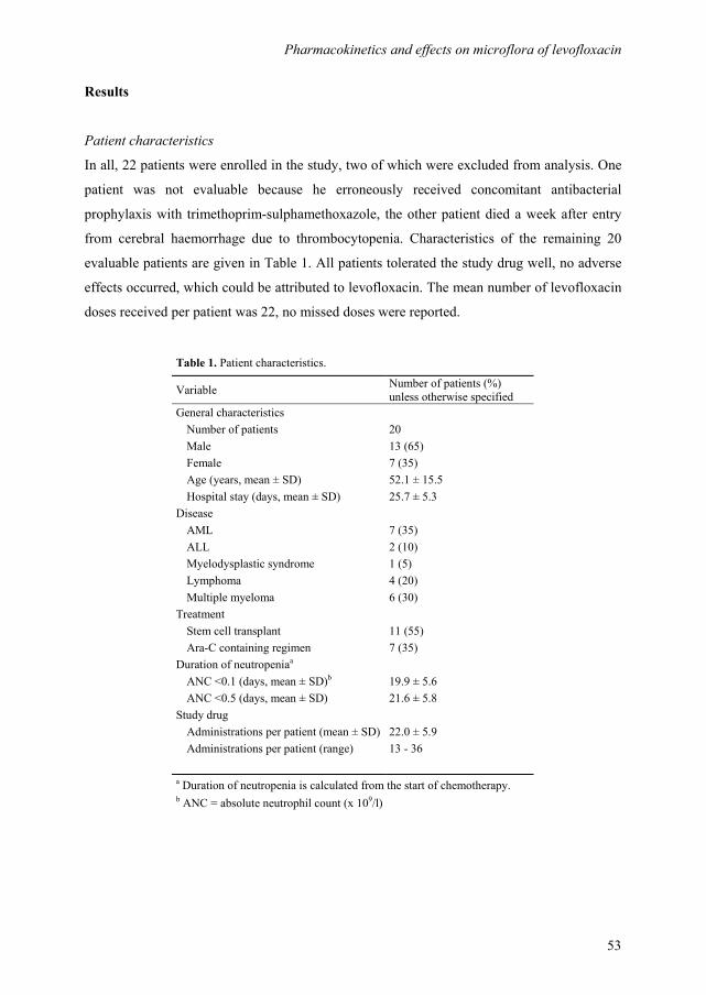
Pharmacokinetics and effects on microflora of levofloxacin
53
Results
Patient characteristics
In all, 22 patients were enrolled in the study, two of which were excluded from analysis. One
patient was not evaluable because he erroneously received concomitant antibacterial
prophylaxis with trimethoprim-sulphamethoxazole, the other patient died a week after entry
from cerebral haemorrhage due to thrombocytopenia. Characteristics of the remaining 20
evaluable patients are given in Table 1. All patients tolerated the study drug well, no adverse
effects occurred, which could be attributed to levofloxacin. The mean number of levofloxacin
doses received per patient was 22, no missed doses were reported.
Table 1. Patient characteristics.
Variable Number of patients (%)
unless otherwise specified
General characteristics
Number of patients 20
Male 13 (65)
Female 7 (35)
Age (years, mean ± SD) 52.1 ± 15.5
Hospital stay (days, mean ± SD) 25.7 ± 5.3
Disease
AML 7 (35)
ALL 2 (10)
Myelodysplastic syndrome 1 (5)
Lymphoma 4 (20)
Multiple myeloma 6 (30)
Treatment
Stem cell transplant 11 (55)
Ara-C containing regimen 7 (35)
Duration of neutropeniaa
ANC <0.1 (days, mean ± SD)b 19.9 ± 5.6
ANC <0.5 (days, mean ± SD) 21.6 ± 5.8
Study drug
Administrations per patient (mean ± SD) 22.0 ± 5.9
Administrations per patient (range) 13 - 36
a Duration of neutropenia is calculated from the start of chemotherapy. b ANC = absolute neutrophil count (x 109/l)

Chapter 3
54
Microbiological results
During the course of the study, a total of 137 throat swabs (mean 6.9/patient) and 115 faecal
samples (mean 5.8/patient) were collected. In all, 17 anal swabs had to be taken because the
patient was not able to produce a stool specimen. Culture data of the throat-swab specimens
are summarized in Table 2. VG streptococci persisted from the start of chemotherapy
throughout the duration of the neutropenic episode. At day 0-1, VG streptococci were highly
susceptible to levofloxacin in 19 patients (MIC ≤1mg/l), in one patient an intermediate-
susceptible strain was found (day 2, MIC =4 mg/l). During the prophylactic administration of
levofloxacin in four patients, VG streptococci were isolated with MIC values increasing in
one step up to ≥32 mg/l, this occurred, respectively, after 11, 13, 14 and 21 days of treatment.
These patients received additional prophylaxis with oral penicillin, 250 mg four times daily.
MIC values of VG streptococci that remained susceptible to levofloxacin (MIC ≤2 mg/l) were
analysed separately, leaving resistant strains out of the calculations. Even in these susceptible
strains there was a gradual but significant increase of MIC values during the study period
(regression coefficient B =0.03, CI95 0.01-0.04, P =0.03).
Culture data of faecal samples are given in Table 3. On admission 18/20 (90%) of patients
were colonized with Enterobacteriaceae or other Gram-negative microorganisms (CFU
>103/g faeces), in two patients S. aureus was identified (CFU >10
3/g faeces). Successful
Table 2. Culture data of throat-swab specimens.
Days on study
0-1 2-7 8-14 15-21 >22
Number of subjects 20 20 20 17 6
Viridans group streptococci (n, %) 19 (95) 16 (80) 15 (75) 9 (53) 2 (33)
MIC (median, mg/l) 0.75 0.50 0.75 2.0 0.5
MIC (range, mg/l) 0.5 - 1.0 0.38 - 4.0 0.38 - ≥32 0.125 - ≥32 0.125 - 2.0
MIC ≥8 mg/l (n) 0 0 3 3 0
β-haemolytic streptococci (n, %) 0 1 (5) 0 0 0
MIC (mg/l) 0.38
Staphylococcus aureus (n, %) 1 (5) 0 0 0 0
MIC (mg/l) 0.064
Gram-negative microorganisms 0 0 0 0 0
Candida spp (n, %) 2 (10) 2 (10) 2 (10) 2 (12) 0
MIC = 4 mg/l indicates intermediate susceptibility, MIC ≥8 mg/l indicates resistance to levofloxacin.

Pharmacokinetics and effects on microflora of levofloxacin
55
eradication of these microorganisms was achieved within 5 days of levofloxacin
administration. Two patients were colonized with a levofloxacin resistant Escherichia coli on
admission (MIC ≥32 and 12 mg/l, respectively). In a third patient, a levofloxacin-resistant
Escherichia coli (MIC ≥32 mg/l) was isolated from a stool specimen during the second week
of his admission (day 13). In addition to levofloxacin, these three patients received oral
colistin, 300 mg three times daily, which provided adequate eradication of levofloxacin-
resistant E. coli. In one patient an Acinetobacter lwoffii was isolated on day 6 and in another
patient a Sphingomonas paucimobilis was found on day 12. Both strains were considered
transient flora because they proved to be susceptible to levofloxacin and were isolated only
once.
Table 3. Culture data of faecal samples and anal-swab specimens.
Days on study
0-1 2-7 8-14 15-21 >22
Number of subjects 20 20 20 17 6
Enterobacteriaceae
Escherichia coli (n, %) 15 (75) 5 (25) 1 (6) 0 0
MIC (median, mg/l) 0.032 0.23 ≥32
MIC (range, mg/l) 0.008 - ≥32 0.016 - ≥32
MIC >8 mg/l (n) 2 2 1a 0
Klebsiella pneumoniae (n, %) 2 (10) 0 0 0 0
MIC (mean, mg/l) 0.262
Enterobacter cloacae (n, %) 1 (5) 0 0 0 0
MIC (mg/l) 0.04
Proteus penneri (n, %) 1 (5) 0 0 0 0
MIC (mg/l) 0.032
Hafnia alvei (n, %) 1 (5) 0 0 0 0
MIC (mg/l) 0.008
S. aureus (n, %) 2 (10) 0 0 0 0
MIC (mean, mg/l) 0.110
Sphingomonas paucimobilis 0 0 1 (5) 0 0
MIC (mg/l) 2.0
Acinetobacter lwoffii 0 1 (5) 0 0 0
MIC (mg/l) 0.25
Non-ferment. Gram neg. rod (n, %) 1 (5) 0 0 0 0
MIC (mg/l) 0.25
Candida spp (n, %) 5 (25) 6 (30) 11 (55) 6 (35) 2 (33)
MIC ≥8 mg/l indicates resistance to levofloxacin. a patient colonized with a resistant E-coli (MIC ≥32 mg/l) on day 13.
Chapter 3
56
Quantitative anaerobic cultures of throat-swab specimens obtained at baseline showed a mean
number of 6.2 × 10⁶ CFU/ml (range 3.0 × 10
6-7.4 × 10
6 CFU/ml). A predominance of
anaerobic Gram-positive bacilli was found in throat samples, subdivided into cocci (21%) and
rods (49%). In faecal samples obtained at baseline, a mean number of anaerobic bacilli of 1.6
× 109 CFU/g (range 1.4 × 10
5-1.3 × 10
10 CFU/g) was found, which were mainly Gram-
negative rods (90%). No significant changes of colonization rates of anaerobes over time were
observed, as expressed by the total number of CFU/g faeces (B=0.02, CI95 -0.01-0.05, P=0.1)
or per ml throat-swab specimen (B=-0.01, CI95 -0.04-0.10, P=0.3). Moreover, there were no
significant shifts from predominance of Gram-positive to Gram-negative anaerobic microflora
or vice versa, during levofloxacin prophylaxis.
Fever, defined as a sustained axillary temperature of >38.5°C, occurred in eight (40%)
patients. Probable causes of fever were a clinically documented infection of the lung (n=2), an
infection of the skin with Absidia spp (n=1) and fever of unknown origin (n=1). Four patients
developed blood-stream infections with Gram-positive bacteria, including coagulase-negative
staphylococcus (n=1), Enterococcus faecalis (n=1), S. oralis (n=1) and E. faecium +
coagulase-negative staphylococcus (n=1). All isolated microorganisms displayed high level
resistance to levofloxacin (MIC ≥32). Patients with fever received imipenem-cilastatin as
initial empirical antibiotic treatment. In two patients vancomycin was added, because of
imipenem-resistant pathogens (E. faecium, coagulase-negative staphylococcus). In another
two patients imipenem-cilastatin was switched to vancomycin in combination with
aztreonam, because of an allergic skin reaction possibly due to imipenem. Amphotericin B
was given to two patients, one with a proven infection with Absidia spp and another because
of persisting fever after 4 × 24 h of treatment with imipenem. All patients with fever
recovered without significant sequelae. There was no mortality attributable to infectious
complications.
Pharmacokinetic data
Urine samples were collected in 10 patients. No in vivo enantioconversion from S-(-)-
ofloxacin (levofloxacin) to R-(+)-ofloxacin was observed. For this reason, it was not
necessary to use an enantio-selective separation method for the analysis of the blood samples.
A total of 170 venous blood samples were drawn in 10 patients. In all, 10 samples per patient
were collected during the 24 h period following first dose administration (phase 1), seven
samples per patient were obtained immediately after neutropenia had been established (phase
2). Phase 2 sampling was initiated at a mean of 13.9±3.8 days from first dose administration.
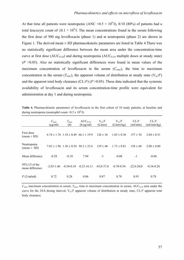
Pharmacokinetics and effects on microflora of levofloxacin
57
At that time all patients were neutropenic (ANC <0.5 × 10⁹/l), 8/10 (80%) of patients had a
total leucocyte count of ≤0.1 × 10⁹/l. The mean concentrations found in the serum following
the first dose of 500 mg levofloxacin (phase 1) and at neutropenia (phase 2) are shown in
Figure 1. The derived mean ± SD pharmacokinetic parameters are listed in Table 4.There was
no statistically significant difference between the mean area under the concentration-time
curve at first dose (AUC0-24) and during neutropenia (AUC0-24 multiple doses at steady state)
(P >0.05). Also no statistically significant differences were found in mean values of the
maximum concentration of levofloxacin in the serum (Cmax), the time to maximum
concentration in the serum (Tmax), the apparent volume of distribution at steady state (Vss/F)
and the apparent total body clearance (CL/F) (P >0.05). These data indicated that the systemic
availability of levofloxacin and its serum concentration-time profile were equivalent for
administration at day 1 and during neutropenia.
Table 4. Pharmacokinetic parameters of levofloxacin in the first cohort of 10 study patients, at baseline and
during neutropenia (neutrophil count <0.5 x 10⁹/l).
Cmax
(µg/ml)
Tmax
(h)
AUC0-24
(h·µg/ml)
Vss/F
(Liters)
Vss/F
(Liters/kg)
CL/F
(ml/min)
CL/F
(ml/min/kg)
First dose
(mean ± SD) 6.74 ± 1.76 1.18 ± 0.49 66.1 ± 19.9 126 ± 36 1.65 ± 0.36 157 ± 54 2.04 ± 0.51
Neutropenia
(mean ± SD) 7.02 ± 1.96 1.36 ± 0.54 58.2 ± 23.6 129 ± 48 1.73 ± 0.81 158 ± 60 2.08 ± 0.80
Mean difference -0.28 -0.18 7.94 -3 -0.08 -1 -0.04
95% CI of the
mean difference -2.03-1.46 -0.54-0.18 -0.23-16.11 -43,0-37,0 -0.70-0.54 -22.0-20,0 -0.34-0.26
P (2-tailed) 0.72 0.28 0.06 0.87 0.78 0.93 0.78
Cmax maximum concentration in serum, Tmax, time to maximum concentration in serum, AUC0-24 area under the
curve for the 24-h dosing interval, Vss/F apparent volume of distribution at steady state, CL/F apparent total
body clearance.
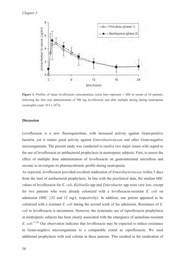
�������
��
����� �� �������� �� ��� ���������� ��������� ������ ���� �� ����� � ��� � ����� �� �� �������
�������� ��� ����� ���� ������������ �� ��� �� ���������� �� ����� ����� �� ����� ����� ����� ���
������ ������������� !���
��������
"��������� �� � �� ������#������� ���� ������� ������$ ������ %���& �������
�������� $�� �� ������ ���� ������$ ������ '(���)*�+���,�+��� �� ����� %���&�������
�������������-�� ���������$�����������������������.��������������������
������������������������������� �� �$����������� �����.���������������������
����� �� ����� �� ���� ������������ �� ���������� � �������������� ��������� ��
�������������������� ������/���� ���������������� ����
0��� ��������������� �������������������������'(���)*�+���,�+�����������$�
������������������������� �� �$������1���������� ��������������������21�
���������������������'3+)4,�54�*6,�44�� ��'(���)*�+���� �������$������� �
��� ��� ������ ��� ���� ������$ ����7�� ���� � ����������&�������� '3 +)4, �
�������� �21� 8�9 �� �9 ��!�� ��� ������$�� 1 �������� �� ����� � ����� �� ��
����7���������������'3+)4,����� ����������/��������������:���������'3
+)4,��������������������;�����������$������������� �������� �� �$�����
������ �����.��������������$�������������������������#������&��������
'3+)4,��<&9�=�����������������������������������$���� ������������������
� %���&������� ������������ �� � �� ������ ����� �� � ��������� >� ����
��������� �� �$������������������� � ����� �������-������������ ��������������
Pharmacokinetics and effects on microflora of levofloxacin
59
the levofloxacin-resistant strains. In a recent publication, the addition of colistin to
ciprofloxacin appeared to reduce the emergence of quinolone-resistant Gram-negative
bacteria and to prevent Gram-negative sepsis in neutropenic patients.18 In our patients no
systemic infections with Gram-negative bacteria occurred.
Colonization with VG streptococci was reduced by the prophylactic administration of
levofloxacin. Of concern was the increase of MIC values for streptococci over time. In four
patients VG streptococci with MICs of ≥32 mg/l were isolated during the course of
antibacterial prophylaxis. In addition, even for VG streptococci that remained susceptible to
levofloxacin, a significant gradual increase of MIC values was found during the study period.
In vitro studies have demonstrated high susceptibility of VG streptococci to levofloxacin,
with MIC values ranging from 0.25 to 1 mg/l.21,22
Our data indicate that resistance of VG
streptococci may develop during long-term administration of levofloxacin. This observation is
in line with recently published clinical data.23 In a series of 37 neutropenic patients receiving
levofloxacin prophylaxis, bacteraemia with VG streptococci occurred in six (16%) patients,
three of whom developed septic shock. All isolates displayed diminished susceptibility to
levofloxacin and cross-resistance to other quinolones. The authors conclude that the
prophylactic administration of levofloxacin may result in the selection of quinolone-resistant
VG streptococci.
Four patients in our study developed blood-stream infections with Gram-positive bacteria,
including coagulase-negative staphylococci, E. faecalis, S. oralis and E. faecium. Preclinical
data show that most strains of enterococci and coagulase-negative staphylococci are
susceptible to levofloxacin, although MIC values are higher than for VG streptococci. MIC
values are reported to range from 0.13 to 4 mg/l for coagulase-negative staphylococci and
from 0.25 to ≥32 mg/l for enterococci.21,22
In a study on haemato-oncology patients in
Switzerland, 65% of strains of coagulase-negative staphylococci were susceptible to
levofloxacin and even 100% of strains of enterococci.24 Despite these compelling data, our
findings indicate that during levofloxacin prophylaxis breakthrough infections with
coagulase-negative staphylococci or enterococci may occur.
Levofloxacin did not affect the anaerobic component of the gut microflora. The number of
anaerobic microorganisms, expressed as the number of CFU/g faeces or per ml throat-swab
specimen, did not change significantly during the course of the prophylactic regimen. This
may be of crucial importance in view of preservation of colonization resistance, which is
based on the concept that selective suppression of the aerobic Gram-negative bacteria in the
Chapter 3
60
digestive tract, without the impairment of the anaerobic flora, prevents the adherence,
proliferation and invasion of potentially pathogenic bacterial and fungal species.25
Our study is the first to report pharmacokinetic data of levofloxacin in neutropenic patients.
The estimated mean values for Cmax, Tmax, AUC(0-24) and CL/F in patients with haematological
malignancies were well comparable with those determined in healthy subjects.13,26
In
addition, we showed that individual pharmacokinetic parameters calculated after the first oral
administration of levofloxacin were not statistically different from those calculated at
neutropenia. This is important because significant changes in the pharmacokinetic profile may
occur in patients receiving intensive chemotherapy. Mucosal damage, for example, may either
decrease or increase the rate of antibiotic absorption, shifts and decreases in the serum
albumin may significantly affect the volume of distribution of the drug. It has been shown
previously that the absorption of oral ciprofloxacin and to a lesser extent of oral ofloxacin are
reduced following cytotoxic chemotherapy.15,16
Pharmacokinetics of levofloxacin appear to be
unaffected by the administration of chemotherapy or neutropenia, indicating that on these
occasions there is no need for dose adjustment.
In conclusion, levofloxacin, administered orally as antibacterial prophylaxis to patients
receiving intensive chemotherapy for haematological malignancies, provides adequate
eradication of Gram-negative microorganisms and S. aureus and preserves the anaerobic
component of the bowel flora. Moreover, the pharmacokinetic properties of levofloxacin are
not altered due to chemotherapy or neutropenia. Levofloxacin is easily administered once
daily and well tolerated. Of concern, however, is the increasing resistance of VG streptococci
during prolonged administration of levofloxacin and a diminished susceptibility of coagulase-
negative staphylococci and enterococci. These findings may hamper its use as a single agent
for prophylaxis in neutropenic subjects. This pilot study, however, with a limited number of
subjects does not allow for definite conclusions on that issue. Hence, the question remains
open as to how levofloxacin prophylaxis compares with standard prophylactic regimens as for
tolerability, efficacy, induction of resistance and the number of break-through infections. To
address these questions, we are currently investigating levofloxacin vs ciprofloxacin plus oral
penicillin in a randomized clinical trial.

Pharmacokinetics and effects on microflora of levofloxacin
61
Acknowledgements
This study was supported in part by a grant from Aventis Pharma BV, Hoevelaken, The
Netherlands.
References
1. Engels EA, Lau J, Barza M. Efficacy of quinolone prophylaxis in neutropenic cancer patients: a
meta-analysis. J Clin Oncol 1998; 16: 1179-1187.
2. Rozenberg-Arska M, Dekker AW, Verhoef J. Ciprofloxacin for selective decontamination of the
alimentary tract in patients with acute leukemia during remission induction treatment: the effect
on fecal flora. J Infect Dis 1985; 152: 104-107.
3. Bow EJ, Rayner E, Louie TJ. Comparison of norfloxacin with cotrimoxazole for infection
prophylaxis in acute leukemia. The trade-off for reduced Gram-negative sepsis. Am J Med
1988; 84: 847-854.
4. Kukuckova E, Spanik S, Ilavska I, Helpianska L, Oravcova E, Lacka J, Krupova I, Grausova S,
Koren P, Bezakova I, Grey E, Balaz M, Studena M, Kunova A, Torfs K, Trupl J, Korec S,
Stopkova K, Krcmery V, Jr. Staphylococcal bacteremia in cancer patients: risk factors and
outcome in 134 episodes prior to and after introduction of quinolones into infection prevention
in neutropenia. Support Care Cancer 1996; 4: 427-434.
5. Bow EJ, Mandell LA, Louie TJ, Feld R, Palmer M, Zee B, Pater J. Quinolone-based
antibacterial chemoprophylaxis in neutropenic patients: effect of augmented Gram-positive
activity on infectious morbidity. National Cancer Institute of Canada Clinical Trials Group. Ann
Intern Med 1996; 125: 183-190.
6. Kern WV, Hay B, Kern P, Marre R, Arnold R. A randomized trial of roxithromycin in patients
with acute leukemia and bone marrow transplant recipients receiving fluoroquinolone
prophylaxis. Antimicrob Agents Chemother 1994; 38: 465-472.
7. Ford CD, Reilly W, Wood J, Classen DC, Burke JP. Oral antimicrobial prophylaxis in bone
marrow transplant recipients: randomized trial of ciprofloxacin versus ciprofloxacin-
vancomycin. Antimicrob Agents Chemother 1998; 42: 1402-1405.
8. International Antimicrobial Therapy Cooperative Group of the European Organization for
Research and Treatment of Cancer. Reduction of fever and streptococcal bacteremia in
granulocytopenic patients with cancer. A trial of oral penicillin V or placebo combined with
pefloxacin. JAMA 1994; 272: 1183-1189.
Chapter 3
62
9. Broun ER, Wheat JL, Kneebone PH, Sundblad K, Hromas RA, Tricot G. Randomized trial of
the addition of Gram-positive prophylaxis to standard antimicrobial prophylaxis for patients
undergoing autologous bone marrow transplantation. Antimicrob Agents Chemother 1994; 38:
576-579.
10. de Jong P, de Jong M, Kuijper E, van der LJ. Evaluation of penicillin G in the prevention of
streptococcal septicaemia in patients with acute myeloid leukaemia undergoing cytotoxic
chemotherapy. Eur J Clin Microbiol Infect Dis 1993; 12: 750-755.
11. Davis R , Bryson HM. Levofloxacin. A review of its antibacterial activity, pharmacokinetics and
therapeutic efficacy. Drugs 1994; 47: 677-700.
12. Fu KP, Lafredo SC, Foleno B, Isaacson DM, Barrett JF, Tobia AJ, Rosenthale ME. In vitro and
in vivo antibacterial activities of levofloxacin (l-ofloxacin), an optically active ofloxacin.
Antimicrob Agents Chemother 1992; 36: 860-866.
13. Chien SC, Rogge MC, Gisclon LG, Curtin C, Wong F, Natarajan J, Williams RR, Fowler CL,
Cheung WK, Chow AT. Pharmacokinetic profile of levofloxacin following once-daily 500-
milligram oral or intravenous doses. Antimicrob Agents Chemother 1997; 41: 2256-2260.
14. Lee LJ, Hafkin B, Lee ID, Hoh J, Dix R. Effects of food and sucralfate on a single oral dose of
500 milligrams of levofloxacin in healthy subjects. Antimicrob Agents Chemother 1997; 41:
2196-2200.
15. Johnson EJ, MacGowan AP, Potter MN, Stockley RJ, White LO, Slade RR, Reeves DS.
Reduced absorption of oral ciprofloxacin after chemotherapy for haematological malignancy. J
Antimicrob Chemother 1990; 25: 837-842.
16. Brown NM, White LO, Blundell EL, Chown SR, Slade RR, MacGowan AP, Reeves DS.
Absorption of oral ofloxacin after cytotoxic chemotherapy for haematological malignancy. J
Antimicrob Chemother 1993; 32: 117-122.
17. Yeh SP, Hsueh EJ, Yu MS, Wu H, Wang YC. Oral ciprofloxacin as antibacterial prophylaxis
after allogeneic bone marrow transplantation: a reappraisal. Bone Marrow Transplant 1999; 24:
1207-1211.
18. Prentice HG, Hann IM, Nazareth B, Paterson P, Bhamra A, Kibbler CC. Oral ciprofloxacin plus
colistin: prophylaxis against bacterial infection in neutropenic patients. A strategy for the
prevention of emergence of antimicrobial resistance. Br J Haematol 2001; 115: 46-52.
19. van Kraaij MG, Dekker AW, Peters E, Fluit A, Verdonck LF, Rozenberg-Arska M. Emergence
and infectious complications of ciprofloxacin-resistant Escherichia coli in haematological cancer
patients. Eur J Clin Microbiol Infect Dis 1998; 17: 591-592.
20. Cometta A, Calandra T, Bille J, Glauser MP. Escherichia coli resistant to fluoroquinolones in
patients with cancer and neutropenia. N Engl J Med 1994; 330: 1240-1241.
Pharmacokinetics and effects on microflora of levofloxacin
63
21. Hoogkamp-Korstanje JA. In-vitro activities of ciprofloxacin, levofloxacin, lomefloxacin,
ofloxacin, pefloxacin, sparfloxacin and trovafloxacin against Gram-positive and Gram-negative
pathogens from respiratory tract infections. J Antimicrob Chemother 1997; 40: 427-431.
22. Bauernfeind A. Comparison of the antibacterial activities of the quinolones Bay 12-8039,
gatifloxacin (AM 1155), trovafloxacin, clinafloxacin, levofloxacin and ciprofloxacin. J
Antimicrob Chemother 1997; 40: 639-651.
23. Razonable RR, Litzow MR, Khaliq Y, Piper KE, Rouse MS, Patel R. Bacteremia due to viridans
group Streptococci with diminished susceptibility to Levofloxacin among neutropenic patients
receiving levofloxacin prophylaxis. Clin Infect Dis 2002; 34: 1469-1474.
24. Siegrist HH, Nepa MC, Jacquet A. Susceptibility to levofloxacin of clinical isolates of bacteria
from intensive care and haematology/oncology patients in Switzerland: a multicentre study. J
Antimicrob Chemother 1999; 43 Suppl C: 51-54.
25. van der Waaij D. Selective decontamination of the digestive tract: general principles. Eur J
Cancer Clin Oncol 1988; 24 Suppl 1: S1-S4.
26. Child J, Mortiboy D, Andrews JM, Chow AT, Wise R. Open-label crossover study to determine
pharmacokinetics and penetration of two dose regimens of levofloxacin into inflammatory fluid.
Antimicrob Agents Chemother 1995; 39: 2749-2751.

Chapter 3
64

65
Levofloxacin versus ciprofloxacin plus phenethicillin for the
prevention of bacterial infections in patients with haematological
malignancies.
Gert Jan Timmers
Alberdina M. Simoons-Smit
Maaike E. Leidekker
Jeroen J.W.M. Janssen
Christina M.J.E. Vandenbroucke-Grauls
Peter C. Huijgens
Department of Haematology, Department of Medical Microbiology and Infection Control,
VU University Medical Center, Amsterdam, The Netherlands.
Clinical Microbiology and Infection 2007;13:497-503.
Chapter 3 3 4
Chapter 4
66
Abstract
An open-label randomized clinical trial was designed to compare the efficacy and tolerance of
levofloxacin and ciprofloxacin plus phenethicillin for the prevention of bacterial infections in
patients with high-risk neutropenia, and to monitor the emergence of antimicrobial resistance.
Adult patients (n = 242) scheduled to receive intensive treatment for haematological
malignancies were assigned randomly to receive oral prophylaxis with either levofloxacin
500 mg once-daily (n = 122), or ciprofloxacin 500 mg twice-daily plus phenethicillin 250 mg
four-times-daily (n = 120). The primary endpoint was failure of prophylaxis, defined as the
first occurrence of either the need to change the prophylactic regimen or the initiation of
intravenous broad-spectrum antibiotics. This endpoint was observed in 89 (73.0%) of 122
levofloxacin recipients and in 85 (70.8%) of 120 ciprofloxacin plus phenethicillin recipients
(RR 1.03, CI95 0.88-1.21, P = 0.71). No differences were noted between the two groups with
respect to secondary outcome measures, including time to endpoint, occurrence of fever, type
and number of microbiologically documented infections, and administration of intravenous
antibiotics. A questionnaire revealed that levofloxacin was tolerated significantly better than
ciprofloxacin plus phenethicillin. Surveillance cultures indicated the emergence of viridans
group (VG) streptococci resistant to levofloxacin in 17 (14%) of 122 levofloxacin recipients;
in these cases, the prophylactic regimen was adjusted. No bacteraemia with VG streptococci
occurred. It was concluded that levofloxacin and ciprofloxacin plus phenethicillin are equally
effective in the prevention of bacterial infections in neutropenic patients, but that levofloxacin
is tolerated better. Emergence of levofloxacin-resistant VG streptococci is of concern, but
appears to be a manageable problem.
Levofloxacin vs ciprofloxacin plus phenethicillin
67
Introduction
Quinolones have been used extensively in the past two decades for the prevention of bacterial
infections during neutropenia in cancer patients. It is evident that this approach reduces the
incidence of Gram-negative bacteraemia significantly.1-3 Unfortunately, this is
counterbalanced by an increased incidence of infections with Gram-positive bacteria. In
particular, viridans group (VG) streptococci and coagulase-negative staphylococci have
emerged as a frequent cause of morbidity and mortality.4,5 To overcome this problem,
quinolone prophylaxis has been combined with other antimicrobial agents, e.g., penicillin,
macrolides and vancomycin, that are active against Gram-positive cocci.6-9
An alternative approach could involve the use of new-generation quinolones, which are more
potent against Gram-positive pathogens. Levofloxacin, as a representative of this group, has
been reported to reduce the incidence of fever and other infection-related outcomes in
neutropenic cancer patients, compared with a placebo,2,10,11
but important issues remain to be
addressed. First, no data are available from controlled clinical trials that allow a direct
comparison between the use of levofloxacin prophylaxis and the use of ciprofloxacin plus an
antibiotic with anti-Gram-positive bacteria activity. Second, there have been some alarming
reports concerning the emergence of levofloxacin-resistant Gram-positive microorganisms, in
particular VG streptococci.12,13
During the past few years, patients admitted to the haematology department of the VU
University Medical Center, Amsterdam, The Netherlands have received ciprofloxacin plus
phenethicillin for the prevention of bacterial infections during neutropenia as the standard of
care. When levofloxacin became available in The Netherlands, the randomized clinical trial
described in this study was conducted to compare levofloxacin with ciprofloxacin plus
phenethicillin with respect to their efficacy as antibacterial prophylaxis for neutropenic
patients. In addition, the trial was designed to investigate the tolerance of these compounds
and to closely monitor emerging antimicrobial resistance.
Patients and methods
Patients
Consecutive adult patients with a haematological malignancy who were hospitalized at the
haematology department of the VU University Medical Center for high-dose combination
Chapter 4
68
chemotherapy, with or without autologous or allogeneic haematopoietic stem-cell
transplantation, were eligible for this study. An anticipated granulocytopenic period
(granulocytes <0.5 × 10⁹/L) of ≥10 days was required. Patients were enrolled only once.
Exclusion criteria were active infection or antibacterial therapy at entry, a history of
hypersensitivity to fluoroquinolones, a creatinine clearance of <15 mL/min, or elevation of
transaminases to greater than three-fold the normal upper limit. The protocol was approved by
the institutional scientific and ethical committees, and all participants provided written
informed consent.
Randomization and prophylactic regimen
The study was a prospective, single-centre, open-label, randomized clinical trial. Patients
were assigned randomly by consecutively drawn, sealed envelopes to receive either
levofloxacin 500-mg tablets once-daily or ciprofloxacin 500-mg tablets twice-daily, plus,
from day 7 after the start of chemotherapy, phenethicillin 250-mg tablets four-times-daily.
Prophylaxis was begun on the first day of chemotherapy and was continued until recovery to
a granulocyte count of >0.5 × 10⁹/L. Phenethicillin was initiated on day 7 because, from that
time on, oropharyngeal mucositis was to be expected, and this has been identified as an
independent risk-factor for infections with streptococci.4 Compliance was monitored by
counting tablets. In addition to the study medication, all patients received fluconazole 50 mg
once-daily and 2 mg nasal amphotericin B spray three-times-daily. A central venous catheter
was inserted before the start of chemotherapy.
Clinical and microbiological evaluation of subjects
Randomized patients were examined daily for clinical signs of infection. Surveillance cultures
for identification of colonizing bacteria and yeasts were taken from throat and anus before the
first dose of the study drugs and once-weekly thereafter. If appropriate, the prophylactic
regimen was adjusted according to the resistance patterns of the microorganisms identified.
If the axillary temperature increased to >38.5°C, or if other signs or symptoms of an infection
occurred without fever, clinical evaluation was performed according to a local protocol,
including a complete physical examination, a chest X-ray, and appropriate samples for
microbiological cultures. At least two separate blood samples were obtained for culture, from
both the central venous catheter and from a peripheral vein. Subsequently, empirical antibiotic
therapy was initiated, consisting of intravenous imipenem-cilastatin 500 mg four-times-daily.
If the fever did not resolve in ≤96 h, patients received antifungal therapy. In case of initiation
Levofloxacin vs ciprofloxacin plus phenethicillin
69
of broad-spectrum antibacterial therapy, levofloxacin or ciprofloxacin were continued, but
phenethicillin was discontinued.
Pathogenic microorganisms, isolated either from surveillance cultures or from cultures
obtained from patients with presumed infection, were identified to the species level by
standard microbiological techniques. MICs of levofloxacin and ciprofloxacin for
staphylococci, streptococci and Gram-negative bacilli were determined by E-tests (AB
Biodisk, Solna, Sweden). Susceptibility of streptococci to phenethicillin was determined by
disk-diffusion tests and was reported as susceptible, intermediately-resistant or resistant.
Breakpoints were defined according to CLSI standards.
Tolerance of the study medication and toxicity
Patients were asked to complete a questionnaire, which recorded a ‘tolerance score’ for the
study medication on a daily basis. Tolerance of the study drug was classified as ‘not able to
take the study drug’, ‘difficult intake’, ‘minor problems on intake’, or ‘intake without any
problem’. Any adverse event that was possibly or probably related to the study medication
was recorded. Routine clinical chemistry tests were performed weekly, and any deterioration
in liver enzymes, bilirubin or kidney function was recorded. All adverse events were
classified using the Common Terminology Criteria for Adverse Events v.3.0 (CTCAE;
National Cancer Institute, Bethesda, MD, USA). Following an adverse event, study
medication was either continued or discontinued, according to the judgement of the
responsible physician.
Outcome
The primary outcome measure of the study was success or failure of the prophylactic
regimen. Failure of prophylaxis was a composite endpoint, defined as the need to change the
prophylactic regimen for any reason, or the initiation of broad-spectrum antibacterial therapy,
whichever event occurred first. The primary endpoint was chosen to reflect the effects of the
prophylactic regimens on the most relevant clinical events. Furthermore, the study was
designed to include patient follow-up beyond this first event, and data were analysed on an
intention-to-treat basis. Secondary outcome measures were the time to primary endpoint, the
occurrence of fever, the type and number of documented infections, the use of antimicrobial
agents, and the tolerance of the study drug. Moreover, the study design provided close
monitoring of the acquisition of antimicrobial resistance by the pathogens isolated.

Chapter 4
70
Statistical evaluation
It was estimated from previous studies in the same ward that c. 30% of patients survive the
neutropenic episode without requiring change of prophylaxis or initiation of broad-spectrum
antibiotics. Thus, according to the definitions used in the present study, 70% of patients were
expected to experience failure of prophylaxis. Sample size was calculated to detect a 25%
reduction (from 70% to 52%) in failure of prophylaxis. To detect such a difference with a
significance level (α) of 0.05 (two-tailed) and a statistical power of 80%, 120 patients per arm
were required. Differences between groups in categorical variables were analysed with the
chi-square test. In case of variables with an ordering or grading scale, the chi-square test for
trend was used. The relative differences between the groups were also expressed as relative
risks (RRs) with 95% confidence intervals (CI95). The Mann-Whitney non-parametric U-test
was used for comparison of means. Differences in survival without failure of prophylaxis
were assessed by the log-rank test, and Kaplan-Meier curves were plotted for each study
group.
Results
Between January 2002 and July 2005, 245 patients were enrolled in the study. Three patients
were excluded from analysis. One patient withdrew informed consent, another patient was
erroneously enrolled twice, and one patient died on the day of randomization because of
disease progression. Of the 242 evaluable patients, 122 were assigned to receive levofloxacin
and 120 to receive ciprofloxacin and phenethicillin. Basic patient characteristics of the two
treatment groups are listed in Table 1. No significant differences were found in gender, age,
type and remission status of the haematological disease and treatment variables, including
stem-cell transplantation procedures. However, the neutropenic episode was significantly
longer in the ciprofloxacin-phenethicillin group (mean difference to a granulocyte count of
>0.5 × 10⁹/L = 1.4 days, P = 0.044; mean difference to granulocyte count >0.1 ×
109/L = 1.6 days, P = 0.017). Patients receiving levofloxacin remained in hospital for a mean
of 25.5 days, compared with 28.1 days for patients receiving ciprofloxacin-phenethicillin, but
this was not statistically significant (mean difference 2.6 days, P = 0.13).

Levofloxacin vs ciprofloxacin plus phenethicillin
71
Table 1. Patient characteristics and clinical course variables.
Levofloxacin Ciprofloxacin
n % n %
Total patients 122 50.4 120 49.6
Age, years
Median (range) 55 (18-71) 54 (19-71)
Gender
Male 76 62.3 79 65.8
Female 46 37.7 41 34.2
Diagnosis
ALL 10 8.2 10 8.3
AML 19 15.6 28 23.3
Multiple Myeloma 46 37.7 39 32.5
Lymphoma 35 28.7 34 28.4
Myelodysplasia 4 3.3 3 2.5
Other 8 6.5 6 5.0
Disease status
Newly diagnosed 81 66.4 79 65.8
Recurrent 41 33.6 41 34.2
Remission status
Active disease 21 17.2 27 22.5
Partial remission 70 57.4 59 49.2
Complete remission 31 25.4 34 28.3
Treatment variables
First course or induction 15 12.3 16 13.3
Second course or consolidation 21 17.2 26 21.7
Stem-cell transplantation 86 70.5 78 65.0
autologous stem-cells 74 86.0 65 83.3
allogeneic stem cells 12 14.0 13 16.7
Laminar airflow room 13 10.7 16 13.3
Haematopoietic growth factor (G-CSF) 10 8.2 8 6.7
Duration of neutropenia
(ANC < 0.5 x 10⁹/L), days
Meana 21.8 23.2
Median (range) 20.5 (15-46) 22.0 (12-55)
Duration of severe neutropenia
(ANC < 0.1 x 10⁹/L), days
Meanb 19.9 21.5
Median (range) 18.0 (13-46) 20.0 (11-53)
Hospital stay, days
Mean 25.5 28.1
Median (range) 23.0 (16-58) 25.0 (14-155)
All differences not significant (NS), except a P = 0.044, b P = 0.017.
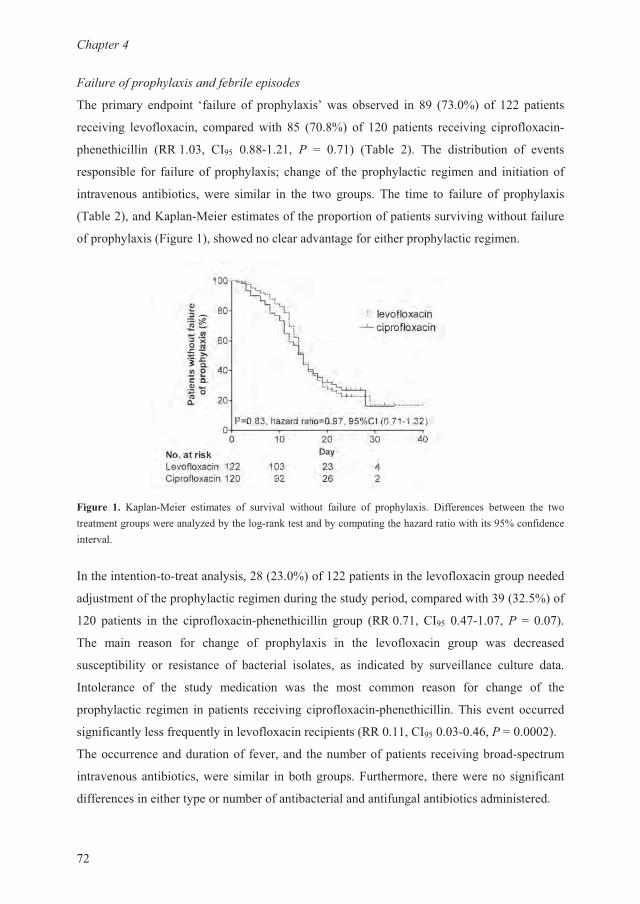
�������?
<9
��,4���)���)���4��,6�(���*�,4���,6)��6
-�� �����$ �� ��� @��������� �� �$�����A����������� � � �<��B���99 ������
�������� ������������ ��� �������� �� �<��B� �� 9� ������ �������� �� ���������&
����������� �::��� �1 � ���&9� C �<� �-���� 9� -�� ����������� �� �����
��� ������ ��� ��������� �� �$�����D������� ��� �� �$������ �������� ����������
��������� ��������������� ������� � ��� ��� ���� � -�� ���� �� ������� �� �� �$�����
�-����9������ ��&2������������������ �� ������� ����������������������������
�� �� �$������������������������������������������� �� �$������������
����� �� �� ��&2���� ��������� �� �������� ������� ������� �� �� �$����� ���������� ������ ��� ��������������� ��������$7���$������&��/�������$��� ���������7��������������� �B���������������
1���������&��&��������$����9��9��B���99 ������������������������� �����
��.����������� �� �$������������������������$ ��������� ��������� ��9�B���
9� ������ � ��� �� ���������& ����������� ���� �::�<��1 � ��<&�<� C ��<�
-�� ��� ����� ��� ����� �� �� �$����� � ��� ����������� ���� ��� ���������
����� �������$ �� ��������� �� ��������� ��������� �� �������� �$ ����������� ������� ����
1�������� �� ��� ����$ ��������� ��� ��� ���� ����� ����� ��� ����� �� ���
�� �$������ ������ � ������ ���������� ���������& �����������-���������������
����������$�������#����$���������������� �����::���1 ����&��E� C����9�
-���������������������� �������� ���������� ������ �������������&� ������
������������������������ ������� ��������� ������������� ���������� ���������
������������������$ �����������������������������������������������������
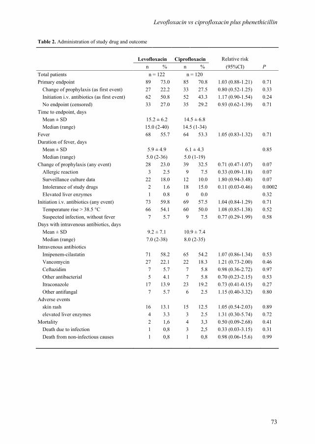
Levofloxacin vs ciprofloxacin plus phenethicillin
73
Table 2. Administration of study drug and outcome
Levofloxacin Ciprofloxacin Relative risk
n % n % (95%CI) P
Total patients n = 122 n = 120 Primary endpoint 89 73.0 85 70.8 1.03 (0.88-1.21) 0.71
Change of prophylaxis (as first event) 27 22.2 33 27.5 0.80 (0.52-1.25) 0.33
Initiation i.v. antibiotics (as first event) 62 50.8 52 43.3 1.17 (0.90-1.54) 0.24
No endpoint (censored) 33 27.0 35 29.2 0.93 (0.62-1.39) 0.71
Time to endpoint, days
Mean ± SD 15.2 ± 6.2 14.5 ± 6.8 Median (range) 15.0 (2-40) 14.5 (1-34) Fever 68 55.7 64 53.3 1.05 (0.83-1.32) 0.71
Duration of fever, days Mean ± SD 5.9 ± 4.9 6.1 ± 4.3 0.85
Median (range) 5.0 (2-36) 5.0 (1-19) Change of prophylaxis (any event) 28 23.0 39 32.5 0.71 (0.47-1.07) 0.07
Allergic reaction 3 2.5 9 7.5 0.33 (0.09-1.18) 0.07
Surveillance culture data 22 18.0 12 10.0 1.80 (0.94-3.48) 0.07
Intolerance of study drugs 2 1.6 18 15.0 0.11 (0.03-0.46) 0.0002
Elevated liver enzymes 1 0.8 0 0.0 0.32
Initiation i.v. antibiotics (any event) 73 59.8 69 57.5 1.04 (0.84-1.29) 0.71
Temperature rise > 38.5 °C 66 54.1 60 50.0 1.08 (0.85-1.38) 0.52
Suspected infection, without fever 7 5.7 9 7.5 0.77 (0.29-1.99) 0.58
Days with intravenous antibiotics, days Mean ± SD 9.2 ± 7.1 10.9 ± 7.4 Median (range) 7.0 (2-38) 8.0 (2-35) Intravenous antibiotics Imipenem-cilastatin 71 58.2 65 54.2 1.07 (0.86-1.34) 0.53
Vancomycin 27 22.1 22 18.3 1.21 (0.73-2.00) 0.46
Ceftazidim 7 5.7 7 5.8 0.98 (0.36-2.72) 0.97
Other antibacterial 5 4.1 7 5.8 0.70 (0.23-2.15) 0.53
Itraconazole 17 13.9 23 19.2 0.73 (0.41-0.15) 0.27
Other antifungal 7 5.7 6 2.5 1.15 (0.40-3.32) 0.80
Adverse events skin rash 16 13.1 15 12.5 1.05 (0.54-2.03) 0.89
elevated liver enzymes 4 3.3 3 2.5 1.31 (0.30-5.74) 0.72
Mortality 2 1,6 4 3,3 0.50 (0.09-2.68) 0.41
Death due to infection 1 0,8 3 2,5 0.33 (0.03-3.15) 0.31
Death from non-infectious causes 1 0,8 1 0,8 0.98 (0.06-15.6) 0.99
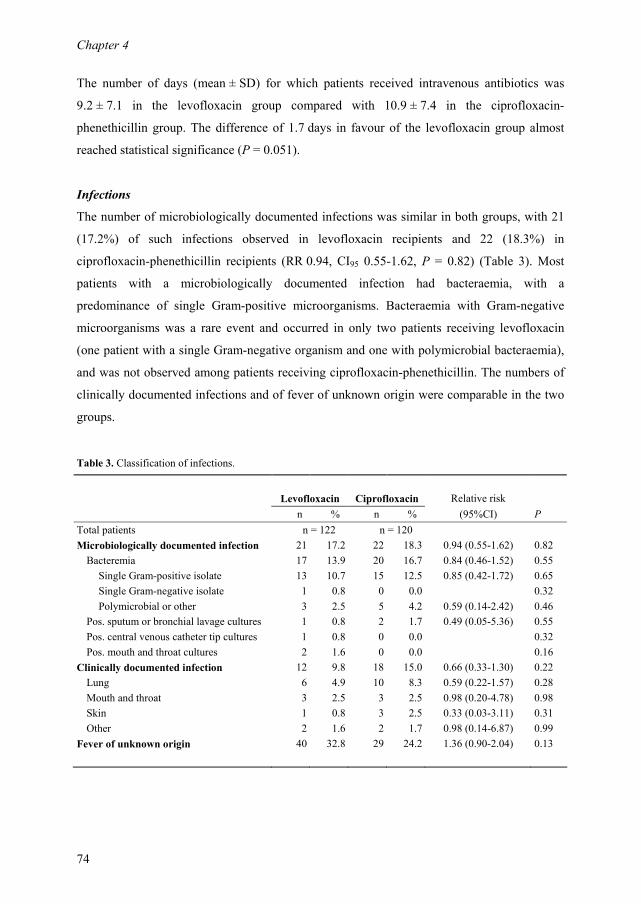
Chapter 4
74
The number of days (mean ± SD) for which patients received intravenous antibiotics was
9.2 ± 7.1 in the levofloxacin group compared with 10.9 ± 7.4 in the ciprofloxacin-
phenethicillin group. The difference of 1.7 days in favour of the levofloxacin group almost
reached statistical significance (P = 0.051).
Infections
The number of microbiologically documented infections was similar in both groups, with 21
(17.2%) of such infections observed in levofloxacin recipients and 22 (18.3%) in
ciprofloxacin-phenethicillin recipients (RR 0.94, CI95 0.55-1.62, P = 0.82) (Table 3). Most
patients with a microbiologically documented infection had bacteraemia, with a
predominance of single Gram-positive microorganisms. Bacteraemia with Gram-negative
microorganisms was a rare event and occurred in only two patients receiving levofloxacin
(one patient with a single Gram-negative organism and one with polymicrobial bacteraemia),
and was not observed among patients receiving ciprofloxacin-phenethicillin. The numbers of
clinically documented infections and of fever of unknown origin were comparable in the two
groups.
Table 3. Classification of infections.
Levofloxacin Ciprofloxacin Relative risk
n % n % (95%CI) P
Total patients n = 122 n = 120
Microbiologically documented infection 21 17.2 22 18.3 0.94 (0.55-1.62) 0.82
Bacteremia 17 13.9 20 16.7 0.84 (0.46-1.52) 0.55
Single Gram-positive isolate 13 10.7 15 12.5 0.85 (0.42-1.72) 0.65
Single Gram-negative isolate 1 0.8 0 0.0 0.32
Polymicrobial or other 3 2.5 5 4.2 0.59 (0.14-2.42) 0.46
Pos. sputum or bronchial lavage cultures 1 0.8 2 1.7 0.49 (0.05-5.36) 0.55
Pos. central venous catheter tip cultures 1 0.8 0 0.0 0.32
Pos. mouth and throat cultures 2 1.6 0 0.0 0.16
Clinically documented infection 12 9.8 18 15.0 0.66 (0.33-1.30) 0.22
Lung 6 4.9 10 8.3 0.59 (0.22-1.57) 0.28
Mouth and throat 3 2.5 3 2.5 0.98 (0.20-4.78) 0.98
Skin 1 0.8 3 2.5 0.33 (0.03-3.11) 0.31
Other 2 1.6 2 1.7 0.98 (0.14-6.87) 0.99
Fever of unknown origin 40 32.8 29 24.2 1.36 (0.90-2.04) 0.13
Levofloxacin vs ciprofloxacin plus phenethicillin
75
Microbiological evaluations
Throat surveillance cultures (levofloxacin, n = 448; ciprofloxacin-phenethicillin, n = 485)
yielded predominantly VG streptococci (Table 4). From day 1 to day 4, VG streptococci were
found in 111 (91%) of 122 patients in the levofloxacin group, compared with 108 (90%) of
120 patients in the ciprofloxacin-phenethicillin group. During the subsequent study period,
the number of patients with throat surveillance cultures that yielded VG streptococci
decreased in both groups. However, the colonization rate of VG streptococci was reduced
faster and to a larger extent over time in the levofloxacin group compared with the
ciprofloxacin-phenethicillin group (chi-square for trend, P = <0.0001). From day 1 to day 4,
107 (96%) of 111 VG streptococcal isolates from levofloxacin recipients were susceptible to
levofloxacin, two (2%) were resistant and two (2%) were intermediately-susceptible. In
contrast, only 25 (23%) of 108 VG streptococcal isolates from ciprofloxacin-phenethicillin
recipients were susceptible to ciprofloxacin, 12 (11%) were resistant and 71 (66%) were
intermediately-susceptible. VG streptococci resistant to phenethicillin were not isolated from
patients receiving ciprofloxacin-phenethicillin prophylaxis, although isolates from 30 patients
had intermediate susceptibility.
Anal surveillance cultures (levofloxacin, n = 469; ciprofloxacin-phenethicillin, n = 503)
yielded predominantly Escherichia coli and other Enterobacteriaceae. Eradication of these
microorganisms was highly efficient with both prophylactic regimens. At baseline, two
patients in each group had quinolone-resistant E. coli. Acquired resistance in E. coli during
the study period was observed for one patient receiving levofloxacin, and for two patients
receiving ciprofloxacin-phenethicillin. Most bacterial isolates from blood cultures and from
cultures of other sites were resistant to levofloxacin and ciprofloxacin (Table 4). The number
and type of bacteria isolated were similar in both groups, with coagulase-negative
staphylococci and enterococci identified most frequently.
Tolerance of study drug and adverse events
The questionnaire concerning daily tolerance of the study medication was completed by 100
patients receiving levofloxacin (response rate 82%) and by 79 patients receiving
ciprofloxacin-phenethicillin (response rate 66%). From day 4, the mean tolerance score per
day was significantly lower for ciprofloxacin-phenethicillin recipients than for patients
receiving levofloxacin (P <0.05), indicating that patients considered the intake of
levofloxacin less problematic than that of ciprofloxacin-phenethicillin.
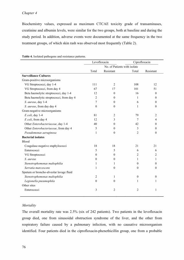
Chapter 4
76
Biochemistry values, expressed as maximum CTCAE toxicity grade of transaminases,
creatinine and albumin levels, were similar for the two groups, both at baseline and during the
study period. In addition, adverse events were documented at the same frequency in the two
treatment groups, of which skin rash was observed most frequently (Table 2).
Table 4. Isolated pathogens and resistance patterns.
Levofloxacin Ciprofloxacin
No. of Patients with isolate
Total Resistant Total Resistant
Surveillance Cultures
Gram-positive microorganisms
VG Streptococci, day 1-4 111 2 108 12
VG Streptococci, from day 4 67 17 101 51
Beta haemolytic streptococci, day 1-4 12 0 16 0
Beta haemolytic streptococci, from day 4 2 0 1 0
S. aureus, day 1-4 7 0 6 0
S. aureus, from day day 4 0 0 1 0
Gram-negative microorganisms
E coli, day 1-4 81 2 79 2
E coli, from day 4 12 3 7 4
Other Enterobacteriaceae, day 1-4 40 0 42 0
Other Enterobacteriaceae, from day 4 5 0 3 0
Pseudomonas aeruginosa 1 0 2 0
Bacterial isolates
Blood
Coagulase-negative staphylococci 18 18 21 21
Enterococci 5 5 6 6
VG Streptococci 0 0 2 2
S. aureus 0 0 1 1
Stenotrophomonas maltophilia 1 1 0 0
Serratia marcescens 1 0 0 0
Sputum or broncho-alveolar lavage fluid
Stenotrophomonas maltophilia 2 1 0 0
Legionella pneumophila 0 0 1 1
Other sites
Enterococci 3 2 2 1
Mortality
The overall mortality rate was 2.5% (six of 242 patients). Two patients in the levofloxacin
group died, one from sinusoidal obstruction syndrome of the liver, and the other from
respiratory failure caused by a pulmonary infection, with no causative microorganism
identified. Four patients died in the ciprofloxacin-phenethicillin group, one from a probable
Levofloxacin vs ciprofloxacin plus phenethicillin
77
infection with Aspergillus fumigatus, a second from cardiac arrest, and two from respiratory
failure. Of the last two patients, the alveolar lavage fluid from one patient yielded
Flavobacteria and Candida albicans, while cultures remained negative for the other patient.
Discussion
The results of this randomized controlled clinical trial demonstrate that levofloxacin and
ciprofloxacin plus phenethicillin are equally successful as antibacterial prophylaxis for
neutropenic patients with haematological malignancies. Failure of prophylaxis, as the primary
outcome measure, was observed at the same frequency in the two treatment groups, as were
its composites: the initiation of broad-spectrum antibacterial antibiotics, and the need for
change of the prophylactic regimen. Other infection-related outcomes, e.g., the time to failure
of prophylaxis, occurrence of fever, the number of patients with a microbiologically
documented infection, and the number of patients who received broad-spectrum intravenous
antibiotics, did not favour either of the prophylactic strategies. However, patients receiving
ciprofloxacin-phenethicillin had a discrete, but significantly longer, duration of neutropenia of
c. 1.6 days. This finding probably accounts for the trend towards a longer duration of hospital
stay for these patients, and may be an explanation for the (almost significant) higher number
of days for which patients in the ciprofloxacin-phenethicillin group needed intravenous
antibiotics. It is well-known that prolonged administration of β-lactam antibiotics may induce
neutropenia, probably because of a direct toxic effect on the bone marrow or an immune-
mediated effect.14,15
Considering the fact that duration of neutropenia has been identified as an
independent risk-factor for the occurrence and severity of infections, and as a critical factor in
a successful outcome, this finding may be of clinical importance.16,17
It was assumed that patients receiving one tablet of levofloxacin per day would tolerate the
study medication better than patients receiving two tablets of ciprofloxacin plus four tablets of
phenethicillin. The results of the questionnaire confirmed this supposition. From day 4, the
mean tolerance score for levofloxacin was significantly higher than the score for
ciprofloxacin-phenethicillin. In line with these results, a change of prophylaxis because of
intolerance of the study drugs was necessary for significantly more patients receiving
ciprofloxacin-phenethicillin than for those receiving levofloxacin. Since these patients are
commonly suffering from discomforting nausea and mucositis, a better tolerance of
prophylactic medication is important and may improve therapy compliance. However, the
Chapter 4
78
results of the questionnaire need to be interpreted with caution. The response rate was 82% in
the levofloxacin group and 66% in the ciprofloxacin-phenethicillin group, which may indicate
selection bias. Patients were sometimes disappointed not to have been assigned to receive
levofloxacin, and other patients became very ill during the study period. It is possible that
these patients, in particular, were less motivated or less able to complete and return the
questionnaire.
The prophylactic administration of both ciprofloxacin-phenethicillin and levofloxacin resulted
in good control over Gram-negative bacteria, and only two patients, both receiving
levofloxacin, developed Gram-negative bacteraemia. The efficacy of quinolone prophylaxis in
reducing Gram-negative infections has been well-documented, although the emergence of
quinolone-resistant bacteria, particularly E. coli, has been reported.18,19
Moreover, prophylaxis
with the older-generation quinolones, e.g., ciprofloxacin, has been associated with an increase
in the number of Gram-positive infections. Levofloxacin and other newer quinolones have
enhanced activity against Gram-positive microorganisms, and may potentially overcome this
problem. However, early reports concerning levofloxacin administered as antibacterial
prophylaxis suggest that its use may be associated with the selection of quinolone-resistant
VG streptococci.12,13
This is a major drawback, as these microorganisms have been reported
to be responsible for up to 39% of cases of bacteraemia in neutropenic patients, and may
result in serious complications, including endocarditis, adult respiratory distress syndrome,
shock and even death.4,13,20
In the present study, surveillance cultures yielded levofloxacin-
resistant VG streptococci from 17 (14%) of 122 levofloxacin recipients. In these patients the
prophylactic regimen was adjusted, in most instances by the addition of penicillin. This
proved to be a valuable approach, as no bacteraemia with VG streptococci occurred in the
levofloxacin group. In agreement with data published previously, the bacteria isolated most
frequently from patients with a bloodstream infection were coagulase-negative staphylococci
and enterococci.20,21
As expected, these bacteria were invariably resistant to levofloxacin and
ciprofloxacin. This finding should be taken into account in the choice of empirical antibiotic
therapy, and underscores the importance of meticulous care of central venous access devices.
In conclusion, levofloxacin was as efficacious as ciprofloxacin plus phenethicillin for the
prevention of bacterial infections in neutropenic patients with cancer. However, levofloxacin
is better-tolerated, which may benefit compliance with therapy. Resistance in VG streptococci
does occur, but this problem appears to be manageable if resistance patterns are monitored
closely. The present study does not answer the question as to which patients with neutropenia
benefit most from the prophylactic administration of levofloxacin and other quinolones, nor

Levofloxacin vs ciprofloxacin plus phenethicillin
79
does it support the unlimited or uncontrolled use of these agents. As outlined in the guidelines
published by the Infectious Diseases Society of America,22 routine quinolone prophylaxis for
all neutropenic patients is not recommended. Based on the estimated infection risk for their
own category of neutropenic patients, and with careful consideration of local antimicrobial
resistance patterns, physicians should weigh the benefits of quinolone prophylaxis against the
potential dangers of this approach.
Acknowledgements
We thank M. Gittelbauer, R. Piqué, F. Tangali, S. Straver, I. Schouten and H. Rouwenhoff
from the Department of Medical Microbiology and Infection Control for their work on culture
data and susceptibility testing. We also thank M. Leisink, Y. den Hartog, C. van Roessel, C.
Eeltink and J. van Wegen from the Haematology Trial Office for their contribution to data
management. This investigator-driven study was sponsored, in part, by a grant from Sanofi
Aventis, Gouda, The Netherlands.
References
1. Cruciani M, Rampazzo R, Malena M, Lazzarini L, Todeschini G, Messori A, Concia E.
Prophylaxis with fluoroquinolones for bacterial infections in neutropenic patients: a meta-
analysis. Clin Infect Dis 1996; 23: 795-805.
2. Reuter S, Kern WV, Sigge A, Dohner H, Marre R, Kern P, von Baum H. Impact of
fluoroquinolone prophylaxis on reduced infection-related mortality among patients with
neutropenia and hematologic malignancies. Clin Infect Dis 2005; 40: 1087-1093.
3. Engels EA, Lau J, Barza M. Efficacy of quinolone prophylaxis in neutropenic cancer patients: a
meta-analysis. J Clin Oncol 1998; 16: 1179-1187.
4. Bochud PY, Calandra T, Francioli P. Bacteremia due to viridans streptococci in neutropenic
patients: a review. Am J Med 1994; 97: 256-264.
5. Zinner SH. Changing epidemiology of infections in patients with neutropenia and cancer:
emphasis on Gram-positive and resistant bacteria. Clin Infect Dis 1999; 29: 490-494.
6. Broun ER, Wheat JL, Kneebone PH, Sundblad K, Hromas RA, Tricot G. Randomized trial of
the addition of Gram-positive prophylaxis to standard antimicrobial prophylaxis for patients
undergoing autologous bone marrow transplantation. Antimicrob Agents Chemother 1994; 38:
576-579.
Chapter 4
80
7. Hidalgo M, Hornedo J, Lumbreras C, Trigo JM, Gomez C, Perea S, Ruiz A, Hitt R, Cortes-
Funes H. Lack of ability of ciprofloxacin-rifampin prophylaxis to decrease infection-related
morbidity in neutropenic patients given cytotoxic therapy and peripheral blood stem cell
transplants. Antimicrob Agents Chemother 1997; 41: 1175-1177.
8. Bow EJ, Mandell LA, Louie TJ, Feld R, Palmer M, Zee B, Pater J. Quinolone-based
antibacterial chemoprophylaxis in neutropenic patients: effect of augmented Gram-positive
activity on infectious morbidity. National Cancer Institute of Canada Clinical Trials Group. Ann
Intern Med 1996; 125: 183-190.
9. Cruciani M, Malena M, Bosco O, Nardi S, Serpelloni G, Mengoli C. Reappraisal with meta-
analysis of the addition of Gram-positive prophylaxis to fluoroquinolone in neutropenic patients.
J Clin Oncol 2003; 21: 4127-4137.
10. Cullen M, Steven N, Billingham L, Gaunt C, Hastings M, Simmonds P, Stuart N, Rea D, Bower
M, Fernando I, Huddart R, Gollins S, Stanley A. Antibacterial prophylaxis after chemotherapy
for solid tumors and lymphomas. N Engl J Med 2005; 353: 988-998.
11. Bucaneve G, Micozzi A, Menichetti F, Martino P, Dionisi MS, Martinelli G, Allione B,
D'Antonio D, Buelli M, Nosari AM, Cilloni D, Zuffa E, Cantaffa R, Specchia G, Amadori S,
Fabbiano F, Deliliers GL, Lauria F, Foa R, Del Favero A. Levofloxacin to prevent bacterial
infection in patients with cancer and neutropenia. N Engl J Med 2005; 353: 977-987.
12. Timmers GJ, Dijstelbloem Y, Simoons-Smit AM, van Winkelhoff AJ, Touw DJ,
Vandenbroucke-Grauls CM, Huijgens PC. Pharmacokinetics and effects on bowel and throat
microflora of oral levofloxacin as antibacterial prophylaxis in neutropenic patients with
haematological malignancies. Bone Marrow Transplant 2004; 33: 847-853.
13. Razonable RR, Litzow MR, Khaliq Y, Piper KE, Rouse MS, Patel R. Bacteremia due to viridans
group Streptococci with diminished susceptibility to Levofloxacin among neutropenic patients
receiving levofloxacin prophylaxis. Clin Infect Dis 2002; 34: 1469-1474.
14. Neftel KA, Hauser SP, Muller MR. Inhibition of granulopoiesis in vivo and in vitro by beta-
lactam antibiotics. J Infect Dis 1985; 152: 90-98.
15. Olaison L, Belin L, Hogevik H, Alestig K. Incidence of beta-lactam-induced delayed
hypersensitivity and neutropenia during treatment of infective endocarditis. Arch Intern Med
1999; 159: 607-615.
16. Bodey GP, Buckley M, Sathe YS, Freireich EJ. Quantitative relationships between circulating
leukocytes and infection in patients with acute leukemia. Ann Intern Med 1966; 64: 328-340.
17. Viscoli C, Varnier O, Machetti M. Infections in patients with febrile neutropenia: epidemiology,
microbiology, and risk stratification. Clin Infect Dis 2005; 40 Suppl 4: S240-S245.
18. Kern WV, Steib-Bauert M, de With K, Reuter S, Bertz H, Frank U, von Baum H.
Fluoroquinolone consumption and resistance in haematology-oncology patients: ecological
analysis in two university hospitals 1999-2002. J Antimicrob Chemother 2005; 55: 57-60.
Levofloxacin vs ciprofloxacin plus phenethicillin
81
19. van Kraaij MG, Dekker AW, Peters E, Fluit A, Verdonck LF, Rozenberg-Arska M. Emergence
and infectious complications of ciprofloxacin-resistant Escherichia coli in haematological
cancer patients. Eur J Clin Microbiol Infect Dis 1998; 17: 591-592.
20. Rolston KV, Yadegarynia D, Kontoyiannis DP, Raad II, Ho DH. The spectrum of Gram-positive
bloodstream infections in patients with hematologic malignancies, and the in vitro activity of
various quinolones against Gram-positive bacteria isolated from cancer patients. Int J Infect Dis
2006; 10: 223-230.
21. Wisplinghoff H, Seifert H, Wenzel RP, Edmond MB. Current trends in the epidemiology of
nosocomial bloodstream infections in patients with hematological malignancies and solid
neoplasms in hospitals in the United States. Clin Infect Dis 2003; 36: 1103-1110.
22. Hughes WT, Armstrong D, Bodey GP, Bow EJ, Brown AE, Calandra T, Feld R, Pizzo PA,
Rolston KV, Shenep JL, Young LS. 2002 guidelines for the use of antimicrobial agents in
neutropenic patients with cancer. Clin Infect Dis 2002; 34: 730-751.

Chapter 4
82

83
Amphotericin B colloidal dispersion (Amphocil) vs fluconazole for
the prevention of fungal infections in neutropenic patients: data of
a prematurely stopped clinical trial.
G.J. Timmers
S. Zweegman
A.M. Simoons-Smit
A.C. van Loenen
D. Touw
P.C. Huijgens
Department of Haematology, Department of Medical Microbiology and Infection Control,
Department of Pharmacy, VU University Medical Center, Amsterdam, The Netherlands.
Bone Marrow Transplantation 2000;25:879-884
Chapter 3 3 5

Chapter 5
84
Abstract
We conducted an open label, randomized clinical trial to compare amphotericin B colloidal
dispersion (ABCD, Amphocil) 2 mg/kg/day intravenously with fluconazole 200 mg/day
orally, for the prevention of fungal disease in neutropenic patients with haematological
malignancies. In the event of unresolved fever after 4 days of empirical antibacterial therapy,
patients in both treatment groups were to receive ABCD, 4 mg/kg/day. However, the study
had to be stopped in an early phase, due to severe side-effects of ABCD. A total of 24 patients
were enrolled, 12 patients were randomly assigned to receive prophylactic ABCD, which was
administered for a mean of 13.9 days. Fluconazole prophylaxis was given to 12 patients for a
mean of 21.2 days. Therapeutic ABCD, 4 mg/kg, was initiated in four patients because of
suspected fungal infection, all of whom had initially received fluconazole. A high rate of
infusion-related toxicity of ABCD was observed. Chills occurred in 15/16 ABCD recipients
(94%), accompanied by a temperature rise of ≥2°C in 4/16 patients and of ≥1°C but <2°c in
10/16 patients. Other ABCD-related adverse events were hypotension (4/16), nausea with
vomiting (5/16), tachycardia (7/16), headache (3/16) and dyspnoea (3/16). For premedication
patients received: antihistamines (12/16), hydrocortisone (9/16) and/or morphine (6/16).
ABCD was discontinued in 8/16 patients (50%) due to side-effects, which ultimately dictated
early termination of the study. we conclude that ABCD is not suitable for antifungal
prophylaxis in neutropenic patients due to severe infusion-related side-effects. Subject
numbers were too low for conclusions on variables of antifungal efficacy.
Amphocil antifungal prophylaxis in neutropenia
85
Introduction
Superficial or invasive fungal infections, most often caused by Candida and Aspergillus
species, are among the most serious complications after chemotherapeutic treatment of
haematological malignancies.1-3 In view of the poor prognosis of disseminated fungal disease,
many centres administer antimycotic agents prophylactically. Fluconazole has emerged as the
most widely used prophylactic agent in neutropenic patients and has been used in doses
varying from 50 to 400 mg.4-10
Its optimal dose in antifungal prophylaxis remains to be
determined.5,6 Not only has fluconazole been found to be highly effective in eliminating
colonization and infection by Candida spp. in patients with leukaemia,11 but interestingly, the
drug has also been proven to significantly reduce the incidence of, and mortality from
systemic fungal infections in allogeneic stem cell transplantation recipients.4,9 An important
limitation to the use of fluconazole however, is its lack of efficacy against Aspergillus spp.
Moreover, some Candida spp. other than albicans, including C. glabrata and C. krusei are
less sensitive or resistant.
Amphotericin B (AmB) remains the gold standard of antifungal therapy and is attractive as a
prophylactic agent because of its broad coverage of most fungal organisms. However, its use
is seriously hampered by dose-limiting toxicity, with acute infusion-related toxicity and renal
failure being the most important adverse effects. To circumvent toxicity, low doses of
intravenous AmB have been used in antifungal prophylaxis with ambiguous results.12-14
Lipid
formulations of AmB have been designed to improve efficacy and to reduce toxicity of the
parent compound, thereby allowing the possibility of administering higher doses of AmB.
Amphotericin B colloidal dispersion (ABCD, Amphocil; Sequus Pharmaceuticals, Brentford,
UK) is a colloidal formulation of amphotericin B complexed to cholesteryl sulphate in a 1:1
ratio, forming disc-shaped particles. In various open-label clinical trials, the use of ABCD
showed promising results with regard to its antifungal efficacy and its safety.15-19
Its assumed
reduced toxicity offers the opportunity of investigating the feasibility of AmB as antifungal
prophylaxis. In a recent randomized trial we reported a 100% mortality in patients with
proven fungal infections due to Aspergillus or Mucor spp., the overall incidence of such
infections in our population being 5%.10 In an attempt to reduce both incidence of, and
mortality from these infections, an open-label, randomized clinical trial was initiated, to
compare the safety and efficacy of intravenous ABCD with oral fluconazole in the prevention
of fungal disease in neutropenic patients with haematological malignancies. For the
prophylactic use of ABCD we decided to administer half of the daily dose that is generally
Chapter 5
86
recommended for antifungal therapy. Unfortunately, due to excessive toxicity of ABCD the
study had to be stopped in an early phase, after the randomization of 24 patients.
Patients and methods
Patients
Consecutive patients, aged 18-75 years, who were to receive treatment for haematological
malignancies with or without peripheral blood stem cell transplantation were enrolled. An
anticipated granulocytopenic period of at least 10 days was required. Patients were excluded
if they had a previous history of allergy or hypersensitivity to any lipid preparation of AmB,
or to other polyene or azole antimycotic agents. Patients were considered not eligible if they
had overt infection requiring treatment at entry, if they had received treatment with systemic
or topical antifungal drugs within 2 weeks prior to enrolment, or if there was hepatic or renal
impairment, defined as elevation of liver enzymes more than three times the upper limit of
normal, or a creatinine clearance of less than 15 ml/min, respectively. The study was
approved by the institutional ethical and scientific committees. Written informed consent was
obtained from all patients.
Study protocol
The study was designed as a randomized, open-label clinical trial. Patients were randomly
assigned to receive once-daily prophylaxis with either fluconazole 200 mg orally, or ABCD 2
mg/kg i.v. ABCD was prepared as recommended by the manufacturer and was administered
at an infusion rate of 1 mg/kg/h. No other drugs were co-administered during ABCD infusion.
Premedications were not given before the first dose of ABCD. In case of infusion-related
toxicity, clemastin, hydrocortisone or morphine were initiated sequentially or in combination
for premedication, depending on the severity of symptoms. Study treatment was started on the
first day of the chemotherapeutic regimen and was continued until neutrophil recovery (ANC
>0.5 × 10⁹/l) or failure of prophylaxis, defined as the necessity of initiating therapeutic
ABCD, treatment interruption due to side-effects or mortality. For prevention purposes all
patients received ciprofloxacin, 500 mg twice daily and azithromycin 250 mg once daily.
Nasal AmB was given, 2 mg three times a day, into both nasal orifices.
Daily clinical assessments were performed, including documentation of signs and symptoms
of infection and of tolerance to the study medication. The highest grade of infusion-related
Amphocil antifungal prophylaxis in neutropenia
87
toxicity that occurred during the study period was recorded. Routine haematology and clinical
chemistry tests were obtained three times weekly. Surveillance cultures for fungal and
bacterial organisms were taken from throat and anus prior to the first dose of the study drug
and, thereafter, once weekly.
When axillary temperature increased above 38.5°C, clinical evaluation was performed and
empirical antibiotic therapy was initiated, as described in detail earlier.10 Empirical treatment
consisted of cefpirome 2000 mg, twice daily i.v. If subsequently, fever did not resolve within
96 h, patients in both treatment groups were to receive ABCD, 4 mg/kg once daily i.v.
Definition of infection
Fungal colonization was considered to be present if one or more surveillance culture yielded a
fungus, in the absence of any clinical symptoms or signs of infection. Suspected systemic
fungal infection was defined as fever, persisting after 96 h of appropriate antibacterial
treatment, necessitating the use of therapeutic ABCD. Proven fungal infection was defined as
a positive culture or characteristic histopathological findings on tissue biopsy specimens
obtained from a normally sterile site.
Statistical analysis
Results from all patients were analysed according to the intention-to-treat principle.
Categorical data were analysed by Fisher's exact test, Student's t-test was used for comparison
of means. All tests were two-sided and in all cases a P value less than 0.05 was considered
statistically significant. To detect significant differences between groups in the number of
proven and suspected fungal infections, it was calculated that the study should include a total
of 220 patients.
Results
Before the early termination of the study, 24 patients with haemato-oncological diseases were
included, 12 receiving ABCD and 12 fluconazole. Patient characteristics are given in Table 1.
The two groups were comparable for sex, type of diagnosis, number of stem-cell transplants
and duration of neutropenia. The ABCD group contained two patients receiving allogeneic or
syngeneic stem cell transplantation. Patients in the fluconazole group were significantly older
than patients in the ABCD group (P <0.006).
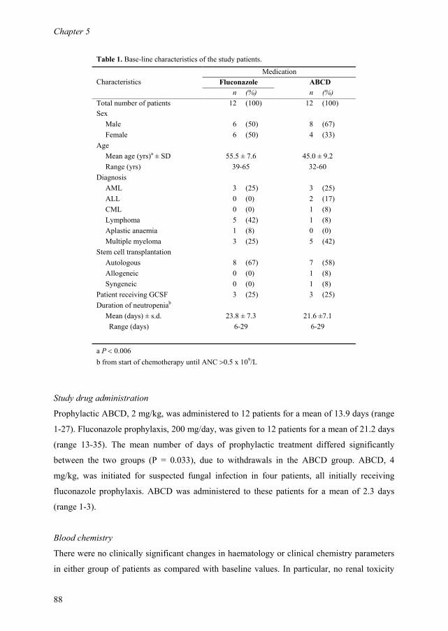
Chapter 5
88
Table 1. Base-line characteristics of the study patients.
Medication
Characteristics Fluconazole ABCD
n (%) n (%)
Total number of patients 12 (100) 12 (100)
Sex
Male 6 (50) 8 (67)
Female 6 (50) 4 (33)
Age
Mean age (yrs)a ± SD 55.5 ± 7.6 45.0 ± 9.2
Range (yrs) 39-65 32-60
Diagnosis
AML 3 (25) 3 (25)
ALL 0 (0) 2 (17)
CML 0 (0) 1 (8)
Lymphoma 5 (42) 1 (8)
Aplastic anaemia 1 (8) 0 (0)
Multiple myeloma 3 (25) 5 (42)
Stem cell transplantation
Autologous 8 (67) 7 (58)
Allogeneic 0 (0) 1 (8)
Syngeneic 0 (0) 1 (8)
Patient receiving GCSF 3 (25) 3 (25)
Duration of neutropeniab
Mean (days) ± s.d. 23.8 ± 7.3 21.6 ±7.1
Range (days) 6-29 6-29
a P < 0.006
b from start of chemotherapy until ANC >0.5 x 10⁹/L
Study drug administration
Prophylactic ABCD, 2 mg/kg, was administered to 12 patients for a mean of 13.9 days (range
1-27). Fluconazole prophylaxis, 200 mg/day, was given to 12 patients for a mean of 21.2 days
(range 13-35). The mean number of days of prophylactic treatment differed significantly
between the two groups (P = 0.033), due to withdrawals in the ABCD group. ABCD, 4
mg/kg, was initiated for suspected fungal infection in four patients, all initially receiving
fluconazole prophylaxis. ABCD was administered to these patients for a mean of 2.3 days
(range 1-3).
Blood chemistry
There were no clinically significant changes in haematology or clinical chemistry parameters
in either group of patients as compared with baseline values. In particular, no renal toxicity
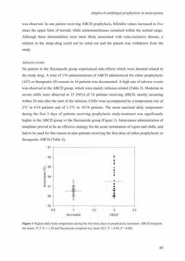
����)+,4�(�,��(��4��)���4��,6,((����)��(,�
�
������������1�� �������������0F�� �� �$����������������������������������
����� ���� �� ��������������������������������������������� �������������
0������� ����� ������������ ���� ���� ��/��$ ���������� ���� ���&��������� �������� �
������� �� ��� ����$&���� ����� �� �� ����� ��� �� ��� ����� ��� �������� ���� ���
����$�
���6����(�6
G� ����� � ��� ������7������� �� ������� ����&���������������� ������ ������� ��
�������$�����0��������<E���������������0F���������������������� �� �$������
��E<�������� ������ ���������E �������������������0����������������������
���������������0F������ ��������������$������&��������-����9��2���������
������������������������ ��� � B����E ������ ��������0F��������$��������
������������������������������������������������� �����$���� �������������
9H� � !�E ������ �����&9H� ���!�E ������� -������������ ����$ ��� �������
����� ��� ����� � ��$� �� ������ �������� �� �$������ ����$&�������� ��� ����������$
����������0F������ �����������7������� ����������1����������������������
��� ��� ��������������������������$���������������������������������������
�������������������������� ���������������������������������� �� �$��������
����� �����0F���-�������
������;����������$���$&��� ���������������������������$�� �� �$��������������I0F������ �����������I�<��H��������������7������� ������������E��H���� ��C������
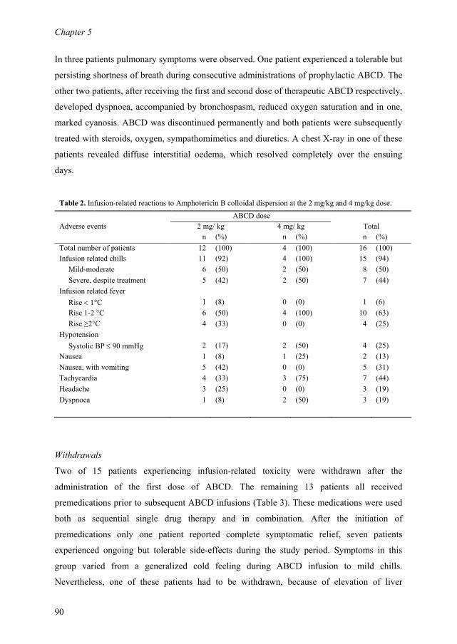
Chapter 5
90
In three patients pulmonary symptoms were observed. One patient experienced a tolerable but
persisting shortness of breath during consecutive administrations of prophylactic ABCD. The
other two patients, after receiving the first and second dose of therapeutic ABCD respectively,
developed dyspnoea, accompanied by bronchospasm, reduced oxygen saturation and in one,
marked cyanosis. ABCD was discontinued permanently and both patients were subsequently
treated with steroids, oxygen, sympathomimetics and diuretics. A chest X-ray in one of these
patients revealed diffuse interstitial oedema, which resolved completely over the ensuing
days.
Table 2. Infusion-related reactions to Amphotericin B colloidal dispersion at the 2 mg/kg and 4 mg/kg dose.
ABCD dose
Adverse events 2 mg/ kg 4 mg/ kg Total
n (%) n (%) n (%)
Total number of patients 12 (100) 4 (100) 16 (100)
Infusion related chills 11 (92) 4 (100) 15 (94)
Mild-moderate 6 (50) 2 (50) 8 (50)
Severe, despite treatment 5 (42) 2 (50) 7 (44)
Infusion related fever
Rise < 1°C 1 (8) 0 (0) 1 (6)
Rise 1-2 °C 6 (50) 4 (100) 10 (63)
Rise ≥2°C 4 (33) 0 (0) 4 (25)
Hypotension
Systolic BP ≤ 90 mmHg 2 (17) 2 (50) 4 (25)
Nausea 1 (8) 1 (25) 2 (13)
Nausea, with vomiting 5 (42) 0 (0) 5 (31)
Tachycardia 4 (33) 3 (75) 7 (44)
Headache 3 (25) 0 (0) 3 (19)
Dyspnoea 1 (8) 2 (50) 3 (19)
Withdrawals
Two of 15 patients experiencing infusion-related toxicity were withdrawn after the
administration of the first dose of ABCD. The remaining 13 patients all received
premedications prior to subsequent ABCD infusions (Table 3). These medications were used
both as sequential single drug therapy and in combination. After the initiation of
premedications only one patient reported complete symptomatic relief, seven patients
experienced ongoing but tolerable side-effects during the study period. Symptoms in this
group varied from a generalized cold feeling during ABCD infusion to mild chills.
Nevertheless, one of these patients had to be withdrawn, because of elevation of liver
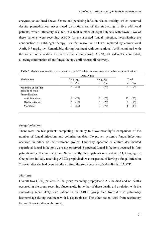
Amphocil antifungal prophylaxis in neutropenia
91
enzymes, as outlined above. Severe and persisting infusion-related toxicity, which occurred
despite premedication, necessitated discontinuation of the study-drug in five additional
patients, which ultimately resulted in a total number of eight subjects withdrawn. Two of
these patients were receiving ABCD for a suspected fungal infection, necessitating the
continuation of antifungal therapy. For that reason ABCD was replaced by conventional
AmB, 0.7 mg/kg i.v. Remarkably, during treatment with conventional AmB, combined with
the same premedication as used while administering ABCD, all side-effects subsided,
allowing continuation of antifungal therapy until neutrophil recovery.
Table 3. Medications used for the termination of ABCD related adverse events and subsequent medications
ABCD dose
Medications 2 mg/ kg 4 mg/ kg Total
n (%) n (%) n (%)
6 (50) 3 (75) 9 (56) Morphine at the first
episode of chills
Premedications
Antihistamines 9 (75) 3 (75) 12 (75)
Hydrocortisone 6 (50) 3 (75) 9 (56)
Morphine 3 (25) 3 (75) 6 (38)
Fungal infections
There were too few patients completing the study to allow meaningful comparison of the
number of fungal infections and colonization data. No proven systemic fungal infections
occurred in either of the treatment groups. Clinically apparent or culture documented
superficial fungal infections were not observed. Suspected fungal infections occurred in four
patients in the fluconazole group. Subsequently, these patients received ABCD, 4 mg/kg i.v.
One patient initially receiving ABCD prophylaxis was suspected of having a fungal infection
2 weeks after she had been withdrawn from the study because of side-effects of ABCD.
Mortality
Overall two (17%) patients in the group receiving prophylactic ABCD died and no deaths
occurred in the group receiving fluconazole. In neither of these deaths did a relation with the
study-drug seem likely; one patient in the ABCD group died from diffuse pulmonary
haemorrhage during treatment with L-asparaginase. The other patient died from respiratory
failure, 3 weeks after withdrawal.

Chapter 5
92
Termination of the study
A variety of strategies not integral to the protocol was used to cope with the high rate of
infusion-related toxicity. Three different batches of ABCD were used to rule out quality
differences of the compound. In individual cases the infusion time was sometimes prolonged
for up to 4 h. However, ultimately all measures failed to increase tolerability and the study
was prematurely closed, according to GCP guidelines.
Discussion
Although the use of antifungal prophylaxis in neutropenic patients, especially in low-risk
autotransplant recipients, is certainly debatable, mortality from these infections remains high.
Lipid formulations of AmB appear attractive as antifungal prophylaxis because these
compounds are claimed to decrease toxicity, while maintaining or even enhancing therapeutic
efficacy. In two randomized, placebo-controlled clinical trials, prophylactic administration of
AmBisome to patients with haematological malignancies was associated with a statistically
significant reduction of fungal colonization.20,21
Both studies, however, failed to show a
significant reduction in proven fungal infections, possibly because patient samples were too
small. Our randomized, open label clinical trial was designed to compare the safety and
efficacy of ABCD with fluconazole in the prevention of fungal infections in neutropenic
patients with haematological malignancies. However, severe and unexpected toxicity of
ABCD resulted in the early withdrawal of eight patients, comprising 50% of the ABCD
recipients. In all instances there was a direct and obvious correlation with the infusion of
ABCD. Infusion-related chills were observed in 94% of the patients receiving ABCD,
necessitating extensive use of premedications. Three patients had pulmonary symptoms, with
marked dyspnoea bronchospasm and hypoxaemia in two. Acute pulmonary toxicity is a
known complication of AmB infusions and has been reported in association with its lipid
formulations before, including ABCD.22-24
We did not anticipate the occurrence of adverse events to such a large extent at the 2 mg/kg
dose level. In early open-label phase I/II trials, escalating ABCD doses of up to 6 mg/kg/day
were administered.15,16
Although the frequency of infusion-related toxicity was considerable,
occurring in about 65% of patients, the rate and severity of adverse events increased
substantially in patients receiving >4 mg/kg/day compared with those receiving 3-4
mg/kg/day. The maximum tolerated dose was set at 7.5 mg/kg/day. Infusion-related events
Amphocil antifungal prophylaxis in neutropenia
93
were reported to be well controllable or to disappear completely, once premedication was
added. Moreover, during the course of the treatment period the proportion of patients
experiencing infusion-related events tended to diminish steadily.15,16,18
These findings are in
sharp contrast with our results. One explanation could be that both patients and treating
physicians are less willing to accept moderate to severe adverse effects when administering
ABCD for prophylactic reasons. However, in the present study, ABCD related toxicity was
also considered intolerable in patients with suspected fungal infections, receiving a
therapeutic dose of ABCD at 4 mg/kg/day. After being withdrawn from the study two of these
patients received conventional AmB without appreciable toxicity. This observation is in line
with data from a recent randomized double blind study, comparing ABCD (4 mg/kg/day) with
conventional AmB (0.8 mg/kg/day) for the empirical treatment of fever in neutropenic
patients.24 The authors report that chills occurred more frequently in ABCD recipients than in
patients receiving conventional AmB, 80% vs 65% respectively, resulting in a more frequent
use of premedication by ABCD recipients.
We have no proper explanation for the high rate of infusion-related toxicity associated with
ABCD, as observed in our patients. The three commercially available lipid formulations
(Amphocil, AmBisome and Abelcet) seem to differ substantially with regard to their infusion-
related toxicity, probably due to major differences in molecular structure.25 In general, the rate
of acute reactions seems to be minimal for AmBisome and highest for ABCD.25,26
Severe and
unexpected toxicity has been reported with the use of Abelcet recently.27 The characteristics
of the adverse events are very much like the well-known toxic effects of conventional AmB,
which have been shown to be associated with increasing plasma levels of TNF-α, IL-1 and IL-
6.28,29
In earlier reports, the use of ABCD is invariably labelled as 'safe'.15-19
Although this may hold
true with regard to its renal sparing effects, our data indicate, in line with recent
observations,24 that infusion-related toxicity of ABCD must not be underestimated and poses
a major drawback to its use. We conclude that ABCD, administered for prophylactic reasons
to patients without life-threatening fungal infections, was associated with major and
intolerable side-effects during infusion of the compound. Therefore, ABCD appears to be
unsuitable for antifungal prophylaxis in neutropenic patients.

Chapter 5
94
Acknowledgements
This study was supported by a grant from Zeneca Farma, Ridderkerk, The Netherlands,
distributor of Amphocil for Europe.
References
1. Walsh TJ, Hiemenz JW, Anaissie E. Recent progress and current problems in treatment of
invasive fungal infections in neutropenic patients. Infect Dis Clin North Am 1996; 10: 365-400.
2. DeGregorio MW, Lee WM, Linker CA, Jacobs RA, Ries CA. Fungal infections in patients with
acute leukemia. Am J Med 1982; 73: 543-548.
3. Bodey G, Bueltmann B, Duguid W, Gibbs D, Hanak H, Hotchi M, Mall G, Martino P, Meunier
F, Milliken S, . Fungal infections in cancer patients: an international autopsy survey. Eur J Clin
Microbiol Infect Dis 1992; 11: 99-109.
4. Goodman JL, Winston DJ, Greenfield RA, Chandrasekar PH, Fox B, Kaizer H, Shadduck RK,
Shea TC, Stiff P, Friedman DJ, . A controlled trial of fluconazole to prevent fungal infections in
patients undergoing bone marrow transplantation. N Engl J Med 1992; 326: 845-851.
5. Huijgens PC, van Loenen AC, Simoons-Smit AM, Prooy E, Ossenkoppele GJ. The prophylactic
use of fluconazole 50 vs. 100 mg daily in haematological malignancies. Eur J Cancer 1993;
29A: 926-927.
6. Alangaden G, Chandrasekar PH, Bailey E, Khaliq Y. Antifungal prophylaxis with low-dose
fluconazole during bone marrow transplantation. The Bone Marrow Transplantation Team.
Bone Marrow Transplant 1994; 14: 919-924.
7. Menichetti F, Del Favero A, Martino P, Bucaneve G, Micozzi A, D'Antonio D, Ricci P,
Carotenuto M, Liso V, Nosari AM, Barbui T, Fasola G, Mandelli F. Preventing fungal infection
in neutropenic patients with acute leukemia: fluconazole compared with oral amphotericin B.
Ann Intern Med 1994; 120: 913-918.
8. Schaffner A , Schaffner M. Effect of prophylactic fluconazole on the frequency of fungal
infections, amphotericin B use, and health care costs in patients undergoing intensive
chemotherapy for hematologic neoplasias. J Infect Dis 1995; 172: 1035-1041.
9. Slavin MA, Osborne B, Adams R, Levenstein MJ, Schoch HG, Feldman AR, Meyers JD,
Bowden RA. Efficacy and safety of fluconazole prophylaxis for fungal infections after marrow
transplantation-a prospective, randomized, double-blind study. J Infect Dis 1995; 171: 1545-
1552.
Amphocil antifungal prophylaxis in neutropenia
95
10. Huijgens PC, Simoons-Smit AM, van Loenen AC, Prooy E, van Tinteren H, Ossenkoppele GJ,
Jonkhoff AR. Fluconazole versus itraconazole for the prevention of fungal infections in
haemato-oncology. J Clin Pathol 1999; 52: 376-380.
11. Winston DJ, Chandrasekar PH, Lazarus HM, Goodman JL, Silber JL, Horowitz H, Shadduck
RK, Rosenfeld CS, Ho WG, Islam MZ, Buell DN. Fluconazole prophylaxis of fungal infections
in patients with acute leukemia. Results of a randomized placebo-controlled, double-blind,
multicenter trial. Ann Intern Med 1993; 118: 495-503.
12. Perfect JR, Klotman ME, Gilbert CC, Crawford DD, Rosner GL, Wright KA, Peters WP.
Prophylactic intravenous amphotericin B in neutropenic autologous bone marrow transplant
recipients. J Infect Dis 1992; 165: 891-897.
13. Rousey SR, Russler S, Gottlieb M, Ash RC. Low-dose amphotericin B prophylaxis against
invasive Aspergillus infections in allogeneic marrow transplantation. Am J Med 1991; 91: 484-
492.
14. Riley DK, Pavia AT, Beatty PG, Petersen FB, Spruance JL, Stokes R, Evans TG. The
prophylactic use of low-dose amphotericin B in bone marrow transplant patients. Am J Med
1994; 97: 509-514.
15. Bowden RA, Cays M, Gooley T, Mamelok RD, van Burik JA. Phase I study of amphotericin B
colloidal dispersion for the treatment of invasive fungal infections after marrow transplant. J
Infect Dis 1996; 173: 1208-1215.
16. Herbrecht R. Safety of Amphotericin B Colloidal Dispersion. Eur J Clin Microbiol Infect Dis
1997; 16: 74-80.
17. Oppenheim BA, Herbrecht R, Kusne S. The safety and efficacy of amphotericin B colloidal
dispersion in the treatment of invasive mycoses. Clin Infect Dis 1995; 21: 1145-1153.
18. Anaissie EJ, Mattiuzzi GN, Miller CB, Noskin GA, Gurwith MJ, Mamelok RD, Pietrelli LA.
Treatment of invasive fungal infections in renally impaired patients with amphotericin B
colloidal dispersion. Antimicrob Agents Chemother 1998; 42: 606-611.
19. Noskin G, Pietrelli L, Gurwith M, Bowden R. Treatment of invasive fungal infections with
amphotericin B colloidal dispersion in bone marrow transplant recipients. Bone Marrow
Transplant 1999; 23: 697-703.
20. Tollemar J, Ringden O, Andersson S, Sundberg B, Ljungman P, Tyden G. Randomized double-
blind study of liposomal amphotericin B (Ambisome) prophylaxis of invasive fungal infections
in bone marrow transplant recipients. Bone Marrow Transplant 1993; 12: 577-582.
21. Kelsey SM, Goldman JM, McCann S, Newland AC, Scarffe JH, Oppenheim BA, Mufti GJ.
Liposomal amphotericin (AmBisome) in the prophylaxis of fungal infections in neutropenic
patients: a randomized, double-blind, placebo-controlled study. Bone Marrow Transplant 1999;
23: 163-168.

Chapter 5
96
22. Arning M, Heer-Sonderhoff AH, Wehmeier A, Schneider W. Pulmonary toxicity during
infusion of liposomal amphotericin B in two patients with acute leukemia. Eur J Clin Microbiol
Infect Dis 1995; 14: 41-43.
23. Ringden O, Andstrom E, Remberger M, Svahn BM, Tollemar J. Allergic reactions and other
rare side-effects of liposomal amphotericin. Lancet 1994; 344: 1156-1157.
24. White MH, Bowden RA, Sandler ES, Graham ML, Noskin GA, Wingard JR, Goldman M, van
Burik JA, McCabe A, Lin JS, Gurwith M, Miller CB. Randomized, double-blind clinical trial of
amphotericin B colloidal dispersion vs. amphotericin B in the empirical treatment of fever and
neutropenia. Clin Infect Dis 1998; 27: 296-302.
25. Mehta J. Do variations in molecular structure affect the clinical efficacy and safety of lipid-
based amphotericin B preparations? Leuk Res 1997; 21: 183-188.
26. Leenders AC , de Marie S. The use of lipid formulations of amphotericin B for systemic fungal
infections. Leukemia 1996; 10: 1570-1575.
27. Ringden O, Jonsson V, Hansen M, Tollemar J, Jacobsen N. Severe and common side-effects of
amphotericin B lipid complex (Abelcet). Bone Marrow Transplant 1998; 22: 733-734.
28. Arning M, Kliche KO, Heer-Sonderhoff AH, Wehmeier A. Infusion-related toxicity of three
different amphotericin B formulations and its relation to cytokine plasma levels. Mycoses 1995;
38: 459-465.
29. Chia JK , McManus EJ. In vitro tumor necrosis factor induction assay for analysis of febrile
toxicity associated with amphotericin B preparations. Antimicrob Agents Chemother 1990; 34:
906-908.
97
Effects of cyclosporin A on single-dose pharmacokinetics of
intravenous itraconazole in patients with haematological
malignancies.
Gert Jan Timmers
Lonneke W. Kessels
Abraham J. Wilhelm
Agnes I. Veldkamp
Tessa M. Bosch
Jos H. Beijnen
Peter C. Huijgens
Department of Haematology, Department of Clinical Pharmacology and Pharmacy VU
University Medical Center, Amsterdam, The Netherlands and Department of Pharmacy,
Slotervaart Hospital, Amsterdam, The Netherlands.
Therapeutic Drug Monitoring, accepted for publication
Chapter 3 3 6
Chapter 6
98
Abstract
An open-label, clinical pilot study was performed to study the effect of cyclosporin A (CsA)
on single-dose pharmacokinetics of itraconazole in patients with a haematological
malignancy. Patients (n=10), admitted for an allogeneic stem-cell transplantation, received a
single dose of 200 mg itraconazole, in an one hour intravenous infusion, during their
treatment period before initiation of CsA. This was repeated during the period that CsA was
administered and a steady state concentration of CsA was achieved (trough whole blood level
200-400 ng/ml). After both administrations of itraconazole, serum pharmacokinetics of
itraconazole and hydroxy (OH) itraconazole were determined during 24 hours. The results
were compared, with each patient acting as his/her own control. Exposure to itraconazole, as
measured by the AUC[0-24h], was not significantly altered when combined with CsA. Large
inter-individual variations were observed in AUC values among patients. In contrast,
exposure to OH-itraconazole was significantly increased when itraconazole was co-
administered with CsA (median increase of AUC[0-24h] 49%), with significant prolongation of
Tmax and T1/2 (median increase of Tmax 37% and T1/2 176%). These differences may be due to
variability in affinity of itraconazole, OH-itraconazole and CsA for the cytochrome P450
(CYP) 3A4 metabolic system and the occurrence of P-gp polymorphisms. In conclusion,
exposure to OH-itraconazole, but not to itraconazole is increased when itraconazole is co-
administered with CsA. Though the interaction profile of itraconazole and CsA remains
complex, these findings may be of importance in patients, in whom monitoring of
itraconazole serum levels is warranted, for example in those with life-threatening fungal
infections, or in those who receive concurrent CYP inducers or inhibitors.

Pharmacokinetics of itraconazole
99
Introduction
Itraconazole is a broad-spectrum triazole antifungal agent, that has proven to be effective in
the prevention and treatment of systemic fungal infections in patients with neutropenia.1-3 The
antifungal activity from itraconazole originates from inhibition of the fungal cytochrome
P450 (CYP) 3A iso-enzyme, which plays a central role in the synthesis of ergosterol, a vital
component of the fungal cell membrane. Although with much lower affinity, itraconazole and
its main metabolite, hydroxy-itraconazole (OH-itraconazole) are also inhibitors of this
enzyme in humans.4-6 Because the CYP3A isoenzymes are involved in the metabolic pathway
of many drugs, itraconazole and OH-itraconazole have the potential to modify the
pharmacokinetics of these medications. In addition, itraconazole is both inhibitor and
substrate of the ATP-dependent cell membrane transporter system P-glycoprotein (P-gp,
multidrug resistance 1 [MDR1]), which is involved in the clearance of many drugs.7,8
Polymorphisms of the MDR1 gene have been reported to be associated with alterations in
disposition kinetics and interaction profiles of clinically useful drugs, including digoxin,
tacrolimus and cyclosporin A, though data on itraconazole are lacking.9
One of the most important drug interactions in which itraconazole is involved, is that with
cyclosporin A (CsA). CsA is a calcineurin inhibitor, frequently used for the prevention of
graft-versus-host disease in patients undergoing allogeneic stem cell transplantation. Like
itraconazole, CsA is both substrate and inhibitor of CYP3A4 and P-gp and it has been well
documented that the combination of these two drugs results in increased CsA serum
concentrations.10-15
This may have important toxic effects, especially for kidney function.
Though it is well documented that administration of itraconazole increases CsA serum
concentrations, there are no data on the effects of CsA on itraconazole pharmacokinetics.
Monitoring of itraconazole serum concentrations may be warranted in patients who use other
drugs that interact with CYP3A4, and when target drug concentrations are to be achieved for
optimal antifungal prophylaxis or therapy.16,17
Moreover, patients receiving CsA are
systematically excluded from studies investigating itraconazole pharmacokinetic parameters,
and the question remains as to whether that is justified. We therefore conducted an open-label,
pilot study in haematological patients receiving allogeneic stem-cell transplantation, to study
the effect of CsA on single-dose pharmacokinetic parameters of itraconazole. In addition, the
P-gp genotype was determined, to study the possible effects of single nucleotide
polymorphisms (SNP) in the MDR1 gene on itraconazole pharmacokinetics.
Chapter 6
100
Materials and methods
Patients
Patients aged 18-75 years, admitted to the department of Haematology of the VU University
Medical Center, Amsterdam, The Netherlands, and scheduled to receive allogeneic stem cell
transplantation for the treatment of haematological malignancies, were eligible. Reasons for
exclusion were a history of previous allergy or known hypersensitivity to itraconazole, known
hepatic impairment as determined by elevation of any liver function test greater than three
times the upper limit of normal, or a creatinine clearance under 15 ml/min. Patients with overt
infection at baseline were not eligible. Moreover, patients who used other drugs known to
affect CYP and P-gp function, for example statins, were excluded.
Due to ethical considerations the number of patients in this pilot study was limited to 10. The
protocol was approved by the institutional scientific and ethical committees and all
participating patients provided written informed consent. The study was performed according
to the recommendations of the Helsinki Declaration.
Study design
The study was designed as a single center, open-label, prospective pharmacokinetic trial and
comprised two phases, with patients acting as their own controls. Phase 1 was scheduled
before initiation of CsA and phase 2 as soon as a steady state concentration of CsA was
achieved within the therapeutic range, defined as a trough whole blood level of 200-400
ng/ml. In both phases patients received a single dose of 200 mg itraconazole (Trisporal, for iv
administration (10 mg/ml) Ortho-Biotech, Tilburg, the Netherlands). Itraconazole was
prepared as recommend by the manufacturer, adding 25 ml (10 mg/ml) itraconazole solution
to 50 ml NaCl 0.9%. Of the dilution obtained 60 ml (200 mg) was administered by an one
hour infusion. Subsequently, serial blood samples were obtained at 1 hour (end of the
infusion);1.25; 1.5; 2; 3; 5; 7; 9; 12; 16 and 24 hours after start of the infusion. Samples were
collected in 10 ml coagulation tubes, via a central venous catheter. Serum was separated from
the clot by centrifugation (3000 rpm for 10 minutes) and subsequently stored at -40°C until
analysis. In order to elucidate the possible effects of the P-gp genotype on pharmacokinetic
parameters, an extra 5 ml EDTA blood sample was drawn at study entry, samples were stored
at -40°C until analysis.
Pharmacokinetics of itraconazole
101
Sample analysis
Itraconazole and its metabolite OH-itraconazole were both quantitatively determined with a
validated reversed-phase high-performance liquid chromatographic assay (RP-HPLC) with
ultraviolet detection at a wavelength of 258 nm, as described previously.18 The analysis took
place at ambient temperature. The assay was linear up to 3.2 mg/L and the lower limit of
quantification was 0.03 mg/L, for both itraconazole and OH-itraconazole. CsA blood
concentrations were measured with a fluorescence polarization immunoassay (FPIA) for the
TDx (Abbott Laboratories, Chicago, USA), following the instructions of the manufacturer.
P glycoprotein pharmacogenetic analysis
For determination of the P-gp genotype, 5 ml EDTA blood was taken. Genomic DNA was
isolated and PCR amplifications were performed by standard techniques. The three most
important single nucleotide polymorphisms (SNP) in the MDR1 gene were determined, as
described previously.19,20
Pharmacokinetic analysis
Pharmacokinetic parameters were first derived by use of noncompartmental methods in
WinNonlin (Version 1.5, Scientific Consulting, Inc). The highest observed serum
concentration was defined as the Cmax, with the corresponding sampling time as Tmax. The
area under the serum itraconazole and OH-itraconazole concentration versus time curve from
0-24 h (AUC[0-24h]) was obtained by use of the linear trapezoidal rule. The concentration at 24
hours after the infusion of itraconazole was defined as the trough concentration (Cmin). The
terminal log-linear period (log C versus T) was defined by the last data points (n≥3) by visual
inspection. The absolute value of this slope (λz) was calculated by least squares regression
analysis. The elimination half-life (T1/2) was calculated using T1/2=ln2/λz. The clearance was
calculated by dividing the dose by the AUC extrapolated to infinity (AUCinf). The volume of
distribution in steady-state (Vss) was estimated from the mean residence time extrapolated to
infinity, times clearance.
Statistical analysis
Statistical calculations were performed with the Statistical Product and Service Solutions
(SPSS) for Windows, version 9.0. To detect a difference in the pharmacokinetic parameters of
itraconazole and OH-itraconazole alone (phase 1) and itraconazole and OH-itraconazole in
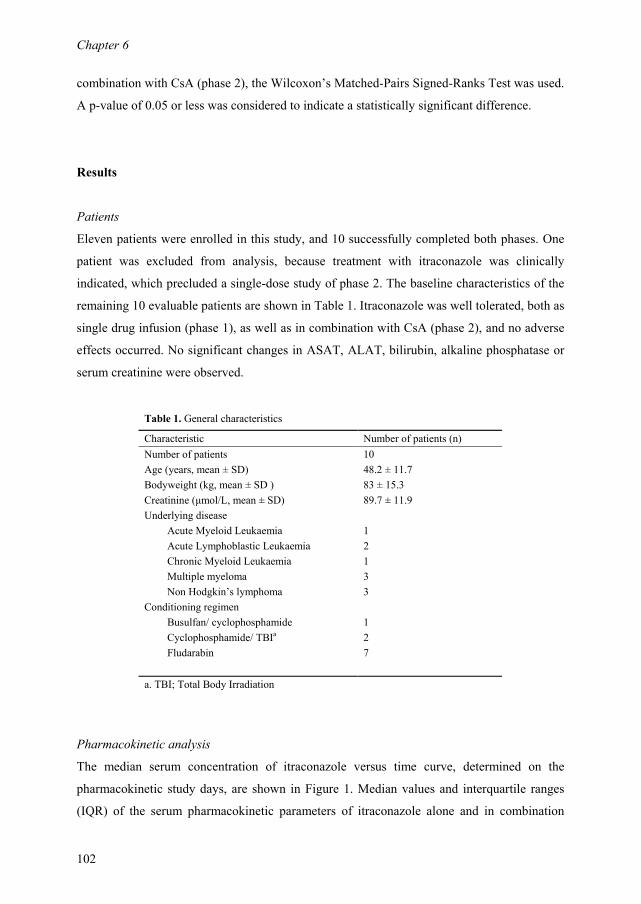
Chapter 6
102
combination with CsA (phase 2), the Wilcoxon’s Matched-Pairs Signed-Ranks Test was used.
A p-value of 0.05 or less was considered to indicate a statistically significant difference.
Results
Patients
Eleven patients were enrolled in this study, and 10 successfully completed both phases. One
patient was excluded from analysis, because treatment with itraconazole was clinically
indicated, which precluded a single-dose study of phase 2. The baseline characteristics of the
remaining 10 evaluable patients are shown in Table 1. Itraconazole was well tolerated, both as
single drug infusion (phase 1), as well as in combination with CsA (phase 2), and no adverse
effects occurred. No significant changes in ASAT, ALAT, bilirubin, alkaline phosphatase or
serum creatinine were observed.
Table 1. General characteristics
Characteristic Number of patients (n)
Number of patients 10
Age (years, mean ± SD) 48.2 ± 11.7
Bodyweight (kg, mean ± SD ) 83 ± 15.3
Creatinine (µmol/L, mean ± SD) 89.7 ± 11.9
Underlying disease
Acute Myeloid Leukaemia 1
Acute Lymphoblastic Leukaemia 2
Chronic Myeloid Leukaemia 1
Multiple myeloma 3
Non Hodgkin’s lymphoma 3
Conditioning regimen
Busulfan/ cyclophosphamide 1
Cyclophosphamide/ TBIa 2
Fludarabin 7
a. TBI; Total Body Irradiation
Pharmacokinetic analysis
The median serum concentration of itraconazole versus time curve, determined on the
pharmacokinetic study days, are shown in Figure 1. Median values and interquartile ranges
(IQR) of the serum pharmacokinetic parameters of itraconazole alone and in combination

������+)�,(��,+6)�,���+)(��)4�
���
������0 ��� ���� � -���� 9� -�� ������� �� ��&������������ ����0 � �������7���
�������/������ � ����� �� �� �����$ � ����������� -���� ��� � ����� ������� ��
0J�K�&9 ��L��� B�1M:& �&9��B��;�����������������������������������$���������
� C��E� �� ��� ������� 9�B �������� �& �B� �� ������� ���� <�B �������� �9��B�
�������������������&�������������������������0J���������� �������� �����
�� ����9�1���������������������$��������������������������������� �����
��9��������� �������/����� ����������������������-����-�!9�����������N���
-������������������������=;&�������7�������������������������������9�
-�������� ���� �������/����� �����������=;&�������7�������������������
������0��������-������-���� �������=;&�������7���������������$���0J�K�&
9 �L�������������$���������������� ����������0������������������� ��������
����� � C����� ��-������� ���������0J�K�&9 �L ���=;&�������7������ B�1M:
9 & �B�� -��� �� -�!9 ���� ���� ����������$ ������� � C�����< �� C����� �
��� �������$��������������������-������<B�1M:�9&���B������������������
-�!9���<EB�1M:���&9 ���-����������������$������������������� C����� ������
���������������E�B�1M: E&<EB��
�������2�����������7���������������������������������� ������������7�������� ����9����
�$���� ���0�-������������� ��������9�B��<�B ���������

��������
��
�������2����=;&�������7���������������������������������� ������������7�������� ����9
�����$���� ���0�-������������� ��������9�B��<�B ���������
��� � �� 2���� �������/����� ��������� �� �������7���� ���� � ���� �� �� � ��������� �����$���� ���0� ����9���� �������-������#����������������� �������� ����������
��������� �����1 �����11 ����� 1���#����������� ����� 1���#����������� &�����0J���&9 ���O��!"� �� ����&�E�9� ��� �E��&��� � ��E9���������!"� �� ����&E�9� �� �9�9&���� ����EE-������ ��� ����&���� ��9 ����&�� � ���99�-�!9��� <�� ����&����� ��E ����&����� �� ��E���������"!�� 9��� ��9�<&��� � ���� ��9�E&9 ��� ���99�N�������������"� ���� �E<��& 9�9� ����< ���E� &9<���� ���99�
��� ���2���� �������/����� ������������$����$&�������7��������� �����������������������$���� ���0� ����9���� �������-������#����������������� �������� ����������
��������� �����1 �����11 ����� 1���#����������� ����� 1���#����������� &�����0J���&9 ���O��!"� ��E �9�E&���� �� � � &E�9� ����� �������!"� ��� ����&�� � ��� ����&�� � �� 9��-������ ��� ����&��E� 9�� ����&9��� �����<-�!9��� <�� ���<&���E� 9��� ��<�E& ���� ����� ���������"!�� ��� �9�� &E ��� � �E � ��&� �E� ����� N�������������"� �E �< � <��&������ E��� ��9<��&<� ��� ������
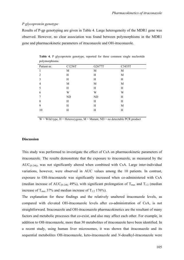
Pharmacokinetics of itraconazole
105
P glycoprotein genotype
Results of P-gp genotyping are given in Table 4. Large heterogeneity of the MDR1 gene was
observed. However, no clear association was found between polymorphisms in the MDR1
gene and pharmacokinetic parameters of itraconazole and OH-itraconazole.
Discussion
This study was performed to investigate the effect of CsA on pharmacokinetic parameters of
itraconazole. The results demonstrate that the exposure to itraconazole, as measured by the
AUC[0-24h], was not significantly altered when combined with CsA. Large inter-individual
variations, however, were observed in AUC values among the 10 patients. In contrast,
exposure to OH-itraconazole was significantly increased when co-administered with CsA
(median increase of AUC[0-24h] 49%), with significant prolongation of Tmax and T1/2 (median
increase of Tmax 37% and median increase of T1/2 176%).
The explanation for these findings and the relatively unaltered itraconazole levels, as
compared with elevated OH-itraconazole levels after co-administration of CsA, is not
straightforward. Itraconazole and OH-itraconazole pharmacokinetics are the resultant of many
factors and metabolic processes that co-exist, and also may affect each other. For example, in
addition to OH-itraconazole, more than 30 metabolites of itraconazole have been identified. In
a recent study, using human liver microsomes, it was shown that itraconazole and its
sequential metabolites OH-itraconazole, keto-itraconazole and N-desalkyl-itraconazole were
Table 4. P glycoprotein genotype, reported for three common single nucleotide
polymorphisms.
Patient nr. C1236T G2677T C3435T
1 M M M
2 H H M
3 H H H
4 M M M
5 H H H
6 W W W
7 ND ND H
8 H H H
9 H H M
10 H H H
W = Wild type, H = Heterozygous, M = Mutant, ND = no detectable PCR product

Chapter 6
106
all high-affinity ligands of CYP3A4. In fact, these metabolites appeared to be as potent as or
more potent CYP3A4 inhibitors than itraconazole itself, and thus may contribute to the
inhibition of CYP3A4 in vivo after itraconazole therapy. In addition, itraconazole, OH-
itraconazole and CsA are not only substrates and inhibitors of the CYP3A4 system, but also
of P-glycoprotein (P-gp).5,7 Since itraconazole, OH-itraconazole and CsA all interact with
CYP3A4 and P-gp, it is difficult to discern their separate effects and dominant avenue of
interaction. Large heterogeneity was found in P-gp genotype in our population of patients. P-
gp genetic polymorphisms may have an effect on disposition of and exposure to itraconazole,
so we hypothesize that this finding may be, at least partially, associated with the pronounced
inter-patient variability of itraconazole concentrations in our patient group. Another
explanation of the large variability of exposure to itraconazole may be the occurrence of
major differences in protein and tissue binding among individual patients. It has been reported
that plasma levels of itraconazole at the same daily dose may differ up to 15-fold.16,17,21
The
explanation of the difference between the unaltered exposure to itraconazole when CsA was
co-administered, as compared with increased OH-itraconazole levels, can only be speculative.
In theory, drugs that require metabolism by the same CYP enzymes compete for binding to-
and metabolism by CYP, and may therefore interact. The clinical significance of this
interaction will depend on the drugs’ relative affinities for binding to these enzymes. In vitro
studies showed that itraconazole and OH-itraconazole were both high affinity ligands to
CYP3A4. However, itraconazole has higher affinity to CYP3A4 than OH-itraconazole, with
Michaelis-menten constants (Km), of 3.9 nM and 27 nM respectively.5 This finding fits our
results, due to the higher affinity of itraconazole for binding to CYP, as compared with that of
OH-itraconazole or CsA, the elimination of itraconazole may prevail and continue, despite the
presence of other competitive substrates, including OH-itraconazole and CsA.
The active metabolite of itraconazole, OH-itraconazole, reaches concentrations in plasma
approximately twice those achieved by the parent drug.6,22
In our study, OH-itraconazole
concentrations were comparable to or even lower than itraconazole concentrations. There may
be several explanations for this finding. First, itraconazole is subject to considerable first pass
metabolism after oral administration. Plasma levels of the first pass metabolite OH-
itraconazole may be higher if itraconazole is administered orally, as compared with
intravenous administration. In a pharmacokinetic study on switching itraconazole from oral to
i.v. use, itraconazole levels continued to rise with a daily dose of 200 mg i.v., while the rate of
rise of OH-itraconazole levels slowed, presumably because the rate of exposure to hepatic
hydroxylation was reduced.23 Second, studies in normal volunteers have shown that OH-
Pharmacokinetics of itraconazole
107
itraconazole accumulates at approximately twice the rate of the parent drug.24,25
Thus, the
ratio OH-itraconazole/ itraconazole increases considerably during multiple dose
administrations and the difference between OH-itraconazole and itraconazole levels becomes
more pronounced with increasing numbers of doses. Our study provides data on a single dose
administration of itraconazole and the results may not be compared or extrapolated to
multiple dose or steady-state pharmacokinetics.
The question remains as to whether monitoring of itraconazole and OH-itraconazole levels is
really of clinical importance. A safe range for itraconazole drug levels has not been defined
yet, and because of its relatively minimal side effects a narrow range is unlikely.17 However,
information on efficacy of itraconazole in the clinical setting is largely dependent upon
clinical judgement. Moreover, variability in itraconazole levels within and between patients is
high and itraconazole pharmacokinetics is unpredictable. Therefore, itraconazole drug level
monitoring may be useful in selected patients, for example in those who receive concurrent
CYP inducers or inhibitors.17 In patients with a life-threatening fungal infection, or in those
who are not responding to treatment, measuring of itraconazole concentrations may be used to
verify the existence of therapeutic levels of the drug.16
In conclusion, our results show a highly variable, but unaltered exposure to itraconazole and
an increased exposure to OH-itraconazole when itraconazole is administered in combination
with CsA. Despite an increased understanding of the pharmacokinetic mechanisms underlying
the interaction profile of itraconazole and CsA, it is still difficult to discern conclusively the
importance and impact of the various metabolic processes that are involved in this interaction.
Acknowledgements
This study was supported by an unrestricted grant from Ortho Biotec, a division of Janssen-
Cilag Pharmaceuticals, Tilburg, The Netherlands.
References
1. Potter M , Donnelly JP. The role of itraconazole in preventing and treating systemic fungal
infections in immunocompromised patients. Acta Haematol. 2004;111:175-180.
Chapter 6
108
2. Glasmacher A, Prentice A, Gorschluter M, et al. Itraconazole prevents invasive fungal infections
in neutropenic patients treated for hematologic malignancies: evidence from a meta-analysis of
3,597 patients. J Clin Oncol. 2003;21:4615-4626.
3. Winston DJ, Maziarz RT, Chandrasekar PH, et al. Intravenous and oral itraconazole versus
intravenous and oral fluconazole for long-term antifungal prophylaxis in allogeneic
hematopoietic stem-cell transplant recipients. A multicenter, randomized trial. Ann Intern Med.
2003;138:705-713.
4. Niwa T, Shiraga T, Takagi A. Effect of antifungal drugs on cytochrome P450 (CYP) 2C9,
CYP2C19, and CYP3A4 activities in human liver microsomes. Biol Pharm Bull. 2005;28:1805-
1808.
5. Isoherranen N, Kunze KL, Allen KE, et al. Role of itraconazole metabolites in CYP3A4
inhibition. Drug Metab Dispos. 2004;32:1121-1131.
6. De Beule K , Van Gestel J. Pharmacology of itraconazole. Drugs. 2001;61 Suppl 1:27-37.
7. Wang EJ, Lew K, Casciano CN, et al. Interaction of common azole antifungals with P
glycoprotein. Antimicrob Agents Chemother. 2002;46:160-165.
8. Miyama T, Takanaga H, Matsuo H, et al. P-glycoprotein-mediated transport of itraconazole
across the blood-brain barrier. Antimicrob Agents Chemother. 1998;42:1738-1744.
9. Ieiri I, Takane H, Otsubo K. The MDR1 (ABCB1) gene polymorphism and its clinical
implications. Clin Pharmacokinet. 2004;43:553-576.
10. Kwan JT, Foxall PJ, Davidson DG, et al. Interaction of cyclosporin and itraconazole. Lancet.
1987;2:282
11. Kramer MR, Marshall SE, Denning DW, et al. Cyclosporine and itraconazole interaction in
heart and lung transplant recipients. Ann Intern Med. 1990;113:327-329.
12. Trenk D, Brett W, Jahnchen E, et al. Time course of cyclosporin/itraconazole interaction.
Lancet. 1987;2:1335-1336.
13. Wimberley SL, Haug MT, III, Shermock KM, et al. Enhanced cyclosporine-itraconazole
interaction with cola in lung transplant recipients. Clin Transplant. 2001;15:116-122.
14. Florea NR, Capitano B, Nightingale CH, et al. Beneficial pharmacokinetic interaction between
cyclosporine and itraconazole in renal transplant recipients. Transplant Proc. 2003;35:2873-
2877.
15. Leather H, Boyette RM, Tian L, et al. Pharmacokinetic evaluation of the drug interaction
between intravenous itraconazole and intravenous tacrolimus or intravenous cyclosporin A in
allogeneic hematopoietic stem cell transplant recipients. Biol Blood Marrow Transplant.
2006;12:325-334.
16. Poirier JM , Cheymol G. Optimisation of itraconazole therapy using target drug concentrations.
Clin Pharmacokinet. 1998;35:461-473.
Pharmacokinetics of itraconazole
109
17. Buchkowsky SS, Partovi N, Ensom MH. Clinical pharmacokinetic monitoring of itraconazole is
warranted in only a subset of patients. Ther Drug Monit. 2005;27:322-333.
18. Compas D, Touw DJ, de Goede PN. Rapid method for the analysis of itraconazole and
hydroxyitraconazole in serum by high-performance liquid chromatography. J Chromatogr B
Biomed Appl. 1996;687:453-456.
19. Kim RB, Leake BF, Choo EF, et al. Identification of functionally variant MDR1 alleles among
European Americans and African Americans. Clin Pharmacol Ther. 2001;70:189-199.
20. Hoffmeyer S, Burk O, von Richter O, et al. Functional polymorphisms of the human multidrug-
resistance gene: multiple sequence variations and correlation of one allele with P-glycoprotein
expression and activity in vivo. Proc Natl Acad Sci U S A. 2000;97:3473-3478.
21. Poirier JM, Berlioz F, Isnard F, et al. Marked intra- and inter-patient variability of itraconazole
steady state plasma concentrations. Therapie. 1996;51:163-167.
22. Boogaerts MA, Maertens J, van der Geest R, et al. Pharmacokinetics and safety of a 7-day
administration of intravenous itraconazole followed by a 14-day administration of itraconazole
oral solution in patients with hematologic malignancy. Antimicrob Agents Chemother.
2001;45:981-985.
23. Prentice AG, Copplestone JA, Hamond MD, et al. Intravenous itraconazole can replace oral
solution in aspergillosis prophylaxis. Br J Haematol. 2000;108:68 (abstract)
24. Meinhof W. Kinetics and spectrum of activity of oral antifungals: the therapeutic implications. J
Am Acad Dermatol. 1993;29:S37-S41.
25. Prentice AG , Glasmacher A. Making sense of itraconazole pharmacokinetics. J Antimicrob
Chemother. 2005;56 Suppl 1:i17-i22.

Chapter 6
110

111
Cefpirome as empirical treatment for febrile neutropenia in
patients with haematological malignancies.
Gert Jan Timmers
Dannis G. van Vuurden
Eleonora L. Swart
Alberdina M. Simoons-Smit
Peter C. Huijgens
Department of Haematology, Department of Pharmacy, Department of Medical Microbiology
and Infection Control, VU University Medical Center, Amsterdam, The Netherlands
Published in a shortened version in Haematologica 2005;90:1005-1006.
Chapter 3 3 7

Chapter 7
112
Abstract
Cefpirome and other fourth generation cephalosporins are used with increasing frequency for
the empirical treatment of febrile neutropenia. The purpose of this study was to assess the
clinical efficacy of cefpirome and its activity against isolated pathogens in neutropenic
patients with haematological malignancies. In addition, pharmacokinetics and optimal dosing
regimen of cefpirome in this population were investigated.
In an open-label, clinical cohort study cefpirome 2 g twice daily was administered during 154
episodes of febrile neutropenia in 106 patients with haematological malignancies. Patients
were evaluated for causes of fever and clinical outcome. Susceptibility patterns of isolated
pathogens were determined. Cefpirome serum levels were measured in a subgroup of 24
patients.
Causes of fever were microbiologically documented infections (MDI) in 55 (36%) episodes,
clinically documented infections (CDI) in 40 (26%) and fever of unknown origin (FUO) in 59
(38%) episodes. In 81 episodes (53%) the patient survived the neutropenic period without the
need of treatment modification. Susceptibility testing of isolated pathogens showed adequate
coverage of a broad range of Gram-positive and Gram-negative microorganisms, including
viridans group streptococci, coagulase-negative staphylococci, Enterobacteriaceae and
Pseudomonas aeruginosa. Our pharmacokinetic data indicate that a dosing regimen of
cefpirome 2 g twice daily resulted in serum levels greater than 4 mg/l for 87.5% of the time
and therefore may be sufficient in this population.
Cefpirome 2 g twice daily, proved to be a valuable addition to the therapeutic arsenal
available for febrile neutropenia.

Cefpirome for febrile neutropenia
113
Introduction
The strategy of prompt intravenous administration of broad-spectrum antibacterial agents to
patients who develop fever during profound neutropenia has become a generally accepted
approach.1-3 This empirical administration of antibiotics has played a crucial role in the
reduction of morbidity and mortality due to infectious complications during neutropenia, as
observed over the past decades. For years, the combination of a beta-lactam antibiotic and an
aminoglycoside was the principal choice.1 This combination offers possible synergism and a
reduced risk of emergence of resistant strains. However, disadvantages of this regimen have
become clear. These include a poor activity against some Gram-positive bacteria such as
Staphylococcus aureus, coagulase-negative staphylococci and viridans group (VG)
streptococci, which have become important pathogens in neutropenic patients. Moreover,
there is a seriuos risk of aminoglycoside related adverse effects, such as nephrotoxicity and
ototoxicity.
With the introduction of third and fourth generation cephalosporins with anti-Pseudomonas
activity, the carbapenems and newer quinolones, single agent therapy has become a viable
option.1,3 Cefpirome is a fourth generation cephalosporin with greater beta-lactamase stability
than the third generation cephalosporins.4 The drug possesses enhanced activity in vitro
against a broad range of Gram-positive microorganisms, including Streptococcus
pneumoniae, VG streptococci and methicillin susceptible Staphylococcus aureus. Cefpirome
is also highly active against Enterobacteriaceae and a variety of other Gram-negative
species.5 The drug is generally well tolerated and it is claimed that its pharmacokinetics are
compatible with a twice daily dosing regimen.5-7 In view of these properties cefpirome might
be a suitable alternative for the empirical treatment of fever in neutropenic patients. Open
trials, as well as some randomized studies have shown promising results.8-10
The dosage of cefpirome, recommended by the manufacturer for infections in neutropenic
patients is 2 g twice daily. However, though pharmacokinetics have been investigated in
healthy volunteers, data in patients treated for cancer are lacking.5,11
Results obtained from
pharmacokinetic evaluations in critically ill patients suggest that the administration of
cefpirome twice daily produced low plasma trough levels in a number of patients and
therefore may be inadequate.12 As yet, the question regarding the optimal dosing regimen in
patients treated for haematological malignancies remains to be answered.

Chapter 7
114
We performed an open label, non-randomized clinical study to monitor the implementation of
cefpirome for the empirical treatment of fever during neutropenia in patients with
haematological malignancies. This study was designed to assess the clinical efficacy of
cefpirome and its activity against isolated pathogens in neutropenic patients with
haematological malignancies. In addition, pharmacokinetics of cefpirome in this population
and the optimal dosing regimen of cefpirome in this population were investigated.
Patients and methods
Patients
Patients with haematological malignancies, admitted to the haematology ward of the VU
University Medical Centre, Amsterdam, The Netherlands, in a two year period, were eligible
if they became febrile while being neutropenic. Fever was defined as two consecutive axillar
temperatures above 38.5°C, taken with an interval of at least one hour and unrelated to
medication or blood products. Neutropenia was defined as an absolute neutrophil count
(ANC) of less than 0.5 x 10⁹/l. Duration of neutropenia was defined as the time between start
of chemotherapy and neutrophil recovery (ANC >0.5 x 10⁹/l). Patients were excluded if they
had received antibacterial or antifungal agents within 72 hours before study entry. Systemic
antimicrobial prophylaxis was allowed, which regularly comprised oral ciprofloxacin 500 mg
two times daily, oral azithromycin 250 mg once daily and oral fluconazole 50 mg once daily,
combined with nasal amphotericin B, 2 mg three times daily, nebulized into both nasal
orifices. Adjustments of this prophylactic regimen were made if indicated by the results of
weekly surveillance cultures of throat and anus.
Evaluations at baseline and cefpirome administration
When a patient fulfilled the entry criteria a complete history was taken and physical
examination as well as a routine chest X ray, one urine culture and at least two blood cultures
were performed. At least one blood culture was drawn from a peripheral vein and if
appropriate a second culture sample was taken from a central venous catheter. Cultures from
other suspected sites of infection were performed as clinically indicated.
After the initial evaluation patients received cefpirome (Cefrom®, Hoechst Marion Roussel,
Hoevelaken, The Netherlands) 2 g two times daily, administered intravenously in 15-minute
infusions. Follow-up studies included daily clinical evaluations, monitoring of body
Cefpirome for febrile neutropenia
115
temperature and repeated blood cultures in case of persisting fever. Chest or sinus radiographs
were performed as clinically indicated. The administration of antibiotics was continued until
the ANC was >0.5 x 10⁹/l and complete resolution of all signs of infection was achieved.
Clinical and microbiological evaluations
Every febrile episode was classified in one of the following categories. A microbiologically
documented infection (MDI) was diagnosed if appropriate cultures proved positive. A
clinically documented infection (CDI) was established if cultures remained negative but
appropriate signs or symptoms were present on clinical evaluation or radiography, which
disappeared together with the fever. The remaining cases of fever were documented as of
unknown origin (FUO). The evaluation of antimicrobial susceptibility to cefpirome and other
antibiotics used, was performed according to standard microbiological techniques, using an in
vitro disk diffusion test, which classified strains as sensitive (S), intermediate susceptible (I)
or resistant (R).
Success of study treatment
Cefpirome treatment was considered to have been successful if the patient survived the
episode of fever and neutropenia without any modification of the cefpirome regimen, and
without signs of remaining active infection. Treatment was considered to have failed if a
patient died during the study period, if one or more antibacterial or antifungal agents had to be
added to the cefpirome regimen, or if cefpirome had to be replaced by other, more appropriate
antimicrobial treatment. The latter either based on available microbiological culture data or
because of unacceptable toxicity.
Pharmacokinetic evaluations
Assessment of pharmacokinetic parameters of cefpirome was performed in 24 subjects,
randomly selected from the total of 154 patients entered in the study. Cefpirome serum levels
were measured in a subgroup of 24 patients, randomly selected from the total of 106 patients
entered in the study. All patients received cefpirome 2 g two times daily, until the final day of
treatment. On the day that cefpirome normally would have been stopped the standard dosing
regimen was switched to one of three different dosing regimens. Group I (n=8) continued to
receive cefpirome 2 g twice daily, group II (n=8) received cefpirome 1 g three times daily and
group III (n=8) received a single dose of 500 mg followed by 3 g cefpirome continuously i.v.
From all patients venous blood was collected immediately prior to and after the first
Chapter 7
116
administration of 2 g cefpirome at the start of cefpirome treatment, at t = -0.25, 0, 0.5, 1, 3, 6,
9 and 12 hours. Subsequently, venous samples were drawn at the final 24 hours of cefpirome
treatment, during the three different dosing regimens, at t = -0.25, 0, 0.5, 1, 3, 6, 12, 18 and 24
hours.
Venous blood samples were centrifuged immediately after collection and the plasma was
stored in plastic tubes at –30°C until analysis. Samples were analysed by reversed phase high-
performance liquid chromatography (HPLC) with diode array detection (Gynkotek,
Germering, Germany). The plasma cefpirome concentration versus time data were fitted to a
two-compartment model using the WinNonlin software package (Scientific Consulting),
yielding the pharmacokinetic parameters elimination half-life (T½), area under the curve
extrapolated to infinity (AUCinf), total body clearance (Cl), and volume of distribution at
steady state (Vss). The model was used to predict the duration of time between cefpirome
administration and the moment that the serum concentration dropped below 4 mg/L, for each
of the different dosing regimens. The concentration of 4 mg/l, being the MIC50 and a
breakpoint of Pseudomonas aeruginosa was defined as a target concentration to be
exceeded.5,12
Statistical analysis
Differences in time to defervescence between the MDI, CDI and FUO group were analysed
using the Kaplan-Meier method. The results of pharmacokinetic parameters were compared
by means of the log-rank test. The P-values are two sided.
Results
Study population
A total of 154 neutropenic episodes with fever were studied, which occurred in 106 eligible
patients. Patient characteristics, including disease and treatment variables are given in Table
1. All patients had profound granulocytopenia at the onset of fever. Mean duration of
neutropenia, calculated from the start of chemotherapy, was 24 days, with a range of 4 to 68
days. Patients received cefpirome for a mean of 9.8 days, with a range of 1 to 42 days. Fifty-
two of 106 patients had acute leukaemia. In 58 of 154 (38%) episodes autologous peripheral
blood stem-cell transplantation was part of the treatment.

Cefpirome for febrile neutropenia
117
Table 1. Characteristics of 154 neutropenic episodes in 106 patients.
Variable Number n (%)
General characterisics
Number of neutropenic episodes 154
Number of patients 106
Male/ female 63/ 43 (59/ 41)
Age (years, mean ± SD) 50.2 ± 13.6
Diagnoses (in 106 patients)
Acute myeloid leukemia 44 (41)
Acute lymphoblastic leukemia 8 (7)
Myelodysplastic syndrome 6 (6)
Lymphoma 21 (20)
Multiple myeloma 20 (19)
Chronic myeloid leukemia 6 (6)
Hairy cell Leukemia 1 (1)
Disease activity (in 154 neutropenic episodes)
Active disease 71 (46)
Partial remission 38 (25)
Complete remission 43 (28)
Not evaluable 2 (1)
Treatment (in 154 neutropenic episodes)
Autologous stem cell transplantation 58 (38)
Central venous access catheter 148 (96)
Neutropenic episodea (days, mean ± SD) 24.8 ± 9.5
Duration of fever (days, mean ± SD) 4.5 ± 4
Duration of fever (days, range) 1-25
Cefpirome
Treatment days (mean ± SD) 9.8 ± 6.2
Treatment days (range) 1-42
a. Duration of the neutropenia is calculated from the start of chemotherapy,
absolute neutrophil count (ANC) <0.5 x 10⁹/l.
Causes of fever
Patients were evaluated for causes of fever. A microbiologically documented infection (MDI)
was evidenced in 55 (36%) of episodes, a clinically documented infection (CDI) in 40 (26%)
and fever of unknown origin (FUO) in 59 (38%) episodes (Table 2). In the majority of cases
an MDI was evidenced by a positive bloodculture(40/55), while in cases with CDI signs and
symptoms of pneumonia (14/40) and sinusitis (10/40) were most frequently found.
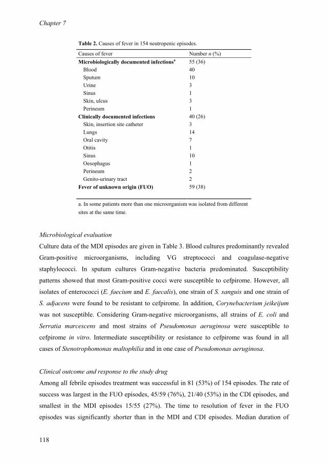
Chapter 7
118
Table 2. Causes of fever in 154 neutropenic episodes.
Causes of fever Number n (%)
Microbiologically documented infectionsa 55 (36)
Blood 40
Sputum 10
Urine 3
Sinus 1
Skin, ulcus 3
Perineum 1
Clinically documented infections 40 (26)
Skin, insertion site catheter 3
Lungs 14
Oral cavity 7
Otitis 1
Sinus 10
Oesophagus 1
Perineum 2
Genito-urinary tract 2
Fever of unknown origin (FUO) 59 (38)
a. In some patients more than one microorganism was isolated from different
sites at the same time.
Microbiological evaluation
Culture data of the MDI episodes are given in Table 3. Blood cultures predominantly revealed
Gram-positive microorganisms, including VG streptococci and coagulase-negative
staphylococci. In sputum cultures Gram-negative bacteria predominated. Susceptibility
patterns showed that most Gram-positive cocci were susceptible to cefpirome. However, all
isolates of enterococci (E. faecium and E. faecalis), one strain of S. sanguis and one strain of
S. adjacens were found to be resistant to cefpirome. In addition, Corynebacterium jeikeijum
was not susceptible. Considering Gram-negative microorganisms, all strains of E. coli and
Serratia marcescens and most strains of Pseudomonas aeruginosa were susceptible to
cefpirome in vitro. Intermediate susceptibility or resistance to cefpirome was found in all
cases of Stenotrophomonas maltophilia and in one case of Pseudomonas aeruginosa.
Clinical outcome and response to the study drug
Among all febrile episodes treatment was successful in 81 (53%) of 154 episodes. The rate of
success was largest in the FUO episodes, 45/59 (76%), 21/40 (53%) in the CDI episodes, and
smallest in the MDI episodes 15/55 (27%). The time to resolution of fever in the FUO
episodes was significantly shorter than in the MDI and CDI episodes. Median duration of

Cefpirome for febrile neutropenia
119
fever in the FUO episodes was 3 days and in the MDI and CDI episodes both 4 days (P =
0.001, by the log-rank test).
The majority of failures, both in the MDI and CDI episodes were classified as such because of
the addition of another antimicrobial agent. Vancomycin was added to cefpirome during 23
episodes. This frequently occurred if blood cultures revealed Gram-positive cocci and the
treating physician was concerned about possible infections with resistant enterococci or
coagulase negative staphylococci. However, subsequent identification of the microorganisms
Table 3. Isolated microorganisms and susceptibility to cefpirome in 55 episodes
with a microbiologically documented infection (MDI).
Causes of fever
No. of
episodes
No. of strains Sensitive
(S), Intermediate (I) or
Resistant (R) to
cefpirome.
S I R
Microbiologically documented infections 55 (36%)
Bloodstream infections 40
Coagulase negative staphylococci 10 10
Viridans group (VG) streptococci 16 14 2
Enterococci 7 7
Corynebebacterium jeikeijum 3 3
Pseudomonas aeruginosa 2 2
Candida tropicalis 1
Other 1 1
Respiratory tract infections 10
Pseudomonas aeruginosa 3 2 1
Stenotrophomonas maltophilia 2 1 1
Serratia marcescens 2 2
E. coli 1 1
Aspergillus fumigatus 2
Urinary tract infections 3
Enterococci 2 2
Proteus mirabilis 1 1
Other 2 1 1
Clinically documented infections 40 (26%)
Lungs 14
Ear, nose, throat 8
Sinuses 10
Skin 3
Genital or (peri) anal 4
Other 1
Fever of unknown origin (FUO) 59 (38%)
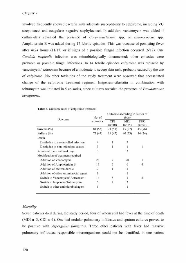
Chapter 7
120
involved frequently showed bacteria with adequate susceptibility to cefpirome, including VG
streptococci and coagulase negative staphylococci. In addition, vancomycin was added if
culture-data revealed the presence of Corynebacterium spp, or Enterococcus spp.
Amphotericin B was added during 17 febrile episodes. This was because of persisting fever
after 4x24 hours (11/17) or if signs of a possible fungal infection occurred (6/17). One
Candida tropicalis infection was microbiologically documented; other episodes were
probable or possible fungal infections. In 14 febrile episodes cefpirome was replaced by
vancomycin/ aztreonam because of a moderate to severe skin rash, probably caused by the use
of cefpirome. No other toxicities of the study treatment were observed that necessitated
change of the cefpirome treatment regimen. Imipenem-cilastatin in combination with
tobramycin was initiated in 5 episodes, since cultures revealed the presence of Pseudomonas
aeruginosa.
Table 4. Outcome rates of cefpirome treatment.
Outcome according to causes of
fever Outcome
No. of
episodes
CDI
(n=40)
MDI
(n=55)
FUO
(n=59)
Success (%) 81 (53) 21 (53) 15 (27) 45 (76)
Failure (%) 73 (47) 19 (47) 40 (73) 14 (24)
Death
Death due to uncontrolled infection 4 1 3
Death due to non-infectious causes 3 1 1 1
Recurrent fever within 4 days 3 3
Modification of treatment required
Addition of Vancomycin 23 2 20 1
Addition of Amphotericin B 17 7 6 4
Addition of Metronidazole 2 1 1
Addition of other antimicrobial agent 1 1
Switch to Vancomycin/ Aztreonam 14 5 1 8
Switch to Imipenem/Tobramycin 5 2 3
Switch to other antimicrobial agent 1 1
Mortality
Seven patients died during the study period, four of whom still had fever at the time of death
(MDI n=3, CDI n=1). One had nodular pulmonary infiltrates and sputum cultures proved to
be positive with Aspergillus fumigatus. Three other patients with fever had massive
pulmonary infiltrates; responsible microorganisms could not be identified, in one patient
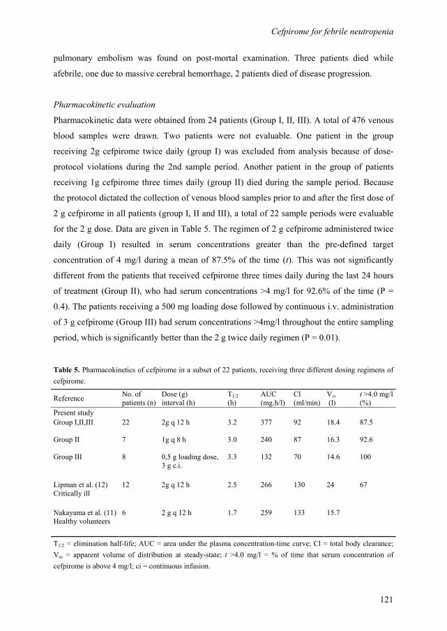
Cefpirome for febrile neutropenia
121
pulmonary embolism was found on post-mortal examination. Three patients died while
afebrile, one due to massive cerebral hemorrhage, 2 patients died of disease progression.
Pharmacokinetic evaluation
Pharmacokinetic data were obtained from 24 patients (Group I, II, III). A total of 476 venous
blood samples were drawn. Two patients were not evaluable. One patient in the group
receiving 2g cefpirome twice daily (group I) was excluded from analysis because of dose-
protocol violations during the 2nd sample period. Another patient in the group of patients
receiving 1g cefpirome three times daily (group II) died during the sample period. Because
the protocol dictated the collection of venous blood samples prior to and after the first dose of
2 g cefpirome in all patients (group I, II and III), a total of 22 sample periods were evaluable
for the 2 g dose. Data are given in Table 5. The regimen of 2 g cefpirome administered twice
daily (Group I) resulted in serum concentrations greater than the pre-defined target
concentration of 4 mg/l during a mean of 87.5% of the time (t). This was not significantly
different from the patients that received cefpirome three times daily during the last 24 hours
of treatment (Group II), who had serum concentrations >4 mg/l for 92.6% of the time (P =
0.4). The patients receiving a 500 mg loading dose followed by continuous i.v. administration
of 3 g cefpirome (Group III) had serum concentrations >4mg/l throughout the entire sampling
period, which is significantly better than the 2 g twice daily regimen (P = 0.01).
Table 5. Pharmacokinetics of cefpirome in a subset of 22 patients, receiving three different dosing regimens of
cefpirome.
Reference No. of
patients (n)
Dose (g)
interval (h)
T1/2
(h)
AUC
(mg.h/l)
Cl
(ml/min)
Vss
(l)
t >4.0 mg/l
(%)
Present study
Group I,II,III
22
2g q 12 h
3.2
377
92
18.4
87.5
Group II
7
1g q 8 h
3.0
240
87
16.3
92.6
Group III
8
0,5 g loading dose,
3 g c.i.
3.3
132
70
14.6
100
Lipman et al. (12)
Critically ill
12
2g q 12 h
2.5
266
130
24
67
Nakayama et al. (11)
Healthy volunteers
6
2 g q 12 h
1.7
259
133
15.7
T1/2 = elimination half-life; AUC = area under the plasma concentration-time curve; Cl = total body clearance;
Vss = apparent volume of distribution at steady-state; t >4.0 mg/l = % of time that serum concentration of
cefpirome is above 4 mg/l; ci = continuous infusion.
Chapter 7
122
Discussion
Cefpirome is a fourth generation cephalosporin, with a broad spectrum of antibacterial
activity, better activity against Gram-positive organisms and greater stability to beta-
lactamases than the third-generation cephalosporins. Moreover, a twice daily dosing regimen
may be sufficient.5 Therefore, cefpirome monotherapy may serve as a suitable alternative in
treatment regimens for infections in neutropenic patients.1 In comparative trials cefpirome has
shown equivalent efficacy and safety to ceftazidime and piperacillin-tazobactam, in the
treatment of suspected bacteraemia or sepsis and febrile neutropenia.8,9
In this open label, non-randomized clinical trial we administered cefpirome 2g twice daily
during 154 episodes of febrile neutropenia, occurring in 106 patients. We were able to
identify the cause of fever in 62% of febrile episodes. In general FUO accounts for
approximately 50% of causes of fever in neutropenic patients.2 Strict adherence to the
investigational protocol may have contributed to the relatively low percentage of FUO (38%)
as observed in our study.
In 81 of 154 episodes the patient survived the episode of neutropenia and fever without any
modification of the cefpirome regimen (overall rate of success 53%). This is slightly lower
than the reported success rates of cefpirome in comparable clinical settings and also lower
than the rate of success achieved with imipenem-cilastatin, as previously reported by our
group and others.8,9,13,14
This finding may be explained by the fact that our protocol provided
rather liberal guidelines for the addition of other antibiotics to cefpirome. Physicians were
reluctant to rely on cefpirome monotherapy and frequently prescribed vancomycin as soon as
baseline culture data revealed Gram-positive microorganisms, in view of possible infection
with enterococci or coagulase negative staphylococci. However, in a number of cases further
determination of these bacteria showed adequate susceptibility to cefpirome. Vancomycin
was discontinued subsequently, but as defined by the protocol these patients were categorized
as failure.
Treatment with cefpirome proved to be most successful in FUO episodes, in which resolution
of fever without treatment modification was achieved in 76%. CDI episodes were
successfully treated in 53% and the lowest rate of success was observed in the MDI episodes
(27%). This is in line with results from large-scale clinical studies on antimicrobial treatment
in neutropenic patients.2,15,16
Patients with an obvious focus of infection clearly represent a
population that is more difficult to treat than those without any focus at all. Infectious deaths
occur in one-fifth of episodes with a focus of infection, in comparison with less than 5% for
Cefpirome for febrile neutropenia
123
episodes without one.2,15
In our study 4 patients died due to uncontrolled infection, 3 with
MDI and one with CDI. No deaths occurred among patients with FUO.
As observed previously, Gram-positive bacteria predominated the isolated microorganisms
(72%).8,14,17
Most strains of VG streptococci and coagulase-negative staphylococci were
susceptible to cefpirome. In vitro studies have shown that up to 100% of strains of beta-
hemolytic streptococci may be expected to be susceptible, and 80-90% of coagulase-negative
staphylococci.10,18
In our study, enterococci were resistant to cefpirome. These findings are in
contrast with data from literature. About 70% of strains of E. faecalis are reported to be
susceptible to cefpirome in vitro. MIC values for enterococci however, are certainly not low
and vary between 4 and 8 mg/l.5,10,18
This observation underscores the importance of gaining
knowledge of local resistance patterns of specific pathogens, especially if borderline
susceptibility is expected. Corynebacterium jeikeijum was resistant to cefpirome. These
Gram-positive rods frequently display multi-resistance, though in general they remain
susceptible to vancomycin.
Considering Gram-negative bacteria, cefpirome was active against E. coli and other
Enterobacteriaceae, including Serratia and Proteus species. MIC values for these
microorganisms are reported to be very low, and vary between 0.4 and 2 mg/l. 5,10,18
Pseudomonas aeruginosa was susceptible to cefpirome, except for one strain with
intermediate susceptibility. In an epidemiological survey in Intensive Care and
Haematological units from 12 large hospitals in the Netherlands, including ours, 82% of
isolated Pseudomonas strains were found to be susceptible to cefpirome.10 However, in other
studies MIC90 values between 12.5 and ≥32 mg/l have been reported, indicating borderline
susceptibility or resistance.5,18
Despite our findings, it is probably right to state that cefpirome
has only moderate activity against Pseudomonas aeruginosa. In our study, physicians
changed cefpirome to imipenem-cilastatin in combination with tobramycin, according to local
protocols, if Pseudomonas aeruginosa was suspected or isolated. This strategy may be
debated, considering the majority of Pseudomonas strains being susceptible to cefpirome,
addition of an aminoglycoside might have been a viable option.
We are the first to report pharmacokinetic parameters of cefpirome in patients with
haematological malignancies. Cefpirome pharmacokinetics have been studied extensively in
healthy volunteers and in patients with renal impairment. The plasma half-life of cefpirome
and the time that the serum levels exceed the MIC of the common pathogens are sustained
enough to recommend a twice-daily dosing regimen.5-7,19
However, in a study in critically ill
patients low trough levels were found, following intravenous administration of 2 g cefpirome
Chapter 7
124
twice daily.12 The authors suggested that this drug regimen may be inadequate in critically ill
patients. Most likely the difference in pharmacokinetics as compared with healthy volunteers
is explained by an increase of volume of distribution, due to extensive fluid therapy in
critically ill patients. This may also hold true for neutropenic patients, who are treated with
intensive chemotherapy and receive frequent intravenous drug and fluid administrations. Our
data however, show that a twice daily dosing regimen of 2 g cefpirome resulted in serum
levels greater than 4 mg/l for 87.5% of the time. This may be explained by our finding that the
volume of distribution of cefpirome in neutropenic subjects is comparable with those found in
healthy volunteers, rather than in critically ill patients.12 Cefpirome 2 g twice daily is
recommended by the manufacturer as a dosing regimen for infections in neutropenic patients
and our data confirm that this may be adequate. However, alternative dosing schedules of 1g
three times daily or a loading dose of 500 mg followed by 3g continuously i.v may even be
better, with serum concentrations above 4 mg/l for 92.6 and 100% of the time respectively.
In common with most other beta-lactam antibiotics cefpirome was well tolerated. In 14 (9%)
episodes cefpirome was changed to vancomycin/ aztreonam because of skin rash, possibly
due to cefpirome. Most likely concomitantly prescribed medications are at least in part
responsible for this relatively high number of failures. In literature, the most common adverse
event is diarrhoea, reported in 1.6% of patients. Rash occurred in 1.4%.4,20
Adverse events are
more frequently reported in patients who were seriously ill, and particularly those with
neutropenia.20
In conclusion, cefpirome proved to be a valuable addition to the therapeutic arsenal available
for febrile neutropenia to date. The drug has broad Gram-positive and Gram-negative
coverage, but gaps in the antimicrobial spectrum involve enterococci, Corynebacterium spp.
and Stenotrophomonas maltophilia. Moreover, a limited activity against Pseudomonas
aeruginosa may in the long run hamper its use as a single agent therapy. Our pharmacokinetic
data indicate that in neutropenic subjects a dosing regimen of cefpirome 2 g twice daily
intravenously is sufficient. Finally, it should be emphasized that extensive use of single-drug
therapy requires vigilance, and close monitoring of the local microbial flora, since success is
largely dependent upon a continued susceptibility to the drug involved.

Cefpirome for febrile neutropenia
125
References
1. Glauser M. Empiric therapy of bacterial infections in patients with severe neutropenia. Diagn
Microbiol Infect Dis 1998; 31: 467-472.
2. Maschmeyer G. Interventional antimicrobial therapy in febrile neutropenic patients. Diagn
Microbiol Infect Dis 1999; 34: 205-212.
3. Viscoli C , Castagnola E. Treatment of febrile neutropenia: what is new? Curr Opin Infect Dis
2002; 15: 377-382.
4. Wilson WR. The role of fourth-generation cephalosporins in the treatment of serious infectious
diseases in hospitalized patients. Diagn Microbiol Infect Dis 1998; 31: 473-477.
5. Wiseman LR , Lamb HM. Cefpirome. A review of its antibacterial activity, pharmacokinetic
properties and clinical efficacy in the treatment of severe nosocomial infections and febrile
neutropenia. Drugs 1997; 54: 117-140.
6. Strenkoski LC , Nix DE. Cefpirome clinical pharmacokinetics. Clin Pharmacokinet 1993; 25:
263-273.
7. Craig WA. The pharmacokinetics of cefpirome--rationale for a twelve-hour dosing regimen.
Scand J Infect Dis 1993; 91: 33-40.
8. Bauduer F, Cousin T, Boulat O, Rigal-Huguet F, Molina L, Fegueux N, Jourdan E, Boiron JM,
Reiffers J. A randomized prospective multicentre trial of cefpirome versus piperacillin-
tazobactam in febrile neutropenia. Leuk Lymphoma 2001; 42: 379-386.
9. Norrby SR, Geddes AM, Shah PM. Randomized comparative trial of cefpirome versus
ceftazidime in the empirical treatment of suspected bacteraemia or sepsis. Multicentre Study
Group. J Antimicrob Chemother 1998; 42: 503-509.
10. Hoogkamp-Korstanje JA, Verduyn LF, Meis JF. Cefpirome: epidemiological survey in intensive
care units and hematological units in The Netherlands. The Dutch Study Group. Diagn
Microbiol Infect Dis 1998; 31: 489-491.
11. Nakayama I, Akieda Y, Yamaji E, Nitta Y, Ohishi M, Katagiri K, Imamura N, Takase K.
Single- and multiple-dose pharmacokinetics of intravenous cefpirome (HR810) to healthy
volunteers. J Clin Pharmacol 1992; 32: 256-266.
12. Lipman J, Wallis SC, Rickard CM, Fraenkel D. Low cefpirome levels during twice daily dosing
in critically ill septic patients: pharmacokinetic modelling calls for more frequent dosing.
Intensive Care Med 2001; 27: 363-370.
13. Norrby SR, Geddes AM, Shah PM. Cefpirome versus ceftazidime as empirical sepsis treatment.
The Study Group. Diagn Microbiol Infect Dis 1998; 31: 499-500.
14. Huijgens PC, Ossenkoppele GJ, Weijers TF, van Loenen AC, Simoons-Smit AM, Wijermans
PW, van Pampus EC, Langenhuijsen MM. Imipenem-cilastatin for empirical therapy in

Chapter 7
126
neutropenic patients with fever: an open study in patients with hematologic malignancies. Eur J
Haematol 1991; 46: 42-46.
15. Rolston KV, Berkey P, Bodey GP, Anaissie EJ, Khardori NM, Joshi JH, Keating MJ, Holmes
FA, Cabanillas FF, Elting L. A comparison of imipenem to ceftazidime with or without
amikacin as empiric therapy in febrile neutropenic patients. Arch Intern Med 1992; 152: 283-
291.
16. Cometta A, Calandra T, Gaya H, Zinner SH, De Bock R, Del Favero A, Bucaneve G, Crokaert
F, Kern WV, Klastersky J, Langenaeken I, Micozzi A, Padmos A, Paesmans M, Viscoli C,
Glauser MP. Monotherapy with meropenem versus combination therapy with ceftazidime plus
amikacin as empiric therapy for fever in granulocytopenic patients with cancer. The
International Antimicrobial Therapy Cooperative Group of the European Organization for
Research and Treatment of Cancer and the Gruppo Italiano Malattie Ematologiche Maligne
dell'Adulto Infection Program. Antimicrob Agents Chemother 1996; 40: 1108-1115.
17. Oppenheim BA. The changing pattern of infection in neutropenic patients. J Antimicrob
Chemother 1998; 41 Suppl D: 7-11.
18. Fernandes C, Pritchard R, Morris A, Benn R. In vitro evaluation of cefpirome: an Australasian
study of isolates from intensive care unit and hematology/oncology patients. The Cefpirome
Study Group. Diagn Microbiol Infect Dis 1998; 31: 493-495.
19. Garau J. The clinical potential of fourth-generation cephalosporins. Diagn Microbiol Infect Dis
1998; 31: 479-480.
20. Rubinstein E, Labs R, Reeves A. A review of the adverse events profile of cefpirome. Drug Saf
1993; 9: 340-345.

127
Outbreak of vancomycin-resistant Enterococcus faecium in a
haematology unit: risk factor assessment and successful control of
the epidemic.
G.J. Timmers
W.C. van der Zwet
A.M. Simoons-Smit
P.H.M. Savelkoul
H.H.M. Meester
C.M.J. Vandenbroucke-Grauls
P.C. Huijgens
Department of Haematology, Department of Medical Microbiology and Infection Control.
Vrije Universiteit Medical Center, Amsterdam, The Netherlands.
British Journal of Haematology 2002;116:826-833.
Chapter 3 3 8
Chapter 8
128
Abstract
We describe an outbreak of vancomycin-resistant Enterococcus faecium (VRE) on the
haematology ward of a Dutch university hospital. After the occurrence of three consecutive
cases of bacteraemia with VRE, strains were genotyped and found to be identical. During the
next 4 months an intensive surveillance programme identified 21 additional patients to be
colonized with VRE, while two more patients developed bacteraemia. A case-control study
was carried out to identify risk factors for VRE acquisition. In comparison with VRE-negative
control patients (n = 49), cases (n = 24) had a longer stay on the ward during the year
preceding the outbreak (25.8 versus 10.1 days, P = 0.02), more cases with acute myeloid
leukaemia (11 versus 4, odds ratio (OR) 9.5, 95% confidence interval (CI95) 2.4-32.2) and
higher grades of mucositis (P = 0.03). Logistic regression analysis identified antibiotic use
within 1 month before admission (OR 13.0, CI95 2.1-80.5, P = 0.006) and low albumin levels
at baseline (OR 1.2, CI95 1.1-1.3, P = 0.02) to be independent risk factors. Four patients with
VRE-bacteraemia were successfully treated with quinupristin/dalfopristin (Synercid®).
Control of the outbreak was achieved by step-wise implementation of intensive infection
control measures, which included the cohorting of patients, allocation of nurses and
reinforcement of hand hygiene.

VRE outbreak in a haematology unit
129
Introduction
With the emergence of resistance to glycopeptides, enterococci have rapidly become
important nosocomial pathogens.1 Over the past decade, vancomycin-resistant enterococci
(VRE) have been reported with increasing frequency as the cause of nosocomial outbreaks,
usually involving high-risk patient populations, such as haemato-oncology patients, organ
transplant recipients and patients in intensive care units.2-8 In general, such outbreaks have a
serious impact on the daily care and treatment of patients on the ward or in the hospital
involved. Extensive infection control measures are mandatory to prevent further spread of the
microorganism and sometimes the ward even has to be temporarily closed to new admissions.
More important, VRE may cause life-threatening systemic infections in immunocompromised
patients. Until recently, effective antibiotic strategies were lacking and systemic infections
with VRE in these patients have been associated with in-hospital mortality rates ranging from
40-100%.9,10
Established risk factors for VRE colonization or infection include neutropenia,
prolonged hospitalization, the frequent use of multiple broad-spectrum antibiotics and the use
of invasive procedures.2,3,5
Therefore, it is not surprising that patients with haematological
malignancies are at a predominant risk for nosocomial acquisition of VRE. Many reports have
included haemato-oncology patients in their analysis of VRE acquisition. However, only a
few have studied strictly haematological populations.11-13
In 1999 a VRE outbreak occurred on our haematology ward. A case-control study was carried
out to identify possible risk factors associated with VRE acquisition. Molecular biological
techniques were used to genotype the VRE isolates. Moreover, successful control of the
outbreak was achieved after step-wise implementation of intensive infection control
measures, which we describe in detail.
Materials and methods
Setting
The outbreak occurred on the 21-bed haematology ward of the VU University Medical
Centre, Amsterdam, the Netherlands. The haematology department is the referral centre for
adult patients with haematological malignancies from the north-western part of the
Netherlands. The unit comprises two private rooms and four rooms with a four-bed capacity.
Patients share a limited number of toilet facilities, two for women and two for men, located in
Chapter 8
130
one sanitary unit. In addition the ward is equipped with three private rooms with controlled-
airflow facilities. According to local protocols, all patients on the ward, who were expected to
be neutropenic (absolute neutrophil count <0.5 × 10⁹/l) for more than 10 d, received
ciprofloxacin 500 mg b.i.d., azitromycin 250 mg b.i.d. and fluconazole 50 mg daily as
infection prophylaxis. Standard care of febrile neutropenic patients on the ward included
empirical treatment with cefpirome 2 g b.i.d. (Cefrom®), a broad-spectrum cephalosporin.
Vancomycin 1 g b.i.d. was added to the antibiotic regimen if culture data so dictated. If no
defervescence occurred within 96 h, amphotericin B 0.7 mg/kg was given intravenously.
Microbiological surveillance
Between November 1998 and May 1999, three patients admitted to the haematology ward
developed bloodstream infections with a VRE strain, which proved to be clonally related.
This prompted microbiological screening of all patients in the ward by biweekly culture of
anal swab specimens. Additionally, rectal swabs were obtained from all newly admitted
patients upon admission and twice weekly thereafter. Environmental samples were taken from
patient-related equipment, housekeeping articles and surfaces of furniture and other room
equipment.
Microbiological methods
Anal swab specimens and environmental swab specimens were cultured for the detection of
VRE in Enterococcosel broth (BBLTM, Becton & Dickinson, Sparks, USA). After incubation
overnight at 37°C, black-coloured broth media were subcultured on Enterococcosel agar
(BBLTM, Becton & Dickinson) for 48 h at 37°C. Black colonies were identified at the
species level using standard microbiological methods. Susceptibility tests were performed
using the disk diffusion method (Rosco tabs, A/S Rosco, Taastrup, Denmark) on Mueller
Hinton agar (Difco, Becton & Dickinson). Vancomycin and teicoplanin resistance was
confirmed with Minimal Inhibitory Concentration (MIC) determination using the E-test (AB
Biodisk, Solna, Sweden). Resistance was defined as MIC ≥32 mg/l.
Molecular typing
All isolates were fingerprinted by amplified fragment length polymorphism (AFLP), which
has been described in detail previously.14 This technique was carried out using fluorescent
primers.15 Levels of similarity of fingerprints were analysed by Pearson correlation, with
specialized software (Gel Compar 4.0, Applied Maths, Kortrijk, Belgium). A dendrogram of

VRE outbreak in a haematology unit
131
percentage similarity was produced by the unweighted pair group method. Isolates with a
homology of >90% were considered to be clonally related.
Case-control study
To identify risk factors for nosocomial acquisition of VRE, we performed a case-control
study. Cases were defined as patients who became colonized or infected with the epidemic
VRE strain. Controls were all patients who were hospitalized on the haematology ward during
the outbreak period and did not acquire VRE. To be eligible, controls had to have at least one
negative culture of a rectal swab specimen. If multiple admissions of one subject occurred
during the outbreak period, the patient was included in the study only once to avoid
disproportional contribution of particular cases or controls to the final analysis. To identify
risk factors for colonization or infection with VRE, a standardized set of data was extracted
from the medical records of cases and controls. Data collected included patient demographics,
disease characteristics and treatment variables, antibiotic use and variables defining the
clinical course. Data for cases were collected up to the day of the first culture that yielded
VRE, and for controls for the total duration of their hospital stay.
Statistical analysis
Univariate analysis of categorical variables was performed by calculating odds ratios (OR)
and 95% confidence intervals (CI95). In case of categorical variables with an ordering or
grading scale, the chi-square test for trend was used. Student's t-test was used for comparison
of means. Logistic regression analysis was performed to identify independent risk factors for
VRE acquisition. All tests were two-tailed, alpha was set at 0.05.
Results
Description of the outbreak
Between November 1998 and May 1999 three patients admitted to the haematology ward
developed VRE bacteraemia. Molecular typing of these VRE showed that the strains were
clonally related. The subsequent microbiological surveillance programme showed that,
between June and September 1999, 21 additional patients acquired the epidemic VRE strain,
resulting in a total number of VRE-positive patients of 24. In addition to the first three
patients with VRE bacteraemia, two patients who became colonized with VRE subsequently
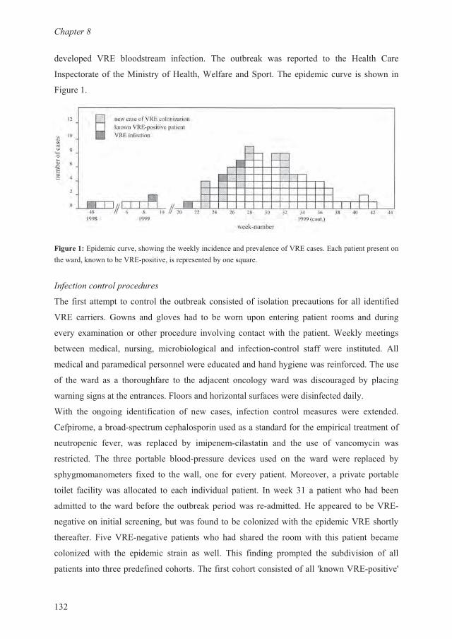
��������
��9
������ �� N:� ����������� �������� -�� �������/ ��� �� ����� �� ��� ;����� ����
1� ���������� ���2�����$��;������>��������� ����-��� ����������� ������ �
��������
�������� ������������������������/�$��������� ����������N:����������� ����� ��������������/������N:�& ������������ ��������$���#�����
�(��+�,)(+)(��)4��)+����6
-�� ���������� � �������� ����������/���������� �������� ��������� ������ ���������
N:� ��������� %��� �� ������ ��� �� �� ��� � � ������ ����� ����� �� �����
����$ ���������������� �������� ������� ���������� ��� ������>��/�$�������
������ �������� ������ ��������������� �� �������&������ ����� ���� ���������� 0��
��������� ���������� ������������������������$�����������������-�����
�� ��� ���� �� � ������������ �� ��� ��.���� ������$���� ��� ����������� �$ �����
����������������������������������7������������������������������$�
>��� ��� ����� ������������ �� �� ������ ������� ������ �������� ���� ��������
��� ������������&� �������� ����� ������������������������� ����������������
����� ��� ������ ��� �� ����� �$ ��� ���&��������� �� ��� ��� �� �����$�� ���
����������� -�� ����� ������� �����& ������� ������� ���� � ��� ���� ���� �� ����� �$
� �$������������ ����� �� ���������� ��� ����$ ������2�������� � ������ �������
������ �������$��� ��������� �� ���� ��������� ������ 1���/�� � ��������������
�������� �� ������������� ����������/ �������� ��&���������;�� ����� ����N:�&
�������� �������������������� ���� ���������7������ ���� ������N:�������$
��������������� N:�&������� ������ ��� ��� ������ ��� �������� ���� ����� ������
�����7�� ���� ��� � ������ ����� �� ����� -��� ����� ��� ��� ��� ���������� �� ���
�������������� ����������������-�������������������������� P/��N:�& �������P
VRE outbreak in a haematology unit
133
patients. Additionally, patients who had been hospitalized between November 1998 and
August 1999 were considered 'possibly VRE-positive', irrespective of culture results. They
were cohorted and barrier nursed. No sharing of any article between patients was allowed in
this patient group. New patients, who had never been admitted to the haematology ward
before, were considered to be a 'true VRE-negative' cohort, and were nursed in separate
rooms, without isolation precautions. On every shift nurses were strictly allocated to either
VRE-positive or -negative patients. Clinicians conducted their rounds visiting the VRE-
negative patients first, followed by the 'possibly colonized patients' and the VRE-positive
patients last. In the third week of August, week 33, the last patient colonized with the
epidemic VRE was identified. Several 'known VRE-positive' patients were re-admitted
thereafter. Occasionally, these patients had negative screening cultures on admission;
however, subsequent VRE cultures invariably became positive again. The last patient known
to be VRE positive was admitted in week 47. After his discharge at the end of December
1999, no known VRE-colonized patients were admitted and no new cases were identified.
After resolve of the outbreak we continued with weekly VRE surveillance cultures as a
standard infection control measure on our ward. Culture data have shown successful control
of the epidemic to date.
Microbiological surveillance and environmental cultures
Between June 1999 and November 1999, 287 anal surveillance cultures were performed
during 115 admissions on the haematology ward. Of these 287 cultures, 76 (26%) yielded
vancomycin-resistant Enterococcus faecium, in 24 patients. All isolates manifested high-level
resistance to vancomycin and teicoplanin (E-test MIC ≥32 mg/l), which is compatible with
the VanA glycopeptide resistance phenotype. In addition the strains were resistant to
amoxycillin, clindamycin and erythromycin. None of the culture samples obtained from 27
environmental sites were positive for VRE. However, at the time of this sampling, intensive
infection control measures had already been installed. AFLP analysis was performed on all
isolated strains, and showed that isolates had > 90% homology of DNA patterns (Figure 2).
This finding was indicative of similarity of strains and provided ultimate proof of the
nosocomial transmission of a single clone. Two patients had VRE isolates that proved to be
unrelated to the epidemic strain (Figure 2, B1).
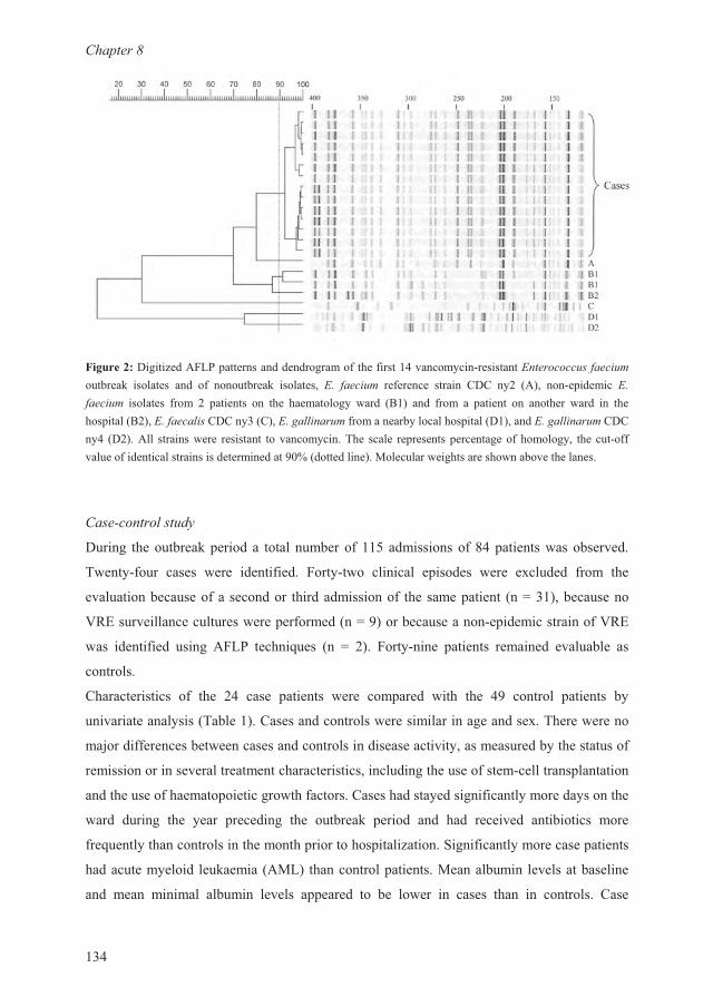
��������
��
�������������7��0�"� ���������������������������� �����$��&��������'(���)+)++�6���+,���������/ �������� �� �� ��������/ ��������� '3 ���+,�� �������� ����� ��� $9 �0�� �&� ������ '3���+,�� �������� ���� 9 ������ � ��� ����������$ ���� �F�� �� ���� � ����� � ������ ���� � ������ �����F9��'3���+�4,6���$�����'3��44,(�������������$�������� �����������'3��44,(�������$ ��9��0�� ���������� �������� �� �����$���-�� ����� �� ������ �����������������$� ������&������������������������������������� �B������������2�����������������������������������
��6��+)(��)46���
����� ����������/ ������ �������������������������� � ������������������
-���$&���� ����� ���� ���������� ����$&��� ������� � ������ ���� �������� ���� ���
�������������������������� ��������������� ������� ����� �C������������
N:������������������������ ���������C ������������&� �������������N:�
��� ��������� ���� 0�"� �����#��� � C 9�� ����$&�� ������ ������� ��������� ��
��������
��������������� �� ��� 9 ���� ������ ���� ��� ���� ���� ��� ������ ������ �$
������������$����-������������������������������������������-���������
��.�����������������������������������������������$������������$�����������
�������������������������������������������������������������&�������� �������
����������������� �������������������������������$������������$������$�����
���� ����� ��� $��� ������� ��� �������/ ����� �� ��� �������� ���������� ����
���#����$������������������ ��������� �����7���������������$�������� ������
���������$����� ���/����� �02"� ��������� �������2�������� ���������������
�� ��� ������ ������ ������ � ����� �� �� ����� � ����� ��� � �������� ����
VRE outbreak in a haematology unit
135
patients were suffering from higher grades of mucositis than control patients. Fever occurred
more frequently in the case group and case patients were having more microbiologically
documented infections, with a predominance of bloodstream infections. During the period
before VRE acquisition, cases received both preventive and therapeutic antibiotics more
frequently, including cephalosporins, vancomycin, imipenem-cilastatin and amphotericin B.
Overall mortality did not differ significantly between the groups.
Variables that were significantly associated with VRE acquisition by univariate analysis and
that were considered to be of clinical relevance were entered in a multivariate model (Table
2). Antibiotic use within 1 month before admission (OR 13.0 (CI95 2.1-80.5), P = 0.006) and
the mean albumin level at baseline (OR 1.2, CI95 1.1-1.3, P = 0.02) appeared to be
independently associated with VRE colonization or infection.
Cases with VRE bacteraemia
Bloodstream infections with VRE developed in five cases. Four patients were diagnosed with
AML and one patient had refractory anaemia with excess blasts in transformation (RAEB-t).
All patients had active disease on admission, except for one patient who was in complete
remission.
The first patient with VRE bacteraemia received teicoplanin and rifampicin and recovered
quickly after regaining a normal neutrophil count. In another patient blood cultures revealed
VRE only shortly after his death. The remaining three patients and one additional patient with
a non-epidemic VRE received quinapristin/dalfopristin (Synercid®) 7.5 mg/kg t.i.d.
intravenously, which had not been officially registered in the Netherlands at that time. All
four patients treated with quinapristin/dalfopristin had a favourable clinical and
bacteriological response. Later, one patient died of respiratory failure of unknown origin, not
related to VRE.
Discussion
Patients with haematological malignancies appear to be at a predominant risk for the
acquisition of VRE, and nosocomial outbreaks involving these patients have been reported
previously.4,5,12,13
The presence of a haematological malignancy has even been identified as an
independent risk factor for infection with VRE.16

Chapter 8
136
Table 1. Univariate analysis of case-control data
Cases Controls
Variable (n= 24) (n= 49) OR (CI95)
Baseline characteristics
Male 12 (50) 30 (61) NS
Female 12 (50) 19 (39) NS
Age (years ± SD) 43.8 ± 14.8 45.7 ± 15.0 NS
Days on ward before outbreak (days ± SD) 25.8 ± 28.1 10.1 ± 21.0 P = 0.02
Diagnoses
AML 11 (46) 4 (8) 9.5 (2.6-34.9)
Myeloma 3 (13) 18 (37) NS
ALL 2 (8) 4 (8) NS
Lymphoma 2 (8) 17 (35) 0.2 (0.04-0.8)
CML 2 (8) 2 (4) NS
CLL 1 (4) 1 (2) NS
Hairy cell leukaemia 0 (0) 3 (6) NS
MDS 3 (13) 0 (0) NC
Disease activity
Complete remission 6 (25) 8 (16)
Partial remission 2 (8) 19 (39) P = 0.5a
Active disease 16 (67) 22 (45)
Treatment
Flow chamber 4 (17) 8 (16) NS
Stemcel transplant 6 (25) 18 (37) NS
Autologous 4 (17) 14 (29) NS
Allogeneic 2 (8) 4 (8) NS
GCSF 4 (17) 8 (16) NS
Steroid use 8 (33) 20 (41) NS
Antibiotics before admission
Within 1 month b.a. 15 (63) 6 (12) 11.9 (3.6-39.2)
Vancomycin within 1 month b.a. 3 (13) 1 (2) NS
Clinical course
Hospital stay (days ± SD) 25.6 ± 8.9 17.9 ± 10.5 P = 0.003
Stay before VRE acquisition (days ± SD) 13.3 ± 6.9 17.9 ± 10.5 P = 0.03
Diarhoea 16 (67) 10 (20) 7.8 (2.6-23.4)
Faecal incontinence 9 (38) 5 (10) 5.3 (1.5-18.3)
Urine incontinence 3 (13) 1 (2) NS
Decubitus 3 (13) 3 (6) NS
Non compliance with the preventive
regimen
5 (21) 7 (14) NS
Nasogastric feeding 0 (0) 0 (0) NS
Antacid medication 12 (50) 26 (53) NS
Neutropenia (ANC <0.5x10⁹/L, days ± SD) 16.3 ± 11.5 9.9 ± 11.3 P = 0.03
Neutropenia before VRE acq. (days ± SD) 10.8 ± 9.9 9.9 ± 11.3 NS
Death 5 (21) 3 (6) NS
Mucositis
None 6 (25) 28 (57)
Grade I 12 (50) 13 (27) P = 0.03a
Grade II 3 (13) 6 (12)
Grade III 3 (13) 2 (4)
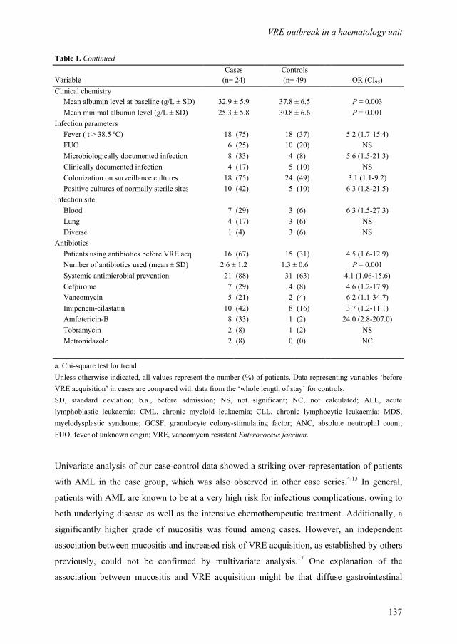
VRE outbreak in a haematology unit
137
Table 1. Continued
Cases Controls
Variable (n= 24) (n= 49) OR (CI95)
Clinical chemistry
Mean albumin level at baseline (g/L ± SD) 32.9 ± 5.9 37.8 ± 6.5 P = 0.003
Mean minimal albumin level (g/L ± SD) 25.3 ± 5.8 30.8 ± 6.6 P = 0.001
Infection parameters
Fever ( t > 38.5 ºC) 18 (75) 18 (37) 5.2 (1.7-15.4)
FUO 6 (25) 10 (20) NS
Microbiologically documented infection 8 (33) 4 (8) 5.6 (1.5-21.3)
Clinically documented infection 4 (17) 5 (10) NS
Colonization on surveillance cultures 18 (75) 24 (49) 3.1 (1.1-9.2)
Positive cultures of normally sterile sites 10 (42) 5 (10) 6.3 (1.8-21.5)
Infection site
Blood 7 (29) 3 (6) 6.3 (1.5-27.3)
Lung 4 (17) 3 (6) NS
Diverse 1 (4) 3 (6) NS
Antibiotics
Patients using antibiotics before VRE acq. 16 (67) 15 (31) 4.5 (1.6-12.9)
Number of antibiotics used (mean ± SD) 2.6 ± 1.2 1.3 ± 0.6 P = 0.001
Systemic antimicrobial prevention 21 (88) 31 (63) 4.1 (1.06-15.6)
Cefpirome 7 (29) 4 (8) 4.6 (1.2-17.9)
Vancomycin 5 (21) 2 (4) 6.2 (1.1-34.7)
Imipenem-cilastatin 10 (42) 8 (16) 3.7 (1.2-11.1)
Amfotericin-B 8 (33) 1 (2) 24.0 (2.8-207.0)
Tobramycin 2 (8) 1 (2) NS
Metronidazole 2 (8) 0 (0) NC
a. Chi-square test for trend.
Unless otherwise indicated, all values represent the number (%) of patients. Data representing variables ‘before
VRE acquisition’ in cases are compared with data from the ‘whole length of stay’ for controls.
SD, standard deviation; b.a., before admission; NS, not significant; NC, not calculated; ALL, acute
lymphoblastic leukaemia; CML, chronic myeloid leukaemia; CLL, chronic lymphocytic leukaemia; MDS,
myelodysplastic syndrome; GCSF, granulocyte colony-stimulating factor; ANC, absolute neutrophil count;
FUO, fever of unknown origin; VRE, vancomycin resistant Enterococcus faecium.
Univariate analysis of our case-control data showed a striking over-representation of patients
with AML in the case group, which was also observed in other case series.4,13
In general,
patients with AML are known to be at a very high risk for infectious complications, owing to
both underlying disease as well as the intensive chemotherapeutic treatment. Additionally, a
significantly higher grade of mucositis was found among cases. However, an independent
association between mucositis and increased risk of VRE acquisition, as established by others
previously, could not be confirmed by multivariate analysis.17 One explanation of the
association between mucositis and VRE acquisition might be that diffuse gastrointestinal

Chapter 8
138
mucosal breakdown promotes the likelihood of colonization of the gut with VRE.
Subsequently, growth of VRE to high numbers may lead to bacteraemia. In line with this
hypothesis, we found a greater frequency of diarrhoea and faecal incontinence in our VRE-
positive population. Like mucositis, diarrhoea may serve as a parameter of changes in
gastrointestinal tract function, increasing the likelihood of colonization with VRE. This
concept has been proposed previously in burn patients.7
Table 2. Variablese entered in the multivariate model
Potential risk factor Cases Controls OR (CI95%) P-value
(n= 24) (n= 49)
AML 11 (46) 4 (8) 6.1 (0.6-58.8) NS
Antibiotics within 1 month before admission 15 (63) 6 (12) 13.0 (2.1-80.5) 0.006
Diarrhoea 17 (71) 10 (20) 3.4 (0.6-19.0) NS
Days on ward before outbreak (days ± SD) 25.8 ± 28.1 10.1 ± 21.0 1.0 (0.9-1.1)
NS
Mucositis (mean grade ± SD) 1.1 ± 0.9 0.6 ± 0.8 1.7 (0.7-4.6) NS
Albumin level at baseline (mean, g/L) 33.2 ± 5.9 37.8 ± 6.5 1.2 (1.1-1.3) 0.02
Fever ( t > 38.5 ºC) 19 (79) 18 (37) 0.6 (0.1-3.8) NS
Values represent the number (%) of patients unless otherwise indicated. SD, standard deviation: NS, not
significant.
Multivariate analysis of our data revealed that antibiotic use within 1 month before admission
was independently associated with VRE colonization or infection. Prior administration of
antibiotics has been consistently recognized as an important risk factor for the acquisition of
VRE, probably because the intensive use of antimicrobial agents provides VRE with a
selective growth advantage. Previous case-control studies have implicated vancomycin, 18
third-generation cephalosporins6,18
and prior exposure to antibiotics with activity against
anaerobes4,19
as significant risk factors. In view of these data, the adjustment of our empirical
antibiotic regimen, replacing cefpirome with imipenem-cilastatin, might be debated, because
the latter possesses anti-anaerobic effects.
A second risk factor independently associated with VRE acquisition in our multivariate model
was the albumin value at baseline, which was significantly lower for cases. It is likely that the
albumin concentration is a highly sensitive indicator of preclinical disease and disease
severity.20 Moreover, in various populations of patients, a low albumin value on admission
has been identified as an independent risk factor for colonization or infection with several
microorganisms, including respiratory-tract pathogens, methicillin-resistant Staphylococcus
aureus (MRSA) and yeasts.21-23

VRE outbreak in a haematology unit
139
A total of five cases developed VRE bloodstream infections. One of the major risk factors for
the development of invasive VRE infection appears to be the presence of gastrointestinal
colonization.4 In our study, prior colonization with VRE was established in both patients who
developed VRE bacteraemia during the period that patients were systematically screened for
colonization. Until recently, no effective antimicrobial agents were available for treating these
patients. We administered quinupristin/dalfopristin (Synercid®), a new streptogramin
antibiotic, to four patients with proven VRE bacteraemia (three cases and one patient with a
non-outbreak VRE).24 The clinical success rate was excellent and all patients became culture
negative. Three patients survived the neutropenic episode, one patient died of respiratory
failure, without signs of active VRE infection.
Our infection control measures were taken according to the Centers for Disease Control
(CDC) recommendations for the prevention of the spread of vancomycin resistance,25
focusing mainly on early detection of VRE carriage, education of hospital staff and a critical
consideration of the standard antibiotic regimen. We restricted the use of vancomycin and
banned all cephalosporins. Strict hand disinfections with an alcoholic hand rub by all
personnel and patients was reinforced. In addition to these general measures, some specific
interventions may have played a key role in the prevention of further spread of VRE.
First, in addition to the cohorting of patients, nurses were strictly allocated to either VRE-
positive or -negative patients. Some of our newly admitted patients became VRE positive
even while being nursed in strict isolation in a single room or in one of the controlled airflow
rooms. It appeared likely that VRE was transmitted via the attending staff, although we did
not perform systematic surveillance cultures in medical personnel to prove this observation.
The transmission of VRE from the hands of healthcare workers to patients appears to play a
major role in the nosocomial spread of the microorganism and exposure to a nurse caring for
another VRE-positive patient has been identified as a significant risk factor for the acquisition
of VRE.3,4 Mathematical analysis of transmission dynamics of VRE has shown that hand
washing and allocation of staff are the most powerful infection control measures in endemic
settings.26
Second, we learned not to rely on negative surveillance culture results in patients who could
have acquired the epidemic VRE during previous admissions. Several patients who had
stayed on the ward before and even patients who were known to be VRE positive, had VRE-
negative surveillance cultures at their re-admission, but subsequently proved to carry the
epidemic VRE. This finding led to the definition of a 'possibly VRE-positive' cohort. We
suggest that the number of VRE in the bowel decreases below detection levels in periods that
Chapter 8
140
the patient is not treated with antimicrobial agents. When appropriate selective antimicrobial
pressure is enforced, VRE numbers may increase accordingly. It has been shown that
colonization with the same strain of VRE may persist for at least a year.27
Third, a measure thought to be essential in the control of the outbreak was the allocation of
private portable toilet facilities to every single patient. Our ward was constructed about 35
years ago and patients had to share a limited number of toilet facilities (two for women, two
for men, shared by 18 patients), located outside the patient rooms. This might have
contributed to the initial spread of VRE. The toilet has been clearly identified as a
transmission route for VRE.28
Until recently, major differences in the epidemiology of VRE infections existed between
Europe and the United States. VRE seems to have become endemic in American hospital
populations and many nosocomial outbreaks of VRE have been reported.2-4,7
To date, the
occurrence of nosocomial infections with VRE in Europe has been rare. However, the
increasing number of reports on VRE outbreaks in Europe and outside the United States may
indicate that a change in epidemiology of VRE towards the American situation is ahead.
Given the threat posed by VRE in hospitals worldwide, much is to be gained by a proper
understanding of issues involving nosocomial VRE infections, especially by physicians
involved in the care of immunocompromised patients.
References
1. Murray BE. Vancomycin-resistant enterococcal infections. N Engl J Med 2000; 342: 710-721.
2. Boyle JF, Soumakis SA, Rendo A, Herrington JA, Gianarkis DG, Thurberg BE, Painter BG.
Epidemiologic analysis and genotypic characterization of a nosocomial outbreak of
vancomycin-resistant enterococci. J Clin Microbiol 1993; 31: 1280-1285.
3. Boyce JM, Opal SM, Chow JW, Zervos MJ, Potter-Bynoe G, Sherman CB, Romulo RL, Fortna
S, Medeiros AA. Outbreak of multidrug-resistant Enterococcus faecium with transferable vanB
class vancomycin resistance. J Clin Microbiol 1994; 32: 1148-1153.
4. Edmond MB, Ober JF, Weinbaum DL, Pfaller MA, Hwang T, Sanford MD, Wenzel RP.
Vancomycin-resistant Enterococcus faecium bacteremia: risk factors for infection. Clin Infect
Dis 1995; 20: 1126-1133.
5. Nourse C, Murphy H, Byrne C, O'Meara A, Breatnach F, Kaufmann M, Clarke A, Butler K.
Control of a nosocomial outbreak of vancomycin resistant Enterococcus faecium in a paediatric
oncology unit: risk factors for colonization. Eur J Pediatr 1998; 157: 20-27.
VRE outbreak in a haematology unit
141
6. Loeb M, Salama S, Armstrong-Evans M, Capretta G, Olde J. A case-control study to detect
modifiable risk factors for colonization with vancomycin-resistant enterococci. Infect Control
Hosp Epidemiol 1999; 20: 760-763.
7. Falk PS, Winnike J, Woodmansee C, Desai M, Mayhall CG. Outbreak of vancomycin-resistant
enterococci in a burn unit. Infect Control Hosp Epidemiol 2000; 21: 575-582.
8. McCarthy KM, Van Nierop W, Duse A, Von Gottberg A, Kassel M, Perovic O, Smego R.
Control of an outbreak of vancomycin-resistant Enterococcus faecium in an oncology ward in
South Africa: effective use of limited resources. J Hosp Infect 2000; 44: 294-300.
9. Lam S, Singer C, Tucci V, Morthland VH, Pfaller MA, Isenberg HD. The challenge of
vancomycin-resistant enterococci: a clinical and epidemiologic study. Am J Infect Control 1995;
23: 170-180.
10. Edmond MB, Ober JF, Dawson JD, Weinbaum DL, Wenzel RP. Vancomycin-resistant
enterococcal bacteremia: natural history and attributable mortality. Clin Infect Dis 1996; 23:
1234-1239.
11. Guiot HF, Peetermans WE, Sebens FW. Isolation of vancomycin-resistant enterococci in
haematologic patients. Eur J Clin Microbiol Infect Dis 1991; 10: 32-34.
12. Chadwick PR, Oppenheim BA, Fox A, Woodford N, Morgenstern GR, Scarffe JH.
Epidemiology of an outbreak due to glycopeptide-resistant Enterococcus faecium on a
leukaemia unit. J Hosp Infect 1996; 34: 171-182.
13. Roghmann MC, McCarter RJ, Jr., Brewrink J, Cross AS, Morris JG, Jr. Clostridium difficile
infection is a risk factor for bacteremia due to vancomycin-resistant enterococci (VRE) in VRE-
colonized patients with acute leukemia. Clin Infect Dis 1997; 25: 1056-1059.
14. Vos P, Hogers R, Bleeker M, Reijans M, van de LT, Hornes M, Frijters A, Pot J, Peleman J,
Kuiper M, . AFLP: a new technique for DNA fingerprinting. Nucleic Acids Res 1995; 23: 4407-
4414.
15. Koeleman JG, Parlevliet GA, Dijkshoorn L, Savelkoul PH, Vandenbroucke-Grauls CM.
Nosocomial outbreak of multi-resistant Acinetobacter baumannii on a surgical ward:
epidemiology and risk factors for acquisition. J Hosp Infect 1997; 37: 113-123.
16. Shay DK, Maloney SA, Montecalvo M, Banerjee S, Wormser GP, Arduino MJ, Bland LA,
Jarvis WR. Epidemiology and mortality risk of vancomycin-resistant enterococcal bloodstream
infections. J Infect Dis 1995; 172: 993-1000.
17. Kuehnert MJ, Jernigan JA, Pullen AL, Rimland D, Jarvis WR. Association between mucositis
severity and vancomycin-resistant enterococcal bloodstream infection in hospitalized cancer
patients. Infect Control Hosp Epidemiol 1999; 20: 660-663.
18. Tornieporth NG, Roberts RB, John J, Hafner A, Riley LW. Risk factors associated with
vancomycin-resistant Enterococcus faecium infection or colonization in 145 matched case
patients and control patients. Clin Infect Dis 1996; 23: 767-772.
Chapter 8
142
19. Donskey CJ, Chowdhry TK, Hecker MT, Hoyen CK, Hanrahan JA, Hujer AM, Hutton-Thomas
RA, Whalen CC, Bonomo RA, Rice LB. Effect of antibiotic therapy on the density of
vancomycin-resistant enterococci in the stool of colonized patients. N Engl J Med 2000; 343:
1925-1932.
20. Goldwasser P , Feldman J. Association of serum albumin and mortality risk. J Clin Epidemiol
1997; 50: 693-703.
21. George DL, Falk PS, Wunderink RG, Leeper KV, Jr., Meduri GU, Steere EL, Corbett CE,
Mayhall CG. Epidemiology of ventilator-acquired pneumonia based on protected bronchoscopic
sampling. Am J Respir Crit Care Med 1998; 158: 1839-1847.
22. Hedderwick SA, Wan JY, Bradley SF, Sangeorzan JA, Terpenning MS, Kauffman CA. Risk
factors for colonization with yeast species in a Veterans Affairs long-term care facility. J Am
Geriatr Soc 1998; 46: 849-853.
23. Onorato M, Borucki MJ, Baillargeon G, Paar DP, Freeman DH, Cole CP, Mayhall CG. Risk
factors for colonization or infection due to methicillin-resistant Staphylococcus aureus in HIV-
positive patients: a retrospective case-control study. Infect Control Hosp Epidemiol 1999; 20:
26-30.
24. Moellering RC, Linden PK, Reinhardt J, Blumberg EA, Bompart F, Talbot GH. The efficacy
and safety of quinupristin/dalfopristin for the treatment of infections caused by vancomycin-
resistant Enterococcus faecium. Synercid Emergency-Use Study Group. J Antimicrob
Chemother 1999; 44: 251-261.
25. Hospital Infection Control Practices Advisory Committee (HICPAC). Recommendations for
preventing the spread of vancomycin resistance. MMWR Recomm Rep 1995; 44: 1-13.
26. Austin DJ, Bonten MJ, Weinstein RA, Slaughter S, Anderson RM. Vancomycin-resistant
enterococci in intensive-care hospital settings: transmission dynamics, persistence, and the
impact of infection control programs. Proc Natl Acad Sci U S A 1999; 96: 6908-6913.
27. Montecalvo MA, de Lencastre H, Carraher M, Gedris C, Chung M, VanHorn K, Wormser GP.
Natural history of colonization with vancomycin-resistant Enterococcus faecium. Infect Control
Hosp Epidemiol 1995; 16: 680-685.
28. Noble MA, Isaac-Renton JL, Bryce EA, Roscoe DL, Roberts FJ, Walker M, Scharf S, Walsh A,
Altamirano-Dimas M, Gribble M. The toilet as a transmission vector of vancomycin-resistant
enterococci. J Hosp Infect 1998; 40: 237-241.
143
Summary and concluding remarks
Chapter 3 3 9
Chapter 9
144
Summary and conclucing remarks
In the Netherlands approximately 7000 patients are diagnosed with a malignancy of the blood
or bone marrow every year. Prospects for these patients have substantially improved over the
past decades. The field of clinical haematology is characterized by rapid and sometimes
exciting innovations in treatment options, achieving higher remission rates and control over
diseases that were deemed incurable until recently. The advent of combination chemotherapy,
dose intensification, autologous and allogeneic stem cell transplantation and immunotherapy
all have contributed substantially to this progress in the treatment of haematological
malignant diseases. Unfortunately, these important and promising developments are
counterbalanced by serious impairment of host immune response mechanisms. This occurs in
a population of patients that is already running a serious risk of infections, because
haematological malignancies reside by definition in the immune system itself. As a
consequence, these patients are highly susceptible to infections with various kinds of
opportunistic pathogens, including viruses, bacteria and fungi.
Infections in patients with aggressive haematological malignancies are associated with
significant morbidity and mortality. Moreover, their initial presentation frequently is erratic,
with sometimes only minimal signs and symptoms. Therefore, it is not surprising that anti-
infective strategies have focused on prevention and early treatment. Antibacterial and
antifungal prophylaxis and early, or ‘empirical’ antibiotic therapy have gained a lot of interest
in the international literature. In chapter 2 an overview is given on current insights and
developments on antibacterial and antifungal prophylaxis and treatment, from a clinical
perspective.
In view of the serious consequences of infections in patients treated for haematological
disease, it is tempting to clinicians to readily introduce newly marketed and promising
antibacterial and antifungal drugs into daily clinical practice, before results of relevant clinical
trials have become available. This entails the risk of neglecting important questions
considering safety, pharmacokinetic behaviour and efficacy of the drug involved, as
prophylaxis or treatment in the specific population of patients with haematological diseases.
The studies described in this thesis aimed to explore the safety and efficacy of a selection of
new antimicrobials, for the prevention of bacterial (chapter 3 and 4) and fungal (chapter 5
Summary and concluding remarks
145
and 6) infections, as well as for the empirical treatment of bacterial infections (chapter 7).
The occurrence of an outbreak of vancomycin-resistant Enterococcus faecium (VRE) led to
an assessment of risk factors for the acquisition of VRE and recommendations for the
prevention and control of such an outbreak (chapter 8).
Main results
Chapter 3. Gram-positive breakthrough infections pose a major drawback to the use of
quinolones for antibacterial prophylaxis in neutropenic patients. Levofloxacin, a relatively
new quinolone, has an augmented Gram-positive spectrum and may potentially overcome this
problem. When administered orally (500 mg, once daily) as antibacterial prophylaxis to
patients receiving intensive chemotherapy for haematological malignancies, levofloxacin
provided adequate eradication of Gram-negative microorganisms and S. aureus and preserved
the anaerobic component of the bowel flora. It was found that the pharmacokinetic properties
of levofloxacin were not altered during the phase of chemotherapy and neutropenia. Minimal
inhibitory concentration (MIC) values for viridans group (VG) streptococci tended to
increase, which cautioned against the occurrence of acquired resistance to levofloxacin.
Chapter 4. Elaborating the results described in chapter 3, the question remained open as to
how levofloxacin prophylaxis compared with standard prophylactic regimens, considering
efficacy, tolerability and induction of resistance among bacterial pathogens. In an attempt to
answer this question, levofloxacin was compared with ciprofloxacin plus phenethicillin as
antibacterial prophylaxis during neutropenia, in a randomized clinical trial. It was found that
levofloxacin and ciprofloxacin-phenethicillin were equally effective in the prevention of
bacterial infections in neutropenic patients. However, levofloxacin was better tolerated, which
may benefit compliance with therapy. In line with the findings described in chapter 3,
resistance to levofloxacin was observed among VG streptococci, but timely adjustments of
the prophylactic regimen were made, based on surveillance culture data and no break-through
infections with VG streptococci occurred.
Chapter 5. In an attempt to improve prophylaxis against the occurrence of invasive fungal
infections in neutropenic patients, a randomized clinical trial was designed to compare a lipid
formulation of amphotericin B intravenously (amphotericin B colloidal dispersion, ABCD)

Chapter 9
146
with oral fluconazole. However, the administration of ABCD for prophylactic reasons to
patients without life-threatening fungal infections, was associated with major and intolerable
side-effects during infusion of the compound. The study was prematurely terminated and it
was concluded that ABCD appeared to be unsuitable for antifungal prophylaxis in
neutropenic patients.
Chapter 6. It is well documented that administration of itraconazole increases cyclosporin A
(CsA) serum concentrations. However, there are no data on the effects of CsA on itraconazole
pharmacokinetics. Itraconazole and hydroxy-itraconazole pharmacokinetic parameters were
studied, before and during administration of cyclosporin A (CsA) in ten patients receiving an
allogeneic stem cell transplantation. It was found that exposure to OH-itraconazole, but not to
itraconazole is increased when itraconazole is co-administered with CsA. Monitoring of
itraconazole serum concentrations is important in patients who use drugs that, like
itraconazole, interact with CYP3A4 and when target drug concentrations are to be achieved
for optimal antifungal efficacy.
Chapter 7. With the introduction of cefpirome, a new fourth generation cephalosporin, as
empirical treatment of febrile neutropenia, a cohort study was conducted to assess the clinical
efficacy of cefpirome and its activity against isolated pathogens. Fifty-three percent of
patients survived the neutropenic episode without the need of treatment modification
(success-rate 53%). Susceptibility testing of isolated pathogens showed adequate coverage of
a broad range of Gram-positive and Gram-negative microorganisms including viridans group
streptococci, coagulase-negative staphylococci, Enterobacteriaceae and Pseudomonas
aeruginosa. In addition, pharmacokinetic data indicated that a dosing regimen of cefpirome 2
g twice daily was sufficient in this population.
Chapter 8. This chapter describes the occurrence of an outbreak of vancomycin-resistant
Enterococcus faecium (VRE) on our haematology ward. A case-control study showed that
cases (n = 24) had a longer stay on the ward during the year preceding the outbreak, as
compared with VRE-negative control patients (n = 49). More cases had acute myeloid
leukaemia and cases had higher grades of mucositis. Logistic regression analysis identified
antibiotic use within 1 month before admission and low albumin levels at baseline to be
independent risk factors for acquisition of VRE. Control of the outbreak was achieved by a

Summary and concluding remarks
147
step-wise implementation of intensive infection control measures, which included the
cohorting of patients, allocation of nurses and reinforcement of hand hygiene.
Conclusions and clinical implications
The studies described in this thesis have originated from questions and problems that were
encountered in daily clinical practice, during the care for patients with haematological
malignancies. From the results reported here, some answers to these questions can be
deduced, which may contribute to actual and future decisions with regard to the prevention
and treatment of infections in these patients:
[1] Levofloxacin may be considered as standard treatment for the prevention of bacterial
infections in neutropenic patients, considering its good tolerability and equal efficacy as
compared with ciprofloxacin-phenethicillin. Surveillance cultures are mandatory, to closely
monitor the emergence of levofloxacin-resistant VG streptococci and adjustments of the
prophylactic regimen must be made accordingly.
[2] Amphotericin B colloidal dispersion (ABCD) is not suitable for antifungal prophylaxis in
neutropenic patients with a haematological malignancy, due to excessive infusion-related
toxicity. If used for therapeutic indications, close pursuit of adverse events is strongly
advised.
[3] Exposure to OH-itraconazole may be increased when itraconazole is co-administered with
Cyclosporin A. This finding is of limited clinical relevance, but may be may be important in
the occasional event that monitoring of serum itraconazole concentrations is warranted.
[4] The use of cefpirome as empirical antibacterial treatment in patients with febrile
neutropenia can not be strongly recommended based on the data presented in this thesis.
Though we found a rather good efficacy, there are some points of concern; (a). Limited
activity against Pseudomonas aeruginosa may in the long run hamper the use of cefpirome as
single agent therapy. (b). The use of cefpirome was thought to be associated with the
occurrence of an outbreak of vancomycin resistant Enterococcus faecium, though chapter 7
nor chapter 8 provide data to fully support this assumption. (c). In a recently published meta
analysis on empirical antibiotic monotherapy for febrile neutropenia, cefepime, which closely
resembles cefpirome, was associated with a higher mortality rate than other antibiotics,
probably due to less efficacy.1
Chapter 9
148
[5] Antibiotic use within 1 month before admission and low albumin levels at baseline are
independent risk factors for acquisition of vancomycin-resistant Enterococcus faecium, during
a nosocomial outbreak. Control of the outbreak is achievable, by intensive infection control
measures.
Future directions
There is now growing evidence that both prophylactic and empirical administration of
antibacterial and antifungal antibiotics may reduce mortality and morbidity among patients
with severe neutropenia. The data presented in this thesis, however, are too limited to add to
the existing evidence on those issues. Our findings rather illustrate the reverse side of the
medal, which displays the concerning emergence of resistant pathogens and increased toxicity
after the introduction of new prophylactic or therapeutic antibiotics. The large number of
reports on anti-microbial resistance and nosocomial outbreaks on haematology or oncology
wards, following the application of new prophylactic or therapeutic strategies and to which
this thesis adds, can not be neglected.2-9 Here, not only the well-being of the individual patient
is at risk, but also of the population at large. Moreover, increased toxicity of antibiotics and
the emergence of resistant microorganisms may have considerable effects on daily care and
management of haemato-oncology units and may substantially increase work load for medical
staff and health-care costs.
Although these concerns argue against the widespread and unlimited use of antibiotics in
patients with neutropenia, a balanced appraisal is needed, that should give direction to future
research.
[1] It is noteworthy that the emergence of resistant strains not necessarily leads to subsequent
infection with the microorganism involved.10-12
The reduction in mortality and infection rates
appears to outweigh the detriments of emerging resistant microorganisms. Future studies
should not only focus on the development of resistance among potential pathogens, but also
on the probability and severity of subsequent invasive infections.
[2] One of the major limitations of the studies described in this thesis is the lack of
subdivision of the population of patients as a whole, into different categories of risk levels for
infectious complications. For example, patients who are treated with an autologous stem cell

Summary and concluding remarks
149
transplantation for Non Hodgkin’s lymphoma are at a different risk and will acquire other
types of infections than patients who receive an allogeneic stem cell transplantation for acute
leukaemia. To date, efforts are made to distinguish categories of risk of infections among
patients with neutropenia.13-15
Future studies should aim to identify the population of patients
that is likely to benefit from a given agent the most.16-18
In selected groups of low-risk
patients therapy may be simplified or even discontinued.
[3] Future studies should focus on the development and improvement of sensitive and rapid
diagnostic techniques, to trace infectious complications in neutropenic patients. Early
detection of bacterial or fungal infections and accurate identification of the pathogens
involved, may lead to earlier and more appropriate antibiotic intervention, thereby increasing
the success of therapy. In selected groups of patients, improved diagnostic procedures and
effective early treatment may even replace prophylaxis and in potential, will reduce the
empirical overtreatment of patients with persisting fever during the neutropenic episode.
So, rather than refraining from the routine use of antibiotics in patients with neutropenia, the
clinician faces a challenge to chose the right antibiotic regimen for the right population of
patients. Not only data from clinical trials should guide these decisions. Other factors, that are
at least as important, include local bacteriological and epidemiological data, with an emphasis
on resistance patterns of predominantly isolated microorganisms, as well as the utility of an
antibiotic in daily practice, its user-friendliness to patients and nursing staff, its toxicity and
costs.

150

Nederlandstalige samenvatting
151
Nederlandstalige samenvatting en conclusies
In Nederland wordt elk jaar bij ongeveer 7000 patiënten een kwaadaardige aandoening van
bloed of beenmerg vastgesteld. De vooruitzichten voor deze patiënten zijn de laatste jaren
sterk verbeterd. Het vakgebied van de klinische hematologie wordt gekenmerkt door snelle en
vaak spectaculaire ontwikkelingen in de behandelingsmogelijkheden, waardoor hogere
genezingspercentages worden bereikt en patienten kunnen worden behandeld met ziektes die
tot recent daarvoor niet in aanmerking kwamen. De toepassing van combinatiechemotherapie,
het intensiveren van de dosis, autologe en allogene stamceltransplantatie en immunotherapie
hebben allen in belangrijke mate bijgedragen aan de vooruitgang van de
behandelingsmogelijkheden in de hemato-oncologie. Een belangrijk nadeel is echter dat deze
op zich veelbelovende ontwikkelingen, zonder uitzondering, het afweersysteem sterk in
negatieve zin beïnvloeden. Bovendien gaat het daarbij om een categorie patiënten die al op
voorhand een verhoogde kans op infecties heeft, omdat een hemato-oncologische ziekte per
definitie zijn oorsprong vindt in het immuunsysteem zelf. Dit alles heeft tot gevolg dat deze
patiënten zeer gevoelig zijn voor infecties met een scala aan opportunistische micro-
organismen, waaronder diverse soorten virussen, bacteriën en schimmels.
Infecties bij patiënten met hematologische maligniteiten kunnen leiden tot ernstige
ziekteverschijnselen en ook sterfte. De eerste presentatie van een dergelijke infectie kan
echter aspecifiek zijn, met soms minimale ziekteverschijnselen. Het is dan ook niet
verwonderlijk dat strategieën ter bestrijding van deze infecties vooral gericht zijn op preventie
en vroege behandeling. Medicamenteuze profylaxe tegen bacteriën en schimmels en vroeg
ingestelde, zogenaamde ‘empirische’ therapie staan sterk in de belangstelling in de
internationale literatuur. In hoofdstuk 2 van dit proefschrift wordt vanuit een klinisch
perspectief een overzicht gegeven van de huidige inzichten en ontwikkelingen op het gebied
van antibacteriële en antifungale profylaxe en behandeling.
Gezien de ernstige gevolgen van infecties voor patiënten die behandeld worden voor een
hemato-oncologische ziekte, hebben behandelend artsen vaak de neiging om veelbelovende
medicamenten, soms net op de markt, snel te introduceren in de dagelijkse praktijk. Dit
gebeurt nogal eens voordat kan worden beschikt over duidelijke gegevens uit klinisch
onderzoek. Daarmee blijven belangrijke vragen onbeantwoord met betrekking tot de
veiligheid van een medicament, het farmacologisch gedrag en de werkzaamheid van het
Hoofdstuk 9
152
betreffende middel als profylaxe of als behandeling, in de bijzondere populatie van patiënten
met hematologische ziekten.
De studies beschreven in dit proefschrift hebben tot doel de veiligheid en werkzaamheid te
onderzoeken van een selectie van nieuwe antibiotica, ingezet voor de preventie van bacteriële-
(hoofdstuk 3 en 4) en schimmelinfecties (hoofdstuk 5 en 6) en voor de empirische
behandeling van bacteriële infecties (hoofdstuk 7). Een uitbraak met vancomycine-resistente
Entrococcus faecium (VRE) gaf aanleiding tot een systematische inventarisatie van
risicofactoren voor besmetting met VRE en leidde tot aanbevelingen voor het voorkomen en
bestrijden van een dergelijke uitbraak (hoofdstuk 8).
Belangrijkste resultaten
Hoofdstuk 3. Het optreden van doorbraakinfecties met Gram-positieve bacteriën vormt een
belangrijk nadeel van het gebruik van quinolonen (antibiotica) als profylaxe bij neutropene
patiënten. Levofloxacin is een relatief nieuw quinolone antibioticum met een betere
antibiotische werking tegen Gram-positieve bacteriën dan eerdere quinolonen en zou daardoor
een oplossing kunnen bieden voor dit probleem. Toediening van levofloxacin (éénmaal daags
500 mg, oraal) als antibacteriële profylaxe, aan patiënten die behandeld werden met
intensieve chemotherapie voor een hematologische maligniteit, resulteerde in een adequate
eradicatie van het Gram-negatieve deel van de darmflora en van Staphylococcus aureus,
waarbij het anaërobe deel van de darmflora in tact bleef. Daarbij kon worden aangetoond dat
de farmacokinetische eigenschappen van levofloxacin niet veranderden gedurende de periode
van toediening van de chemotherapie en de neutropene fase. De waarden van de minimaal
remmende concentratie (MIC) voor viridans streptokokken bleek geleidelijk te stijgen,
hetgeen wijst op het optreden van resistentie tegen levofloxacin.
Hoofdstuk 4. Met de resultaten zoals beschreven in hoofdstuk 3, bleef de vraag
onbeantwoord hoe levofloxacin zich verhoudt tot de huidige antibacteriële profylaxe, vooral
wat betreft werkzaamheid, bijwerkingen en tolerantie en het optreden van resistentie bij
bacteriën. Om deze vraag te beantwoorden werd een klinische gerandomiseerde studie
verricht, waarbij levofloxacin werd vergeleken met ciprofloxacin plus phenethicilline als
antibacteriële profylaxe gedurende neutropenie. Beide middelen bleken even effectief in het

Nederlandstalige samenvatting
153
voorkomen van bacteriële infecties bij neutropene patiënten. Levofloxacin werd echter beter
verdragen, hetgeen gunstig zal zijn voor de therapietrouw. Zoals ook beschreven in hoofdstuk
3, bleken viridans streptokokken soms resistentie te ontwikkelen tegen levofloxacin. Op basis
van de resultaten van inventarisatiekweken kon in die gevallen het profylactische antibiotica
schema tijdig worden aangepast en doorbraakinfecties met viridans streptokokken werden niet
waargenomen.
Hoofdstuk 5. In een poging om de profylaxe tegen schimmelinfecties te verbeteren werd een
gerandomiseerde klinische studie ontworpen, om intraveneuze toediening van een lipide-vorm
van amfotericine B (amfotericine B in colloïdale dispersie, ABCD) te vergelijken met
fluconazol oraal. De toediening van ABCD als profylaxe, aan patiënten die geen
levensbedreigende schimmelinfecties hadden, bleek echter gepaard te gaan met ernstige en
onacceptabele bijwerkingen tijdens de infusie van het middel. De studie werd dan ook
voortijdig gestaakt en geconcludeerd werd dat ABCD niet geschikt is als antifungale
profylaxe bij neutropene patiënten.
Hoofdstuk 6. Het is een bekend gegeven dat toediening van itraconazol de bloedspiegel van
ciclosporine (cyclosporine A) verhoogt. Er zijn echter geen gegevens over het effect van
ciclosporine op de farmacokinetiek van itraconazol. De farmacokinetische eigenschappen van
itraconazol en hydroxy (OH) itraconazol werden bestudeerd vóór en tijdens toediening van
ciclosporine, bij tien patiënten die een allogene stamcel transplantatie ondergingen. De
blootstelling aan OH-itraconazol bleek significant te zijn toegenomen wanneer itraconazol
gelijktijdig werd toegediend met ciclosporine, dit bleek niet het geval te zijn voor de
blootstelling aan itraconazol zelf. Het meten van itraconazol spiegels kan van belang zijn als
patiënten daarnaast andere medicamenten gebruiken, die net als itraconazol worden
afgebroken via het cytochroom P450 (CYP) 3A enzym systeem. Dit is vooral van toepassing
wanneer bepaalde concentraties van dit medicament moeten worden behaald om een optimaal
antischimmel effect te verkrijgen.
Hoofdstuk 7. De introductie van cefpirom, een destijds nieuw vierde-generatie cefalosporine,
voor de empirische behandeling van neutropene patiënten met koorts, op de afdeling
hematologie van het VU medisch centrum, leidde tot de uitvoering van een klinisch cohort
onderzoek om de effectiviteit van cefpirom te onderzoeken. Ook werd de werkzaamheid tegen
de meest geïsoleerde ziekteverwekkers onderzocht. Drieënvijftig procent van de patiënten

Hoofdstuk 9
154
overleefde de neutropene episode zonder dat wijziging van antibiotica nodig was
(succespercentage 53%). Gevoeligheidsbepalingen bij geïsoleerde bacteriën toonde aan dat
een breed scala aan Gram-positieve en Gram-negatieve bacteriën gevoelig was voor cefpirom,
met name viridans streptokokken, coagulase-negatieve stafylokokken, Enterobacteriae en
Pseudomonas aeruginosa. Daarbij liet farmacokinetisch onderzoek zien dat toediening van
twee maal daags 2 gram cefpirom afdoende zou moeten zijn in deze populatie.
Hoofdstuk 8. Dit hoofdstuk beschrijft een uitbraak van vancomycine-resistente Enterococcus
faecium (VRE) op de afdeling hematologie van het VU medisch centrum. Een patiënt-
controle onderzoek toonde aan dat besmette patiënten (n = 24) langer opgenomen waren
geweest op de afdeling gedurende het jaar voorafgaand aan de uitbraak in vergelijking met
VRE-negatieve controle patiënten (n = 49). Onder de besmette patiënten waren meer gevallen
van acute myeloide leukemie en in deze groep was ook de mate van slijmvliesbeschadiging
door chemotherapie (mucositis) hoger dan in de controlegroep. Door middel van logistische
regressie analyse kon worden aangetoond dat antibiotica gebruik in de maand voorafgaand
aan de opname en een lage albuminewaarde bij aanvang onafhankelijke risicofactoren waren
voor besmetting met VRE. De uitbraak kon succesvol worden bestreden door een
stapsgewijze inzet van intensieve infectie beperkende maatregelen, waaronder het indelen van
de patiëntenpopulatie in drie groepen (besmet, onbesmet of verdacht), het toewijzen van
specifieke verpleegkundigen aan zo’n categorie en het verplichten van betere handhygiëne.
Conclusies en klinische toepasbaarheid
De studies beschreven in dit proefschrift zijn gebaseerd op vraagstukken en problemen die
voortkwamen uit de dagelijkse klinische praktijk, tijdens de zorg voor patiënten met een
hemato-oncologische ziekte. Uit de in dit proefschrift beschreven resultaten van deze studies
kunnen antwoorden op deze vragen worden afgeleid, die richting kunnen geven aan actuele en
toekomstige beslissingen ten aanzien van preventie en behandeling van infecties bij deze
patiënten.
[1] Het gebruik van levofloxacin als standaard antibacteriële profylaxe bij neutropene
patiënten is het overwegen waard, gezien het feit dat het middel uitstekend verdragen wordt
en de werkzaamheid vergelijkbaar is met die van ciprofloxacin en phenethicilline. Het
Nederlandstalige samenvatting
155
verrichten van inventarisatiekweken is daarbij een vereiste, om het optreden van levofloxacin-
resistente viridans streptokokken te signaleren en het antibacteriële profylactische schema
tijdig te kunnen aanpassen.
[2] Amfotericine B in colloïdale dispersie (ABCD) is niet geschikt voor de preventie van
schimmelinfecties bij neutropene patiënten met een hematologische maligniteit vanwege
ernstige, infusiegerelateerde bijwerkingen. Indien dit middel gebruikt wordt voor
therapeutische doeleinden is het nauwkeurig observeren van bijwerkingen sterk aan te raden.
[3] Blootstelling aan OH-itraconazol kan verhoogd zijn als itraconazol en ciclosporine
gelijktijdig worden toegediend. Deze bevinding heeft beperkte klinische betekenis, maar kan
van belang zijn als het meten van itraconazol spiegels is aangewezen.
[4] De toepassing van cefpirom als empirische antibacteriële therapie bij de behandeling van
neutropene koorts kan alleen op basis van de gegevens beschreven in dit proefschrift niet
zonder meer worden aanbevolen. Hoewel een redelijke effectiviteit van het middel kon
worden aangetoond zijn er een aantal zaken die de aandacht behoeven; (a). Een beperkte
werkzaamheid tegen Pseudomonas aeruginosa is waarschijnlijk een belangrijk nadeel van het
gebruik van cefpirom als monotherapie. (b). Het gebruik van cefpirom heeft mogelijk een rol
gespeeld bij het optreden van de uitbraak met vancomycine-resistente Enterococcus faecium,
hoewel hoofdstuk 7 en 8 geen gegevens bevatten die deze aanname kunnen ondersteunen. (c)
In een recent gepubliceerde meta-analyse van de toepassing van monotherapie op empirische
gronden bij neutropene koorts wordt gemeld dat het gebruik van cefepime, een cefalosporine
dat sterke gelijkenis vertoont met cefpirom, gepaard gaat met een hoger sterftepercentage dan
andere antibiotica. Dit is waarschijnlijk het gevolg van een mindere werkzaamheid dan de
andere geteste antibiotica.1
[5] Het gebruik van antibiotica binnen één maand voor opname en een lage albumine waarde
bij aanvang van de behandeling van een hemato-oncologische ziekte zijn onafhankelijke
risicofactoren voor de besmetting met vancomycine-resistente Enterococcus faecium
gedurende een ziekenhuisuitbraak. Een dergelijke uitbraak is te bestrijden door toepassing van
intensieve infectie beperkende maatregelen.
Toekomstperspectief
Er is toenemend wetenschappelijk bewijs dat de toepassing van profylactische en
therapeutische antibacteriële en antifungale antibiotica daadwerkelijk leidt tot een reductie
Hoofdstuk 9
156
van ziekteverschijnselen en ook sterfte bij patiënten met ernstige neutropenie. De gegevens
beschreven in dit proefschrift zijn echter te beperkt om een belangrijke bijdrage te kunnen
leveren aan de bewijsvoering van deze bewering. Onze gegevens illustreren eerder de
keerzijde van de medaille, die gekenmerkt wordt door een zorgwekkende toename van
resistente bacteriën en het optreden van ernstige bijwerkingen als nieuwe profylactische of
therapeutische antibiotica worden ingezet. De grote hoeveelheid wetenschappelijke artikelen
gewijd aan ziekenhuisuitbraken op hematologie- of oncologieafdelingen en aan bacteriële
resistentie tegen antibiotica illustreert dit nog eens extra.2-9 Het gaat daarbij niet langer en
alleen om het welzijn van de individuele patiënt maar ook van diens directe omgeving en
uiteindelijk van de bevolking in zijn geheel. Daarbij komt dat het optreden van ernstige
bijwerkingen van antibiotica en de toename van resistente micro-organismen aanzienlijke
gevolgen hebben voor de dagelijkse zorg en werkzaamheden op hemato-oncologie afdelingen.
Uiteindelijk leidt dit alles tot een verminderde genezingskans voor de patiënt, een toename
van de werklast voor medisch personeel en tot stijging van kosten van de gezondheidszorg.
Hoewel deze argumenten pleiten tegen een wijdverbreid en ongelimiteerd gebruik van
antibiotica bij neutropene patiënten is een afgewogen oordeel op zijn plaats, hetgeen ook
richting kan geven aan onderwerpen van toekomstig onderzoek.
[1] Uit literatuurgegevens en uit de studies beschreven in dit proefschrift blijkt dat het
optreden van resistentie niet noodzakelijkerwijs wordt gevolgd door een infectie met het
betreffende micro-organisme.10-12
De afname van sterfte en infectiekans lijken ruimschoots op
te wegen tegen de nadelen die de aanwezigheid van resistente micro-organismen met zich
meebrengt. Toekomstig onderzoek zou zich naast onderzoek naar resistentiepatronen, ook
dienen te richten op de kans dat daadwerkelijk een infectie met het betreffende micro-
organisme volgt, met een beoordeling van de ernst daarvan.
[2] Eén van de belangrijkste beperkingen van de studies beschreven in dit proefschrift is het
ontbreken van een indeling van de patientenpopulatie in verschillende categorieën, naar het
risico op het optreden van infecties. Patiënten die bijvoorbeeld worden behandeld met een
autologe stamcel transplantatie voor een Non-Hodgkin lymfoom lopen een ander risico op
infecties, met andersoortige verwekkers, dan patiënten die een allogene stamcel transplantatie
ondergaan vanwege acute leukemie. Tegenwoordig worden pogingen gedaan om een
dergelijke risicoindeling te maken.13-15
Toekomstig onderzoek zou ten doel moeten hebben
om juist die populatie te identificeren die het meeste baat heeft bij toediening van een bepaald

Nederlandstalige samenvatting
157
medicament.16-18
In geselecteerde groepen van patiënten met een laag infectierisico, is het
denkbaar dat het voorschrijven van antibiotica sterk wordt teruggebracht of zelfs geheel
achterwege wordt gelaten.
[3] Toekomstig onderzoek zou zich dienen te richten op de ontwikkeling en verbetering van
diagnostische technieken om infecties bij patiënten met neutropenie op te sporen.
Vroegtijdige ontdekking van een bacteriële- of schimmelinfectie en identificatie van het
betrokken micro-organisme kan bijdragen aan een snellere en beter afgestemde behandeling,
waardoor het succes daarvan zal toenemen. In geselecteerde patiëntengroepen kunnen
verbeterde diagnostische procedures, zo nodig gevolgd door tijdig ingestelde therapie,
wellicht het profylactisch gebruik van antibiotica overbodig maken en de overbehandeling
met antibiotica op empirische basis reduceren.
Dus, in plaats van volledig af te zien van het routinematig gebruik van antibiotica bij
patiënten met een hematologische maligniteit, ziet de behandelend arts zich gesteld voor de
uitdaging om het juiste antibioticum voor te schrijven aan de juiste categorie patiënten. Niet
alleen gegevens uit klinisch onderzoek zijn daarbij van belang. Andere factoren, die minstens
zo belangrijk zijn bij deze beslissing, zijn bijvoorbeeld plaatselijke microbiologische en
epidemiologische gegevens. Daarbij dient gelet te worden op resistentiepatronen van micro-
organismen die in het betreffende centrum frequent worden geïsoleerd. Ook het
gebruiksgemak van een antibioticum is van belang, zowel voor patiënten als voor
verpleegkundigen. Tenslotte zal de keuze voor een bepaald middel worden bepaald door de
bijwerkingen van het middel en de kosten.
Referenties
1. Paul M, Yahav D, Fraser A, Leibovici L. Empirical antibiotic monotherapy for febrile
neutropenia: systematic review and meta-analysis of randomized controlled trials. J Antimicrob
Chemother 2006; 57: 176-189.
2. Oppenheim BA, Hartley JW, Lee W, Burnie JP. Outbreak of coagulase negative staphylococcus
highly resistant to ciprofloxacin in a leukaemia unit. BMJ 1989; 299: 294-297.

Hoofdstuk 9
158
3. Shaw BE, Boswell T, Byrne JL, Yates C, Russell NH. Clinical impact of MRSA in a stem cell
transplant unit: analysis before, during and after an MRSA outbreak. Bone Marrow Transplant
2007; 39: 623-629.
4. Weinstock DM, Conlon M, Iovino C, Aubrey T, Gudiol C, Riedel E, Young JW, Kiehn TE,
Zuccotti G. Colonization, bloodstream infection, and mortality caused by vancomycin-resistant
enterococcus early after allogeneic hematopoietic stem cell transplant. Biol Blood Marrow
Transplant 2007; 13: 615-621.
5. Timmers GJ, van der Zwet WC, Simoons-Smit IM, Savelkoul PH, Meester HH,
Vandenbroucke-Grauls CM, Huijgens PC. Outbreak of vancomycin-resistant Enterococcus
faecium in a haematology unit: risk factor assessment and successful control of the epidemic. Br
J Haematol 2002; 116: 826-833.
6. Sample ML, Gravel D, Oxley C, Toye B, Garber G, Ramotar K. An outbreak of vancomycin-
resistant enterococci in a hematology-oncology unit: control by patient cohorting and terminal
cleaning of the environment. Infect Control Hosp Epidemiol 2002; 23: 468-470.
7. Kern WV, Klose K, Jellen-Ritter AS, Oethinger M, Bohnert J, Kern P, Reuter S, von Baum H,
Marre R. Fluoroquinolone resistance of Escherichia coli at a cancer center: epidemiologic
evolution and effects of discontinuing prophylactic fluoroquinolone use in neutropenic patients
with leukemia. Eur J Clin Microbiol Infect Dis 2005; 24: 111-118.
8. Prentice HG, Hann IM, Nazareth B, Paterson P, Bhamra A, Kibbler CC. Oral ciprofloxacin plus
colistin: prophylaxis against bacterial infection in neutropenic patients. A strategy for the
prevention of emergence of antimicrobial resistance. Br J Haematol 2001; 115: 46-52.
9. Myoken Y, Kyo T, Sugata T, Murayama SY, Mikami Y. Breakthrough fungemia caused by
fluconazole-resistant Candida albicans with decreased susceptibility to voriconazole in patients
with hematologic malignancies. Haematologica 2006; 91: 287-288.
10. Gafter-Gvili A, Fraser A, Paul M, Leibovici L. Meta-analysis: antibiotic prophylaxis reduces
mortality in neutropenic patients. Ann Intern Med 2005; 142: 979-995.
11. Gafter-Gvili A, Paul M, Fraser A, Leibovici L. Effect of quinolone prophylaxis in afebrile
neutropenic patients on microbial resistance: systematic review and meta-analysis. J Antimicrob
Chemother 2007; 59: 5-22.
12. Leibovici L, Paul M, Cullen M, Bucaneve G, Gafter-Gvili A, Fraser A, Kern WV. Antibiotic
prophylaxis in neutropenic patients: new evidence, practical decisions. Cancer 2006; 107: 1743-
1751.
13. Klastersky J , Paesmans M. Risk-adapted strategy for the management of febrile neutropenia in
cancer patients. Support Care Cancer 2007; 15: 477-482.
14. Paesmans M. Risk factors assessment in fabrile neutropenia. Int J Antimicrob Agents 2000; 16:
107-111.

Nederlandstalige samenvatting
159
15. Kern WV. Risk assessment and risk-based therapeutic strategies in febrile neutropenia. Curr
Opin Infect Dis 2001; 14: 415-422.
16. Prentice HG, Kibbler CC, Prentice AG. Towards a targeted, risk-based, antifungal strategy in
neutropenic patients. Br J Haematol 2000; 110: 273-284.
17. De Pauw BE , Donnelly JP. Prophylaxis and aspergillosis--has the principle been proven? N
Engl J Med 2007; 356: 409-411.
18. Klastersky J. Antifungal therapy in patients with fever and neutropenia--more rational and less
empirical? N Engl J Med 2004; 351: 1445-1447.

Hoofdstuk 9
160

Dankwoord
161
Dankwoord
Het verrichten van patiëntgebonden klinisch onderzoek is een stuk eenvoudiger als de
onderzoeker werkzaam is in het instituut waar die patiënten zich ook feitelijk bevinden.
Omdat ik het grootste deel van mijn dagen doorbreng in een ziekenhuis ongeveer 5 kilometer
bezuiden het VUMC, ben ik veel dank verschuldigd aan hen die, zowel geografisch als
inhoudelijk dichtbij het onderzoeksterrein, bereid waren zich in te zetten. Hun bijdrage en
hulp zijn onmisbaar geweest bij het totstandkomen van dit proefschrift. Een aantal van deze
personen wil ik hier met name noemen.
Prof. dr. P.C. Huijgens, beste Peter, jouw in mijn co-assistententijd (met licht Amsterdams
accent) uitgesproken woorden “seg Timmers, wat wil jij eigenlijk in het leven?”, hebben
richting gegeven aan mijn verdere ontwikkeling en loopbaan als arts. Jij gaf mij de
gelegenheid om hematoloog te worden, binnen een groep mensen voor wie maximale en
professionele inzet voor elke individuele patiënt hand in hand gaan met humor en
relativeringsvermogen. Naar dat voorbeeld wil ik tot op de dag van vandaag werken. Dank
voor je vertrouwen en de kansen die je me hebt gegeven om dit proefschrift in eigen tempo af
te ronden.
Mw. dr. A.M. Simoons-Smit, beste Ina, van het begin tot het einde heb jij me bijgestaan bij
het vormgeven van wat uiteindelijk dit proefschrift is geworden. Zonder jouw inhoudelijke
sturing van de verschillende onderwerpen, en daarna je altijd nauwkeurige en heldere
commentaar op de artikelen was dit zeker niet in zijn huidige vorm tot stand gekomen. In
duistere dalen, waar het pad der wetenschap kennelijk al te vaak doorheen leidt, wist jij me
steeds weer te motiveren of de helpende hand te bieden. Ik dank je zeer en verheug me op het
voortzetten van onze samenwerking, binnen het toekomstige samenwerkingsverband tussen
de afdeling Medische Microbiologie van het VUMC en Ziekenhuis Amstelland.
Prof. dr. G.J. Ossenkoppele, beste Gert, jij bent voor mijn vorming tot hematoloog van
cruciale betekenis geweest. Van jou leerde ik over ‘paprikapoeder in sigarettenrook’, van
‘billocyten’ en nog veel meer. Tot op de dag van vandaag is het een genoegen om met je te
mogen overleggen in de consultatieve sfeer. Ik hecht sterk aan je heldere en altijd overwogen
adviezen, vaak compleet met een uit het hoofd geciteerd relevant artikel (met tijdschrift en
jaargang). Dat jij wel het Elfstedenkruis (zelfs meerdere) bezit, terwijl ik ’t moet doen met een

Hoofdstuk 9
162
‘speldje’ van de Weissensee voegt alleen maar toe aan mijn respect. Dank voor je bereidheid
om in de promotiecommissie zitting te nemen.
Prof. dr. C.M.J.E. Vandenbroucke-Grauls, beste Christina, het verrichten van wetenschap
betekende voor mij vooral nog veel te moeten leren. Ik heb veel van je geleerd met name op
het methodologisch en epidemiologisch vlak. Zonder jouw inbreng en zonder de menskracht
en middelen van jouw afdeling was de uitvoering van de meeste studies in dit proefschrift niet
mogelijk geweest. Dank daarvoor en ook voor je bereidheid om te opponeren tijdens de
promotie.
De overige leden van de promotiecommissie, prof. dr. J.H. Beijnen, dr. A.W. Dekker, drs.
A.C. van Loenen, prof. dr. M.H.J. van Oers en zijn vervanger in de oppositie dr. B.J.
Biemond, wil ik danken voor hun bereidheid om aandacht te besteden aan mijn proefschrift
en om zitting te nemen in de promotiecommissie.
De stafleden van de afdeling hematologie wil ik bedanken voor hun bijdrage aan dit
proefschrift. Josée Zijlstra, Sonja Zweegman, Mariëlle van Wondergem, Jeroen Janssen, Otto
Visser, Arjan van de Loosdrecht en Roel Jonkhoff, dank voor jullie bereidheid om de
klinische studies ook daadwerkelijk tot uitvoering te brengen en de passerende afdelingsartsen
daar telkens weer voor te motiveren. Dank voor jullie betrokkenheid en medeleven, ik ben er
trots op dat ik destijds deel mocht zijn van het ‘team’ en voel mij nog steeds nauw met jullie
verbonden.
Alle verpleegkundigen van de afdeling hematologie (3b, in mijn tijd 7 west) dank ik voor hun
inspanningen om alle pilletjes op tijd uit te delen en keelwatten, poepjes, plasjes en
bloedmonsters op tijd te verzamelen. Ik denk nog altijd met veel plezier terug aan mijn tijd op
de afdeling.
De hoofden van de apotheek en afdeling klinische farmacologie van het VUMC, drs. A.C. van
Loenen en dr. E.L. Swart, beste Arie en Noortje, ik dank jullie voor je inhoudelijke bijdrage
aan de artikelen in dit proefschrift en voor alle faciliteiten die door de apotheek ter
beschikking van het onderzoek werden gesteld. Veel dank ben ik verschuldigd aan Yvonne
Dijstelbloem, Agnes Veldkamp, Bram Wilhelm en Daan Touw, voor hun bijdrage aan de
farmacologische inhoud van de studies en voor het feit dat ze geprobeerd hebben mij wegwijs

Dankwoord
163
te maken in het ingewikkelde vakgebied van de klinische farmacologie. Ik wil de
medewerkers van het apotheek-laboratorium hartelijk danken voor alle spiegelbepalingen.
De medewerkers van het laboratorium van de medische microbiologie wil ik danken voor al
hun inspanningen bij het uitvoeren van de kweken en resistentiebepalingen van al die ‘vieze
beestjes’ die in dit proefschrift de revue passeren. Ook een woord van dank aan dr. P.H.M.
Savelkoul, beste Paul, dank voor je tijd en energie die je hebt gestoken in de moleculaire
typering van de VRE stammen en de vertaling daarvan, naar ook voor mij begrijpelijke
resultaten.
Ik dank de overige medeauteurs van de artikelen voor hun inzet en inspirerende bijdrage. Met
name wil ik noemen dr. W.C. van der Zwet, beste Wil, het samen uitpuzzelen van de statistiek
bij het ‘VRE artikel’ was buitengewoon leerzaam voor mij. Drs. D.G. van Vuurden, beste
Dannis, dank voor je trouwe inzet bij het cefpirom onderzoek en voor het feit dat je me
wegwijs hebt gemaakt in de wereld die ‘MS Access’ heet. Drs. L.W. Kessels, beste Lonneke,
je was deelgenoot van het onderwerp in dit proefschrift dat met afstand de meeste
hoofdbrekens heeft gekost, de itraconazole studie, dank voor al je hulp.
Grote waardering heb ik gekregen voor de ‘dames van het hematologie trialbureau’, Yvonne
den Hartog, Marjan Leisink, Cinthy van Roessel, Corien Eeltink, Joke van Wegen en Maaike
Leidekker. Ik heb vele uren doorgebracht bij jullie in het hok, achter de PC, om stapels
dossiers door te ploegen, altijd op jacht naar ‘data’. Ik werd door jullie met vele kopjes koffie
op de been gehouden en aan het einde van de dag was ik meestal blij dat ik de volgende dag
weer wat anders kon gaan doen. Jullie doen dit werk dagelijks en vol overgave. Een speciaal
woord van dank aan Maaike Leidekker, beste Maaike, zonder jouw hulp zou ik de
‘levostudie’ nooit tot een goed einde hebben kunnen brengen. Dank voor je enorme inzet, je
nauwkeurigheid bij het includeren van patiënten en de hulp bij het verzamelen van alle
gegevens.
De secretaresses van de afdeling hematologie Annette Bredero, Maartje Ens en Marjan Jalink,
dank ik voor hun bereidheid briefjes te plakken op piepers, promotors op te sporen, dossiers
op te vragen en verder voor alle hand- en spandiensten verricht ten bate van mijn proefschrift.

Hoofdstuk 9
164
De groep vrienden die ik mijn maatschapsleden mag noemen, dr. C. Rustemeijer, dr. C van
Groeningen, dr. L. A. Noach, D. Cahen en dr. H.J. Voerman, dank ik voor de goede sfeer en
veelal prettige omstandigheden waaronder ik mijn dagelijkse bezigheden kan uitoefenen.
Beste Cees, Cees, Leslie, Djuna en Bert, ik ben er trots op deel uit te maken van een groep
internisten die zich volgens de Elsvier 2007 ‘de beste van Nederland’ mag noemen. De unieke
combinatie van bloedserieuze patiëntenzorg en een grote dosis humor is een omgeving waarin
ik met veel plezier functioneer. Hoewel geen deel meer van de maatschap wil ik dr. ir. J.J.M.
van der Hoeven hier noemen. Beste Koos, het was een genoegen om tot je vertrek naar
Alkmaar met je samen te werken. Ik heb veel van je geleerd, vooral op het voor mij
grotendeels onbekende terrein van de geneeskundige oncologie. Je inzet en persoonlijke zorg
voor je patiënten (en collega’s), die nooit tevergeefs een beroep op je zullen doen, is mij nog
steeds tot voorbeeld.
De secretaresses van onze maatschap, Marga Claasen, Carla Bakker, Henny van der Vis,
Simone Walstra, Anja Heineman, Tonny van Weerdenburg en Esther Kunnen, dragen in hoge
mate bij aan de prettige werkomgeving in Ziekenhuis Amstelland. Ik wil hen daarvoor
danken. Mijn speciale dank gaat daarbij uit naar Kitty van der Sluis, beste Kitty, door de jaren
heen ben je steun en toeverlaat geworden niet alleen voor mij, maar vooral ook voor de
patiënten in ‘onze’ praktijk. Je overzichtelijke manier van werken en je nauwkeurigheid
helpen mij om het hoofd te kunnen bieden aan het ‘verschrikkelijke postkantoor’ dat de
dagelijkse praktijkvoering met zich meebrengt. Dit heeft mij de ruimte gegeven, die nodig
was om dit proefschrift te kunnen afronden.
Ik dank Martin van Putten voor zijn hulp bij de voorbereiding op de verdediging en voor hele
dikke sigaren.
Ik dank alle patiënten, die in een moeilijke periode van hun leven bereid waren om deel te
nemen aan de studies in dit proefschrift.
Ik dank mijn beide paranimfen, Cees Rustemeijer en Jesse van Akkeren, voor hun bereidheid
mij tijdens het reeds lang gevreesde uur ter zijde te staan. Cees, ik hoop dat jouw rustige
overwogenheid, die ik bewonder, een klein beetje op mij zal afstralen die dag. Jesse, goede
vriend, medeontwerper van het ‘stegosaurus model’ voor de IC patiënt, ik ben blij dat je me

Dankwoord
165
wilt ondersteunen tijdens de ceremonie, hoewel ik liever een rondje met je rijd op de
mountainbike door de Schoorlse duinen.
Ik dank mijn ouders, Jan en Tineke Timmers, voor hun liefde en steun en voor het feit dat zij
het waren die mij stimuleerden en de kans boden om te gaan studeren en arts te worden.
Lieve Jozien en Annemarieke, ik ben blij dat ik hier niet hoef te schrijven dat ik nu weer tijd
krijg om naar jullie hockeywedstrijden te komen kijken. Ik heb er niet veel gemist en blijf
jullie trouwste supporter. Ik ben trots op jullie! Jootje, dank voor je hulp bij de voorkant van
dit proefschrift.
Lieve Erica, dat wij elkaar begrijpen ook zonder woorden en voelen wat de ander voelt zelfs
zonder elkaar aan te kijken, is mij zo veel waard. Dank voor je liefde, vertrouwen en vooral
geduld met mij. Het kortstondig geluk dat het voltooien van dit proefschrift met zich
meebrengt weegt niet op tegen de Landerumer heide in de avondzon in jouw gezelschap.
Tenslotte, maar niet op de laatste plaats dank ik God, die aan het begin en einde is van alle
dingen.

Hoofdstuk 9
166
Curiculum vitae
167
Curiculum vitae
Gert Jan Timmers werd in 1966 in Leiden geboren. Na het behalen van het VWO diploma aan
het Chr. Lyceum Dr. W.A. Visser ’t Hooft te Leiden, begon hij in 1984 met de studie
geneeskunde aan de Vrije Universiteit te Amsterdam. Het artsexamen werd in 1992 (cum
laude) afgelegd. Vanaf 1993 specialiseerde hij zich in de interne geneeskunde, aanvankelijk in
het Andreas Ziekenhuis te Amsterdam (opleiders dr E.H. Nauta, later dr. E. Monasch) en
vervolgens in het VU Medisch Centrum te Amsterdam (opleider prof. dr. J. van der Meer).
Het laaste jaar van de opleiding werd al besteed aan werkzaamheden binnen het
aandachtsgebied hematologie (hoofd van de afdeling prof. dr. P.C. Huijgens). In 1999 werd
hij geregistreerd als internist en in 2000 als hematoloog. In datzelfde jaar werd begonnen met
het onderzoek zoals beschreven in dit proefschrift en volgde toetreding tot de maatschap
internisten in Ziekenhuis Amstelland (destijds Ziekenhuis Amstelveen). Binnen dat
ziekenhuis is hij momenteel werkzaam als algemeen internist met als aandachtsgebied de
hematologie en oncologie. De afgelopen jaren vervulde hij binnen het ziekenhuis
verschillende bestuurlijke functies. Momenteel is hij lid van het bestuur van de medische staf,
is hij ondermeer betrokken bij de vormgeving van het samenwerkingsverband tussen
huisartsen en het ziekenhuis in het kader van een nieuw op te zetten Eerste Hulp afdeling en
bij de implementatie van een electronisch patientendossier (EPD) in Ziekenhuis Amstelland.

Hoofdstuk 9
168


