1420 • CID 2004:38 (15 May) • EMERGING INFECTIONS EMERGING INFECTIONS INVITED ARTICLE Larry J. Strausbaugh, Section Editor Severe Acute Respiratory Syndrome Michael D. Christian, 1 Susan M. Poutanen, 2,3 Mona R. Loutfy, 5 Matthew P. Muller, 2,4 and Donald E. Low 2,3,4 1 Toronto General Hospital, University Health Network, 2 Toronto Medical Laboratories & Mount Sinai Hospital Department of Microbiology, and Departments of 3 Laboratory Medicine and Pathobiology and 4 Medicine, University of Toronto, Toronto, Ontario; 5 Department of Medicine, McGill University, Montreal, Quebec The first cases of severe acute respiratory syndrome (SARS) occurred in China in November 2002. The agent causing this illness has been identified as a novel coronavirus, SARS-coronavirus. Since its introduction !1 year ago, this virus has infected 8098 people in 26 countries, killing 774 of them. We present an overview of the epidemiology, clinical presentation,diagnosis, and treatment of SARS based on the current state of knowledge derived from published studies and our own personal experience. On 11 February 2003, the Program for Monitoring Emerging Diseases (http://www.promedmail.org) reported that, since No- vember 2002, an unidentified agent had caused some 300 cases of pneumonia in persons in the south of China. On 12 March 2003, the World Health Organization (WHO) issued a global alert regarding these and similar cases in Hong Kong and Vi- etnam. This clinical syndrome subsequently became known as “severe acute respiratory syndrome” (SARS). Since then, 8098 people in 26 countries have had probable SARS diagnosed, 774 of whom have died (figure 1), yielding a global case-fatality rate of ∼10% [1, 2]. On 5 July 2003, the WHO reported that the last known human chain of transmission of SARS had been broken [3]. A newly discovered coronavirus (SARS-CoV) has been iden- tified as the cause of SARS [4–7]. SARS-CoV–like viruses have been detected in Himalayan palm civets and a raccoon-dog in a market in southern China, suggesting that the origin of SARS- CoV may have been from these or other wild animals [8]. Given the possibility that human or animal reservoirs of SARS-CoV may still exist and that SARS may have a seasonal predilection, there is concern that SARS may return in upcoming respiratory seasons. WHO guidelines emphasize the need for all countries to remain vigilant and to maintain their capacity to detect and respond to the potential reemergence of SARS [9]. Received 22 October 2003; accepted 22 January 2004; electronically published 29 April 2004. Reprints or correspondence: Dr. Donald E. Low, Mount Sinai Hospital, 600 University Ave., Rm. 1487, Toronto, Ontario, Canada M5G 1X5 ([email protected]). Clinical Infectious Diseases 2004; 38:1420–7 2004 by the Infectious Diseases Society of America. All rights reserved. 1058-4838/2004/3810-0016$15.00 EPIDEMIOLOGY SARS remained isolated in China from November 2002 until 21 February 2003, when a physician with SARS traveled from Guangdong province to a hotel in Hong Kong, infecting 10 other guests [9]. The movements of these 11 individuals re- sulted in the spread of SARS worldwide and sparked all of the major epicenters outside of China [1] (figure 2). The rate of spread of an epidemic and whether it is self- sustaining depend on the basic reproduction number (R 0 ). R 0 is defined as the average number of secondary cases generated by 1 primary case in a susceptible population [10]. This quan- tity determines the potential for an infectious agent to start an outbreak, the extent of transmission in the absence of control measures, and the ability of control measures to reduce spread. During the course of an epidemic, R t, the effective reproduction number, decreases in comparison with R 0 as a result of the depletion of susceptible persons in the population, death or recovery with subsequent immunity, and the implementation of specific control measures. To stop an outbreak, R t must be maintained below 1. Mathematical modeling of the early phase of the Singapore and Hong Kong outbreaks, before the insti- tution of control measures and during which time it was oc- curring primarily in the hospital setting, estimated that the R 0 was 2.2–3.7, indicating that the virus is moderately infective [11, 12]. The attack rate for SARS-CoV ranges from 10.3% to 60% or 2.4 to 31.3 cases/1000 exposure-hours, depending on the clinical setting and the unit of measurement [13]. A sig- nificant limitation of these calculations is that these data are based on diagnoses made with a clinical case definition. Rean- alysis will be required once the results of seroprevalence studies are completed and will provide a more accurate estimate of R 0 . Downloaded from https://academic.oup.com/cid/article/38/10/1420/345616 by guest on 09 July 2022
Transcript
1420 • CID 2004:38 (15 May) • EMERGING INFECTIONS
E M E R G I N G I N F E C T I O N S I N V I T E D A R T I C L ELarry J. Strausbaugh, Section Editor
Severe Acute Respiratory Syndrome
Michael D. Christian,1 Susan M. Poutanen,2,3 Mona R. Loutfy,5 Matthew P. Muller,2,4 and Donald E. Low2,3,4
1Toronto General Hospital, University Health Network, 2Toronto Medical Laboratories & Mount Sinai Hospital Department of Microbiology, and Departmentsof 3Laboratory Medicine and Pathobiology and 4Medicine, University of Toronto, Toronto, Ontario; 5Department of Medicine, McGill University, Montreal, Quebec
The first cases of severe acute respiratory syndrome (SARS) occurred in China in November 2002. The agent causing this
illness has been identified as a novel coronavirus, SARS-coronavirus. Since its introduction !1 year ago, this virus has infected
8098 people in 26 countries, killing 774 of them. We present an overview of the epidemiology, clinical presentation, diagnosis,
and treatment of SARS based on the current state of knowledge derived from published studies and our own personal
experience.
On 11 February 2003, the Program for Monitoring Emerging
Diseases (http://www.promedmail.org) reported that, since No-
vember 2002, an unidentified agent had caused some 300 cases
of pneumonia in persons in the south of China. On 12 March
2003, the World Health Organization (WHO) issued a global
alert regarding these and similar cases in Hong Kong and Vi-
etnam. This clinical syndrome subsequently became known as
“severe acute respiratory syndrome” (SARS). Since then, 8098
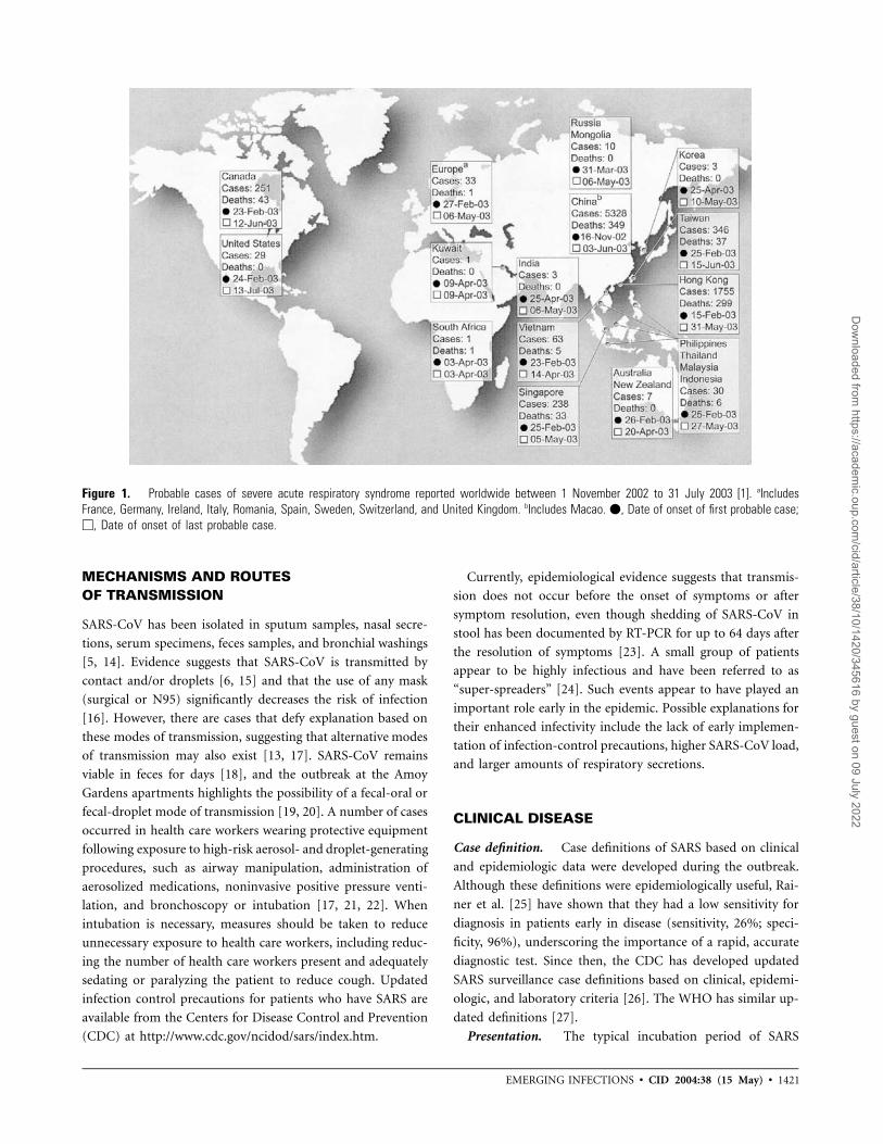
people in 26 countries have had probable SARS diagnosed, 774
of whom have died (figure 1), yielding a global case-fatality
rate of ∼10% [1, 2]. On 5 July 2003, the WHO reported that
the last known human chain of transmission of SARS had been
broken [3].
A newly discovered coronavirus (SARS-CoV) has been iden-
tified as the cause of SARS [4–7]. SARS-CoV–like viruses have
been detected in Himalayan palm civets and a raccoon-dog in
a market in southern China, suggesting that the origin of SARS-
CoV may have been from these or other wild animals [8]. Given
the possibility that human or animal reservoirs of SARS-CoV
may still exist and that SARS may have a seasonal predilection,
there is concern that SARS may return in upcoming respiratory
seasons. WHO guidelines emphasize the need for all countries
to remain vigilant and to maintain their capacity to detect and
respond to the potential reemergence of SARS [9].
Received 22 October 2003; accepted 22 January 2004; electronically published 29 April2004.
Reprints or correspondence: Dr. Donald E. Low, Mount Sinai Hospital, 600 University Ave.,Rm. 1487, Toronto, Ontario, Canada M5G 1X5 ([email protected]).
Clinical Infectious Diseases 2004; 38:1420–7� 2004 by the Infectious Diseases Society of America. All rights reserved.1058-4838/2004/3810-0016$15.00
EPIDEMIOLOGY
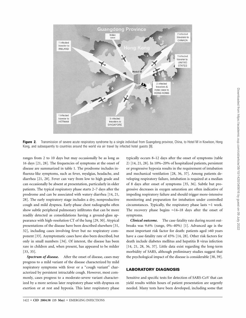
SARS remained isolated in China from November 2002 until
21 February 2003, when a physician with SARS traveled from
Guangdong province to a hotel in Hong Kong, infecting 10
other guests [9]. The movements of these 11 individuals re-
sulted in the spread of SARS worldwide and sparked all of the
major epicenters outside of China [1] (figure 2).
The rate of spread of an epidemic and whether it is self-
sustaining depend on the basic reproduction number (R0). R0
is defined as the average number of secondary cases generated
by 1 primary case in a susceptible population [10]. This quan-
tity determines the potential for an infectious agent to start an
outbreak, the extent of transmission in the absence of control
measures, and the ability of control measures to reduce spread.
During the course of an epidemic, Rt, the effective reproduction
number, decreases in comparison with R0 as a result of the
depletion of susceptible persons in the population, death or
recovery with subsequent immunity, and the implementation
of specific control measures. To stop an outbreak, Rt must be
maintained below 1. Mathematical modeling of the early phase
of the Singapore and Hong Kong outbreaks, before the insti-
tution of control measures and during which time it was oc-
curring primarily in the hospital setting, estimated that the R0
was 2.2–3.7, indicating that the virus is moderately infective
[11, 12]. The attack rate for SARS-CoV ranges from 10.3% to
60% or 2.4 to 31.3 cases/1000 exposure-hours, depending on
the clinical setting and the unit of measurement [13]. A sig-
nificant limitation of these calculations is that these data are
based on diagnoses made with a clinical case definition. Rean-
alysis will be required once the results of seroprevalence studies
are completed and will provide a more accurate estimate of R0.
Dow
nloaded from https://academ
ic.oup.com/cid/article/38/10/1420/345616 by guest on 09 July 2022
EMERGING INFECTIONS • CID 2004:38 (15 May) • 1421
Figure 1. Probable cases of severe acute respiratory syndrome reported worldwide between 1 November 2002 to 31 July 2003 [1]. aIncludesFrance, Germany, Ireland, Italy, Romania, Spain, Sweden, Switzerland, and United Kingdom. bIncludes Macao. �, Date of onset of first probable case;�, Date of onset of last probable case.
MECHANISMS AND ROUTESOF TRANSMISSION
SARS-CoV has been isolated in sputum samples, nasal secre-
tions, serum specimens, feces samples, and bronchial washings
[5, 14]. Evidence suggests that SARS-CoV is transmitted by
contact and/or droplets [6, 15] and that the use of any mask
(surgical or N95) significantly decreases the risk of infection
[16]. However, there are cases that defy explanation based on
these modes of transmission, suggesting that alternative modes
of transmission may also exist [13, 17]. SARS-CoV remains
viable in feces for days [18], and the outbreak at the Amoy
Gardens apartments highlights the possibility of a fecal-oral or
fecal-droplet mode of transmission [19, 20]. A number of cases
occurred in health care workers wearing protective equipment
following exposure to high-risk aerosol- and droplet-generating
procedures, such as airway manipulation, administration of
lation, and bronchoscopy or intubation [17, 21, 22]. When
intubation is necessary, measures should be taken to reduce
unnecessary exposure to health care workers, including reduc-
ing the number of health care workers present and adequately
sedating or paralyzing the patient to reduce cough. Updated
infection control precautions for patients who have SARS are
available from the Centers for Disease Control and Prevention
(CDC) at http://www.cdc.gov/ncidod/sars/index.htm.
Currently, epidemiological evidence suggests that transmis-
sion does not occur before the onset of symptoms or after
symptom resolution, even though shedding of SARS-CoV in
stool has been documented by RT-PCR for up to 64 days after
the resolution of symptoms [23]. A small group of patients
appear to be highly infectious and have been referred to as
“super-spreaders” [24]. Such events appear to have played an
important role early in the epidemic. Possible explanations for
their enhanced infectivity include the lack of early implemen-
tation of infection-control precautions, higher SARS-CoV load,
and larger amounts of respiratory secretions.
CLINICAL DISEASE
Case definition. Case definitions of SARS based on clinical
and epidemiologic data were developed during the outbreak.
Although these definitions were epidemiologically useful, Rai-
ner et al. [25] have shown that they had a low sensitivity for
diagnosis in patients early in disease (sensitivity, 26%; speci-
ficity, 96%), underscoring the importance of a rapid, accurate
diagnostic test. Since then, the CDC has developed updated
SARS surveillance case definitions based on clinical, epidemi-
ologic, and laboratory criteria [26]. The WHO has similar up-
dated definitions [27].
Presentation. The typical incubation period of SARS
Dow
nloaded from https://academ
ic.oup.com/cid/article/38/10/1420/345616 by guest on 09 July 2022
1422 • CID 2004:38 (15 May) • EMERGING INFECTIONS
Figure 2. Transmission of severe acute respiratory syndrome by a single individual from Guangdong province, China, to Hotel M in Kowloon, HongKong, and subsequently to countries around the world via air travel by infected hotel guests [9].
ranges from 2 to 10 days but may occasionally be as long as
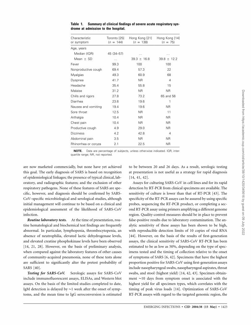
16 days [21, 28]. The frequencies of symptoms at the onset of
disease are summarized in table 1. The prodrome includes in-
fluenza-like symptoms, such as fever, myalgias, headache, and
diarrhea [21, 28]. Fever can vary from low to high grade and
can occasionally be absent at presentation, particularly in older
patients. The typical respiratory phase starts 2–7 days after the
prodrome and can be associated with watery diarrhea [14, 21,
28]. The early respiratory stage includes a dry, nonproductive
cough and mild dyspnea. Early-phase chest radiographs often
show subtle peripheral pulmonary infiltrates that can be more
readily detected as consolidations having a ground-glass ap-
pearance with high-resolution CT of the lung [29, 30]. Atypical
presentations of the disease have been described elsewhere [31,
32], including cases involving fever but no respiratory com-
ponent [33]. Asymptomatic cases have also been described, but
only in small numbers [34]. Of interest, the disease has been
rare in children and, when present, has appeared to be milder
[33, 35].
Spectrum of disease. After the onset of disease, cases may
progress to a mild variant of the disease characterized by mild
respiratory symptoms with fever or a “cough variant” char-
acterized by persistent intractable cough. However, most com-
monly, cases progress to a moderate-severe variant character-
ized by a more serious later respiratory phase with dyspnea on
exertion or at rest and hypoxia. This later respiratory phase
typically occurs 8–12 days after the onset of symptoms (table
2) [14, 21, 28]. In 10%–20% of hospitalized patients, persistent
or progressive hypoxia results in the requirement of intubation
and mechanical ventilation [28, 36, 37]. Among patients de-
veloping respiratory failure, intubation is required at a median
of 8 days after onset of symptoms [35, 36]. Subtle but pro-
gressive decreases in oxygen saturation are often indicative of
impeding respiratory failure and should trigger more-intensive
monitoring and preparation for intubation under controlled
circumstances. Typically, the respiratory phase lasts ∼1 week.
The recovery phase begins ∼14–18 days after the onset of
symptoms.
Clinical outcome. The case-fatality rate during recent out-
breaks was 9.6% (range, 0%–40%) [1]. Advanced age is the
most important risk factor for death: patients aged 160 years
have a case-fatality rate of 45% [14, 28]. Other risk factors for
death include diabetes mellitus and hepatitis B virus infection
[14, 21, 28, 36, 37]. Little data exist regarding the long-term
morbidity of SARS, although preliminary studies suggest that
the psychological impact of the disease is considerable [38, 39].
LABORATORY DIAGNOSIS
Sensitive and specific tests for detection of SARS-CoV that can
yield results within hours of patient presentation are urgently
needed. Many tests have been developed, including some that
Dow
nloaded from https://academ
ic.oup.com/cid/article/38/10/1420/345616 by guest on 09 July 2022
EMERGING INFECTIONS • CID 2004:38 (15 May) • 1423
Table 1. Summary of clinical findings of severe acute respiratory syn-drome at admission to the hospital.
Characteristicor symptom
Toronto [25](n p 144)
Hong Kong [21](n p 138)
Hong Kong [14](n p 75)
Age, years
Median (IQR) 45 (34–57) … …
Mean � SD … 39.3 � 16.8 39.8 � 12.2
Fever 99.3 100 100
Nonproductive cough 69.4 57.3 22
Myalgias 49.3 60.9 68
Dyspnea 41.7 NR 4
Headache 35.4 55.8 15
Malaise 31.2 NR NR
Chills and rigors 27.8 73.2 65 and 56
Diarrhea 23.6 19.6 1
Nausea and vomiting 19.4 19.6 NR
Sore throat 12.5 NR 11
Arthalgia 10.4 NR NR
Chest pain 10.4 NR NR
Productive cough 4.9 29.0 NR
Dizziness 4.2 42.8 4
Abdominal pain 3.5 NR NR
Rhinorrhea or coryza 2.1 22.5 NR
NOTE. Data are percentage of subjects, unless otherwise indicated. IQR, inter-quartile range; NR, not reported.
are now marketed commercially, but none have yet achieved
this goal. The early diagnosis of SARS is based on recognition
of epidemiological linkages; the presence of typical clinical, lab-
oratory, and radiographic features; and the exclusion of other
respiratory pathogens. None of these features of SARS are spe-
cific, however, and diagnosis should be confirmed by SARS-
CoV–specific microbiological and serological studies, although
initial management will continue to be based on a clinical and
epidemiological assessment of the likelihood of SARS-CoV
infection.
Routine laboratory tests. At the time of presentation, rou-
tine hematological and biochemical test findings are frequently
abnormal. In particular, lymphopenia, thrombocytopenia, an
absence of neutrophilia, elevated lactic dehydrogenase levels,
and elevated creatine phosphokinase levels have been observed
[14, 21, 28]. However, on the basis of preliminary analysis,
when compared against the laboratory features of other causes
of community-acquired pneumonia, none of these tests alone
are sufficient to significantly alter the pretest probability of
SARS [40].
Testing for SARS-CoV. Serologic assays for SARS-CoV
include immunofluorescent assays, ELISAs, and Western blot
assays. On the basis of the limited studies completed to date,
IgM detection is delayed by ∼1 week after the onset of symp-
toms, and the mean time to IgG seroconversion is estimated
to be between 20 and 26 days. As a result, serologic testing
at presentation is not useful as a strategy for rapid diagnosis
[14, 41, 42].
Assays for culturing SARS-CoV in cell lines and for its rapid
detection by RT-PCR from clinical specimens are available. The
sensitivity of culture is lower than that of RT-PCR [43]. The
specificity of the RT-PCR assays can be assured by using specific
probes, sequencing the RT-PCR product, or completing a sec-
ond RT-PCR assay using primers amplifying a different genome
region. Quality-control measures should be in place to prevent
false-positive results due to laboratory contamination. The an-
alytic sensitivity of these assays has been shown to be high,
with reproducible detection limits of 10 copies of viral RNA
[44]. However, on the basis of the results of first-generation
assays, the clinical sensitivity of SARS-CoV RT-PCR has been
estimated to be as low as 50%, depending on the type of spec-
imens tested and the timing of collection relative to the onset
of symptoms of SARS [6, 42]. Specimens that have the highest
proportion positive for SARS-CoV using first-generation assays
include nasopharyngeal swabs, nasopharyngeal aspirates, throat
swabs, and stool (highest yield) [14, 42, 43]. Specimen obtain-
ment ∼10 days from symptom onset is associated with the
highest yield for all specimen types, which correlates with the
timing of peak virus loads [14]. Optimization of SARS-CoV
RT-PCR assays with regard to the targeted genomic region, the
Dow
nloaded from https://academ
ic.oup.com/cid/article/38/10/1420/345616 by guest on 09 July 2022
1424 • CID 2004:38 (15 May) • EMERGING INFECTIONS
Table 2. Duration of clinical phases of the mild and moderatelysevere variants of severe acute respiratory syndrome.
Time
Phase
Prodrome
Respiratory
RecoveryEarly Late
From onset, days 0 2–7 8–12 14–18
Duration, days 2–7 1–10 5–10 5–7
timing of specimen obtainment, the type of specimens, and the
extraction methodology is ongoing. A recent report describes
a second generation assay with a clinical sensitivity of 80%
using a modified extraction method on nasopharyngeal aspi-
rates obtained in the first 3 days of illness [45].
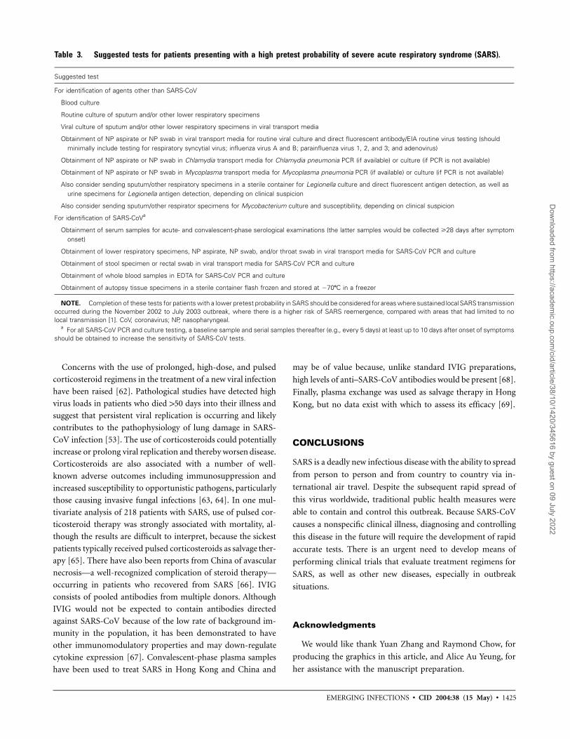
A summary of samples and tests to investigate possible SARS
appears in table 3. Coinfections with SARS-CoV and other
infectious agents can exist, and, as a result, finding an infectious
agent other that SARS-CoV should not be used to rule out
SARS [46].
TREATMENT
At present, there is not sufficient evidence to recommend any
specific therapy for the treatment of SARS. Because SARS can-
not be easily distinguished from other causes of pneumonia,
patients who are suspected of having SARS who have pul-
monary infiltrates should receive appropriate antibiotic cov-
erage [47]. Respiratory failure is the primary cause of acute
morbidity and mortality due to SARS-CoV infection and occurs
in 20%–25% of cases [21, 28, 37]. When mechanical ventilation
is required, a “lung protective” ventilation strategy should be
used based on an analogy to data for the treatment of acute
respiratory distress syndrome (ARDS) and the current intensive
care unit experience managing SARS [36, 37, 48]. In fact, bar-
otrauma appears to be one of the most frequent complications
of severe SARS-CoV infection, with pneumothorax and/or
pneumomediastinum occurring in 20%–34% of ventilated pa-
tients, a rate that is much higher than the rate of 2.5% observed
in a large study of ARDS [36, 37, 48].
Antiviral therapy. Antiviral agents used in the therapy of
SARS include ribavirin, IFN-a, and lopinavir-ritonavir. Riba-
virin is a nucleoside analogue with in vitro activity against a
number of RNA and DNA viruses, including some animal co-
ronaviruses [49]. Ribavirin was widely used for the treatment
of SARS. Initial reports noted improvement in surrogate mark-
ers of outcome, such as resolution of fever and improvement
in oxygenation and radiographic appearance [15, 21, 50]. These
studies were not controlled, and most patients also received
corticosteroids [15, 21, 50]. Other reports failed to identify
improvement with ribavirin [28, 51], and one report identified
a high frequency of adverse events among patients treated with
high-dose ribavirin, including severe hemolysis (in 49% of pa-
tients) [28]. In vitro testing of SARS-CoV indicated that ri-
bavirin does not have activity against this virus at clinically
achievable concentrations [52]. Postmortem findings for some
patients demonstrated that high virus loads persisted despite
treatment with ribavirin [53].
IFNs are cytokines with well-described antiviral activity [54].
IFNs, particularly IFN-b, inhibit SARS-CoV in vitro [55]. An
open-labelled study using IFN-alfacon-1 and high-dose meth-
ylprednisolone demonstrated more-rapid improvement in ra-
diographic appearance and oxygenation in recipients, com-
pared with a historic cohort that received a lower dose of
corticosteroids alone [56]. A complex 4-arm trial examining
ribavirin and IFN and differing doses of corticosteroids also
demonstrated improvement in surrogate end points, such as
radiographic appearance, but these improvements only oc-
curred in the IFN recipients who also received high-dose cor-
ticosteroids [57].
Lopinavir-ritonavir is a combination drug consisting of 2
protease inhibitors with proven efficacy in the treatment of
HIV. Lopinavir-ritonavir was studied in a nonrandomized open
label study in Hong Kong as initial and rescue therapy for SARS.
It was added to local standard therapy consisting of ribavirin
and corticosteroids, and, when used as initial therapy, recipients
had a significant reduction in the overall death rate and in-
tubation rate, compared with a matched control group who
received standard treatment alone. The control group, however,
had lower rates of steroid use at lower mean doses, making
definitive conclusions difficult [58].
Anti-inflammatory therapy. Anti-inflammatory or im-
munomodulatory therapies include corticosteroids, intrave-
nous immunoglobulin (IVIG), and convalescent-phase serum
and plasma exchange. Corticosteroids were widely used for
loads and increasing antibody titers during the second week of
illness, at a time when the respiratory disease typically pro-
gresses [14]. These results suggest that lung damage in patients
with SARS-CoV infection may be immune mediated and pro-
vides the rationale for corticosteroid therapy. Pathological find-
ings are consistent with cytokine dysregulation and provide
further support for the theory that lung damage is immune
mediated [52]. Initial case reports described resolution of fever
and improvements in oxygenation and radiographic appear-
ance in some patients treated with ribavirin and corticosteroids
[59]. Subsequently, clinicians noted that cases in many patients
progress despite receiving treatment with corticosteroids, and
higher doses or pulsed steroid regimens were required as rescue
therapy [60, 61]. A trial comparing early use of pulsed versus
nonpulsed corticosteroids did not note any difference in the
requirement for ventilation or mortality, but it did reveal im-
provements in oxygenation and radiographic appearance [62].
Dow
nloaded from https://academ
ic.oup.com/cid/article/38/10/1420/345616 by guest on 09 July 2022
EMERGING INFECTIONS • CID 2004:38 (15 May) • 1425
Table 3. Suggested tests for patients presenting with a high pretest probability of severe acute respiratory syndrome (SARS).
Suggested test
For identification of agents other than SARS-CoV
Blood culture
Routine culture of sputum and/or other lower respiratory specimens
Viral culture of sputum and/or other lower respiratory specimens in viral transport media
Obtainment of NP aspirate or NP swab in viral transport media for routine viral culture and direct fluorescent antibody/EIA routine virus testing (shouldminimally include testing for respiratory syncytial virus; influenza virus A and B; parainfluenza virus 1, 2, and 3; and adenovirus)
Obtainment of NP aspirate or NP swab in Chlamydia transport media for Chlamydia pneumonia PCR (if available) or culture (if PCR is not available)
Obtainment of NP aspirate or NP swab in Mycoplasma transport media for Mycoplasma pneumonia PCR (if available) or culture (if PCR is not available)
Also consider sending sputum/other respiratory specimens in a sterile container for Legionella culture and direct fluorescent antigen detection, as well asurine specimens for Legionella antigen detection, depending on clinical suspicion
Also consider sending sputum/other respirator specimens for Mycobacterium culture and susceptibility, depending on clinical suspicion
For identification of SARS-CoVa
Obtainment of serum samples for acute- and convalescent-phase serological examinations (the latter samples would be collected �28 days after symptomonset)
Obtainment of lower respiratory specimens, NP aspirate, NP swab, and/or throat swab in viral transport media for SARS-CoV PCR and culture
Obtainment of stool specimen or rectal swab in viral transport media for SARS-CoV PCR and culture
Obtainment of whole blood samples in EDTA for SARS-CoV PCR and culture
Obtainment of autopsy tissue specimens in a sterile container flash frozen and stored at �70�C in a freezer
NOTE. Completion of these tests for patients with a lower pretest probability in SARS should be considered for areas where sustained local SARS transmissionoccurred during the November 2002 to July 2003 outbreak, where there is a higher risk of SARS reemergence, compared with areas that had limited to nolocal transmission [1]. CoV, coronavirus; NP, nasopharyngeal.
a For all SARS-CoV PCR and culture testing, a baseline sample and serial samples thereafter (e.g., every 5 days) at least up to 10 days after onset of symptomsshould be obtained to increase the sensitivity of SARS-CoV tests.
Concerns with the use of prolonged, high-dose, and pulsed
corticosteroid regimens in the treatment of a new viral infection
have been raised [62]. Pathological studies have detected high
virus loads in patients who died 150 days into their illness and
suggest that persistent viral replication is occurring and likely
contributes to the pathophysiology of lung damage in SARS-
CoV infection [53]. The use of corticosteroids could potentially
increase or prolong viral replication and thereby worsen disease.
Corticosteroids are also associated with a number of well-
known adverse outcomes including immunosuppression and
increased susceptibility to opportunistic pathogens, particularly
those causing invasive fungal infections [63, 64]. In one mul-
tivariate analysis of 218 patients with SARS, use of pulsed cor-
ticosteroid therapy was strongly associated with mortality, al-
though the results are difficult to interpret, because the sickest
patients typically received pulsed corticosteroids as salvage ther-
apy [65]. There have also been reports from China of avascular
necrosis—a well-recognized complication of steroid therapy—
occurring in patients who recovered from SARS [66]. IVIG
consists of pooled antibodies from multiple donors. Although
IVIG would not be expected to contain antibodies directed
against SARS-CoV because of the low rate of background im-
munity in the population, it has been demonstrated to have
other immunomodulatory properties and may down-regulate
have been used to treat SARS in Hong Kong and China and
may be of value because, unlike standard IVIG preparations,
high levels of anti–SARS-CoV antibodies would be present [68].
Finally, plasma exchange was used as salvage therapy in Hong
Kong, but no data exist with which to assess its efficacy [69].
CONCLUSIONS
SARS is a deadly new infectious disease with the ability to spread
from person to person and from country to country via in-
ternational air travel. Despite the subsequent rapid spread of
this virus worldwide, traditional public health measures were
able to contain and control this outbreak. Because SARS-CoV
causes a nonspecific clinical illness, diagnosing and controlling
this disease in the future will require the development of rapid
accurate tests. There is an urgent need to develop means of
performing clinical trials that evaluate treatment regimens for
SARS, as well as other new diseases, especially in outbreak
situations.
Acknowledgments
We would like thank Yuan Zhang and Raymond Chow, for
producing the graphics in this article, and Alice Au Yeung, for
her assistance with the manuscript preparation.
Dow
nloaded from https://academ
ic.oup.com/cid/article/38/10/1420/345616 by guest on 09 July 2022
1426 • CID 2004:38 (15 May) • EMERGING INFECTIONS
References
1. World Health Organization. Summary of probable SARS cases withonset of illness from 1 November 2002 to 31 July 2003. 23 September2003. Available at: http://www.who.int/csr/sars/country/table2003_09_23/en/. Accessed on 25 September 2003.
2. World Health Organization. Biosafety and SARS incident in SingaporeSeptember 2003: report of the Review Panel on New SARS Case andBiosafety. Singapore Ministry of Health. 24 September 2003. Availableat: http://www.moh.gov.sg/sars/pdf/Report_SARS_Biosafety.pdf. Ac-cessed on 25 September 2003.
3. World Health Organization. SARS: breaking the chains of transmission.5 July 2003. Available at: http://www.who.int/features/2003/07/en/. Ac-cessed on 5 September 2003.
4. Ksiazek TG, Erdman D, Goldsmith CS, et al. A novel coronavirusassociated with severe acute respiratory syndrome. N Engl J Med2003; 348:1953–66.
5. Drosten C, Gunther S, Preiser W, et al. Identification of a novel co-ronavirus in patients with severe acute respiratory syndrome. N EnglJ Med 2003; 348:1967–76.
6. Peiris JS, Lai ST, Poon LL, et al. Coronavirus as a possible cause ofsevere acute respiratory syndrome. Lancet 2003; 361:1319–25.
7. Kuiken T, Fouchier RAM, Schutten M, et al. Newly discovered coron-avirus as the primary cause of severe acute respiratory syndrome. Lan-cet 2003; 362:263–70.
8. Guan Y, Zheng BJ, He YQ, et al. Isolation and characterization ofviruses related to the SARS coronavirus from animals in southernChina. Science 2003; 302:276–8.
9. Centers for Disease Control and Prevention. Update: outbreak of severeacute respiratory syndrome—worldwide, 2003. MMWR Morb MortalWkly Rep 2003; 52:241–8.
10. Donnelly CA, Ghani AC, Leung GM, et al. Epidemiological determi-nants of spread of causal agent of severe acute respiratory syndromein Hong Kong. Lancet 2003; 361:1761–6.
11. Lipsitch M, Cohen T, Cooper B, et al. Transmission dynamics andcontrol of severe acute respiratory syndrome. Science 2003; 300:1966–70.
12. Riley S, Fraser C, Donnelly CA, et al. Transmission dynamics of theetiological agent of SARS in Hong Kong: impact of public health in-terventions. Science 2003; 300:1961–6.
13. Varia M, Wilson S, Sarwal S, et al. Investigation of a nosocomial out-break of severe acute respiratory syndrome (SARS) in Toronto, Canada.CMAJ 2003; 169:285–92.
14. Peiris J, Chu C, Cheng V, et al. Clinical progression and viral load ina community outbreak of coronavirus-associated SARS pneumonia: aprospective study. Lancet 2003; 361:1767–72.
15. Poutanen SM, Low DE, Henry B, et al. Identification of severe acuterespiratory syndrome in Canada. N Engl J Med 2003; 348:1995–2005.
16. Seto WH, Tsang D, Yung RW, et al. Effectiveness of precautions againstdroplets and contact in prevention of nosocomial transmission of se-vere acute respiratory syndrome (SARS). Lancet 2003; 361:1519–20.
17. Christian MD, Loutfy M, McDonald, et al. Possible SARS coronavirustransmission during cardiopulmonary resuscitation. Emerg Infect Dis[serial online] 2004. Available at: http://www.cdc.gov/ncidod/EID/vol10no2/03-0700.htm.
18. World Health Organization. First data on stability and resistance ofSARS coronavirus compiled by members of WHO laboratory network.2003. Available at: http://www.who.int/csr/sars/survival_2003_05_04/en/. Accessed on 15 October 2003.
19. World Health Organization. Update 15—situation in Hong Kong, ac-tivities of WHO team in China. 2003. Available at: http://www.who.int/csr/sars/infectioncontrol/en/. Accessed on 15 October 2003.
20. World Health Organization. Inadequate plumbing systems likely con-tributed to SARS transmission. 2003. Available at: http://www.who.int/mediacentre/releases/2003/pr70/en/. Accessed on 15 October 2003.
21. Lee N, Hui D, Wu A, et al. A major outbreak of severe acute respiratorysyndrome in Hong Kong. N Engl J Med 2003; 348:1986–94.
22. Ofner M, Lem M, Sarwal S, et al. Cluster of severe acute respiratorysyndrome cases among protected health care workers—Toronto, April2003. Can Commun Dis Rep 2003; 29:93–7.
23. Ren Y, Ding HG, Wu QF, et al. Detection of SARS-CoV RNA in stoolsamples of SARS patients by nest RT-PCR and its clinical value [inChinese]. Zhongguo Yi Xue Ke Xue Yuan Xue Bao 2003; 25:368–71.
24. Centers for Disease Control and Prevention. Severe acute respiratorysyndrome—Singapore, 2003. MMWR Morb Mortal Wkly Rep 2003;52:405–11.
25. Rainer TH, Cameron PA, Smit D, et al. Evaluation of WHO criteriafor identifying patients with severe acute respiratory syndrome out ofhospital: prospective observational study. Brit Med J 2003; 326:1354–8.
26. Centers for Disease Control and Prevention. Revised US surveillancecase definition for severe acute respiratory syndrome (SARS) and up-date on SARS cases—United States and worldwide, December 2003.MMWR Morb Mortal Wkly Rep 2003; 52:1202–6.
27. World Health Organization. Alert, verification and public health man-agement of SARS in the post-outbreak period. 14 August 2003. Avail-able at: http://www.who.int/csr/sars/postoutbreak/en/. Accessed on 26April 2004.
28. Booth CM, Matukas LM, Tomlinson GA, et al. Clinical features andshort-term outcomes of 144 patients with SARS in the greater Torontoarea. JAMA 2003; 289:2801–9.
29. Wong K, Antonio G, Hui D, et al. Think-section CT of severe acuterespiratory syndrome: evaluation of 73 patients exposed to or with thedisease. Radiology 2003; 228:395–400.
30. Wong K, Antonio G, Hui D, et al. Severe acute respiratory syndrome:radiographic appearances and pattern of progression in 138 patients.Radiology 2003; 228:401–6.
31. Fisher DA, Lim TK, Lim YT, et al. Atypical presentations of SARS.Lancet 2003; 361:1740.
32. Wu EB, Sung JJ. Haemorrhagic-fever–like changes and normal chestradiograph in a doctor with SARS. Lancet 2003; 361:1520–1.
33. Li G, Zhao ZX, Chen LB, et al. Mild severe acute respiratory syndrome.Emerg Infect Dis 2003; 9:1182–3.
34. Gold WL, Mederski B, Rose D, et al. Prevalence of asymptomatic (AS)infection by severe acute respiratory syndrome coronavirus (SARS-CoV) in exposed healthcare workers (HCW) [abstract K-1315c]. In:Program and abstracts of the 43rd Meeting of the Interscience Con-ference on Antimicrobial Agents and Chemotherapy (Chicago). Wash-ington, DC: American Society for Microbiology, 2003.
35. Hon K, Leung C, Cheng W, et al. Clinical presentations and outcomeof severe acute respiratory syndrome in children. Lancet 2003; 361:1701–3.
36. Fowler RA, Lapinsky SE, Hallett D, et al. Critically ill patients withsevere acute respiratory syndrome. JAMA 2003; 290:367–73.
37. Lew TW, Kwek TK, Tai D, et al. Acute respiratory distress syndromein critically ill patients with severe acute respiratory syndrome. JAMA2003; 290:374–80.
38. Maunder R, Hunter J, Vincent L, et al. The immediate psychologicaland occupational impact of the 2003 SARS outbreak in a teachinghospital. CMAJ 2003; 168:1245–51.
39. Styra R, Gold W, Robinson S. Post-traumatic stress disorder and qualityof life in patients diagnosed with SARS [abstract V-796a]. In: Programand abstracts of the 43rd Meeting of the Interscience Conference onAntimicrobial Agents and Chemotherapy (Chicago). Washington, DC:American Society for Microbiology, 2003.
40. Muller M, Tomlinson G, Marrie T, et al. Discriminatory ability oflaboratory parameters in severe acute respiratory syndrome (SARS)[abstract V-796b]. In: Program and abstracts of the 43rd Meeting ofthe Interscience Conference on Antimicrobial Agents and Chemo-therapy (Chicago). Washington, DC: American Society for Microbi-ology, 2003.
41. Li G, Chen X, Xu A. Profile of specific antibodies to the SARS-asso-ciated coronavirus. N Engl J Med 2003; 349:508–9.
42. Tang P, Louie M, Richardson S, et al. Laboratory diagnosis of severeacute respiratory syndrome (SARS) in Canada [abstract V-485a]. In:
Dow
nloaded from https://academ
ic.oup.com/cid/article/38/10/1420/345616 by guest on 09 July 2022
EMERGING INFECTIONS • CID 2004:38 (15 May) • 1427
Program and abstracts of the 43rd Meeting of the Interscience Con-ference on Antimicrobial Agents and Chemotherapy (Chicago). Wash-ington, DC: American Society for Microbiology, 2003.
43. Hsueh P-R, Hsiao C-H, Yeh S-H, et al. Microbiologic characteristics,serologic responses, and clinical manifestations in severe acute respi-ratory syndrome, Taiwan. Emerg Infect Dis 2003; 9:1163–7.
44. Mahony J, Petrich A, Louie L, et al. Comparison of the cost andperformance of seven RT-PCR assays for detecting SARS coronavirusRNA [abstract V-796d]. In: Program and abstracts of the 43rd Meetingof the Interscience Conference on Antimicrobial Agents and Che-motherapy (Chicago). Washington, DC: American Society for Micro-biology, 2003.
45. Poon LLM, Chan KH, Wong OK, et al. Early diagnosis of SARS co-ronavirus infection by real time RT-PCR. J Clin Virol 2003; 28:233–8.
46. Chang P, Tam J, Lam C-W, et al. Human metapneumovirus detectionin patients with severe acute respiratory syndrome. Emerg Infect Dis2003; 9:1058–63.
47. Bartlett JG, Dowell SF, Mandell LA, et al. Practice guidelines for themanagement of community-acquired pneumonia in adults. Clin InfectDis 2000; 31:347–82.
48. The Acute Respiratory Distress Syndrome Network. Ventilation withlower tidal volumes as compared with traditional tidal volumes foracute lung injury and the acute respiratory distress syndrome. N EnglJ Med 2000; 342:1301–8.
49. Koren G, King S, Knowles S, et al. Ribavirin in the treatment of SARS:a new trick for an old drug? CMAJ 2003; 168:1289–92.
50. Tsang KW, Ho PL, Ooi GC, et al. A cluster of cases of severe acuterespiratory syndrome in Hong Kong. N Engl J Med 2003; 348:1977–85.
51. Hsu LY, Lee CC, Green JA, et al. Severe acute respiratory syndrome(SARS) in Singapore: clinical features of index patient and initial con-tacts. Emerg Infect Dis 2003; 9:713–7.
52. Charles M. Severe acute respiratory syndrome (SARS) and coronavirustesting—United States, 2003. The CDC SARS Investigative Team.MMWR Morb Mortal Wkly Rep 2003; 52:297–302.
53. Mazzulli T, Farcas GA, Poutanen SM, et al. Severe acute respiratorysyndrome–associated coronavirus in lung tissue. Emerg Infect Dis2004; 10. Available at: http://www.cdc.gov/ncidod/EID/vol10no1/03-0404.htm. Accessed on 5 April 2004.
54. Samuel CE. Antiviral actions of interferons. Clin Microbiol Rev2001; 14:778–809.
55. Cinatl J, Morgenstern B, Bauer G, et al. Treatment of SARS with humaninterferons. Lancet 2003; 362:293–4.
56. Loutfy M, Blatt L, Ward S, et al. Preliminary results on the potentialtherapeutic benefit of interferon alfacon-1 plus steroids in severe acuterespiratory syndrome [abstract K-1315e]. In: Program and abstracts ofthe meeting of the Interscience Conference on Antimicrobial Agentsand Chemotherapy (Chicago). Washington, DC: American Society forMicrobiology, 2003.
57. Zhao Z, Zhang F, Xu M, et al. Description and clinical treatment ofan early outbreak of severe acute respiratory syndrome (SARS) inGuangzhou, PR China. J Med Microbiol 2003; 52:715–20.
58. Chan KS, Lai ST, Chu CM, et al. Treatment of severe acute respiratorysyndrome with lopinavir/ritonavir: a multicentre retrospective matchedcohort study. Hong Kong Med J 2003; 9:399–406.
59. Nicholls JM, Poon LL, Lee KC, et al. Lung pathology of fatal severeacute respiratory syndrome. Lancet 2003; 361:1773–8.
60. So LK, Lau AC, Yam LY, et al. Development of a standard treatmentprotocol for severe acute respiratory syndrome. Lancet 2003; 361:1615–7.
61. Ho JC, Ooi GC, Mok TY, et al. High dose pulse versus nonpulsecorticosteroid regimens in severe acute respiratory syndrome. Am JRespir Crit Care Med 2003; 15:1449–56.
62. Oba Y. The use of corticosteroids in SARS. N Engl J Med 2003; 348:2034–5.
63. Wang H, Ding Y, Li X, et al. Fatal aspergillosis in a patient with SARSwho was treated with corticosteroids. N Engl J Med 2003; 349:507–8.
64. Tsang KW, Lam WK. Management of severe acute respiratory syn-drome: the Hong Kong University experience. Am J Respir Crit CareMed 2003; 168:417–24.
65. Tsang O, Chau TN, Choi KW. Coronavirus-positive nasopharyngealaspirate as predictor for severe acute respiratory syndrome mortality.Emerg Infect Dis 2003; 9:1381–7.
67. Ballow M. Mechanisms of action of intravenous immune serum glob-ulin in autoimmune and inflammatory diseases. J Allergy Clin Im-munol 1997; 100:151–7.
68. Wong VW, Dai D, Wu AK, et al. Treatment of severe acute respiratorysyndrome with convalescent plasma. Hong Kong Med J 2003; 9:199–201.
69. Tsang K, Zhong NS. SARS: pharmacotherapy. Respirology 2003; 8:S25–30.
Dow
nloaded from https://academ
ic.oup.com/cid/article/38/10/1420/345616 by guest on 09 July 2022
academic.oup.com/cid of 1 4
Please excuse the presence of this and the following test pages, which have been
added to a small number of article PDFs for a limited time as part of our process of
continual development and improvement.
academic.oup.com/cid
Lorem ipsum dolor sit amet, consectetur adipiscing elit, sed do eiusmod tempor incididunt ut labore et dolore magna aliqua. Ut enim ad minim veniam, quis nostrud exercitation ullamco laboris nisi ut aliquip ex ea commodo consequat. Duis aute irure dolor in reprehenderit in voluptate velit esse cillum dolore eu fugiat nulla pariatur. Excepteur sint occaecat cupidatat non proident, sunt in culpa qui officia deserunt mollit anim id est laborum. Lorem ipsum dolor sit amet, consectetur adipiscing elit, sed do eiusmod tempor incididunt ut labore et dolore magna aliqua. Ut enim ad minim veniam, quis nostrud exercitation ullamco laboris nisi ut aliquip ex ea commodo consequat. Duis aute irure dolor in reprehenderit in voluptate velit esse cillum dolore eu fugiat nulla pariatur. Excepteur sint occaecat cupidatat non proident, sunt in culpa qui officia deserunt mollit anim id est laborum. Lorem ipsum dolor sit amet, consectetur adipiscing elit, sed do eiusmod tempor incididunt ut labore et dolore magna aliqua. Ut enim ad minim veniam, quis nostrud exercitation ullamco laboris nisi ut aliquip ex ea commodo consequat. Duis aute irure dolor in reprehenderit in voluptate velit esse cillum dolore eu fugiat nulla pariatur. Excepteur sint occaecat cupidatat non proident, sunt in culpa qui officia deserunt mollit anim id est laborum. Lorem ipsum dolor sit amet, consectetur adipiscing elit, sed do eiusmod tempor incididunt ut labore et dolore magna aliqua. Ut enim ad minim veniam, quis nostrud exercitation ullamco laboris nisi ut aliquip ex ea commodo consequat. Duis aute irure dolor in reprehenderit in voluptate velit esse cillum dolore eu fugiat nulla pariatur. Excepteur sint occaecat cupidatat non proident, sunt in culpa qui officia deserunt mollit anim id est laborum. Lorem ipsum dolor sit amet, consectetur adipiscing elit, sed do eiusmod tempor incididunt ut labore et dolore magna aliqua. Ut enim ad minim veniam, quis nostrud exercitation ullamco laboris nisi ut aliquip ex ea commodo consequat. Duis aute irure dolor in reprehenderit in voluptate velit esse cillum dolore eu fugiat nulla pariatur. Excepteur sint occaecat cupidatat non proident, sunt in culpa qui officia deserunt mollit anim id est laborum. Lorem ipsum dolor sit amet, consectetur adipiscing elit, sed do eiusmod tempor incididunt ut labore et dolore magna aliqua. Ut enim ad minim veniam, quis nostrud exercitation ullamco laboris nisi ut aliquip ex ea commodo consequat. Duis aute irure dolor in reprehenderit in voluptate velit esse cillum dolore eu fugiat nulla pariatur. Excepteur sint occaecat cupidatat non proident, sunt in culpa qui officia deserunt mollit anim id est laborum. Lorem ipsum dolor sit amet, consectetur adipiscing elit, sed do academic.oup.com/cid of 2 4
eiusmod tempor incididunt ut labore et dolore magna aliqua. Ut enim ad minim veniam, quis nostrud exercitation ullamco laboris nisi ut aliquip ex ea commodo consequat. Duis aute irure dolor in reprehenderit in voluptate velit esse cillum dolore eu fugiat nulla pariatur. Excepteur sint occaecat cupidatat non proident, sunt in culpa qui officia deserunt mollit anim id est laborum. Lorem ipsum dolor sit amet, consectetur adipiscing elit, sed do eiusmod tempor incididunt ut labore et dolore magna aliqua. Ut enim ad minim veniam, quis nostrud exercitation ullamco laboris nisi ut aliquip ex ea commodo consequat. Duis aute irure dolor in reprehenderit in voluptate velit esse cillum dolore eu fugiat nulla pariatur. Excepteur sint occaecat cupidatat non proident, sunt in culpa qui officia deserunt mollit anim id est laborum. Lorem ipsum dolor sit amet, consectetur adipiscing elit, sed do eiusmod tempor incididunt ut labore et dolore magna aliqua. Ut enim ad minim veniam, quis nostrud exercitation ullamco laboris nisi ut aliquip ex ea commodo consequat. Duis aute irure dolor in reprehenderit in voluptate velit esse cillum dolore eu fugiat nulla pariatur. Excepteur sint occaecat cupidatat non proident, sunt in culpa qui officia deserunt mollit anim id est laborum. Lorem ipsum dolor sit amet, consectetur adipiscing elit, sed do eiusmod tempor incididunt ut labore et dolore magna aliqua. Ut enim ad minim veniam, quis nostrud exercitation ullamco laboris nisi ut aliquip ex ea commodo consequat. Duis aute irure dolor in reprehenderit in voluptate velit esse cillum dolore eu fugiat nulla pariatur. Excepteur sint occaecat cupidatat non proident, sunt in culpa qui officia deserunt mollit anim id est laborum. Lorem ipsum dolor sit amet, consectetur adipiscing elit, sed do eiusmod tempor incididunt ut labore et dolore magna aliqua. Ut enim ad minim veniam, quis nostrud exercitation ullamco laboris nisi ut aliquip ex ea commodo consequat. Duis aute irure dolor in reprehenderit in voluptate velit esse cillum dolore eu fugiat nulla pariatur. Excepteur sint occaecat cupidatat non proident, sunt in culpa qui officia deserunt mollit anim id est laborum. Lorem ipsum dolor sit amet, consectetur adipiscing elit, sed do eiusmod tempor incididunt ut labore et dolore magna aliqua. Ut enim ad minim veniam, quis nostrud exercitation ullamco laboris nisi ut aliquip ex ea commodo consequat. Duis aute irure dolor in reprehenderit in voluptate velit esse cillum dolore eu fugiat nulla pariatur. Excepteur sint occaecat cupidatat non proident, sunt in culpa qui officia deserunt mollit anim id est laborum. Lorem ipsum dolor sit amet, consectetur adipiscing elit, sed do eiusmod tempor incididunt ut labore et dolore magna aliqua. academic.oup.com/cid of 3 4
Ut enim ad minim veniam, quis nostrud exercitation ullamco laboris nisi ut aliquip ex ea commodo consequat. Duis aute irure dolor in reprehenderit in voluptate velit esse cillum dolore eu fugiat nulla pariatur. Excepteur sint occaecat cupidatat non proident, sunt in culpa qui officia deserunt mollit anim id est laborum. Lorem ipsum dolor sit amet, consectetur adipiscing elit, sed do eiusmod tempor incididunt ut labore et dolore magna aliqua. Ut enim ad minim veniam, quis nostrud exercitation ullamco laboris nisi ut aliquip ex ea commodo consequat. Duis aute irure dolor in reprehenderit in voluptate velit esse cillum dolore eu fugiat nulla pariatur. Excepteur sint occaecat cupidatat non proident, sunt in culpa qui officia deserunt mollit anim id est laborum. Lorem ipsum dolor sit amet, consectetur adipiscing elit, sed do eiusmod tempor incididunt ut labore et dolore magna aliqua. Ut enim ad minim veniam, quis nostrud exercitation ullamco laboris nisi ut aliquip ex ea commodo consequat. Duis aute irure dolor in reprehenderit in voluptate velit esse cillum dolore eu fugiat nulla pariatur. Excepteur sint occaecat cupidatat non proident, sunt in culpa qui officia deserunt mollit anim id est laborum. Lorem ipsum dolor sit amet, consectetur adipiscing elit, sed do eiusmod tempor incididunt ut labore et dolore magna aliqua. Ut enim ad minim veniam, quis nostrud exercitation ullamco laboris nisi ut aliquip ex ea commodo consequat. Duis aute irure dolor in reprehenderit in voluptate velit esse cillum dolore eu fugiat nulla pariatur. Excepteur sint occaecat cupidatat non proident, sunt in culpa qui officia deserunt mollit anim id est laborum. Lorem ipsum dolor sit amet, consectetur adipiscing elit, sed do eiusmod tempor incididunt ut labore et dolore magna aliqua. Ut enim ad minim veniam, quis nostrud exercitation ullamco laboris nisi ut aliquip ex ea commodo consequat. Duis aute irure dolor in reprehenderit in voluptate velit esse cillum dolore eu fugiat nulla pariatur. Excepteur sint occaecat cupidatat non proident, sunt in culpa qui officia deserunt mollit anim id est laborum. Lorem ipsum dolor sit amet, consectetur adipiscing elit, sed do eiusmod tempor incididunt ut labore et dolore magna aliqua. Ut enim ad minim veniam, quis nostrud exercitation ullamco laboris nisi ut aliquip ex ea commodo consequat. Duis aute irure dolor in reprehenderit in voluptate velit esse cillum dolore eu fugiat nulla pariatur. Excepteur sint occaecat cupidatat non proident, sunt in culpa qui officia deserunt mollit anim id est laborum.