Peanut allergies are one of the leading causes of fatal and near-fatal food induced allergic reactions,
and are estimated to induce hypersensitivity reactions in 8% of children and 2% of adults (Sicherer,
2011; Sicherer et al., 1999; Sicherer et al., 2010; Sicherer & Sampson, 2014). This allergy attracts
the attention of many due to its increasing prevalence and its identification as one of the few food-
allergies that starts in childhood and continues through life, as opposed to others that resolve with
age (Burks & Sampson, 1993). There is evidence that approximately 20% and 10% of peanut and
tree nut allergic individuals, respectively, outgrow their allergy and approximately 8% of the
peanut allergic individuals may suffer a recurrence (Fleischer, 2007). Currently the only treatment
L. Offermann, M. Perdue, J. He, B. Hurlburt, S. Maleki, and M. Chruszcz / Journal of Contemporary Immunology
(2015) Vol. 2 No. 1 pp. 1-26
2
plan for those suffering from the allergy is avoidance (Burks et al., 2012; Lieberman & Sicherer,
2011), which is a stressful task for families (Kim & Sicherer, 2011) especially as peanuts become
increasingly popular as an economical protein source in processed foods (Shin et al., 1998). While
individuals avoid foods made directly with peanuts, accidental ingestion is common due to the high
risks of cross-contamination (Sicherer et al., 1999). Moreover, peanut and tree nut allergens are
often cross-reactive which further increases a risk of an adverse reaction (Bublin & Breiteneder,
2014; Kulis et al., 2009; Teuber & Beyer, 2004).
Table 1 All registered peanut allergens grouped according to the family to which they belong.
Please note: Ara h 4 is not included because it was renamed as a variant of Ara h 3.
Protein Family
Allergens
(including
variants)
Uniprot ID Genbank
Nucleotide
MW
(kDa) pI
PDB
Codes
Cupin (Vicillin-type,
7s globulin)
Ara h 1.0101 P43238 L34402 68.8 6.4 3SMH
3S7E
3S7I
Conglutin
(2s albumin)
Ara h 2.0101 Q6PSU2 AY007229 18.0 5.5 3OB4
Ara h 2.0201 Q6PSU2 AY158467 17.7 5.3
Ara h 6.0101 Q647G9 AF092846 14.8 5.5 1W2Q
Ara h 7.0101 Q9SQH1 AF091737 16.3 5.6
Ara h 7.0201 B4XID4 EU046325 17.4 7.5
Cupin (Legumin-type,
11s globulin, Glycinin)
Ara h 3.0101 O82580 AF093541 58.3 5.7 3C3V
Ara h 3.0201 Q9SQH7 AF086821 61.0 5.5
Profilin Ara h 5.0101 Q9SQI9 AF059616 14.1 4.6 4ESP
PR-10 (Pathogenesis
related Protein)
Ara h 8.0101 Q6VT83 AY328088 17.0 5.0 4MAP
4M9B
4M9W
4MA6
Ara h 8.0201 B0YIU5 EF436550 16.4 5.1
Nonspecific Lipid-
transfer protein 1
(nsLTP1)
Ara h 9.0101 B6CEX8 EU159429 9.1 9.5 N/A
Ara h 9.0201 B6CG41 EU161278 9.1 9.3 N/A
Oleosin Ara h 10.0101 Q647G5 AY722694 17.8 9.6 N/A
Ara h 10.0201 Q647G4 AY722695 15.5 9.4 N/A
Ara h 11.0101 Q45W87 DQ097716 14.3 10.1 N/A
Defensin Ara h 12.0101 N/A EY396089 7.9 7.7 N/A
Ara h 13.0101 N/A EY396019 8.4 7.5 N/A
L. Offermann, M. Perdue, J. He, B. Hurlburt, S. Maleki, and M. Chruszcz / Journal of Contemporary Immunology
(2015) Vol. 2 No. 1 pp. 1-26
3
Upon ingestion of peanuts, IgE-allergen complexes form and facilitate cross-linking amongst mast
cell receptors and induce a signal transduction cascade that elicits the allergic reaction in patients
(Fung-Leung et al., 1996; Sanchez-Mejorada & Rosales, 1998). Therefore thorough characterization and structural analysis of peanut allergens and their complexes with antibodies may be critical for the identification of IgE binding epitopes. It is anticipated that such studies may lead to the design of recombinant variants of allergens with decreased IgE binding capacity for application in immunotherapy. In this review we focus on the current status of the structural studies of peanut allergens, however we do not discuss in detail the cross-reactivity of these allergens, as this topic was recently reviewed (Bublin and Breiteneder, 2014). Despite the fact that peanuts are well studied and several peanut allergens have had their structures experimentally determined, more research is required in order to shed light on the biological function of these proteins as well as the contribution of their structural and physicochemical properties to allergenicity (Schein et al., 2005). Twelve peanut allergens are officially registered to the Allergen Nomenclature Sub-Committee of the International Union of Immunological Societies (IUIS), and like other allergens they belong to just a few protein families (Breiteneder & Radauer, 2004; Radauer et al., 2008) with half of them belonging to just two superfamilies: cupin and prolamin (Table 1).
2. Materials and Methods According to the Allergen Nomenclature Sub-Committee of the International Union of Immunological Societies (IUIS) (www.allergen.org) (Radauer et al., 2014), twelve allergens belong to the Arachis hypogaea (Peanut) species. Protein sequences of allergens, including their isoforms, were downloaded from UniProt (UniProt, 2014). Ara h 12 and 13 only had nucleotide sequences that were downloaded from GenBank (Benson et al., 2013), and then translated using ExPASy (Artimo et al., 2012). Sequence alignment was done with ClustalW (Thompson et al., 1994) and visualized with ESPript 3.0 (Robert & Gouet, 2014). For each alignment, the output was submitted to the SIAS server, which calculated sequence identity (blue), those resides that are identical between the two proteins, and sequence similarity (red), those residues that have similar sidechains, using the default parameters (http://imed.med.ucm.es/Tools/sias.html). These data are shown in Tables 2-7. The peanut allergens (Ara h 1, 2, 3, 5, 6, and 8) whose structures have been deposited to the Protein Data Bank (PDB) were downloaded (Berman et al., 2002). Ara h 7, and 9-13 whose structures have yet to be determined experimentally, were submitted for modeling to the Protein Homology/analogy Recognition Engine v 2.0 (Phyre2) server (Kelley & Sternberg, 2009) using the intensive modeling mode. Modeling of oleosins (Ara h 10, 11) was attempted, but failed due to lack of a good template. Once all structures (experimental or models) were obtained, they were submitted to the ConSurf (Celniker et al., 2013) server to map sequence conservation on the surface of the allergen. All structural figures were created with Pymol (DeLano). Structural alignements were performed with COOT (Emsley et al., 2010) using secondary structure matching algorithm (Krissinel & Henrick, 2004).
L. Offermann, M. Perdue, J. He, B. Hurlburt, S. Maleki, and M. Chruszcz / Journal of Contemporary Immunology
(2015) Vol. 2 No. 1 pp. 1-26
4
3. Cupins Ara h 1 (vicilin, 7S globulin) (Table 2) and Ara h 3 (legumin, 11S globulin) (Table 3) share the same overall fold and belong to cupin superfamily (Dunwell et al., 2004). While these proteins are mainly recognized as storage proteins and an energy source for plants during germination, some reports point to their role in plant defense as well (Candido Ede et al., 2011). Together, 7S and 11S globulins represent the majority of the protein in many seeds that are part of the human diet. High abundance of Ara h 1 in peanuts may allow for reliable monitoring of peanut allergen presence in food products (Pomes et al., 2003). However, Ara h 1 was shown to become highly aggregated and insoluble following thermal processes such as boiling, roasting and frying and therefore may not be reliable for detection of peanut in food products with methods that depend on allergen solubility, such as ELISA (Schmitt et al., 2010). Table 2 Sequence identity (blue) and similarity (red) between peanut, tree nut, and soy 7S
globulins, which are registered as allergens. Higher percentages correspond with darker shading.
Both Ara h 1 and Ara h 3 are classified as bicupins (Fig. 1), as they have two characteristic β-barrel domains present in their structures. These two proteins are also the largest peanut allergens with molecular weights of 68.8 kDa and 58.3 kDa, per a single chain of Ara h 1 and Ara h 3 respectively. Although Ara h 3 is synthesized as a single chain/protein, it is later cleaved by an endopepsidase into two chains. After Ara h 6 (Lehmann et al., 2006), Ara h 3 is the second peanut allergen that had its structure determined experimentally (Jin et al., 2009), and it is currently the only peanut allergen for which the structure was determined using protein originating from the natural source. The structure of core Ara h 1 fragment 1 was determined later and protein used for these studies corresponds to a truncated version of the allergen (Cabanos et al., 2011b; Chruszcz et al., 2011). As, both Ara h 1 and Ara h 3 are bicupins, their molecules may be described as having two modules related by a pseudo two-fold axis (Fig. 1). For example, superposition of N- and C-terminal Ara h 1 domains results in an rmsd of 1.9 Å (over 153 aligned residues), while the sequence identity of the superposed fragments is only 15% (Chruszcz et al., 2011). Helical fragments flanking the cupin domains are involved in the formation of oligomeric assemblies in both Ara h 1 and Ara h 3. This type of overall fold is characteristics for all 7S and 11S globulins (Tandang-Silvas et al., 2010).
Similarity (%)
Ide
nti
ty (
%)
Jug n 2.01 Jug r 2.01 Gly m 5.01 Gly m 5.02 Gly m 5.03 Ara h 1.01 Ana o 1.01 Ana o 1.02
Jug n 2.01 100 81 47 47 55 38 48 48
Jug r 2.01 81 100 44 46 42 37 45 45
Gly m 5.01 37 33 100 88 70 49 44 44
Gly m 5.02 38 35 84 100 68 50 43 43
Gly m 5.03 45 32 66 63 100 47 41 42
Ara h 1.01 28 28 40 40 39 100 34 34
Ana o 1.01 38 34 34 32 31 25 100 99.7
Ana o 1.02 38 34 34 32 31 25 99.5 100
L. Offermann, M. Perdue, J. He, B. Hurlburt, S. Maleki, and M. Chruszcz / Journal of Contemporary Immunology
(2015) Vol. 2 No. 1 pp. 1-26
5
Table 3 Sequence identity (blue) and similarity (red) between peanut, tree nut, and soy 11S globulins, which are registered as allergens. Higher percentages correspond with darker shading.
Fig. 1. Top: Overall structure of Ara h 1 monomer (PDB ID: 3S7I). Secondary structural elements are colored separately where α-helices are colored cyan, β-sheets are colored in red, and loops are colored magenta. Bottom: Ribbon representation of Ara h 1 showing residue conservation derived from an alignment of related protein sequences. Conservation was calculated using the ConSurf server. The most conserved residues are shown in blue while the more variable residues are shown in red.
Similarity (%)
Ide
nti
ty (
%)
Ara h 3.01 Ara h 3.02 Gly m 6.01 Gly m 6.03 Gly m 6.02 Pis v 2.01 Pis v 2.02 Ber e 2.01 Jug r 4.01 Car i 4.01 Cor a 9.01 Ana o 2.01 Pis v 5.01 Pru du 6.01 Gly m 6.04 Gly m 6.05
Ara h 3.01 100 90 70 71 72 53 55 53 59 60 58 61 60 53 49 54
Ara h 3.02 90 100 70 72 71 53 55 54 59 60 59 60 59 52 49 54
L. Offermann, M. Perdue, J. He, B. Hurlburt, S. Maleki, and M. Chruszcz / Journal of Contemporary Immunology
(2015) Vol. 2 No. 1 pp. 1-26
6
The high molecular weight and the fact that both Ara h 1 and Ara h 3 form oligomeric assemblies distinguish these cupins from other peanut allergens. Ara h 1 was shown to form stable trimers, while Ara h 3 forms hexamers (Fig. 2) (Shin et al., 1998). The oligomers are stabilized by large interfaces between monomers (Maleki et al., 2000b). The unusual stability of this form of protein allows them to survive during food digestion and has a direct link to their allergenicity (Koppelman et al., 1999; Maleki et al., 2000a; Sen et al., 2002; van Boxtel et al., 2008). In solution, and possibly in peanuts too, trimers of Ara h 1 undergo an additional association, and they form assemblies that may be described as trimers of trimers and/or as tetramers of trimers (Chruszcz et al., 2011; Schmitt et al., 2010; van Boxtel et al., 2006). The ability of Ara h 1 to form such higher order oligomeric assemblies is not affected by protein glycosylation (Kolarich & Altmann, 2000), and the core fragment of Ara h 1 is sufficient to promote formation of such assemblies (Chruszcz et al., 2011).
Fig. 2. Top: structure of Ara h 1 trimer. Bottom: structure of Ara h 3 hexamer. Secondary structural
elements are colored separately where α-helices are colored cyan, β-sheets are colored in red, and loops are colored magenta. Molecular surface of the oligomers is shown in grey.
4. Conglutins (2S Albumin) Conglutins (2S albumins) together with non-specific lipid transport proteins (nsLTP1) belong to the prolamin superfamily and are one of the biggest groups of plant allergens (Breiteneder & Radauer, 2004; Radauer & Breiteneder, 2007). 2S albumins form a major group of storage proteins, which is characteristic for Dicotyledons. These proteins are water-soluble and are rich in glutamine and cysteine residues. They are encoded by a multigene family, which together with posttranslational
L. Offermann, M. Perdue, J. He, B. Hurlburt, S. Maleki, and M. Chruszcz / Journal of Contemporary Immunology
(2015) Vol. 2 No. 1 pp. 1-26
7
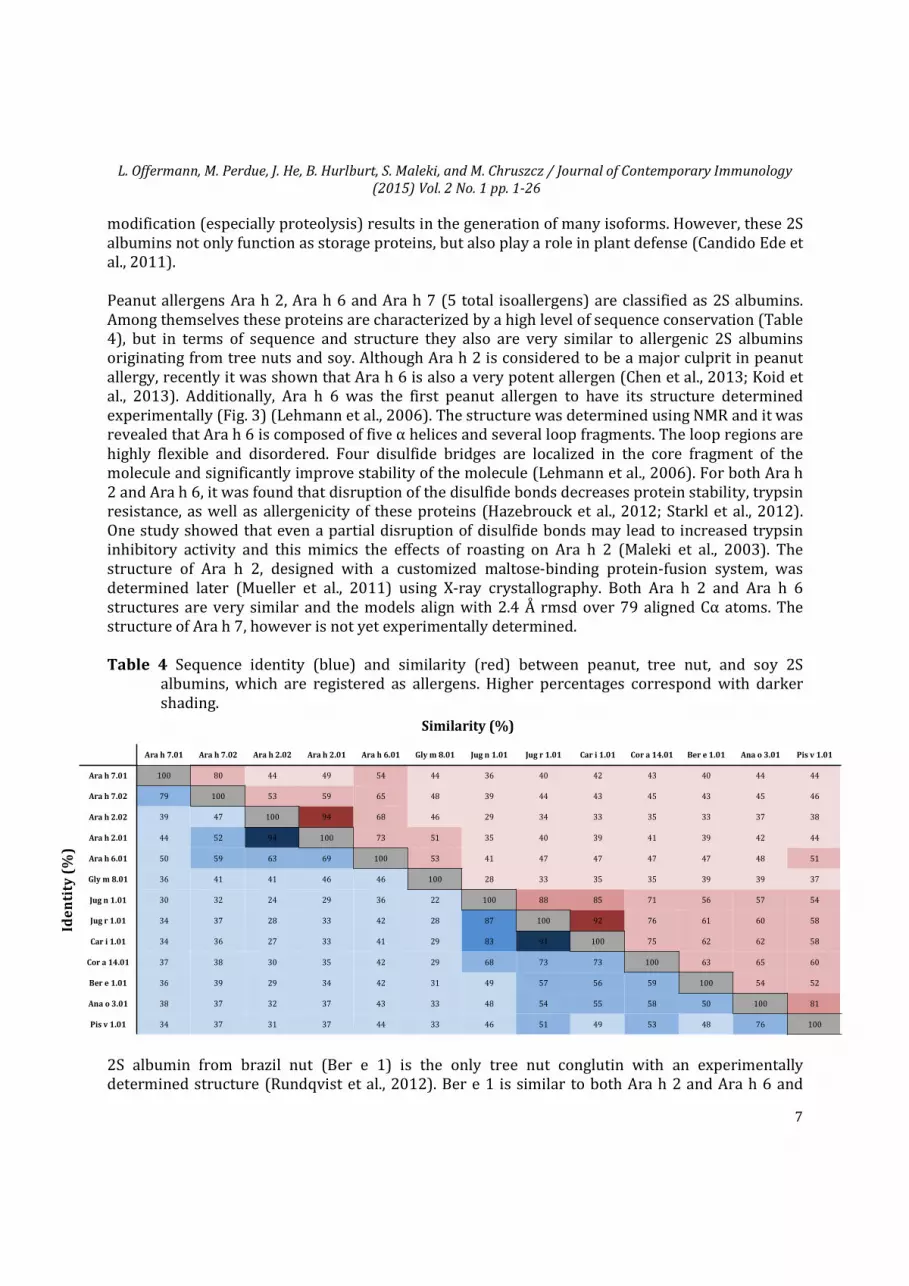
modification (especially proteolysis) results in the generation of many isoforms. However, these 2S albumins not only function as storage proteins, but also play a role in plant defense (Candido Ede et al., 2011). Peanut allergens Ara h 2, Ara h 6 and Ara h 7 (5 total isoallergens) are classified as 2S albumins. Among themselves these proteins are characterized by a high level of sequence conservation (Table 4), but in terms of sequence and structure they also are very similar to allergenic 2S albumins originating from tree nuts and soy. Although Ara h 2 is considered to be a major culprit in peanut allergy, recently it was shown that Ara h 6 is also a very potent allergen (Chen et al., 2013; Koid et al., 2013). Additionally, Ara h 6 was the first peanut allergen to have its structure determined experimentally (Fig. 3) (Lehmann et al., 2006). The structure was determined using NMR and it was revealed that Ara h 6 is composed of five α helices and several loop fragments. The loop regions are highly flexible and disordered. Four disulfide bridges are localized in the core fragment of the molecule and significantly improve stability of the molecule (Lehmann et al., 2006). For both Ara h 2 and Ara h 6, it was found that disruption of the disulfide bonds decreases protein stability, trypsin resistance, as well as allergenicity of these proteins (Hazebrouck et al., 2012; Starkl et al., 2012). One study showed that even a partial disruption of disulfide bonds may lead to increased trypsin inhibitory activity and this mimics the effects of roasting on Ara h 2 (Maleki et al., 2003). The structure of Ara h 2, designed with a customized maltose-binding protein-fusion system, was determined later (Mueller et al., 2011) using X-ray crystallography. Both Ara h 2 and Ara h 6 structures are very similar and the models align with 2.4 Å rmsd over 79 aligned Cα atoms. The structure of Ara h 7, however is not yet experimentally determined.
Table 4 Sequence identity (blue) and similarity (red) between peanut, tree nut, and soy 2S albumins, which are registered as allergens. Higher percentages correspond with darker shading.
2S albumin from brazil nut (Ber e 1) is the only tree nut conglutin with an experimentally determined structure (Rundqvist et al., 2012). Ber e 1 is similar to both Ara h 2 and Ara h 6 and
Ide
nti
ty (
%)
Similarity (%)
Ara h 7.01 Ara h 7.02 Ara h 2.02 Ara h 2.01 Ara h 6.01 Gly m 8.01 Jug n 1.01 Jug r 1.01 Car i 1.01 Cor a 14.01 Ber e 1.01 Ana o 3.01 Pis v 1.01
Ara h 7.01 100 80 44 49 54 44 36 40 42 43 40 44 44
Ara h 7.02 79 100 53 59 65 48 39 44 43 45 43 45 46
Ara h 2.02 39 47 100 94 68 46 29 34 33 35 33 37 38
Ara h 2.01 44 52 94 100 73 51 35 40 39 41 39 42 44
Ara h 6.01 50 59 63 69 100 53 41 47 47 47 47 48 51
Ber e 1.01 36 39 29 34 42 31 49 57 56 59 100 54 52
Ana o 3.01 38 37 32 37 43 33 48 54 55 58 50 100 81
Pis v 1.01 34 37 31 37 44 33 46 51 49 53 48 76 100
L. Offermann, M. Perdue, J. He, B. Hurlburt, S. Maleki, and M. Chruszcz / Journal of Contemporary Immunology
(2015) Vol. 2 No. 1 pp. 1-26
8
their models superpose with rmsds of 2.7 Å (over 82 aligned Cα atoms) and 3.1 Å (over 82 aligned Cα atoms), respectively. Interestingly, due to a high content of methionine and cysteine Ber e 1 was used to boost sulfur content in transgenic plants. This resulted in a transfer of this major Brazil nut allergen into soy (Nordlee et al., 1996). Structural studies of Ber e 1 also revealed that this protein binds Cu2+ ions with a 1:1 stoichiometry. This feature of Ber e 1 is quite unusual as 2S albumins were not associated with aligand binding properties. As already mentioned, both 2S albumins and nsLTP1 belong to the same protein superfamily. Their structure is formed by four or five α helices linked by disulfide bonds. For example, despite very low sequence identity and similarity, structures of Ara h 2 and Pru du 3 (nsLPT1 from almond) superpose well with 3.1 Å rmsd over 67 aligned Cα atoms.
The biological function of Ara h 2, Ara h 6 and Ara h 7 is still not fully understood. While Ara h 2 and Ara h 6 are very abundant, Ara h 7 is present in peanuts only in small quantities and the natural forms of the allergen were identified only recently (Schmidt et al., 2010). Analysis of sequence and structures of peanut 2S albumins suggests that they may function as trypsin or/and amylase inhibitors (Maleki et al., 2003; Schmidt et al., 2010). It was experimentally shown that Ara h 2 is a weak trypsin inhibitor and roasting increased this activity (Maleki et al., 2003). Ara h 2 and Ara h 6 purified from roasted peanuts not only retain their structure and function, but also retain IgE-binding activity of the native protein (Vissers et al., 2011).
Fig. 3. Top: Overall structure of Ara h 6 (PDB ID: 1W2Q). First model from an ensemble deposited to PDB is shown. Secondary structural elements are colored separately where α-helices are colored cyan, and loops are colored magenta. Bottom: Ribbon representation of Ara h 6 showing residue conservation derived from an alignment of related protein sequences. Conservation was calculated using the ConSurf server. The most conserved residues are shown in blue while the more variable residues are shown in red.
L. Offermann, M. Perdue, J. He, B. Hurlburt, S. Maleki, and M. Chruszcz / Journal of Contemporary Immunology
(2015) Vol. 2 No. 1 pp. 1-26
9
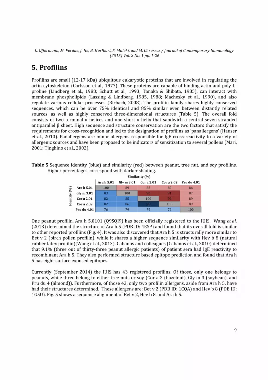
5. Profilins Profilins are small (12-17 kDa) ubiquitous eukaryotic proteins that are involved in regulating the actin cytoskeleton (Carlsson et al., 1977). These proteins are capable of binding actin and poly-L-proline (Lindberg et al., 1988; Schutt et al., 1993; Tanaka & Shibata, 1985), can interact with membrane phospholipids (Lassing & Lindberg, 1985, 1988; Machesky et al., 1990), and also regulate various cellular processes (Birbach, 2008). The profilin family shares highly conserved sequences, which can be over 75% identical and 85% similar even between distantly related sources, as well as highly conserved three-dimensional structures (Table 5). The overall fold consists of two terminal α-helices and one short α-helix that sandwich a central seven-stranded antiparallel β sheet. High sequence and structure conservation are the two factors that satisfy the requirements for cross-recognition and led to the designation of profilins as ‘panallergens’ (Hauser et al., 2010). Panallergens are minor allergens responsible for IgE cross-reactivity to a variety of allergenic sources and have been proposed to be indicators of sensitization to several pollens (Mari, 2001; Tinghino et al., 2002). Table 5 Sequence identity (blue) and similarity (red) between peanut, tree nut, and soy profilins.
Higher percentages correspond with darker shading.
One peanut profilin, Ara h 5.0101 (Q9SQI9) has been officially registered to the IUIS. Wang et al. (2013) determined the structure of Ara h 5 (PDB ID: 4ESP) and found that its overall fold is similar to other reported profilins (Fig. 4). It was also discovered that Ara h 5 is structurally more similar to Bet v 2 (birch pollen profilin), while it shares a higher sequence similarity with Hev b 8 (natural rubber latex profilin)(Wang et al., 2013). Cabanos and colleagues (Cabanos et al., 2010) determined that 9.1% (three out of thirty-three peanut allergic patients) of patient sera had IgE reactivity to recombinant Ara h 5. They also performed structure based epitope prediction and found that Ara h 5 has eight-surface exposed epitopes. Currently (September 2014) the IUIS has 43 registered profilins. Of those, only one belongs to peanuts, while three belong to either tree nuts or soy (Cor a 2 (hazelnut), Gly m 3 (soybean), and Pru du 4 (almond)). Furthermore, of those 43, only two profilin allergens, aside from Ara h 5, have had their structures determined. These allergens are: Bet v 2 (PDB ID: 1CQA) and Hev b 8 (PDB ID: 1G5U). Fig. 5 shows a sequence alignment of Bet v 2, Hev b 8, and Ara h 5.
Similarity (%)
Ide
nti
ty (
%)
Ara h 5.01 Gly m 3.01 Cor a 2.01 Cor a 2.02 Pru du 4.01
Ara h 5.01 100 89 88 89 86
Gly m 3.01 83 100 90 91 87
Cor a 2.01 82 85 100 99 89
Cor a 2.02 82 86 98 100 89
Pru du 4.01 76 79 79 79 100
L. Offermann, M. Perdue, J. He, B. Hurlburt, S. Maleki, and M. Chruszcz / Journal of Contemporary Immunology
(2015) Vol. 2 No. 1 pp. 1-26
10
Fig. 4. Top: Overall structure of Ara h 5 (PDB ID: 4ESP). Secondary structural elements are colored separately where α-helices are colored cyan, β-sheets are colored in red, and loops are colored magenta. Bottom: Ribbon representation of Ara h 5 showing residue conservation derived from an alignment of profilin related protein sequences. Conservation was calculated using the ConSurf server. The most conserved residues are shown in blue while the more variable residues are shown in red.
Fig. 5. Sequence alignment of profilin allergens, whose structures have been determined. Out of 43 registered profilins to the IUIS, only three, Bet v 2 (birch), Hev b 8 (natural rubber latex), and Ara h 5 (peanut) have had their structures determined.
L. Offermann, M. Perdue, J. He, B. Hurlburt, S. Maleki, and M. Chruszcz / Journal of Contemporary Immunology
(2015) Vol. 2 No. 1 pp. 1-26
11
6. Pathogenesis Related Class 10 (PR-10) Proteins Pathogenesis-related protein class 10 class of proteins, or PR10s, belongs to the birch pollen Bet v 1-like superfamily of proteins, which are extensively dispersed among higher plants (Liscombe & Facchini, 2008). Protein expression occurs in high concentrations in reproductive tissues such as pollen, seeds and fruits and is thought to be induced by pathogen attack or abiotic stress (Fernandes et al., 2013). The function of PR10s however, is not fully understood. It has been suggested that some PR10s may play a role in ribonuclease activity (Moiseyev et al., 1997; Park et al., 2004), while other PR10s may be steroid hormone carriers (Hurlburt et al., 2013; Markovic-Housley et al., 2003). The overall structure of PR10s is highly conserved and generally consists of three α-helices that flank a seven-stranded, anti-parallel β-sheet. These proteins typically have a large, hydrophobic cavity that is able to bind small hydrophobic ligands (Fernandes et al., 2013). Table 6 indicates that among peanut, tree nut, and soy PR10s, the sequence similarity is 59% or greater, while the sequence identity is 41% or greater. Table 6 Sequence identity (blue) and similarity (red) between peanut, tree nut, and soy PR10s.
Higher percentages correspond with darker shading.
Allergens belonging to the Bet v 1 allergen family are the main cause of pollen-related food allergies. Contact with these allergens can result in many kinds of allergic reactions, ranging from isolated oral allergy syndrome to life-threatening anaphylactic shock (Glaumann et al., 2013; Kleine-Tebbe et al., 2002). In general, Bet v 1 related allergens are characterized as labile proteins, in contrast to other food allergens, which are more stable upon heating and digestion (Bollen et al., 2010). However, it is worth mentioning, that despite this general characteristic, Ara h 8 which is a peanut PR-10 is stable during purification which involves heating up to 70°C (Hurlburt et al., 2013; Petersen et al., 2014). Two PR-10 proteins, which originate from peanut, are officially registered to the IUIS. These proteins are Ara h 8.0101 (Q6VT83) and Ara h 8.0201 (B0YIU5). Ara h 8 is considered to be a minor peanut allergen (Riecken et al., 2008). However, in the case of birch pollen allergic patients, Ara h 8 is more significant due to its cross-reactivity with Bet v 1 (Mittag et al., 2004). Interestingly, isolated Ara h 8 sensitization was suggested as being associated with no or mild symptoms among peanut-sensitized patients (Asarnoj et al., 2012; Glaumann et al., 2013; Klemans et al., 2014). However, severe reactions have been reported with Ara h 8 recognition by IgE. Also, it is important to note that reactivity to Ara h 8 and 9 are geographically dependent and the severity of the reaction in monosensitized patients to either of these cannot be predicted accurately (Glaumann et al., 2013;
Similarity (%)
Ide
nti
ty (
%)
Ara h 8.01 Gly m 4.01 Cor a 1.0401 Cor a 1.0404 Cor a 1.0402 Cor a 1.0403 Cor a 1.01 Cor a 1.02 Cor a 1.03 Cas s 1.01 Ara h 8.02
Ara h 8.01 100 79 65 65 65 65 60 63 64 63 70 Gly m 4.01 71 100 68 68 67 66 63 65 63 63 66
Cor a 1.01 43 47 63 63 62 62 100 85 77 69 59 Cor a 1.02 49 50 70 70 69 69 75 100 81 70 60 Cor a 1.03 50 52 71 71 70 70 65 71 100 74 61 Cas s 1.01 47 50 59 58 59 59 58 58 64 100 61 Ara h 8.02 53 49 42 42 42 42 41 42 44 46 100
L. Offermann, M. Perdue, J. He, B. Hurlburt, S. Maleki, and M. Chruszcz / Journal of Contemporary Immunology
(2015) Vol. 2 No. 1 pp. 1-26
12
Klemans et al., 2014). Until recently, the function of Ara h 8 was completely unknown. Hurlburt et
al., demonstrated that Ara h 8.0101 has indeed an overall fold similar to Bet v 1 (Fig. 6) and is able to bind different ligands (Hurlburt et al., 2013). Ara h 8.0201, the second isoform, was discovered relatively late (Riecken et al., 2008) and is not well characterized.
Fig. 6. Top: Overall structure of Ara h 8 (PDB ID: 4M9B). Secondary structural elements are colored separately where α-helices are colored cyan, β-sheets are colored in red, and loops are colored magenta. Bottom: Ribbon representation of Ara h 8 showing conserved residues derived from an alignment of PR10 related protein sequences. Conservation was calculated using the ConSurf server. The most conserved residues are shown in blue while the more variable residues are shown in red.
As of September 2014, the IUIS has 23 registered PR10 proteins and an additional 18 registered proteins that belong to the Bet v 1 superfamily. Of the PR10s, Ara h 8.0101 and Ara h 8.0201 belong to peanuts, while three belong to either tree nuts or soy (Cor a 1 (hazelnut), Cas s 4 (chestnut), and Gly m 4 (soybean)). Furthermore, only six PR10 allergens, aside from Ara h 8.0101, have had their structures determined. These allergens are: Api g 1 (celery) (PDB ID: 2BK0), Bet v 1 (birch) (PDB ID: 4A88, 4BK7, 4A84, 1B6F, 4A81, 4A83, 4A86, 4A87, 4A8G), Dau c 1 (carrot) (PDB ID: 2WQL), Fra a 1 (strawberry) (PDB IDs: 2LPX, 4C9C, and 4C9I), Gly m 4 (soybean) (PDB ID: 2K7H), and Pru av 1 (sweet cherry) (PDB Ids: 1E09, and 1H2O). Fig. 7 shows a sequence alignment of PR10 proteins that are allergens and have had their structures determined.
L. Offermann, M. Perdue, J. He, B. Hurlburt, S. Maleki, and M. Chruszcz / Journal of Contemporary Immunology
(2015) Vol. 2 No. 1 pp. 1-26
13
Fig. 7. Sequence alignment of PR10s allergens, whose structures have been determined. Out of 23
registered PR10s to the IUIS, only seven, Ara h 8.0101 (peanut), Gly m 4 (soybean), Fra a 1 (strawberry), Pru av 1 (sweet cherry), Bet v 1 (birch), Api g 1 (celery), and Dau c 1 (carrot) have had their structures determined. Ara h 8.0201 was included to show the differences between isoforms of Ara h 8.
7. Nonspecific Lipid-transfer Proteins (nsLTP1) Nonspecific lipid-transfer proteins (nsLTP), first named for their ability to mediate the transfer of phospholipids between membranes in vitro, are basic proteins with molecular masses of 9 kDa (nsLTP1) and 7 kDa (nsLTP2), which are stabilized by four disulfide bonds (Kader et al, 1984; Douliez et al., 2000). The nsLTP1 family is expressed throughout plants, most notably in the peripheral cell layers surrounding aerial organs (Salcedo et al., 2007). It has been suggested that both families of nonspecific lipid-transfer proteins are involved with defense mechanisms, more specifically the formation of the waxy and polymeric cutin and suberin layers of tissues, and are believed to play a crucial role in protecting plants from bacterial and fungal pathogens (Egger et al., 2010). The 9 kDa plant nsLTPs, or nsLTP1s, became correlated with human allergies after two IgE-binding components in peach and apple were identified as part of the nsLTP1 family, which lead to the identification of allergenic nsLTP1s in other fruits of the Rosacea family (Salcedo et al., 2007). Additional members of the family have been detected in other foods including citrus fruits, tomato, vegetables, nuts, and maize and found to participate in IgE cross-reactions; however, sensitization
L. Offermann, M. Perdue, J. He, B. Hurlburt, S. Maleki, and M. Chruszcz / Journal of Contemporary Immunology
(2015) Vol. 2 No. 1 pp. 1-26
14
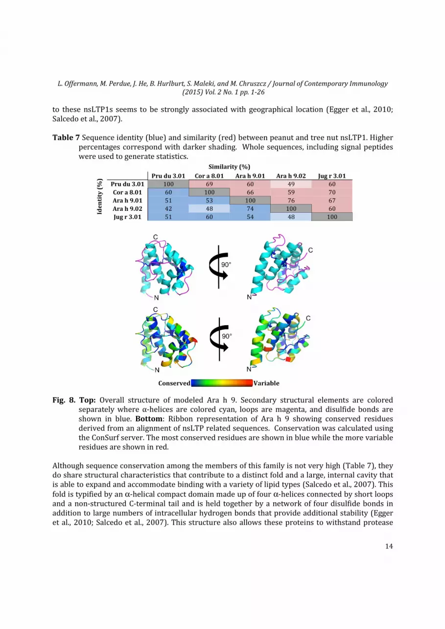
to these nsLTP1s seems to be strongly associated with geographical location (Egger et al., 2010; Salcedo et al., 2007). Table 7 Sequence identity (blue) and similarity (red) between peanut and tree nut nsLTP1. Higher
percentages correspond with darker shading. Whole sequences, including signal peptides were used to generate statistics.
Fig. 8. Top: Overall structure of modeled Ara h 9. Secondary structural elements are colored separately where α-helices are colored cyan, loops are magenta, and disulfide bonds are shown in blue. Bottom: Ribbon representation of Ara h 9 showing conserved residues derived from an alignment of nsLTP related sequences. Conservation was calculated using the ConSurf server. The most conserved residues are shown in blue while the more variable residues are shown in red.
Although sequence conservation among the members of this family is not very high (Table 7), they do share structural characteristics that contribute to a distinct fold and a large, internal cavity that is able to expand and accommodate binding with a variety of lipid types (Salcedo et al., 2007). This fold is typified by an α-helical compact domain made up of four α-helices connected by short loops and a non-structured C-terminal tail and is held together by a network of four disulfide bonds in addition to large numbers of intracellular hydrogen bonds that provide additional stability (Egger et al., 2010; Salcedo et al., 2007). This structure also allows these proteins to withstand protease
Ide
nti
ty (
%)
Similarity (%)
Pru du 3.01 Cor a 8.01 Ara h 9.01 Ara h 9.02 Jug r 3.01
Pru du 3.01 100 69 60 49 60 Cor a 8.01 60 100 66 59 70 Ara h 9.01 51 53 100 76 67 Ara h 9.02 42 48 74 100 60 Jug r 3.01 51 60 54 48 100
L. Offermann, M. Perdue, J. He, B. Hurlburt, S. Maleki, and M. Chruszcz / Journal of Contemporary Immunology
(2015) Vol. 2 No. 1 pp. 1-26
15
and thermal treatment, which is the proposed reasoning behind the prolonged systemic reactions in affected patients (Krause et al., 2009). The nsLTP from peanuts is Ara h 9. Due to its molecular mass of 9.1 kDa, Ara h 9 belongs to the nsLTP1 family. Two isoforms of the protein, designated as Ara h 9.0101 and Ara h 9.0201, were found to have 90% sequence identity (without signal peptides) and reported to have an important role in peanut allergies, especially in people of the Mediterranean area (Bublin & Breiteneder, 2014; Lauer et al., 2009). Although the structure of Ara h 9 is yet to be determined, its sequence was submitted for modeling and the modeled three-dimensional structure is shown in Fig. 8. The IUIS (as of September 2014) has 34 registered nsLTP1 proteins. Of the nsLTP1s, Ara h 9.0101 and Ara h 9.0201 belong to peanuts, while five allergens belong to tree nut (Jug r 3 (English walnut), Pru du 3 (almond), Cor a 8 (hazelnut), and Cas s 8 (chestnut)).
8. Oleosins
Fig. 9. Sequence alignment of oleosins from peanuts (Ara h 10.0101 (Q647G5), Ara h 10.0102
(Q647G4) and Ara h 11.0101 (Q45W87)). The green bar corresponds to SDQTRTGY sequence of IgE epitope responsible for cross-reactivity between peanut and buckwheat. Light blue and purple bars (respectively) correspond to GXSXG and HX4D motifs found in oleosin 3. Blue bar corresponds to proline knot (PX5SPX3P), and orange bar to a transmembrane region predicted for Q647G5 by TopPred (von Heijne, 1992).
L. Offermann, M. Perdue, J. He, B. Hurlburt, S. Maleki, and M. Chruszcz / Journal of Contemporary Immunology
(2015) Vol. 2 No. 1 pp. 1-26
16
Oleosins, are small plant proteins which are responsible for the formation and stability of oil bodies containing triacylglycerides, and they may make up to 20% of total protein in oil rich seeds (Abell et al., 2004). These proteins form a barrier on the surface of oil bodies and prevent them from contacting and coalescing with other oil droplets. Oleosins are composed of three distinct parts which include N-terminal and C-terminal hydrophilic fragments and the central hydrophobic core that is usually composed of ~70 conserved residues (Fig. 9). The central hydrophobic part contain so-called proline knot motif (PX5SPX3P) that is highly conserved across oleosins (Ratnayake & Huang, 1996). This motif has been show to be critical for oil body targeting and insertion (Abell et al., 2004). The N- and C-terminal domains are significantly less conserved than the hydrophobic domain, however sequence analysis of the C-terminal lead recently to a new classification of oleosins (Fang et al., 2014). In the case of peanut oleosin (OLE3; Q647G3) it was demonstrated that the protein has both monoacylglycerol acyltransferase and phospholipase activities (Parthibane et al., 2012). These activities were correlated with the presence of HX4D motif (signature of an acyltransferase) in the C-terminal part and a GXSXG motif (signature motif of phospholipase) in the N-terminal part of the protein (Fig. 9). It was shown that 14 kDa and 16 kDa peanut oleosins melt at approximately 50 °C, while 18 kDa oleosins melt at 59 °C (Cabanos et al., 2011a). This indicates that the thermal stability of these peanut allergens is not as pronounced as for Ara h 1 and Ara h 2. Unfortunately, currently there is no experimental 3D model of any oleosin reported to the PDB. Three oleosins that are currently registered as allergens by the WHO/IUIS Allergen Nomenclature Sub-committee and originate from peanuts are: Ara h 10.0101 (Q647G5), Ara h 10.0102 (Q647G4) and Ara h 11.0101 (Q45W87). The other officially registered allergens from this family include hazelnut allergens (Cor a 12 and Cor a 13) and sesame (Ses i 4 and Ses i 5). Some peanut oleosins were clearly identified as candidates for IgE-mediated reactions (Pons et al., 2002) there are other peanut oleosins that are still not well characterized in terms of their immunologic properties. OLE3 (Q647G3), which is not yet officially registered as an allergen, contains in the N-terminal part a peptide SDQTRTGY that was identified as an IgE epitope (Kobayashi et al., 2012). In addition, it was shown that this peptide is responsible for cross-reactivity between peanut and buckwheat. Oleosins do not only pose a challenge for structural studies, but due to their hydrophobicity are also problematic from the diagnostic point of view, as they are underrepresented in diagnostic extracts (Zuidmeer-Jongejan et al., 2014). Therefore, recombinant versions of these allergens (Cabanos et al., 2011a; Pons et al., 2005) may become especially important in diagnosis of peanut, hazelnut and sesame allergies.
9. Defensins Plant defensins are cysteine-rich, highly stable, small peptides that make up part of the innate immune system against fungal pathogens (Lay & Anderson, 2005). They are expressed in a variety of organs and act as the first line of protection against pathogen attack. Furthermore, they possess antibacterial, antifungal, insect amylase inhibitory, or protease inhibitory activity. Aside from these activities, defensins are also involved in cellular signaling, and growth regulation (Okuda et al., 2009; Stotz et al., 2009; Takayama et al., 2001).
Ara h 12.0101 and Ara h 13.0101 are the two peanut defensins that are registered by the WHO/IUIS Allergen Nomenclature Sub-committee. To date, neither Ara h 12 nor Ara h 13 have had their 3D structures determined experimentally, so both were modeled using Phyre2 (Kelley & Sternberg,
L. Offermann, M. Perdue, J. He, B. Hurlburt, S. Maleki, and M. Chruszcz / Journal of Contemporary Immunology
(2015) Vol. 2 No. 1 pp. 1-26
17
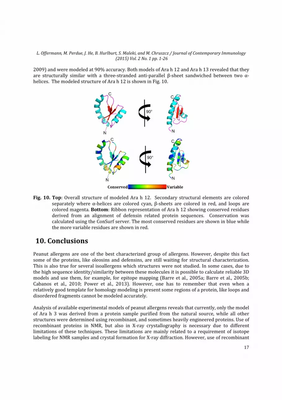
2009) and were modeled at 90% accuracy. Both models of Ara h 12 and Ara h 13 revealed that they are structurally similar with a three-stranded anti-parallel β-sheet sandwiched between two α-helices. The modeled structure of Ara h 12 is shown in Fig. 10.
Fig. 10. Top: Overall structure of modeled Ara h 12. Secondary structural elements are colored separately where α-helices are colored cyan, β-sheets are colored in red, and loops are colored magenta. Bottom: Ribbon representation of Ara h 12 showing conserved residues derived from an alignment of defensin related protein sequences. Conservation was calculated using the ConSurf server. The most conserved residues are shown in blue while the more variable residues are shown in red.
10. Conclusions Peanut allergens are one of the best characterized group of allergens. However, despite this fact some of the proteins, like oleosins and defensins, are still waiting for structural characterization. This is also true for several isoallergens which structures were not studied. In some cases, due to the high sequence identity/similarity between these molecules it is possible to calculate reliable 3D models and use them, for example, for epitope mapping (Barre et al., 2005a; Barre et al., 2005b; Cabanos et al., 2010; Power et al., 2013). However, one has to remember that even when a relatively good template for homology modeling is present some regions of a protein, like loops and disordered fragments cannot be modeled accurately. Analysis of available experimental models of peanut allergens reveals that currently, only the model of Ara h 3 was derived from a protein sample purified from the natural source, while all other structures were determined using recombinant, and sometimes heavily engineered proteins. Use of recombinant proteins in NMR, but also in X-ray crystallography is necessary due to different limitations of these techniques. These limitations are mainly related to a requirement of isotope labeling for NMR samples and crystal formation for X-ray diffraction. However, use of recombinant
L. Offermann, M. Perdue, J. He, B. Hurlburt, S. Maleki, and M. Chruszcz / Journal of Contemporary Immunology
(2015) Vol. 2 No. 1 pp. 1-26
18
proteins does not allow for easy explanation of the impact of posttranslational modifications on protein structure and molecular properties. In addition, some allergens carry various small molecule ligands or metal ions that may additionally affect protein properties like stability and allergenicity. Identification of such physiological ligands is not easy and requires a complex, multi-technique approach. Currently, there is no single structure of a peanut allergen in complex with an antibody or its fragment. Studies of such complexes in combination with information on ability of the complexed antibody to interfere with IgE binding provides an elegant method for characterization of antigenic determinants involved in IgE antibody binding. This approach not only allows for identification of allergen residues that are critical for IgE binding, but also provides an excellent starting point to design recombinant versions of allergens for application in immunotherapy. Having this in mind, we believe that the next step in structural characterization of peanut allergens should involve studies of peanut allergens-antibodies complexes, which is especially important after the recent discovery of IgE cross-reactivity between major peanut allergen Ara h 2 and nonhomologous Ara h 1 and Ara h 3 (Bublin & Breiteneder, 2014).
Acknowledgements We would like to thank Lukasz Lebioda for reading the manuscript and his valuable comments. This work was supported by internal funds from the University of South Carolina and the U.S. Department of Agriculture.
References
Abell, B. M., Hahn, M., Holbrook, L. A., & Moloney, M. M. (2004). Membrane topology and sequence requirements for oil body targeting of oleosin. Plant J, 37(4), 461-470. http://dx.doi.org/10.1111/j.1365-313X.2003.01988.x
Artimo, P., Jonnalagedda, M., Arnold, K., Baratin, D., Csardi, G., de Castro, E., Duvaud, S., Flegel, V., Fortier, A., Gasteiger, E., Grosdidier, A., Hernandez, C., Ioannidis, V., Kuznetsov, D., Liechti, R., Moretti, S., Mostaguir, K., Redaschi, N., Rossier, G., Xenarios, I., & Stockinger, H. (2012). ExPASy: SIB bioinformatics resource portal. Nucleic Acids Res, 40(Web Server issue), W597-603. http://dx.doi.org/10.1093/nar/gks400
Asarnoj, A., Nilsson, C., Lidholm, J., Glaumann, S., Ostblom, E., Hedlin, G., van Hage, M., Lilja, G., & Wickman, M. (2012). Peanut component Ara h 8 sensitization and tolerance to peanut. J Allergy Clin Immunol, 130(2), 468-472. http://dx.doi.org/10.1016/j.jaci.2012.05.019
Barre, A., Borges, J. P., Culerrier, R., & Rouge, P. (2005a). Homology modelling of the major peanut allergen Ara h 2 and surface mapping of IgE-binding epitopes. Immunol Lett, 100(2), 153-158. http://dx.doi.org/10.1016/j.imlet.2005.03.014
Barre, A., Borges, J. P., & Rouge, P. (2005b). Molecular modelling of the major peanut allergen Ara h 1 and other homotrimeric allergens of the cupin superfamily: a structural basis for their IgE-binding cross-reactivity. Biochimie, 87(6), 499-506. http://dx.doi.org/10.1016/j.biochi.2005.02.011
Benson, D. A., Cavanaugh, M., Clark, K., Karsch-Mizrachi, I., Lipman, D. J., Ostell, J., & Sayers, E. W. (2013).
L. Offermann, M. Perdue, J. He, B. Hurlburt, S. Maleki, and M. Chruszcz / Journal of Contemporary Immunology
Berman, H. M., Battistuz, T., Bhat, T. N., Bluhm, W. F., Bourne, P. E., Burkhardt, K., Feng, Z., Gilliland, G. L., Iype, L., Jain, S., Fagan, P., Marvin, J., Padilla, D., Ravichandran, V., Schneider, B., Thanki, N., Weissig, H., Westbrook, J. D., & Zardecki, C. (2002). The Protein Data Bank. Acta Crystallogr D Biol Crystallogr, 58(Pt 6 No 1), 899-907. http://dx.doi.org/10.1107/S0907444902003451
Birbach, A. (2008). Profilin, a multi-modal regulator of neuronal plasticity. Bioessays, 30(10), 994-1002. http://dx.doi.org/10.1002/bies.20822
Bollen, M. A., Wichers, H. J., Helsper, J. P. F. G., Savelkoul, H. F. J., & van Boekel, M. A. J. S. (2010). Thermodynamic characterization of the PR-10 allergens Bet v 1, Api g 1 and Dau c 1 and pH-dependence of nApi g 1 and nDau c 1. Food Chemistry, 119(1), 241-248. http://dx.doi.org/10.1016/j.foodchem.2009.06.013
Breiteneder, H., & Radauer, C. (2004). A classification of plant food allergens. J Allergy Clin Immunol, 113(5), 821-830; quiz 831. http://dx.doi.org/10.1016/j.jaci.2004.01.779
Bublin, M., & Breiteneder, H. (2014). Cross-reactivity of peanut allergens. Curr Allergy Asthma Rep, 14(4), 426.
http://dx.doi.org/10.1007/s11882-014-0426-8
Burks, A. W., & Sampson, H. A. (1993). Food allergies in children. Curr Prob Pediatr, 23, 230-252. http://dx.doi.org/10.1016/0097-3165(93)90015-Z
Burks, A. W., Tang, M., Sicherer, S., Muraro, A., Eigenmann, P. A., Ebisawa, M., Fiocchi, A., Chiang, W., Beyer, K., Wood, R., Hourihane, J., Jones, S. M., Lack, G., & Sampson, H. A. (2012). ICON: food allergy. J Allergy Clin Immunol, 129(4), 906-920.
http://dx.doi.org/10.1016/j.jaci.2012.02.001
Cabanos, C., Katayama, H., Tanaka, A., Utsumi, S., & Maruyama, N. (2011a). Expression and purification of peanut oleosins in insect cells. Protein J, 30(7), 457-463. doi: 10.1007/s10930-011-9351-z http://dx.doi.org/10.1007/s10930-011-9351-z
Cabanos, C., Tandang-Silvas, M. R., Odijk, V., Brostedt, P., Tanaka, A., Utsumi, S., & Maruyama, N. (2010). Expression, purification, cross-reactivity and homology modeling of peanut profilin. Protein Expr Purif, 73(1), 36-45. http://dx.doi.org/10.1016/j.pep.2010.03.005
Cabanos, C., Urabe, H., Tandang-Silvas, M. R., Utsumi, S., Mikami, B., & Maruyama, N. (2011b). Crystal structure of the major peanut allergen Ara h 1. Mol Immunol, 49(1-2), 115-123. http://dx.doi.org/10.1016/j.molimm.2011.08.004
Candido Ede, S., Pinto, M. F., Pelegrini, P. B., Lima, T. B., Silva, O. N., Pogue, R., Grossi-de-Sa, M. F., & Franco, O. L. (2011). Plant storage proteins with antimicrobial activity: novel insights into plant defense mechanisms. FASEB J, 25(10), 3290-3305. http://dx.doi.org/10.1096/fj.11-184291
Carlsson, L., Nystrom, L. E., Sundkvist, I., Markey, F., & Lindberg, U. (1977). Actin polymerizability is influenced by profilin, a low molecular weight protein in non-muscle cells. J Mol Biol, 115(3), 465-483. http://dx.doi.org/10.1016/0022-2836(77)90166-8
Celniker, G., Nimrod, G., Ashkenazy, H., Glaser, F., Martz, E., Mayrose, I., Pupko, T., & Ben-Tal, N. (2013). ConSurf: Using Evolutionary Data to Raise Testable Hypotheses about Protein Function. Israel Journal of Chemistry, 53(3-4), 199-206. http://dx.doi.org/10.1002/ijch.201200096
Chen, X., Wang, Q., El-Mezayen, R., Zhuang, Y., & Dreskin, S. C. (2013). Ara h 2 and Ara h 6 have similar allergenic activity and are substantially redundant. Int Arch Allergy Immunol, 160(3), 251-258.
L. Offermann, M. Perdue, J. He, B. Hurlburt, S. Maleki, and M. Chruszcz / Journal of Contemporary Immunology
(2015) Vol. 2 No. 1 pp. 1-26
20
http://dx.doi.org/10.1159/000341642
Chruszcz, M., Maleki, S. J., Majorek, K. A., Demas, M., Bublin, M., Solberg, R., Hurlburt, B. K., Ruan, S., Mattison, C. P., Breiteneder, H., & Minor, W. (2011). Structural and immunologic characterization of Ara h 1, a major peanut allergen. J Biol Chem, 286(45), 39318-39327. http://dx.doi.org/10.1074/jbc.M111.270132
DeLano, W.L. . The PyMOL Molecular Graphics System. Retrieved from http://www.pymol.org
Douliez, J. P., Michon, T., Elmorjani, K., & Marion, D. (2000). Structure, biological and technological functions of lipid transfer proteins and indolines, the major lipid binding proteins from cereal kernels. J Cereal Sci, 32, 1–20. http://dx.doi.org/10.1006/jcrs.2000.0315
Dunwell, J. M., Purvis, A., & Khuri, S. (2004). Cupins: the most functionally diverse protein superfamily? Phytochemistry, 65(1), 7-17. http://dx.doi.org/10.1016/j.phytochem.2003.08.016
Egger, M., Hauser, M., Mari, A., Ferreira, F., & Gadermaier, G. (2010). The role of lipid transfer proteins in allergic diseases. Curr Allergy Asthma Rep, 10(5), 326-335.
http://dx.doi.org/10.1007/s11882-010-0128-9
Emsley, P., Lohkamp, B., Scott, W. G., & Cowtan, K. (2010). Features and development of Coot. Acta Crystallogr, D66(Pt 4), 486-501. http://dx.doi.org/10.1107/S0907444910007493
Fang, Y., Zhu, R. L., & Mishler, B. D. (2014). Evolution of oleosin in land plants. PLoS One, 9(8), e103806. http://dx.doi.org/10.1371/journal.pone.0103806
Fernandes, H., Michalska, K., Sikorski, M., & Jaskolski, M. (2013). Structural and functional aspects of PR-10 proteins. FEBS J, 280(5), 1169-1199.
http://dx.doi.org/10.1111/febs.12114
Fleischer, D. M. (2007). The natural history of peanut and tree nut allergy. Curr Allergy Asthma Rep, 7(3), 175-181. http://dx.doi.org/10.1007/s11882-007-0018-y
Fung-Leung, W. P., De Sousa-Hitzler, J., Ishaque, A., Zhou, L., Pang, J., Ngo, K., Panakos, J. A., Chourmouzis, E., Liu, F. T., & Lau, C. Y. (1996). Transgenic mice expressing the human high-affinity immunoglobulin (Ig) E receptor alpha chain respond to human IgE in mast cell degranulation and in allergic reactions. J Exp Med, 183(1), 49-56. http://dx.doi.org/10.1084/jem.183.1.49
Glaumann, S., Nopp, A., Johansson, S. G., Borres, M. P., Lilja, G., & Nilsson, C. (2013). Anaphylaxis to peanuts in a 16-year-old girl with birch pollen allergy and with monosensitization to Ara h 8. J Allergy Clin Immunol Pract, 1(6), 698-699. http://dx.doi.org/10.1016/j.jaip.2013.08.010
Hauser, M., Roulias, A., Ferreira, F., & Egger, M. (2010). Panallergens and their impact on the allergic patient. Allergy Asthma Clin Immunol, 6(1), 1.
http://dx.doi.org/10.1186/1710-1492-6-1
Hazebrouck, S., Guillon, B., Drumare, M. F., Paty, E., Wal, J. M., & Bernard, H. (2012). Trypsin resistance of the major peanut allergen Ara h 6 and allergenicity of the digestion products are abolished after selective disruption of disulfide bonds. Mol Nutr Food Res, 56(4), 548-557. http://dx.doi.org/10.1002/mnfr.201100614
Hurlburt, B. K., Offermann, L. R., McBride, J. K., Majorek, K. A., Maleki, S. J., & Chruszcz, M. (2013). Structure and function of the peanut panallergen Ara h 8. J Biol Chem, 288(52), 36890-36901. http://dx.doi.org/10.1074/jbc.M113.517797
Jin, T., Guo, F., Chen, Y. W., Howard, A., & Zhang, Y. Z. (2009). Crystal structure of Ara h 3, a major allergen in
L. Offermann, M. Perdue, J. He, B. Hurlburt, S. Maleki, and M. Chruszcz / Journal of Contemporary Immunology
Kader, J. C., Julienne, M., & Vergnolle, C. (1984). Purification and characterization of a spinach-leaf protein capable of transferring phospholipids from liposomes to mitochondria or chloroplasts. Eur J Biochem, 139(2), 411-416. http://dx.doi.org/10.1111/j.1432-1033.1984.tb08020.x
Kelley, L. A., & Sternberg, M. J. (2009). Protein structure prediction on the Web: a case study using the Phyre server. Nat Protoc, 4(3), 363-371 http://dx.doi.org/10.1038/nprot.2009.2
Kim, J. S., & Sicherer, S. H. (2011). Living with food allergy: allergen avoidance. Pediatr Clin North Am, 58(2), 459-470, xi. http://dx.doi.org/10.1016/j.pcl.2011.02.007
Kliene-Tebbe, J., Wangorsch, A., Vogel, L., Crowell, D. N., Haustein, U-F., & Vieths, S. (2002). Severe oral allergy syndrome and anaphylactic reactions caused by a Bet v 1– related PR-10 protein in soybean, SAM22. J Allergy Clin Immunol, 110(5), 797-804. http://dx.doi.org/10.1067/mai.2002.128946
Klemans, R. J., van Os-Medendorp, H., Blankestijn, M., Bruijnzeel-Koomen, C. A., Knol, E. F., & Knulst, A. C. (2014). Diagnostic accuracy of specific IgE to components in diagnosing peanut allergy: a systematic review. Clin Exp Allergy. http://dx.doi.org/10.1111/cea.12412
Kobayashi, S., Katsuyama, S., Wagatsuma, T., Okada, S., & Tanabe, S. (2012). Identification of a new IgE-binding epitope of peanut oleosin that cross-reacts with buckwheat. Biosci Biotechnol Biochem, 76(6), 1182-1188.
Koid, A. E., Chapman, M. D., Hamilton, R. G., Van Ree, R., Versteeg, S. A., Dreskin, S. C., Koppelman, S. J., & Wuenschmann, S. (2013). Ara h 6 Complements Ara h 2 as an Important Marker for IgE Reactivity to Peanut. J Agric Food Chem. http://dx.doi.org/10.1021/jf4022509
Kolarich, D., & Altmann, F. (2000). N-Glycan analysis by matrix-assisted laser desorption/ionization mass spectrometry of electrophoretically separated nonmammalian proteins: application to peanut allergen Ara h 1 and olive pollen allergen Ole e 1. Anal Biochem, 285(1), 64-75. http://dx.doi.org/10.1006/abio.2000.4737
Koppelman, S. J., Bruijnzeel-Koomen, C. A., Hessing, M., & de Jongh, H. H. (1999). Heat-induced conformational changes of Ara h 1, a major peanut allergen, do not affect its allergenic properties. J Biol Chem, 274(8), 4770-4777. http://dx.doi.org/10.1074/jbc.274.8.4770
Krause, S., Reese, G., Randow, S., Zennaro, D., Quaratino, D., Palazzo, P., Ciardiello, M. A., Petersen, A., Becker, W. M., & Mari, A. (2009). Lipid transfer protein (Ara h 9) as a new peanut allergen relevant for a Mediterranean allergic population. J Allergy Clin Immunol, 124(4), 771-778 e775. http://dx.doi.org/10.1016/j.jaci.2009.06.008
Krissinel, E., & Henrick, K. (2004). Secondary-structure matching (SSM), a new tool for fast protein structure alignment in three dimensions. Acta Crystallogr D Biol Crystallogr, 60(Pt 12 Pt 1), 2256-2268. http://dx.doi.org/10.1107/S0907444904026460
Kulis, M., Pons, L., & Burks, A. W. (2009). In vivo and T cell cross-reactivity between walnut, cashew and peanut. Int Arch Allergy Immunol, 148(2), 109-117. http://dx.doi.org/10.1159/000155741
Lassing, I., & Lindberg, U. (1985). Specific interaction between phosphatidylinositol 4,5-bisphosphate and profilactin. Nature, 314(6010), 472-474. http://dx.doi.org/10.1038/314472a0
L. Offermann, M. Perdue, J. He, B. Hurlburt, S. Maleki, and M. Chruszcz / Journal of Contemporary Immunology
(2015) Vol. 2 No. 1 pp. 1-26
22
Lassing, I., & Lindberg, U. (1988). Specificity of the interaction between phosphatidylinositol 4,5-bisphosphate and the profilin:actin complex. J Cell Biochem, 37(3), 255-267. http://dx.doi.org/10.1002/jcb.240370302
Lauer, I., Dueringer, N., Pokoj, S., Rehm, S., Zoccatelli, G., Reese, G., Miguel-Moncin, M. S., Cistero-Bahima, A., Enrique, E., Lidholm, J., Vieths, S., & Scheurer, S. (2009). The non-specific lipid transfer protein, Ara h 9, is an important allergen in peanut. Clin Exp Allergy, 39(9), 1427-1437. http://dx.doi.org/10.1111/j.1365-2222.2009.03312.x
Lay, F. T., & Anderson, M. A. (2005). Defensins--components of the innate immune system in plants. Curr Protein Pept Sci, 6(1), 85-101. http://dx.doi.org/10.2174/1389203053027575
Lehmann, K., Schweimer, K., Reese, G., Randow, S., Suhr, M., Becker, W. M., Vieths, S., & Rosch, P. (2006). Structure and stability of 2S albumin-type peanut allergens: implications for the severity of peanut allergic reactions. Biochem J, 395(3), 463-472. http://dx.doi.org/10.1042/BJ20051728
Lieberman, J. A., & Sicherer, S. H. (2011). Quality of life in food allergy. Curr Opin Allergy Clin Immunol, 11(3), 236-242. http://dx.doi.org/10.1097/ACI.0b013e3283464cf0
Lindberg, U., Schutt, C. E., Hellsten, E., Tjader, A. C., & Hult, T. (1988). The use of poly(L-proline)-Sepharose in the isolation of profilin and profilactin complexes. Biochim Biophys Acta, 967(3), 391-400. http://dx.doi.org/10.1016/0304-4165(88)90102-X
Liscombe, D. K., & Facchini, P. J. (2008). Evolutionary and cellular webs in benzylisoquinoline alkaloid biosynthesis. Curr Opin Biotechnol, 19(2), 173-180. http://dx.doi.org/10.1016/j.copbio.2008.02.012
Machesky, L. M., Goldschmidt-Clermont, P. J., & Pollard, T. D. (1990). The affinities of human platelet and Acanthamoeba profilin isoforms for polyphosphoinositides account for their relative abilities to inhibit phospholipase C. Cell Regul, 1(12), 937-950.
Maleki, S. J., Chung, S. Y., Champagne, E. T., & Raufman, J. P. (2000a). The effects of roasting on the allergenic properties of peanut proteins. J Allergy Clin Immunol, 106(4), 763-768. http://dx.doi.org/10.1067/mai.2000.109620
Maleki, S. J., Kopper, R. A., Shin, D. S., Park, C. W., Compadre, C. M., Sampson, H., Burks, A. W., & Bannon, G. A. (2000b). Structure of the major peanut allergen Ara h 1 may protect IgE-binding epitopes from degradation. J Immunol, 164(11), 5844-5849. http://dx.doi.org/10.4049/jimmunol.164.11.5844
Maleki, S. J., Viquez, O., Jacks, T., Dodo, H., Champagne, E. T., Chung, S. Y., & Landry, S. J. (2003). The major peanut allergen, Ara h 2, functions as a trypsin inhibitor, and roasting enhances this function. J Allergy Clin Immunol, 112(1), 190-195. http://dx.doi.org/10.1067/mai.2003.1551
Mari, A. (2001). Multiple pollen sensitization: a molecular approach to the diagnosis. Int Arch Allergy Immunol, 125(1), 57-65. http://dx.doi.org/10.1159/000053797
Markovic-Housley, Z., Degano, M., Lamba, D., von Roepenack-Lahaye, E., Clemens, S., Susani, M., Ferreira, F., Scheiner, O., & Breiteneder, H. (2003). Crystal structure of a hypoallergenic isoform of the major birch pollen allergen Bet v 1 and its likely biological function as a plant steroid carrier. J Mol Biol, 325(1), 123-133. http://dx.doi.org/10.1016/S0022-2836(02)01197-X
Mittag, D., Akkerdaas, J., Ballmer-Weber, B. K., Vogel, L., Wensing, M., Becker, W. M., Koppelman, S. J., Knulst, A. C., Helbling, A., Hefle, S. L., Van Ree, R., & Vieths, S. (2004). Ara h 8, a Bet v 1-homologous allergen from peanut, is a major allergen in patients with combined birch pollen and peanut allergy. J Allergy Clin
L. Offermann, M. Perdue, J. He, B. Hurlburt, S. Maleki, and M. Chruszcz / Journal of Contemporary Immunology
Moiseyev, G. P., Fedoreyeva, L. I., Zhuravlev, Y. N., Yasnetskaya, E., Jekel, P. A., & Beintema, J. J. (1997). Primary structures of two ribonucleases from ginseng calluses - New members of the PR-10 family of intracellular pathogenesis-related plant proteins. Febs Letters, 407(2), 207-210. http://dx.doi.org/10.1016/S0014-5793(97)00337-2
Mueller, G. A., Gosavi, R. A., Pomes, A., Wunschmann, S., Moon, A. F., London, R. E., & Pedersen, L. C. (2011). Ara h 2: crystal structure and IgE binding distinguish two subpopulations of peanut allergic patients by epitope diversity. Allergy, 66(7), 878-885. http://dx.doi.org/10.1111/j.1398-9995.2010.02532.x
Nordlee, J. A., Taylor, S. L., Townsend, J. A., Thomas, L. A., & Bush, R. K. (1996). Identification of a Brazil-nut allergen in transgenic soybeans. N Engl J Med, 334(11), 688-692. http://dx.doi.org/10.1056/NEJM199603143341103
Okuda, S., Tsutsui, H., Shiina, K., Sprunck, S., Takeuchi, H., Yui, R., Kasahara, R. D., Hamamura, Y., Mizukami, A., Susaki, D., Kawano, N., Sakakibara, T., Namiki, S., Itoh, K., Otsuka, K., Matsuzaki, M., Nozaki, H., Kuroiwa, T., Nakano, A., Kanaoka, M. M., Dresselhaus, T., Sasaki, N., & Higashiyama, T. (2009). Defensin-like polypeptide LUREs are pollen tube attractants secreted from synergid cells. Nature, 458(7236), 357-361. http://dx.doi.org/10.1038/nature07882
Park, C. J., Kim, K. J., Shin, R., Park, J. M., Shin, Y. C., & Paek, K. H. (2004). Pathogenesis-related protein 10 isolated from hot pepper functions as a ribonuclease in an antiviral pathway. Plant J, 37(2), 186-198. http://dx.doi.org/10.1046/j.1365-313X.2003.01951.x
Parthibane, V., Rajakumari, S., Venkateshwari, V., Iyappan, R., & Rajasekharan, R. (2012). Oleosin is bifunctional enzyme that has both monoacylglycerol acyltransferase and phospholipase activities. J Biol Chem, 287(3), 1946-1954. http://dx.doi.org/10.1074/jbc.M111.309955
Petersen, A., Rennert, S., Kull, S., Becker, W. M., Notbohm, H., Goldmann, T., & Jappe, U. (2014). Roasting and lipid binding provide allergenic and proteolytic stability to the peanut allergen Ara h 8. Biol Chem, 395(2), 239-250. http://dx.doi.org/10.1515/hsz-2013-0206
Pomes, A., Helm, R. M., Bannon, G. A., Burks, A. W., Tsay, A., & Chapman, M. D. (2003). Monitoring peanut allergen in food products by measuring Ara h 1. J Allergy Clin Immunol, 111(3), 640-645. http://dx.doi.org/10.1067/mai.2003.118
Pons, L., Chery, C., Mrabet, N., Schohn, H., Lapicque, F., & Gueant, J. L. (2005). Purification and cloning of two high molecular mass isoforms of peanut seed oleosin encoded by cDNAs of equal sizes. Plant Physiol Biochem, 43(7), 659-668. http://dx.doi.org/10.1016/j.plaphy.2005.06.002
Pons, L., Chery, C., Romano, A., Namour, F., Artesani, M. C., & Gueant, J. L. (2002). The 18 kDa peanut oleosin is a candidate allergen for IgE-mediated reactions to peanuts. Allergy, 57 Suppl 72, 88-93. http://dx.doi.org/10.1034/j.1398-9995.57.s72.16.x
Power, T. D., Ivanciuc, O., Schein, C. H., & Braun, W. (2013). Assessment of 3D models for allergen research. Proteins, 81(4), 545-554. http://dx.doi.org/10.1002/prot.24239
Radauer, C., & Breiteneder, H. (2007). Evolutionary biology of plant food allergens. J Allergy Clin Immunol, 120(3), 518-525. http://dx.doi.org/10.1016/j.jaci.2007.07.024
Radauer, C., Bublin, M., Wagner, S., Mari, A., & Breiteneder, H. (2008). Allergens are distributed into few protein families and possess a restricted number of biochemical functions. J Allergy Clin Immunol,
L. Offermann, M. Perdue, J. He, B. Hurlburt, S. Maleki, and M. Chruszcz / Journal of Contemporary Immunology
Radauer, C., Nandy, A., Ferreira, F., Goodman, R. E., Larsen, J. N., Lidholm, J., Pomes, A., Raulf-Heimsoth, M., Rozynek, P., Thomas, W. R., & Breiteneder, H. (2014). Update of the WHO/IUIS Allergen Nomenclature Database based on analysis of allergen sequences. Allergy, 69(4), 413-419. http://dx.doi.org/10.1111/all.12348
Ratnayake, C., & Huang, A. H. (1996). Oleosins and oil bodies in plant seeds have postulated structures. Biochem J, 317 (Pt 3), 956-958.
Riecken, S., Lindner, B., Petersen, A., Jappe, U., & Becker, W. M. (2008). Purification and characterization of natural Ara h 8, the Bet v 1 homologous allergen from peanut, provides a novel isoform. Biol Chem, 389(4), 415-423. http://dx.doi.org/10.1515/BC.2008.038
Robert, X., & Gouet, P. (2014). Deciphering key features in protein structures with the new ENDscript server. Nucleic Acids Res, 42(Web Server issue), W320-324. http://dx.doi.org/10.1093/nar/gku316
Rundqvist, L., Tengel, T., Zdunek, J., Bjorn, E., Schleucher, J., Alcocer, M. J., & Larsson, G. (2012). Solution structure, copper binding and backbone dynamics of recombinant Ber e 1-the major allergen from Brazil nut. PLoS One, 7(10), e46435. http://dx.doi.org/10.1371/journal.pone.0046435
Salcedo, G., Sanchez-Monge, R., Barber, D., & Diaz-Perales, A. (2007). Plant non-specific lipid transfer proteins: an interface between plant defence and human allergy. Biochim Biophys Acta, 1771(6), 781-791. http://dx.doi.org/10.1016/j.bbalip.2007.01.001
Sanchez-Mejorada, G., & Rosales, C. (1998). Signal transduction by immunoglobulin Fc receptors. J Leukoc Biol, 63(5), 521-533.
Schein, C. H., Ivanciuc, O., & Braun, W. (2005). Common physical-chemical properties correlate with similar structure of the IgE epitopes of peanut allergens. J Agric Food Chem, 53(22), 8752-8759. http://dx.doi.org/10.1021/jf051148a
Schmidt, H., Krause, S., Gelhaus, C., Petersen, A., Janssen, O., & Becker, W. M. (2010). Detection and structural characterization of natural Ara h 7, the third peanut allergen of the 2S albumin family. J Proteome Res, 9(7), 3701-3709. http://dx.doi.org/10.1021/pr1002406
Schmitt, D. A., Nesbit, J. B., Hurlburt, B. K., Cheng, H., & Maleki, S. J. (2010). Processing can alter the properties of peanut extract preparations. J Agric Food Chem, 58(2), 1138-1143. http://dx.doi.org/10.1021/jf902694j
Schutt, C. E., Myslik, J. C., Rozycki, M. D., Goonesekere, N. C., & Lindberg, U. (1993). The structure of crystalline profilin-beta-actin. Nature, 365(6449), 810-816. http://dx.doi.org/10.1038/365810a0
Sen, M., Kopper, R., Pons, L., Abraham, E. C., Burks, A. W., & Bannon, G. A. (2002). Protein structure plays a critical role in peanut allergen stability and may determine immunodominant IgE-binding epitopes. J Immunol, 169(2), 882-887. http://dx.doi.org/10.4049/jimmunol.169.2.882
Shin, D. S., Compadre, C. M., Maleki, S. J., Kopper, R. A., Sampson, H., Huang, S. K., Burks, A. W., & Bannon, G. A. (1998). Biochemical and structural analysis of the IgE binding sites on ara h1, an abundant and highly allergenic peanut protein. J Biol Chem, 273(22), 13753-13759. http://dx.doi.org/10.1074/jbc.273.22.13753
Sicherer, S. H. (2011). Epidemiology of food allergy. J Allergy Clin Immunol, 127(3), 594-602. http://dx.doi.org/10.1016/j.jaci.2010.11.044
Sicherer, S. H., Munoz-Furlong, A., Burks, A. W., & Sampson, H. A. (1999). Prevalence of peanut and tree nut
L. Offermann, M. Perdue, J. He, B. Hurlburt, S. Maleki, and M. Chruszcz / Journal of Contemporary Immunology
(2015) Vol. 2 No. 1 pp. 1-26
25
allergy in the US determined by a random digit dial telephone survey. J Allergy Clin Immunol, 103(4), 559-562. http://dx.doi.org/10.1016/S0091-6749(99)70224-1
Sicherer, S. H., Munoz-Furlong, A., Godbold, J. H., & Sampson, H. A. (2010). US prevalence of self-reported peanut, tree nut, and sesame allergy: 11-year follow-up. J Allergy Clin Immunol, 125(6), 1322-1326. http://dx.doi.org/10.1016/j.jaci.2010.03.029
Sicherer, S. H., & Sampson, H. A. (2014). Food allergy: Epidemiology, pathogenesis, diagnosis, and treatment. J Allergy Clin Immunol, 133(2), 291-307; quiz 308. http://dx.doi.org/10.1016/j.jaci.2013.11.020
Starkl, P., Felix, F., Krishnamurthy, D., Stremnitzer, C., Roth-Walter, F., Prickett, S. R., Voskamp, A. L., Willensdorfer, A., Szalai, K., Weichselbaumer, M., O'Hehir, R. E., & Jensen-Jarolim, E. (2012). An unfolded variant of the major peanut allergen Ara h 2 with decreased anaphylactic potential. Clin Exp Allergy, 42(12), 1801-1812. http://dx.doi.org/10.1111/cea.12031
Stotz, H. U., Spence, B., & Wang, Y. (2009). A defensin from tomato with dual function in defense and development. Plant Mol Biol, 71(1-2), 131-143. http://dx.doi.org/10.1007/s11103-009-9512-z
Takayama, S., Shimosato, H., Shiba, H., Funato, M., Che, F. S., Watanabe, M., Iwano, M., & Isogai, A. (2001). Direct ligand-receptor complex interaction controls Brassica self-incompatibility. Nature, 413(6855), 534-538 http://dx.doi.org/10.1038/35097104
Tanaka, M., & Shibata, H. (1985). Poly(L-proline)-binding proteins from chick embryos are a profilin and a profilactin. Eur J Biochem, 151(2), 291-297. http://dx.doi.org/10.1111/j.1432-1033.1985.tb09099.x
Tandang-Silvas, M. R., Fukuda, T., Fukuda, C., Prak, K., Cabanos, C., Kimura, A., Itoh, T., Mikami, B., Utsumi, S., & Maruyama, N. (2010). Conservation and divergence on plant seed 11S globulins based on crystal structures. Biochim Biophys Acta, 1804(7), 1432-1442. http://dx.doi.org/10.1016/j.bbapap.2010.02.016
Teuber, S. S., & Beyer, K. (2004). Peanut, tree nut and seed allergies. Curr Opin Allergy Clin Immunol, 4(3), 201-203. http://dx.doi.org/10.1097/00130832-200406000-00011
Thompson, J. D., Higgins, D. G., & Gibson, T. J. (1994). CLUSTAL W: improving the sensitivity of progressive multiple sequence alignment through sequence weighting, position-specific gap penalties and weight matrix choice. Nucleic Acids Res, 22(22), 4673-4680. http://dx.doi.org/10.1093/nar/22.22.4673
Tinghino, R., Twardosz, A., Barletta, B., Puggioni, E. M., Iacovacci, P., Butteroni, C., Afferni, C., Mari, A., Hayek, B., Di Felice, G., Focke, M., Westritschnig, K., Valenta, R., & Pini, C. (2002). Molecular, structural, and immunologic relationships between different families of recombinant calcium-binding pollen allergens. J Allergy Clin Immunol, 109(2), 314-320. http://dx.doi.org/10.1067/mai.2002.121528
UniProt, Consortium. (2014). Activities at the Universal Protein Resource (UniProt). Nucleic Acids Res, 42(Database issue), D191-198. http://dx.doi.org/10.1093/nar/gkt1140
van Boxtel, E. L., Koppelman, S. J., van den Broek, L. A., & Gruppen, H. (2008). Determination of pepsin-susceptible and pepsin-resistant epitopes in native and heat-treated peanut allergen Ara h 1. J Agric Food Chem, 56(6), 2223-2230. http://dx.doi.org/10.1021/jf072907n
van Boxtel, E. L., van Beers, M. M., Koppelman, S. J., van den Broek, L. A., & Gruppen, H. (2006). Allergen Ara h
L. Offermann, M. Perdue, J. He, B. Hurlburt, S. Maleki, and M. Chruszcz / Journal of Contemporary Immunology
(2015) Vol. 2 No. 1 pp. 1-26
26
1 occurs in peanuts as a large oligomer rather than as a trimer. J Agric Food Chem, 54(19), 7180-7186. http://dx.doi.org/10.1021/jf061433+
Vissers, Y. M., Blanc, F., Skov, P. S., Johnson, P. E., Rigby, N. M., Przybylski-Nicaise, L., Bernard, H., Wal, J. M., Ballmer-Weber, B., Zuidmeer-Jongejan, L., Szepfalusi, Z., Ruinemans-Koerts, J., Jansen, A. P., Savelkoul, H. F., Wichers, H. J., Mackie, A. R., Mills, C. E., & Adel-Patient, K. (2011). Effect of heating and glycation on the allergenicity of 2S albumins (Ara h 2/6) from peanut. PLoS One, 6(8), e23998. http://dx.doi.org/10.1371/journal.pone.0023998
von Heijne, G. (1992). Membrane protein structure prediction. Hydrophobicity analysis and the positive-inside rule. J Mol Biol, 225(2), 487-494. http://dx.doi.org/10.1016/0022-2836(92)90934-C
Wang, Y., Fu, T. J., Howard, A., Kothary, M. H., McHugh, T. H., & Zhang, Y. (2013). Crystal structure of peanut (Arachis hypogaea) allergen Ara h 5. J Agric Food Chem, 61(7), 1573-1578. http://dx.doi.org/10.1021/jf303861p
Zuidmeer-Jongejan, L., Fernandez-Rivas, M., Winter, M. G., Akkerdaas, J. H., Summers, C., Lebens, A., Knulst, A. C., Schilte, P., Briza, P., Gadermaier, G., & van Ree, R. (2014). Oil body-associated hazelnut allergens including oleosins are underrepresented in diagnostic extracts but associated with severe symptoms. Clin Transl Allergy, 4(1), 4. http://dx.doi.org/10.1186/2045-7022-4-4