ORIGINAL RESEARCH ARTICLE published: 26 September 2014 doi: 10.3389/fncel.2014.00301 Synaptic function is modulated by LRRK2 and glutamate release is increased in cortical neurons of G2019S LRRK2 knock-in mice Dayne A. Beccano-Kelly 1,2 † , Naila Kuhlmann 1,2,3 † , Igor Tatarnikov 1† , Mattia Volta 1,2 , Lise N. Munsie 1,2 , Patrick Chou 1 , Li-Ping Cao 1,2 , Heather Han 1,2 , Lucia Tapia 1,2 , Matthew J. Farrer 1,2,4 † and Austen J. Milnerwood 1,2,5 * † 1 Centre for Applied Neurogenetics, Medical Genetics, University of British Columbia, Vancouver, BC, Canada 2 Djavad Mowafaghian Centre for Brain Health, Faculty of Medicine, University of British Columbia, Vancouver, BC, Canada 3 Graduate Program in Neuroscience, University of British Columbia, Vancouver, BC, Canada 4 Department of Medical Genetics, University of British Columbia, Vancouver, BC, Canada 5 Division of Neurology, University of British Columbia, Vancouver, BC, Canada Edited by: Arianna Maffei, State University of Reviewed by: Ilya Bezprozvanny, University of Texas Southwestern Medical Center at Dallas, USA Joshua L. Plotkin, Northwestern University, USA *Correspondence: Austen J. Milnerwood, Centre for Applied Neurogenetics, Djavad Mowafaghian Centre for Brain Health, University of British Columbia, 2215 Wesbrook Mall, 5th Floor, Vancouver, BC V6T 1Z3, Canada e-mail: [email protected]† These authors have contributed equally to this work. Mutations in Leucine-Rich Repeat Kinase-2 (LRRK2) result in familial Parkinson’s disease and the G2019S mutation alone accounts for up to 30% in some ethnicities. Despite this, the function of LRRK2 is largely undetermined although evidence suggests roles in phosphorylation, protein interactions, autophagy and endocytosis. Emerging reports link loss of LRRK2 to altered synaptic transmission, but the effects of the G2019S mutation upon synaptic release in mammalian neurons are unknown. To assess wild type and mutant LRRK2 in established neuronal networks, we conducted immunocytochemical, electrophysiological and biochemical characterization of >3 week old cortical cultures of LRRK2 knock-out, wild-type overexpressing and G2019S knock-in mice. Synaptic release and synapse numbers were grossly normal in LRRK2 knock-out cells, but discretely reduced glutamatergic activity and reduced synaptic protein levels were observed. Conversely, synapse density was modestly but significantly increased in wild-type LRRK2 overexpressing cultures although event frequency was not. In knock-in cultures, glutamate release was markedly elevated, in the absence of any change to synapse density, indicating that physiological levels of G2019S LRRK2 elevate probability of release. Several pre-synaptic regulatory proteins shown by others to interact with LRRK2 were expressed at normal levels in knock-in cultures; however, synapsin 1 phosphorylation was significantly reduced. Thus, perturbations to the pre-synaptic release machinery and elevated synaptic transmission are early neuronal effects of LRRK2 G2019S. Furthermore, the comparison of knock-in and overexpressing cultures suggests that one copy of the G2019S mutation has a more pronounced effect than an ∼3-fold increase in LRRK2 protein. Mutant-induced increases in transmission may convey additional stressors to neuronal physiology that may eventually contribute to the pathogenesis of Parkinson’s disease. Keywords: LRRK2, LRRK2 mutation, G2019S, Parkinson disease, electrophysiology, cortical culture, transgenic mice INTRODUCTION Despite intense research efforts in the context of Parkinson’s dis- ease (PD), the basic neurophysiology of LRRK2 remains largely unknown. Progress is possibly confounded by numerous poten- tial roles, resulting from LRRK2 being a large multi-domain protein containing ROC, COR, kinase, WD40, and leucine-rich repeats (Cookson, 2010). Roc and COR domains are characteris- tic of the Ras/GTPase (ROCO) signal transductase superfamily, involved in cytoskeleton reorganization and membrane traffic (Mizuno-Yamasaki et al., 2012). Evidence suggests LRRK2 kinase substrates include tau (Kawakami et al., 2012), endophilin A (Matta et al., 2012), 4E-BP (Lee et al., 2010, 2012) and LRRK2 itself; autophosphorylation regulates its GTPase and kinase activities (Webber et al., 2011). There is consensus between sev- eral neuronal culture studies regarding LRRK2-dependent neu- ritic regeneration phenotypes; axon/dendrite outgrowth are most often exaggerated by LRRK2 loss and retarded by mutant over- expression (MacLeod et al., 2006; Plowey et al., 2008; Parisiadou et al., 2009; Dachsel et al., 2010; Lee et al., 2010; Lin et al., 2010; Chan et al., 2011; Ramonet et al., 2011; Winner et al., 2011; Kawakami et al., 2012). However, others have found that neu- rite phenotypes are robust only during the first week in vitro (Sepulveda et al., 2013). Comparatively little LRRK2 is expressed during this period, whereas LRRK2 levels ∼double between the first and second week, both in vitro and in vivo (Biskup et al., 2007; Piccoli et al., 2011), during which time glutamatergic Frontiers in Cellular Neuroscience www.frontiersin.org September 2014 | Volume 8 | Article 301 | 1 CELLULAR NEUROSCIENCE New York Stony Brook, USA
Transcript
ORIGINAL RESEARCH ARTICLEpublished: 26 September 2014doi: 10.3389/fncel.2014.00301
Synaptic function is modulated by LRRK2 and glutamaterelease is increased in cortical neurons of G2019S LRRK2knock-in miceDayne A. Beccano-Kelly1,2 †, Naila Kuhlmann1,2,3 †, Igor Tatarnikov1†, Mattia Volta1,2, Lise N. Munsie1,2,
Patrick Chou1, Li-Ping Cao1,2, Heather Han1,2, Lucia Tapia1,2, Matthew J. Farrer1,2,4 † and
Austen J. Milnerwood1,2,5*†
1 Centre for Applied Neurogenetics, Medical Genetics, University of British Columbia, Vancouver, BC, Canada2 Djavad Mowafaghian Centre for Brain Health, Faculty of Medicine, University of British Columbia, Vancouver, BC, Canada3 Graduate Program in Neuroscience, University of British Columbia, Vancouver, BC, Canada4 Department of Medical Genetics, University of British Columbia, Vancouver, BC, Canada5 Division of Neurology, University of British Columbia, Vancouver, BC, Canada
Edited by:
Arianna Maffei, State University of
Reviewed by:
Ilya Bezprozvanny, University ofTexas Southwestern Medical Centerat Dallas, USAJoshua L. Plotkin, NorthwesternUniversity, USA
*Correspondence:
Austen J. Milnerwood, Centre forApplied Neurogenetics, DjavadMowafaghian Centre for BrainHealth, University of BritishColumbia, 2215 Wesbrook Mall,5th Floor, Vancouver, BC V6T 1Z3,Canadae-mail: [email protected]
†These authors have contributedequally to this work.
Mutations in Leucine-Rich Repeat Kinase-2 (LRRK2) result in familial Parkinson’s diseaseand the G2019S mutation alone accounts for up to 30% in some ethnicities. Despitethis, the function of LRRK2 is largely undetermined although evidence suggests roles inphosphorylation, protein interactions, autophagy and endocytosis. Emerging reports linkloss of LRRK2 to altered synaptic transmission, but the effects of the G2019S mutationupon synaptic release in mammalian neurons are unknown. To assess wild type andmutant LRRK2 in established neuronal networks, we conducted immunocytochemical,electrophysiological and biochemical characterization of >3 week old cortical cultures ofLRRK2 knock-out, wild-type overexpressing and G2019S knock-in mice. Synaptic releaseand synapse numbers were grossly normal in LRRK2 knock-out cells, but discretelyreduced glutamatergic activity and reduced synaptic protein levels were observed.Conversely, synapse density was modestly but significantly increased in wild-type LRRK2overexpressing cultures although event frequency was not. In knock-in cultures, glutamaterelease was markedly elevated, in the absence of any change to synapse density,indicating that physiological levels of G2019S LRRK2 elevate probability of release. Severalpre-synaptic regulatory proteins shown by others to interact with LRRK2 were expressedat normal levels in knock-in cultures; however, synapsin 1 phosphorylation was significantlyreduced. Thus, perturbations to the pre-synaptic release machinery and elevated synaptictransmission are early neuronal effects of LRRK2 G2019S. Furthermore, the comparisonof knock-in and overexpressing cultures suggests that one copy of the G2019S mutationhas a more pronounced effect than an ∼3-fold increase in LRRK2 protein. Mutant-inducedincreases in transmission may convey additional stressors to neuronal physiology that mayeventually contribute to the pathogenesis of Parkinson’s disease.
INTRODUCTIONDespite intense research efforts in the context of Parkinson’s dis-ease (PD), the basic neurophysiology of LRRK2 remains largelyunknown. Progress is possibly confounded by numerous poten-tial roles, resulting from LRRK2 being a large multi-domainprotein containing ROC, COR, kinase, WD40, and leucine-richrepeats (Cookson, 2010). Roc and COR domains are characteris-tic of the Ras/GTPase (ROCO) signal transductase superfamily,involved in cytoskeleton reorganization and membrane traffic(Mizuno-Yamasaki et al., 2012). Evidence suggests LRRK2 kinasesubstrates include tau (Kawakami et al., 2012), endophilin A(Matta et al., 2012), 4E-BP (Lee et al., 2010, 2012) and LRRK2itself; autophosphorylation regulates its GTPase and kinase
activities (Webber et al., 2011). There is consensus between sev-eral neuronal culture studies regarding LRRK2-dependent neu-ritic regeneration phenotypes; axon/dendrite outgrowth are mostoften exaggerated by LRRK2 loss and retarded by mutant over-expression (MacLeod et al., 2006; Plowey et al., 2008; Parisiadouet al., 2009; Dachsel et al., 2010; Lee et al., 2010; Lin et al., 2010;Chan et al., 2011; Ramonet et al., 2011; Winner et al., 2011;Kawakami et al., 2012). However, others have found that neu-rite phenotypes are robust only during the first week in vitro(Sepulveda et al., 2013). Comparatively little LRRK2 is expressedduring this period, whereas LRRK2 levels ∼double between thefirst and second week, both in vitro and in vivo (Biskup et al.,2007; Piccoli et al., 2011), during which time glutamatergic
Frontiers in Cellular Neuroscience www.frontiersin.org September 2014 | Volume 8 | Article 301 | 1
Beccano-Kelly et al. Mutant LRRK2 alters glutamate release
synaptogenesis and synapse maturation occur. In light of this, wesought to investigate LRRK2 manipulations in relatively matureneuronal networks containing functional glutamatergic synapses(>18 days in vitro; DIV18).
Synaptic transmission and LRRK2 have been studied at theDrosophila neuromuscular junction where LRRK2 loss results insynaptic bouton overgrowth, and overexpression has the oppositeeffect (Lee et al., 2010) leading to exaggerated phosphorylationof the vesicle cycle regulator endophilin A (endoA), impairedendocytosis and a reduced capacity for repeated synaptic release(Matta et al., 2012). LRRK2 binds several synaptic vesicle cycleproteins, including adaptor proteins 1 and 2, alpha-actinin 2,the clathrin coat assembly protein AP180, synapsin 1, VAMP2,SNAP25, dynamin 1 and synaptophysin (Piccoli et al., 2011,2014). In cultured cortical neurons, acute RNAi knock-down ofLRRK2 increases glutamatergic release probability (Pr), vesiclemotility and recycling (Piccoli et al., 2011); conversely decreasedglutamate release is reported in LRRK2 KO mouse pups at post-natal day 15 (Parisiadou et al., 2014). The reports of knock-downand KO in mammalian cortical neurons and brain slices sug-gest that LRRK2 acts at the synaptic terminal altering glutamaterelease (Piccoli et al., 2011; Parisiadou et al., 2014); however, thefact that these reports are directly contradictory necessitates fur-ther examination. Here we aimed to investigate LRRK2 synapticphysiology in the context of loss of function and gain of function,against which to compare and contrast LRRK2 mutant effects.The data presented here are, to the best of our knowledge, the firstinvestigation of glutamatergic transmission in primary neuronalcultures derived from LRRK2 transgenic overexpressing (OE),knock-out (KO) and knock-in (KI) mice.
We found that LRRK2 levels differentially regulate glutamater-gic synapse density and activity in neuronal cultures from KO andOE mice. Furthermore, glutamate release was increased, and pre-synaptic regulatory protein chemistry was disturbed, in corticalneurons from KI mice. The data demonstrate that endogenousexpression of the most common known genetic cause of PDhas marked effects upon neuronal physiology. Such neuronaldysfunction, manifested as increased activity, may place an exces-sive demand upon the neuronal network that may eventuallycontribute to the pathogenesis of PD.
EXPERIMENTAL METHODSTRANSGENIC MICE AND CULTURE PREPARATIONNon-transgenic littermate (NT) C57BL/6J mice and transgenichuman wild-type LRRK2 BAC overexpressing (OE) (Melroseet al., 2010), knock-out (KO) (Hinkle et al., 2012) and G2019Sknock-in (KI) mice were maintained according to CanadianCouncil on Animal Care regulations. KI mice were generated byinserting a floxed PGK neomycin (PGK-neo) cassette betweenmurine LRRK2 exons 41 and 42, using C57BL/6 genomic DNAtemplate. In this process the targeting 5′ homology arm replacedtwo nucleotides c.6055G>A & c.6057G>C (numbered from theATG start codon of mouse reference sequence NM_025730).Hence, the native nucleotide sequence of murine exon 41 GACTAC GGG ATC GCA (encoding -DYGIA-) was mutated to GACTAC AGC ATC GCA (mutated nucleotides underlined; encod-ing -DYSIA-) to encode the p.G2019S pathogenic substitution.
Targeting, ES clone selection, blastocyst injection and breedingof ES cell chimeras was performed to obtain germline trans-mission by Ozgene Pty Ltd. (www.ozgene.com). The PGK-neocassette was removed by crossing with Cre-deletor mice and theCre transgene was removed in subsequent breeding. The constitu-tive KI animals have subsequently been maintained on C57BL/6Jstock (>10 generations). Heterozygote (HET) KO × HET KObreeds yielded homozygous KO and NT pups, HET OE × NTyielded HET OE pups and NT littermates. HET KI × HET KIbreeds yielded homozygous, heterozygous KI and NT littermates.Primary neuronal cultures were prepared from timed pregnancydames from background strain C57BL/6 mice with the aforemen-tioned transgenic crosses. Tails from each embryo were genotypedduring the single-pup neuronal isolation, before cells were pooledby genotype and plated. Only heterozygous KI mice that faithfullyreproduce the human condition were included and are hereinreferred to as KI. Cultures were prepared as previously described(Milnerwood et al., 2012); briefly, cortical neurons were isolatedfrom pups at E16.5, brains were removed and placed on ice inHank’s Balanced Salt Solution (HBSS, GIBCO). 24-well plateswere seeded at 115 k cells/well in 1 ml plating medium (PM,2% B27+1/100 penicillin/streptomycin, Invitrogen; 0.5 mM α-glutamine; neurobasal medium, GIBCO). From DIV4, 10% ofmedia was added every 3–5 days.
IMMUNOSTAINING AND IMAGE ANALYSISFor immunocytochemistry DIV21-26 cells were fixed in 4%paraformaldehyde (PFA) + 4% sucrose for 10 min then per-meabilized with methanol (3 min at −20◦C), and blocked (3 ×20 min washes of 10% normal goat serum, NGS, in PBS). Primaryantibodies were incubated overnight with agitation at 4◦C inPBST plus 2% NGS before blocking again (10% NGS+PBS)for 1 h at RT, then applying secondary antibody (in PBST +2% NGS) for 0.5 h (incubated at RT for 1 h, then washed3× with PBST before 1.5 h at RT with secondary antibodies(α-mouse Alexa-488, α-rabbit Alexa 568, α-guinea pig Alexa633 and α-chicken AMCA from Molecular probes and JacksonLaboratories). Primary antibodies were α-microtubule associ-ated protein 2 (MAP2, chicken, abcam, ab5392, 1:2500), α-post-synaptic density protein 95 (PSD-95, mouse, Thermo Scientific,MA1045, 1:1000), α-vesicular glutamate transporter 1 (VGLUT1,guinea pig, Chemicon, AB 5905, 1:4000) and α-synapsin1(Synapsin1, rabbit, Millipore, ab1543p, 1:500). Coverslips wereslide mounted with fluoromount (SouthernBiotech) and allimages were acquired on an Olympus Fluoview 1000 confo-cal microscope as 0.4 μm z-stacks at 60× magnification (flat-tened with the max projection function for cluster analysis)and at 40× for cell density counts. Single 60× images (clus-ter analyses) or mosaics of 40× images were used for anal-yses. Excitation and acquisition parameters were constrainedacross all paired (culture) acquisitions. For co-localization anal-ysis, stacks in all three channels contained all visible dendriticMAP2 staining and images were manually thresholded and bina-rised with experimenter blind, and quantification was conductedon masked dendritic image fields (MAP2 was expanded 5 pix-els to catch pre-synaptic elements). Cluster colocalization wascalculated as percentage of PSD95 clusters overlapping with
Frontiers in Cellular Neuroscience www.frontiersin.org September 2014 | Volume 8 | Article 301 | 2
Beccano-Kelly et al. Mutant LRRK2 alters glutamate release
VGluT1 clusters in Cell Profiler (analysis pipelines availableon request) and data expressed as mean ± s.e.m., where nis average per image field (5–15 images per culture) from aminimum of 3 independent cultures (culture n in brackets).For neuronal densities, soma counts (by MAP2) were manuallyproduced with experimenter blind to genotype on 5–6 imagefields from a minimum of 3 independent cultures (culture n inbrackets).
ELECTROPHYSIOLOGYWhole-cell patch-clamp recordings were performed on corticalcells at DIV21–26 in voltage clamp at Vh −70 mV and the mem-brane test function was used to determine intrinsic membraneproperties ∼1 min after obtaining whole-cell configuration, asdescribed previously (Tapia et al., 2011; Kaufman et al., 2012;Milnerwood et al., 2012; Brigidi et al., 2014). Briefly, neurons wereperfused at room temperature with extracellular solution (ECS)containing (in mM unless stated): 167 NaCl, 2.4 KCl, 1 MgCl2,10 glucose, 10 HEPES, 2 CaCl2, pH 7.4, 300 mOsm. Tetrodotoxin(TTX, 0.2 μM), and picrotoxin (PTX, 100 μM, except when ana-lyzing GABA currents) were added before use. Pipette resistance(Rp) was 3–6 M� when filled with (in mM): 130 Cs methanesul-fonate, 5 CsCl, 4 NaCl, 1 MgCl2, 5 EGTA, 10 HEPES, 5 QX-314,0.5 GTP, 10 Na2-phosphocreatine, and 5 MgATP, 0.1 spermine,pH 7.2, 290 mOsm. Data were acquired by Multiclamp 700 Bamplifier and signals were filtered at 2 kHz, digitized at 10 kHz,and analyzed in Clampfit10 (Molecular Devices). Tolerance forseries resistance (Rs) was <25 M� and uncompensated; �Rs tol-erance cut-off was <10%. mEPSCs and mIPSCs were analyzedwith experimenter blind to genotype using Clampfit10 (threshold5pA mEPSC, 10pA mIPSC); all events were checked by eye andmonophasic events were used for amplitude and decay kinetics,while others were suppressed but included in frequency counts,as in Tapia et al. (2011), Kaufman et al. (2012), Milnerwood et al.(2012), Brigidi et al. (2014). Data are presented as mean ± s.e.m.where n is cells from a minimum of 3 separate cultures (culture nin brackets).
WESTERN BLOTCultures were scraped from individual coverslips at DIV21 in100 μl of LDS loading buffer (Life Technologies). 15 μl of lysatewas resolved using SDS-PAGE on a NuPage 4–12% Bis-Tris gel(Life technologies) and transferred onto a PVDF membrane(Millipore) at 25 V for 100 min at room temperature. Membraneswere incubated overnight in 2% BSA/TBST with the followingprimary antibodies; α-Endophilin A (EndoA, mouse, ThermoScientific, WH0006456M1, 1:1000), α-vesicle associated mem-brane protein 1 (VAMP1, rabbit, Synaptic Systems, 104 002,1:2500), α-vesicle associated membrane protein 2 (VAMP2, rab-bit, Synaptic Systems, 104 202, 1:2500), α-synaptojanin 1 (SynJ1,rabbit, Synaptic Systems, 145 003, 1:1000), α-dynamin 1 (DNM1,rabbit, Thermo Scientific, PA1-660, 1:1000), α-synapsin 1 (rab-bit, Millipore, AB1543P, 1:5000), α-phophoserine 9 synapsin 1(rabbit, Thermo Scientific, PA1-4604, 1:2500), α-phosphoserine603 synapsin 1 (rabbit, Cell Signaling, 2311, 1:2500). Secondaryantibodies (HRP conjugated α-mouse and α-rabbit, Santa CruzBiotechnology) were used at 1:5000. BIO-RAD ChemiDoc™ MP
Imaging System was used to detect the signal, which was quanti-fied using inbuilt Image Lab software (Bio-Rad).
STATISTICAL ANALYSESAnalyses were performed using Prism6 software (Graphpad,Inc.). Direct comparisons were made by Student’s t-test (2- tailed,herein t-test) and multiple comparisons by appropriate analysis ofvariance (ANOVA) and post-tests as detailed in the text.
RESULTSLRRK WILD TYPE, OVEREXPRESSION AND G2019S MUTANT LEVELS INCORTICAL NEURON CULTURESAs LRRK2 is implicated in PD, a disease characterized by nigros-triatal dysfunction, we concluded it would be appropriate to studyLRRK2’s synaptic activity in cortical cells (CTX) given their inputinto the striatum is modulated by nigrostriatal dopamine.
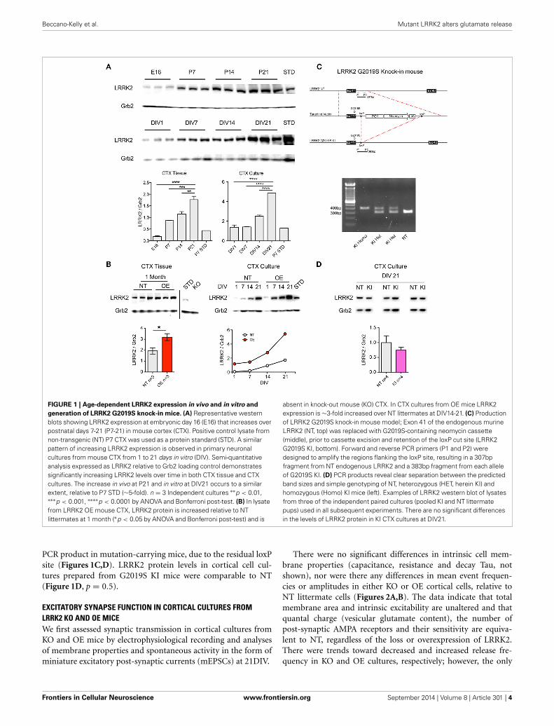
Similarly to increases previously reported in whole-brain lysate(Biskup et al., 2007), western blotting for LRRK2 protein showedthat levels increase rapidly during the 2nd and 3rd postnatalweek in the cortex of non-transgenic (NT) mice (Figure 1A). Weobserved that this pattern is preserved in primary neuronal cul-tures of CTX cells over the first 3 weeks in vitro (Figure 1A), inagreement with others (Piccoli et al., 2011). As LRRK2 proteinlevels are relatively low over the first week, and because neuritephenotypes may be lost by the second week in vitro (Sepulvedaet al., 2013), we decided to investigate the effects of LRRK2manipulations upon synaptic function in neuronal networks ofcortical cultures aged >21DIV. LRRK2 is absent in cortical tissuefrom LRRK2 knock out (KO) mice (Figure 1B) and LRRK2 levelsare 2–3-fold increased in human wild-type LRRK2 overexpress-ing (OE) mouse cortex (p = 0.04) and this pattern is maintainedin cortical cell cultures (Figure 1B). In order to study the effects ofLRRK2 mutations in a genetically and physiologically appropriatemanner, we generated G2019S knock-in (KI) mice (Figure 1C).Founders were backcrossed onto our in-house strain >20 gener-ations and, as predicted from similar lines (Herzig et al., 2011);our LRRK2 KI mice are viable, healthy and breed well. Successfulmutation of the endogenous mouse LRRK2 gene, by insertionand subsequent removal of the cassette, results in a slightly longer
Frontiers in Cellular Neuroscience www.frontiersin.org September 2014 | Volume 8 | Article 301 | 3
Beccano-Kelly et al. Mutant LRRK2 alters glutamate release
FIGURE 1 | Age-dependent LRRK2 expression in vivo and in vitro and
generation of LRRK2 G2019S knock-in mice. (A) Representative westernblots showing LRRK2 expression at embryonic day 16 (E16) that increases overpostnatal days 7-21 (P7-21) in mouse cortex (CTX). Positive control lysate fromnon-transgenic (NT) P7 CTX was used as a protein standard (STD). A similarpattern of increasing LRRK2 expression is observed in primary neuronalcultures from mouse CTX from 1 to 21 days in vitro (DIV). Semi-quantitativeanalysis expressed as LRRK2 relative to Grb2 loading control demonstratessignificantly increasing LRRK2 levels over time in both CTX tissue and CTXcultures. The increase in vivo at P21 and in vitro at DIV21 occurs to a similarextent, relative to P7 STD (∼5-fold). n = 3 Independent cultures ∗∗p < 0.01,∗∗∗p < 0.001, ∗∗∗∗p < 0.0001 by ANOVA and Bonferroni post-test. (B) In lysatefrom LRRK2 OE mouse CTX, LRRK2 protein is increased relative to NTlittermates at 1 month (∗p < 0.05 by ANOVA and Bonferroni post-test) and is
absent in knock-out mouse (KO) CTX. In CTX cultures from OE mice LRRK2expression is ∼3-fold increased over NT littermates at DIV14-21. (C) Productionof LRRK2 G2019S knock-in mouse model; Exon 41 of the endogenous murineLRRK2 (NT, top) was replaced with G2019S-containing neomycin cassette(middle), prior to cassette excision and retention of the loxP cut site (LRRK2G2019S KI, bottom). Forward and reverse PCR primers (P1 and P2) weredesigned to amplify the regions flanking the loxP site, resulting in a 307bpfragment from NT endogenous LRRK2 and a 383bp fragment from each alleleof G2019S KI. (D) PCR products reveal clear separation between the predictedband sizes and simple genotyping of NT, heterozygous (HET, herein KI) andhomozygous (Homo) KI mice (left). Examples of LRRK2 western blot of lysatesfrom three of the independent paired cultures (pooled KI and NT littermatepups) used in all subsequent experiments. There are no significant differencesin the levels of LRRK2 protein in KI CTX cultures at DIV21.
PCR product in mutation-carrying mice, due to the residual loxPsite (Figures 1C,D). LRRK2 protein levels in cortical cell cul-tures prepared from G2019S KI mice were comparable to NT(Figure 1D, p = 0.5).
EXCITATORY SYNAPSE FUNCTION IN CORTICAL CULTURES FROMLRRK2 KO AND OE MICEWe first assessed synaptic transmission in cortical cultures fromKO and OE mice by electrophysiological recording and analysesof membrane properties and spontaneous activity in the form ofminiature excitatory post-synaptic currents (mEPSCs) at 21DIV.
There were no significant differences in intrinsic cell mem-brane properties (capacitance, resistance and decay Tau, notshown), nor were there any differences in mean event frequen-cies or amplitudes in either KO or OE cortical cells, relative toNT littermate cells (Figures 2A,B). The data indicate that totalmembrane area and intrinsic excitability are unaltered and thatquantal charge (vesicular glutamate content), the number ofpost-synaptic AMPA receptors and their sensitivity are equiva-lent to NT, regardless of the loss or overexpression of LRRK2.There were trends toward decreased and increased release fre-quency in KO and OE cultures, respectively; however, the only
Frontiers in Cellular Neuroscience www.frontiersin.org September 2014 | Volume 8 | Article 301 | 4
Beccano-Kelly et al. Mutant LRRK2 alters glutamate release
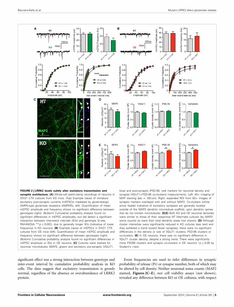
FIGURE 2 | LRRK2 levels subtly alter excitatory transmission and
synaptic architecture. (A) Whole-cell patch-clamp recordings of neurons inDIV21 CTX cultures from KO mice. (Top) Example traces of miniatureexcitatory post-synaptic currents (mEPSCs) mediated by glutamatergicAMPA-type glutamate receptors (AMPARs, left). Quantification of meanmEPSC amplitude and frequency shows no significant difference betweengenotypes (right). (Bottom) Cumulative probability analysis found nosignificant differences in mEPSC amplitudes, but did detect a significantinteraction between inter-event intervals (IEIs) and genotype (2-wayRM-ANOVA ∗∗∗p ≤ 0.001), due to generally longer IEIs (indicative of lowerfrequency) in KO neurons. (B) Example traces of mEPSCs in DIV21 CTXcultures from OE mice (left). Quantification of mean mEPSC amplitude andfrequency shows no significant difference between genotypes (right).(Bottom) Cumulative probability analysis found no significant differences inmEPSC amplitues or IEIs in OE neurons. (C) Cultures were stained forneuronal microtubules (MAP2, green) and excitatory pre-synaptic (VGluT1,
blue) and post-synaptic (PSD-95, red) markers for neuronal density andsynapse (VGluT1+PSD-95 co-clusters) measurements. Left: 40× imaging ofMAP staining (bar = 100 um). Right: expanded ROI from 63× images ofsynaptic markers overlayed with and without MAP2. Co-clusters (whitearrow heads) indicative of excitatory synapses are generally locatedoutside of the MAP2 dendritic microtubule scaffold, upon dendritic spinesthat do not contain microtubules. (D,E) Both KO and OE neuronal densitieswere similar to those of their respective NT littermate cultures (by MAP2soma counts) as were their total dendritic areas (not shown). (D) Althoughcluster intensities were significantly reduced in KO cultures (see text) andthey exhibited a trend toward fewer synapses, there were no significantdifferences in the density or size of VGluT1 clusters, PSD-95 clusters orco-clusters. (E) In OE neurons, there was no significant difference inVGluT1 cluster density, despite a strong trend. There were significantlymore PSD95 clusters and synaptic co-clusters in OE neurons ∗p ≤ 0.05 byStudent’s t-test.
significant effect was a strong interaction between genotype andinter-event interval by cumulative probability analysis in KOcells. The data suggest that excitatory transmission is grosslynormal, regardless of the absence or overabundance of LRRK2protein.
Event frequencies are used to infer differences in synapticprobability of release (Pr) or synapse number, both of which maybe altered by cell density. Neither neuronal soma counts (MAP2stained, Figures 2C–E), nor cell viability assays (not shown),revealed any difference between KO or OE cultures, with respect
Frontiers in Cellular Neuroscience www.frontiersin.org September 2014 | Volume 8 | Article 301 | 5
Beccano-Kelly et al. Mutant LRRK2 alters glutamate release
to their NT controls. In order to conclude that a similar event fre-quency is attributable to a similar Pr, synapse density must alsobe determined. In cultures from KO mice, immunocytochemi-cal staining to label pre-synaptic (vesicular glutamate transporter1, VGluT1) and post-synaptic (post-synaptic density protein 95,PSD-95) structures showed no significant change in the meandendritic density of either marker, or mean synapse density (esti-mated by VGluT1+PSD-95 co-localization). Although the sizeand density of VGluT1 and PSD-95 clusters was equivalent, wefound that the mean signal intensity of both markers was sig-nificantly reduced in KO mice (VGluT1: NT = 0.21 ± 0.01a.u., KO = 0.16 ± 0.01 a.u., p = 0.0026, MW U = 309. PSD-95:NT = 0.14 ± 0.01 a.u., KO = 0.11 ± 0.01 a.u., p = 0.0001, MWU = 163). Conversely, in OE cultures we observed a significantincrease in the density of PSD-95 clusters, relative to NT con-trols, that was accompanied by a significant increase (17.6%) insynapse density (p = 0.017, Figures 2C–E) but no alteration tosignal intensity.
Together, the data demonstrate that constitutive loss of LRRK2does not prevent neuronal survival or synaptic network matu-ration, but does result in subtle negative alterations to synapticproteins and release probability. Furthermore, the 2–3 fold over-expression of human wild-type LRRK2 had no marked effectupon neuronal survival or synaptic network maturation but didproduce an increase in excitatory synapse density in 3-week-oldcortical neurons.
INCREASED SYNAPTIC TRANSMISSION G2019S KNOCK-IN MOUSECULTURESThe data suggest that chronic loss of LRRK2 function induces onlymodest negative effects upon glutamate synapses, and that LRRK2overexpression produces an increase in synapse connectivity. Thisinformation provides the requisite foundation against which toinfer gain- or loss-of function effects in PD mutants, which wasthe primary goal of this study. To investigate the specific effectsof LRRK2 mutations we prepared cortical cultures from G2019Sknock-in mice (Figures 1C,D) that carry the most commondisease-linked mutation in the endogenous murine protein underappropriate expression patterns and levels (Figures 1C,D).
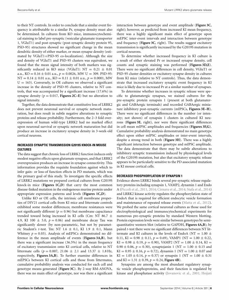
Unlike KO or OE cells, the intrinsic cell membrane proper-ties of DIV21 cortical cells from KI mice and littermate controlsexhibited some modest differences; membrane resistances werenot significantly different (p = 0.96) but membrane capacitancetrended toward being increased in KI cells (Cm: NT 86.7 ±4.9, KI 100 ± 5.0, p = 0.06) and membrane decay Tau wassignificantly slower (by non-parametric, but not by paramet-ric Student’s t-test. Tm: NT 1.6 ± 0.1, KI 1.9 ± 0.1, MannWhitney p = 0.03). Analysis of mEPSCs demonstrated no dif-ference in the mean amplitude of events (Figures 3A,B), butthere was a significant increase (36.5%) in the mean frequencyof excitatory transmission onto KI cortical cells, relative to NTlittermate cells (p = 0.007, 21.06 ± 2.1 and 13.37 ± 1.8 Hz,respectively, Figures 3A,B). To further examine differences inmEPSCs between KI cortical cells and those from littermates,cumulative probability analysis was conducted for each cell andgenotype means generated (Figure 3C). By 2-way RM-ANOVA,there was no main effect of genotype, nor was there a significant
interaction between genotype and event amplitude (Figure 3C,right); however, as predicted from increased KI mean frequency,there was a highly significant main effect of genotype uponmEPSC inter-event intervals and interaction between genotypeand frequency (Figure 3C, right). The results suggest excitatorytransmission is significantly increased by the G2019S mutation incortical neurons.
To determine whether increased frequency in KI culture isa result of either elevated Pr or increased synapse density, cellcounts and synaptic staining was performed (Figures 3D,E).There were no significant differences in cell density, VGluT1 orPSD-95 cluster densities or excitatory synapse density in culturesfrom KI mice (relative to NT controls). Thus, the data demon-strate that increased excitatory synaptic event frequency in KImice is likely due to increased Pr at a similar number of synapses.
To determine whether increases in synaptic release were spe-cific to glutamatergic synapses, we stained cultures for thepre-synaptic protein synapsin 1 (present at both glutamater-gic and GABAergic terminals) and recorded GABAergic minia-ture inhibitory post-synaptic currents (mIPSCs, Figures 3E–H).There were no significant differences in the number (or inten-sity; not shown) of synapsin 1 clusters in cultured KI neu-rons (Figure 3E, right), nor were there significant differencesin cell mean mIPSC amplitudes and frequencies (Figures 3F,G).Cumulative probability analysis demonstrated no main genotypeeffect upon either mIPSC amplitudes or inter-event intervals,despite a strong trend in both (Figure 3H). There was a highlysignificant interaction between genotype and mIPSC amplitude.The data demonstrate that there may be subtle alterations toinhibitory synaptic transmission induced by physiological levelsof the G2019S mutation, but also that excitatory synaptic releaseappears to be particularly sensitive to the PD associated mutationin KI mouse cortical cells.
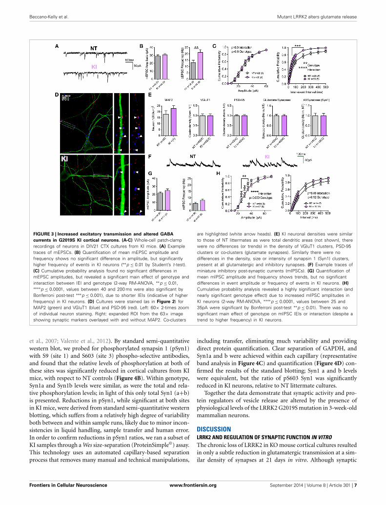
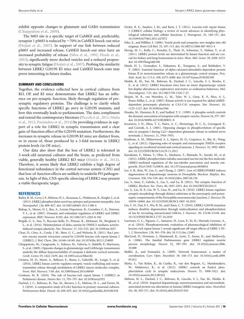
DECREASED PHOSPHORYLATION OF SYNAPSIN 1Evidence shows LRRK2 binds several pre-synaptic release regula-tory proteins including synapsin 1, VAMP2, dynamin 1 and EndoA (Piccoli et al., 2011, 2014; Cirnaru et al., 2014; Stafa et al., 2014)and LRRK2 kinase activity regulates the phosphorylation state ofEndoA that is required for efficient endocytic vesicle formationand maintenance of repeated release events (Matta et al., 2012).We probed the same cortical neuronal cultures as those used forelectrophysiological and immunocytochemical experiments fornumerous pre-synaptic proteins by standard Western blotting.Protein expression levels were similar between genotypes by semi-quantitative western blot (relative to GAPDH, Figures 4A,B); bypaired t-test there were no significant differences between NT lit-termate and KI cultures in the levels of EndoA (NT = 1.00 ±0.11, KI = 0.98 ± 0.11, p = 0.69), VAMP1 (NT = 1.00 ± 0.22,KI = 0.98 ± 0.19, p = 0.90), VAMP2 (NT = 1.00 ± 0.14, KI =0.90 ± 0.06, p = 0.30), synaptojanin 1 (NT = 1.00 ± 0.15 andKI = 0.95 ± 0.16, p = 0.72), dynamin 1 (NT = 1.00 ± 0.07 andKI = 1.05 ± 0.14, p = 0.37) or synapsin 1 (NT = 1.00 ± 0.31and KI = 1.31 ± 0.39, p = 0.24, Figure 4B).
Synapsins are among the most abundant regulatory synap-tic vesicle phosphoproteins, and their function is regulated bykinase and phosphatase activity (Jovanovic et al., 2001; Hojjati
Frontiers in Cellular Neuroscience www.frontiersin.org September 2014 | Volume 8 | Article 301 | 6
Beccano-Kelly et al. Mutant LRRK2 alters glutamate release
FIGURE 3 | Increased excitatory transmission and altered GABA
currents in G2019S KI cortical neurons. (A-C) Whole-cell patch-clamprecordings of neurons in DIV21 CTX cultures from KI mice. (A) Exampletraces of mEPSCs. (B) Quantification of mean mEPSC amplitude andfrequency shows no significant difference in amplitude, but significantlyhigher frequency of events in KI neurons (∗∗p ≤ 0.01 by Student’s t-test).(C) Cumulative probability analysis found no significant differences inmEPSC amplitudes, but revealed a significant main effect of genotype andinteraction between IEI and genotype (2-way RM-ANOVA, ∗∗p ≤ 0.01,∗∗∗∗p ≤ 0.0001, values between 40 and 200 ms were also significant byBonferroni post-test ∗∗∗p ≤ 0.001), due to shorter IEIs (indicative of higherfrequency) in KI neurons. (D) Cultures were stained (as in Figure 2) forMAP2 (green) and VGluT1 (blue) and PSD-95 (red). Left: 60× 2-times zoomof individual neuron staining. Right: expanded ROI from the 63× imageshowing synaptic markers overlayed with and without MAP2. Co-clusters
are highlighted (white arrow heads). (E) KI neuronal densities were similarto those of NT littermates as were total dendritic areas (not shown), therewere no differences (or trends) in the density of VGluT1 clusters, PSD-95clusters or co-clusters (glutamate synapses). Similarly there were nodifferences in the density, size or intensity of synapsin 1 (Syn1) clusters,present at all glutamatergic and inhibitory synapses. (F) Example traces ofminiature inhibitory post-synaptic currents (mIPSCs). (G) Quantification ofmean mIPSC amplitude and frequency shows trends, but no significantdifferences in event amplitude or frequency of events in KI neurons. (H)
Cumulative probability analysis revealed a highly significant interaction (andnearly significant genotype effect) due to increased mIPSC amplitudes inKI neurons (2-way RM-ANOVA, ∗∗∗∗p ≤ 0.0001, values between 25 and35pA were significant by Bonferroni post-test ∗∗p ≤ 0.01). There was nosignificant main effect of genotype on mIPSC IEIs or interaction (despite atrend to higher frequency) in KI neurons.
including transfer, eliminating much variability and providingdirect protein quantification. Clear separation of GAPDH, andSyn1a and b were achieved within each capillary (representativeband analysis in Figure 4C) and quantification (Figure 4D) con-firmed the results of the standard blotting; Syn1 a and b levelswere equivalent, but the ratio of pS603 Syn1 was significantlyreduced in KI neurons, relative to NT littermate cultures.
Together the data demonstrate that synaptic activity and pro-tein regulators of vesicle release are altered by the presence ofphysiological levels of the LRRK2 G2019S mutation in 3-week-oldmammalian neurons.
DISCUSSIONLRRK2 AND REGULATION OF SYNAPTIC FUNCTION IN VITROThe chronic loss of LRRK2 in KO mouse cortical cultures resultedin only a subtle reduction in glutamatergic transmission at a sim-ilar density of synapses at 21 days in vitro. Although synaptic
Frontiers in Cellular Neuroscience www.frontiersin.org September 2014 | Volume 8 | Article 301 | 7
Beccano-Kelly et al. Mutant LRRK2 alters glutamate release
FIGURE 4 | Reduced Synapsin 1 phosphorylation in KI cortical
neurons. Levels of pre-synaptic proteins in DIV21 CTX cultures wereassayed by standard western blotting and verified via WES automatedcapillary-based size sorting system. (A) Representative western blots ofEndophilinA (EndoA), vesicle associated membrane protein 1 (VAMP1),vesicle associated membrane protein 2 (VAMP2), dynamin 1, synapsin1(Syn1), phosphoserine 9 synapsin1 (pS9 Syn1), and phosphoserine 603synapsin1 (pS603 Syn1). (B) Quantification of synapsin1 levels andassociated phosphorylation sites. Synapsin1 levels were similar between
NT and KI however the ratio of phosphorylated synapsin1 wassignificantly reduced at both sites. (C) Standard western blot resultswere verified using the WES automated capillary-based size sortingsystem for the S603 phosphorylation site. Representative pseudo-gels(left) and electropherograms (right) exported from the WES compassanalysis software. (D) Quantification of synapsin1 and pS603 synapsin1confirmed significant reductions pS603 synapsin1. Data expressedrelative to GAPDH and normalized to NT, ∗p ≤ 0.05 ∗∗p ≤ 0.01 by pairedStudent’s t-test.
cluster densities were unaltered in KO cells, there was a markedgeneral reduction in both VGluT1 and PSD95 signal intensity,which may reflect reduced synaptic protein levels. In light of this,a harsh image threshold would produce a reduction in the den-sity of both markers and synapses in KOs, but careful (blinded)thresholding demonstrated that the size and density of synapsesis equivalent in NT and KO cells. KOs cells have been shown tohave (at least at some point in development) longer dendrites(MacLeod et al., 2006; Parisiadou et al., 2009; Dachsel et al.,2010; Sepulveda et al., 2013). If they were similarly longer inthis study, with equivalent synapse densities, elevations in totalsynapse number might be predicted to result in an increasedevent frequency. The opposite trend was observed here in KOcells. Together, the data provide further evidence that LRRK2 actsat glutamatergic synapses in mammalian neurons (Piccoli et al.,2011; Parisiadou et al., 2014) and, along with recent Drosophilastudies (Lee et al., 2010; Matta et al., 2012), strengthens the casefor LRRK2s role in the regulation of synaptic release machinery.That said, understanding the experimental and synaptic contextof LRRK2 manipulations is crucial to attempts at extrapolatingits physiological role, as both increases (Piccoli et al., 2011) anddecreases (Matta et al., 2012; Parisiadou et al., 2014) in synapticactivity have been observed following LRRK2 loss of function indifferent systems.
Increasing LRRK2 levels 2–3 fold resulted in an increase inthe density of synaptic markers and synapse numbers. Therewere non-significant trends toward increased event frequency and
reduced inter-event intervals that may reflect the ∼17% increasein synapse density. Alternatively, homeostatic compensation maymask the increased synapse density by reducing probability ofrelease at a greater number of synapses. This is in agreement witha lack of effect observed in DA cell degeneration in Drosophila(Lee et al., 2007); however, overexpression has been shown todecrease pre-synaptic bouton numbers (Lee et al., 2010) and haveexactly the same effect as LRRK2 loss of function on synap-tic release in Drosophila (Matta et al., 2012). Together the datastrongly suggest that the synaptic consequences of LRRK2 manip-ulations may be model-, cell- and context-dependent; thus itmay be of paramount importance to determine the compara-tive effects of knock-out, overexpression and mutation within thesame system to enable interpretation of the results.
PHYSIOLOGICAL LEVELS OF G2019S LRRK2 INCREASE GLUTAMATERELEASE AND ALTER PRE-SYNAPTIC FUNCTIONExamination of overexpression and knock-out of LRRK2 in ourprimary cortical cultures provided the platform against which tocompare the effects of mutant LRRK2. We observed a marked∼37% increase in the frequency of glutamate excitatory currentsin G2019S KI cultures, in the absence of any change to synapsedensity. Importantly, this demonstrated that the LRRK2 muta-tion produces effects that are distinct from those of simple loss-offunction or gain-of-function. We did detect a (non-significant)13% increase in KI membrane capacitance that could be pre-dictive of increased dendritic membrane area, longer dendrites
Frontiers in Cellular Neuroscience www.frontiersin.org September 2014 | Volume 8 | Article 301 | 8
Beccano-Kelly et al. Mutant LRRK2 alters glutamate release
and more numerous (equally dense) synapses. However, cor-relation analysis between membrane capacitance and mEPSCevent frequency in KI cells showed absolutely no relationship(Pearson’s R = 0.002, P = 0.99), whereas there was a significantpositive correlation in NT cells (R = 0.49, P = 0.007). We takethis as evidence that the increase in KI frequency is indepen-dent of potential difference in total dendrite length especiallyas it supersedes the usual correlation—i.e. smaller cells withsimilar synapse densities also have high event frequency, pre-sumably from overactive pre-synaptic elements. Also, there isconsensus from several studies that wild-type overexpression andmutant overexpression both result in shortened dendritic length(MacLeod et al., 2006; Parisiadou et al., 2009; Dachsel et al., 2010;Ramonet et al., 2011; Winner et al., 2011; Sepulveda et al., 2013).If OE and KI cells here also have shortened dendrites, with equalsynapse densities, then total synapse number would be reduced.Consequently the observed increase in KI event frequency wouldbe an underestimate for increased Pr.
There was also a significant slowing of membrane responses todirect current injection in KI cells, which correlated with signifi-cantly slower mEPSCs rise times. This, in concert with trends incapacitance values, suggests altered intrinsic membrane proper-ties that are likely related and may be explored in future studies.However, like capacitance, there was no significant correlationbetween mEPSC rise time and event frequency in either NT(R = 0.1, p = 0.09) or KI cells (R = 0.004, p = 0.7), arguing thatpost-synaptic membrane alterations do not account for increasedtransmission. It appears that a single copy of G2019S LRRK2 issufficient to dramatically alter excitatory synaptic release, in amanner distinct from a loss of LRRK2 and in excess of any changesproduced by a 3-fold increase in LRRK2 levels.
The G2019S mutation resides in LRRK2’s kinase domain, andhas been shown to augment LRRK2 kinase activity in vitro,demonstrating a gain-of-function for LRRK2 autophosporylationand phosphorylation of a generic substrate (West et al., 2005;Nichols et al., 2010). This has led to a major push for iden-tification of LRRK2 substrates and the development of kinaseinhibitors as they may offer therapeutic potential (Webber et al.,2011). The list of candidate substrates is growing, and includesTau (Kawakami et al., 2012; Bailey et al., 2013), 4E-BP (Leeet al., 2010), and EndoA (Matta et al., 2012). Unfortunately,interpretation of many of these findings is hampered by bind-ing relationships potentially forced in vitro by non-physiologicalconcentrations of substrate and enzyme (Webber et al., 2011),failure of supporting evidence in vivo (Trancikova et al., 2012)and reliance on inhibitors that exhibit off-target and/or sys-temic effects (Drolet et al., 2011; Cirnaru et al., 2014; Luermanet al., 2014) even in LRRK2 knock-out cells. That said, it isclear that many of the proposed LRRK2 interactors and sub-strates are directly linked to the synaptic vesicle cycle, notablysyntaxin 1A, dynamin1, synapsin1 and VAMP2 (Piccoli et al.,2014) and EndoA; phosphorylation of EndoA by LRRK2 hasbeen demonstrated to regulate transmitter release (Matta et al.,2012). If the cause of increased release is as simple as the ∼3-fold increase in LRRK2 kinase activity (West et al., 2005; Nicholset al., 2010), other factors must be at play to account for synapticalterations in KI mouse cells well above those seen in OE cultures
(expressing 3-fold more LRRK2) and preferential effects uponglutamatergic, rather than GABAergic release. LRRK2 localiza-tion and kinase activity are regulated by its own phosphorylationstate and through dimerization by co-chaperone 14-3-3 (Senet al., 2009; Nichols et al., 2010; Rudenko and Cookson, 2010);thus greater effects may be engendered by G2019S upon kinaseactivity in living neuronal systems under appropriate regulation.We assayed the protein levels of several interactors but foundnone to be significantly altered. The phosphorylation state ofvesicle cycle regulators has direct consequences for their activ-ity and we sought to assay the phosphorylation status of EndoA,pertinent to LRRK2 activity and vesicle release in Drosophila(Matta et al., 2012); unfortunately, the only phosphoantibodycurrently specific to the pertinent EndoA serine 75 site is inef-fective in mammalian tissue (personal communication, Dr. PatrikVerstreken). We therefore turned our attention to reasonably well-characterized phosphorylation sites on synapsin 1, one of themost abundant of all pre-synaptic vesicle proteins.
Synapsins are believed to regulate the balance between thereserve and the readily releasable (docked) vesicle pools and actas modulators of vesicle exocytosis (Fdez and Hilfiker, 2006). Ithas been suggested that phosphorylated synapsin 1 binds vesi-cles and tethers them to the actin cytoskeleton for retention inthe reserve pool; thus, phospho-synapsin 1 reduces the num-ber of vesicles available for release from the readily releasablepool, whereas dephosphorylated synapsin dissociates from vesi-cles, thereby freeing them to dock for ready release (Hosakaet al., 1999). However, this supposition is not universally sup-ported; some reports describe synapsin depletion when vesiclesundergo active zone docking (Pieribone et al., 1995) but oth-ers show that synapsins remain associated with vesicles duringexo- and endocytosis (Torri-Tarelli et al., 1990). Furthermore,with several studies showing the mature vesicle cluster containsvirtually no cytoskeleton, the original hypothesis is unlikely toexplain synapsin function there (reviewed in Sudhof, 2004; Fdezand Hilfiker, 2006). Although the mechanism by which synapsin1 regulates vesicle release remains elusive, synapsin 1 phosphory-lation states are indicative of pre-synaptic regulation and releaseactivity (reviewed in Sudhof, 2004; Fdez and Hilfiker, 2006;Valente et al., 2012; Verstegen et al., 2014). In KI cortical culturesthat exhibited a marked increase in synaptic release, we founda clear reduction in synapsin 1 phosphorylation at both S6 andS603 by traditional western blot and confirmed reduced pS603 byproteinsimple Wes size separation.
It is interesting to note that we observed significant effectsupon glutamate frequency and significant effects upon GABAamplitudes. An increase in excitatory synaptic transmissionacross the neuronal network in the culture might be predictedto alter the GABAergic inhibitory neurons within it and subse-quently the post-synaptic responsiveness to their activity (Wangand Maffei, 2014). Homeostatic mechanisms may also exag-gerate GABAergic inhibition, in order to counteract the effectsof increased glutamate release (Shepherd et al., 2006; Maffeiand Fontanini, 2009), but network interactions are very hardto predict or interpret. This dichotomy may even be explainedby alterations to synapsin 1, as it has been shown that neu-rons in cortical cultures prepared from synapsin 1 null mice
Frontiers in Cellular Neuroscience www.frontiersin.org September 2014 | Volume 8 | Article 301 | 9
Beccano-Kelly et al. Mutant LRRK2 alters glutamate release
exhibit opposite changes to glutamate and GABA transmission(Chiappalone et al., 2009).
The S603 site is a specific target of CaMKII and, predictably,synapsin 1 pS603 is reduced by ∼70% in CaMKII knock-out mice(Hojjati et al., 2007). In support of our link between reducedpS603 and increased release, CaMKII knock-out mice have anincreased probability of release (Silva et al., 1992; Hinds et al.,2003), significantly more docked vesicles and a reduced propen-sity to synaptic fatigue (Hojjati et al., 2007). Probing the similaritybetween LRRK2 G2019S KI mice and CaMKII knock-outs mayprove interesting in future studies.
SUMMARY AND CONCLUSIONSTogether, the evidence collected here in cortical cultures fromKO, OE and KI mice demonstrates that LRRK2 has an influ-ence on pre-synaptic function, likely through regulation of pre-synaptic regulatory proteins. The challenge is to clarify whichspecific functions of LRRK2 go awry in G2019S mutants, andhow this eventually leads to parkinsonism. The data complimentand extend the contemporary literature (Piccoli et al., 2011; Mattaet al., 2012; Parisiadou et al., 2014) by providing evidence in sup-port of a role for LRRK2 in synaptic transmission and a cleargain-of-function effect of the G2019S mutation. Furthermore, theincreases in synaptic release in G2019S KI mice are distinct from,or in excess of, those produced by a 3-fold increase in LRRK2protein levels (in OE mice).
Our data also show that the loss of LRRK2 is tolerated in3-week-old neuronal cultures and the brain, as predicted fromviable, generally healthy LRRK2 KO mice (Hinkle et al., 2012).Therefore, it seems likely that LRRK2 exhibits a high degree offunctional redundancy in the central nervous system (CNS) andthat loss-of-function effects are unlikely to underlie PD pathogen-esis. In light of this, CNS-specific silencing of LRRK2 may presenta viable therapeutic target.
REFERENCESBailey, R. M., Covy, J. P., Melrose, H. L., Rousseau, L., Watkinson, R., Knight, J., et al.
(2013). LRRK2 phosphorylates novel tau epitopes and promotes tauopathy. ActaNeuropathol. 126, 809–827. doi: 10.1007/s00401-013-1188-4
Biskup, S., Moore, D. J., Rea, A., Lorenz-Deperieux, B., Coombes, C. E., Dawson,V. L., et al. (2007). Dynamic and redundant regulation of LRRK2 and LRRK1expression. BMC Neurosci. 8:102. doi: 10.1186/1471-2202-8-102
Brigidi, G. S., Sun, Y., Beccano-Kelly, D., Pitman, K., Mobasser, M., Borgland, S.L., et al. (2014). Palmitoylation of delta-catenin by DHHC5 mediates activity-induced synapse plasticity. Nat. Neurosci. 17, 522–532. doi: 10.1038/nn.3657
Chan, D., Citro, A., Cordy, J. M., Shen, G. C., and Wolozin, B. (2011). Rac1 pro-tein rescues neurite retraction caused by G2019S leucine-rich repeat kinase 2(LRRK2). J. Biol. Chem. 286, 16140–16149. doi: 10.1074/jbc.M111.234005
Chiappalone, M., Casagrande, S., Tedesco, M., Valtorta, F., Baldelli, P., Martinoia,S., et al. (2009). Opposite changes in glutamatergic and GABAergic transmissionunderlie the diffuse hyperexcitability of synapsin I-deficient cortical networks.Cereb. Cortex 19, 1422–1439. doi: 10.1093/cercor/bhn182
Cirnaru, M. D., Marte, A., Belluzzi, E., Russo, I., Gabrielli, M., Longo, F., et al.(2014). LRRK2 kinase activity regulates synaptic vesicle trafficking and neuro-transmitter release through modulation of LRRK2 macro-molecular complex.Front. Mol. Neurosci. 7:49. doi: 10.3389/fnmol.2014.00049
Cookson, M. R. (2010). The role of leucine-rich repeat kinase 2 (LRRK2) inParkinson’s disease. Nature Rev. 11, 791–797. doi: 10.1038/nrn2935
Dachsel, J. C., Behrouz, B., Yue, M., Beevers, J. E., Melrose, H. L., and Farrer, M.J. (2010). A comparative study of Lrrk2 function in primary neuronal cultures.Parkinsonism Relat. Disord. 16, 650–655. doi: 10.1016/j.parkreldis.2010.08.018
Drolet, R. E., Sanders, J. M., and Kern, J. T. (2011). Leucine-rich repeat kinase2 (LRRK2) cellular biology: a review of recent advances in identifying phys-iological substrates and cellular functions. J. Neurogenet. 25, 140–151. doi:10.3109/01677063.2011.627072
Fdez, E., and Hilfiker, S. (2006). Vesicle pools and synapsins: new insights into oldenigmas. Brain Cell Biol. 35, 107–115. doi: 10.1007/s11068-007-9013-4
Herzig, M. C., Kolly, C., Persohn, E., Theil, D., Schweizer, T., Hafner, T., et al.(2011). LRRK2 protein levels are determined by kinase function and are cru-cial for kidney and lung homeostasis in mice. Hum. Mol. Genet. 20, 4209–4223.doi: 10.1093/hmg/ddr348
Hinds, H. L., Goussakov, I., Nakazawa, K., Tonegawa, S., and Bolshakov, V.Y. (2003). Essential function of alpha-calcium/calmodulin-dependent proteinkinase II in neurotransmitter release at a glutamatergic central synapse. Proc.Natl. Acad. Sci. U.S.A. 100, 4275–4280. doi: 10.1073/pnas.0530202100
Hinkle, K. M., Yue, M., Behrouz, B., Dachsel, J. C., Lincoln, S. J., Bowles, E.E., et al. (2012). LRRK2 knockout mice have an intact dopaminergic systembut display alterations in exploratory and motor co-ordination behaviors. Mol.Neurodegener. 7:25. doi: 10.1186/1750-1326-7-25
Hojjati, M. R., van Woerden, G. M., Tyler, W. J., Giese, K. P., Silva, A. J.,Pozzo-Miller, L., et al. (2007). Kinase activity is not required for alphaCaMKII-dependent presynaptic plasticity at CA3-CA1 synapses. Nat. Neurosci. 10,1125–1127. doi: 10.1038/nn1946
Hosaka, M., Hammer, R. E., and Sudhof, T. C. (1999). A phospho-switch controlsthe dynamic association of synapsins with synaptic vesicles. Neuron 24, 377–387.doi: 10.1016/S0896-6273(00)80851-X
Jovanovic, J. N., Sihra, T. S., Nairn, A. C., Hemmings, H. C. Jr., Greengard, P.,and Czernik, A. J. (2001). Opposing changes in phosphorylation of specificsites in synapsin I during Ca2+-dependent glutamate release in isolated nerveterminals. J. Neurosci. 21, 7944–7953.
Kaufman, A. M., Milnerwood, A. J., Sepers, M. D., Coquinco, A., She, K., Wang,L., et al. (2012). Opposing roles of synaptic and extrasynaptic NMDA receptorsignaling in cocultured striatal and cortical neurons. J. Neurosci. 32, 3992–4003.doi: 10.1523/JNEUROSCI.4129-11.2012
Kawakami, F., Yabata, T., Ohta, E., Maekawa, T., Shimada, N., Suzuki, M., et al.(2012). LRRK2 phosphorylates tubulin-associated tau but not the free molecule:LRRK2-mediated regulation of the tau-tubulin association and neurite out-growth. PLoS ONE 7:e30834. doi: 10.1371/journal.pone.0030834
Lee, S. B., Kim, W., Lee, S., and Chung, J. (2007). Loss of LRRK2/PARK8 inducesdegeneration of dopaminergic neurons in Drosophila. Biochem. Biophys. Res.Commun. 358, 534–539. doi: 10.1016/j.bbrc.2007.04.156
Lee, S., Imai, Y., Gehrke, S., Liu, S., and Lu, B. (2012). The synaptic function ofLRRK2. Biochem. Soc. Trans. 40, 1047–1051. doi: 10.1042/BST20120113
Lee, S., Liu, H. P., Lin, W. Y., Guo, H., and Lu, B. (2010). LRRK2 kinase regulatessynaptic morphology through distinct substrates at the presynaptic and postsy-naptic compartments of the Drosophila neuromuscular junction. J. Neurosci.30,16959–16969. doi: 10.1523/JNEUROSCI.1807-10.2010
Lin, C. H., Tsai, P. I., Wu, R. M., and Chien, C. T. (2010). LRRK2 G2019S mutationinduces dendrite degeneration through mislocalization and phosphorylationof tau by recruiting autoactivated GSK3ss. J. Neurosci. 30, 13138–13149. doi:10.1523/JNEUROSCI.1737-10.2010
Luerman, G. C., Nguyen, C., Samaroo, H., Loos, P., Xi, H., Hurtado-Lorenzo, A.,et al. (2014). Phosphoproteomic evaluation of pharmacological inhibition ofleucine-rich repeat kinase 2 reveals significant off-target effects of LRRK-2-IN-1. J. Neurochem. 128, 561–576. doi: 10.1111/jnc.12483
MacLeod, D., Dowman, J., Hammond, R., Leete, T., Inoue, K., and Abeliovich,A. (2006). The familial Parkinsonism gene LRRK2 regulates neuriteprocess morphology. Neuron 52, 587–593. doi: 10.1016/j.neuron.2006.10.008
Maffei, A., and Fontanini, A. (2009). Network homeostasis: a matter ofcoordination. Curr. Opin. Neurobiol. 19, 168–173. doi: 10.1016/j.conb.2009.05.012
Matta, S., Van Kolen, K., da Cunha, R., van den Bogaart, G., Mandemakers,W., Miskiewicz, K., et al. (2012). LRRK2 controls an EndoA phos-phorylation cycle in synaptic endocytosis. Neuron 75, 1008–1021. doi:10.1016/j.neuron.2012.08.022
Melrose, H. L., Dachsel, J. C., Behrouz, B., Lincoln, S. J., Yue, M., Hinkle, K.M., et al. (2010). Impaired dopaminergic neurotransmission and microtubule-associated protein tau alterations in human LRRK2 transgenic mice. Neurobiol.Dis. 40, 503–517. doi: 10.1016/j.nbd.2010.07.010
Frontiers in Cellular Neuroscience www.frontiersin.org September 2014 | Volume 8 | Article 301 | 10
Beccano-Kelly et al. Mutant LRRK2 alters glutamate release
Milnerwood, A. J., Kaufman, A. M., Sepers, M. D., Gladding, C. M.,Zhang, L., Wang, L., et al. (2012). Mitigation of augmented extrasynap-tic NMDAR signaling and apoptosis in cortico-striatal co-cultures fromHuntington’s disease mice. Neurobiol. Dis. 48, 40–51. doi: 10.1016/j.nbd.2012.05.013
Mizuno-Yamasaki, E., Rivera-Molina, F., and Novick, P. (2012). GTPase networksin membrane traffic. Annu. Rev. Biochem. 81, 637–659. doi: 10.1146/annurev-biochem-052810-093700
Nichols, R. J., Dzamko, N., Morrice, N. A., Campbell, D. G., Deak, M., Ordureau,A., et al. (2010). 14-3-3 binding to LRRK2 is disrupted by multiple Parkinson’sdisease-associated mutations and regulates cytoplasmic localization. Biochem. J.430, 393–404. doi: 10.1042/BJ20100483
O’Neill, R. A., Bhamidipati, A., Bi, X., Deb-Basu, D., Cahill, L., Ferrante, J.,et al. (2006). Isoelectric focusing technology quantifies protein signaling in 25cells. Proc. Natl. Acad. Sci. U.S.A. 103, 16153–16158. doi: 10.1073/pnas.0607973103
Parisiadou, L., Xie, C., Cho, H. J., Lin, X., Gu, X. L., Long, C. X., et al. (2009).Phosphorylation of ezrin/radixin/moesin proteins by LRRK2 promotes the rear-rangement of actin cytoskeleton in neuronal morphogenesis. J. Neurosci. 29,13971–13980. doi: 10.1523/JNEUROSCI.3799-09.2009
Piccoli, G., Condliffe, S. B., Bauer, M., Giesert, F., Boldt, K., De Astis, S., et al.(2011). LRRK2 controls synaptic vesicle storage and mobilization within therecycling pool. J. Neurosci. 31, 2225–2237. doi: 10.1523/JNEUROSCI.3730-10.2011
Piccoli, G., Onofri, F., Cirnaru, M. D., Kaiser, C. J., Jagtap, P., Kastenmuller, A.,et al. (2014). Leucine-rich repeat kinase 2 binds to neuronal vesicles throughprotein interactions mediated by its C-terminal WD40 domain. Mol. Cell. Biol.34, 2147–2161. doi: 10.1128/MCB.00914-13
Pieribone, V. A., Shupliakov, O., Brodin, L., Hilfiker-Rothenfluh, S., Czernik, A. J.,and Greengard, P. (1995). Distinct pools of synaptic vesicles in neurotransmitterrelease. Nature 375, 493–497. doi: 10.1038/375493a0
Plowey, E. D., Cherra, S. J. 3rd., Liu, Y. J., and Chu, C. T. (2008). Role ofautophagy in G2019S-LRRK2-associated neurite shortening in differentiatedSH-SY5Y cells. J. Neurochem. 105, 1048–1056. doi: 10.1111/j.1471-4159.2008.05217.x
Ramonet, D., Daher, J. P., Lin, B. M., Stafa, K., Kim, J., Banerjee, R., et al. (2011).Dopaminergic neuronal loss, reduced neurite complexity and autophagicabnormalities in transgenic mice expressing G2019S mutant LRRK2. PLoS ONE6:e18568. doi: 10.1371/journal.pone.0018568
Rudenko, I. N., and Cookson, M. R. (2010). 14-3-3 proteins are promising LRRK2interactors. Biochem. J. 430, e5–e6. doi: 10.1042/BJ20101200
Sen, S., Webber, P. J., and West, A. B. (2009). Dependence of leucine-richrepeat kinase 2 (LRRK2) kinase activity on dimerization. J. Biol. Chem. 284,36346–36356. doi: 10.1074/jbc.M109.025437
Sepulveda, B., Mesias, R., Li, X., Yue, Z., and Benson, D. L. (2013). Short- and long-term effects of LRRK2 on axon and dendrite growth. PLoS ONE 8:e61986. doi:10.1371/journal.pone.0061986
Shepherd, J. D., Rumbaugh, G., Wu, J., Chowdhury, S., Plath, N., Kuhl, D., et al.(2006). Arc/Arg3.1 mediates homeostatic synaptic scaling of AMPA receptors.Neuron 52, 475–484. doi: 10.1016/j.neuron.2006.08.034
Silva, A. J., Stevens, C. F., Tonegawa, S., and Wang, Y. (1992). Deficient hippocam-pal long-term potentiation in alpha-calcium-calmodulin kinase II mutant mice.Science 257, 201–206. doi: 10.1126/science.1378648
Stafa, K., Tsika, E., Moser, R., Musso, A., Glauser, L., Jones, A., et al. (2014).Functional interaction of Parkinson’s disease-associated LRRK2 with members
of the dynamin GTPase superfamily. Hum. Mol. Genet. 23, 2055–2077. doi:10.1093/hmg/ddt600
Sudhof, T. C. (2004). The synaptic vesicle cycle. Annu. Rev. Neurosci. 27, 509–547.doi: 10.1146/annurev.neuro.26.041002.131412
Tapia, L., Milnerwood, A., Guo, A., Mills, F., Yoshida, E., Vasuta, C., et al.(2011). Progranulin deficiency decreases gross neural connectivity but enhancestransmission at individual synapses. J. Neurosci. 31, 11126–11132. doi:10.1523/JNEUROSCI.6244-10.2011
Torri-Tarelli, F., Villa, A., Valtorta, F., De Camilli, P., Greengard, P., and Ceccarelli,B. (1990). Redistribution of synaptophysin and synapsin I during alpha-latrotoxin-induced release of neurotransmitter at the neuromuscular junction.J. Cell Biol. 110, 449–459. doi: 10.1083/jcb.110.2.449
Trancikova, A., Mamais, A., Webber, P. J., Stafa, K., Tsika, E., Glauser, L., et al.(2012). Phosphorylation of 4E-BP1 in the mammalian brain is not alteredby LRRK2 expression or pathogenic mutations. PLoS ONE 7:e47784. doi:10.1371/journal.pone.0047784
Valente, P., Casagrande, S., Nieus, T., Verstegen, A. M., Valtorta, F., Benfenati, F.,et al. (2012). Site-specific synapsin I phosphorylation participates in the expres-sion of post-tetanic potentiation and its enhancement by BDNF. J. Neurosci. 32,5868–5879. doi: 10.1523/JNEUROSCI.5275-11.2012
Verstegen, A. M., Tagliatti, E., Lignani, G., Marte, A., Stolero, T., Atias, M.,et al. (2014). Phosphorylation of synapsin I by cyclin-dependent kinase-5 setsthe ratio between the resting and recycling pools of synaptic vesicles at hip-pocampal synapses. J. Neurosci. 34, 7266–7280. doi: 10.1523/JNEUROSCI.3973-13.2014
Wang, L., and Maffei, A. (2014). Inhibitory plasticity dictates the sign of plasticity atexcitatory synapses. J. Neurosci. 34, 1083–1093. doi: 10.1523/JNEUROSCI.4711-13.2014
Webber, P. J., Smith, A. D., Sen, S., Renfrow, M. B., Mobley, J. A., and West, A.B. (2011). Autophosphorylation in the leucine-rich repeat kinase 2 (LRRK2)GTPase domain modifies kinase and GTP-binding activities. J. Mol. Biol. 412,94–110. doi: 10.1016/j.jmb.2011.07.033
West, A. B., Moore, D. J., Biskup, S., Bugayenko, A., Smith, W. W., Ross, C. A., et al.(2005). Parkinson’s disease-associated mutations in leucine-rich repeat kinase2 augment kinase activity. Proc. Natl. Acad. Sci. U.S.A. 102, 16842–16847. doi:10.1073/pnas.0507360102
Winner, B., Melrose, H. L., Zhao, C., Hinkle, K. M., Yue, M., Kent, C., et al. (2011).Adult neurogenesis and neurite outgrowth are impaired in LRRK2 G2019Smice. Neurobiol. Dis. 41, 706–716. doi: 10.1016/j.nbd.2010.12.008
Conflict of Interest Statement: The authors declare that the research was con-ducted in the absence of any commercial or financial relationships that could beconstrued as a potential conflict of interest.