Page 1
�������� ����� ��
The pesticide adjuvant, toximulTM, alters hepatic metabolism through effectson downstream targets of PPAR
Jacqueline Upham, Philip D. Acott, Patrick O’Regan, Christopher J.Sinal, John F.S. Crocker, Laurette Geldenhuys, Mary G. Murphy
PII: S0925-4439(07)00143-3DOI: doi: 10.1016/j.bbadis.2007.06.003Reference: BBADIS 62738
To appear in: BBA - Molecular Basis of Disease
Received date: 22 February 2007Revised date: 1 June 2007Accepted date: 8 June 2007
Please cite this article as: Jacqueline Upham, Philip D. Acott, Patrick O’Regan, Christo-pher J. Sinal, John F.S. Crocker, Laurette Geldenhuys, Mary G. Murphy, The pesticideadjuvant, toximulTM, alters hepatic metabolism through effects on downstream targetsof PPAR, BBA - Molecular Basis of Disease (2007), doi: 10.1016/j.bbadis.2007.06.003
This is a PDF file of an unedited manuscript that has been accepted for publication.As a service to our customers we are providing this early version of the manuscript.The manuscript will undergo copyediting, typesetting, and review of the resulting proofbefore it is published in its final form. Please note that during the production processerrors may be discovered which could affect the content, and all legal disclaimers thatapply to the journal pertain.
peer
-005
6279
2, v
ersi
on 1
- 4
Feb
2011
Author manuscript, published in "Biochimica et Biophysica Acta (BBA) – Molecular Basis of Disease 1772, 9 (2007) 1057" DOI : 10.1016/j.bbadis.2007.06.003
Page 2
ACC
EPTE
D M
ANU
SCR
IPT
ACCEPTED MANUSCRIPT 1
The Pesticide Adjuvant, Toximul™, Alters Hepatic Metabolism Through Effects on 1
Downstream Targets of PPARα 2
3
Jacqueline Uphama, Philip D. Acottb,c, Patrick O’Reganb, Christopher J. Sinalc, John F.S. 4
Crockerb, Laurette Geldenhuysd and Mary G. Murphya,* 5
6
Departments of Physiology & Biophysicsa and Pharmacologyc, 5850 College Street, 7
Dalhousie University, Halifax, Nova Scotia Canada B3H 1X5 8
bDepartment of Pediatrics, Dalhousie University and the IWK-Grace Health Centre, 9
5850-5980 University Avenue, Halifax, Nova Scotia Canada B3K 6R8 10
dDepartment of Pathology (Anatomical Pathology), 5788 University Avenue, Dalhousie 11
University, Halifax, Nova Scotia Canada B3H 1V8 12
13
Keywords: pesticide adjuvant, PPARα, PPARα-null mice, mRNA and protein 14
expression, Cyp4A, peroxisomal fatty-acid oxidation 15
16
*Corresponding Author: Mary G. Murphy, Ph.D. 17
Department of Physiology & Biophysics 18
Sir Charles Tupper Medical Building 19
Dalhousie University 20
Halifax, Nova Scotia Canada B3H 1X5 21
Fax: (902) 494-1685 22
Email: [email protected] 23
peer
-005
6279
2, v
ersi
on 1
- 4
Feb
2011
Page 3
ACC
EPTE
D M
ANU
SCR
IPT
ACCEPTED MANUSCRIPT 2
Abstract Previous studies demonstrated that chronic dermal exposure to the 1
pesticide adjuvant (surfactant), Toximul (Tox), has significant detrimental effects on 2
hepatic lipid metabolism. This study demonstrated that young mice dermally exposed to 3
Tox for 12 days have significant increases in expression of peroxisomal acyl-CoA 4
oxidase (mRNA and protein), bifunctional enzyme (mRNA) and thiolase (mRNA), as 5
well as the P450 oxidizing enzymes Cyp4A10 and Cyp4A14 (mRNA and protein). Tox 6
produced a similar pattern of increases in wild type adult female mice but did not induce 7
these responses in PPARα-null mice. These data support the hypothesis that Tox, a 8
heterogeneous blend of non-ionic and anionic surfactants, modulates hepatic metabolism 9
at least in part through activation of PPARα. Notably, all three groups of Tox-treated 10
mice had increased relative liver weights due to significant accumulation of lipid. This 11
could be endogenous in nature and/or a component(s) of Tox or a metabolite thereof. 12
The ability of Tox and other hydrocarbon pollutants to induce fatty liver despite being 13
PPARα agonists indicate a novel consequence of exposure to this class of chemicals, and 14
may provide a new understanding of fatty liver in populations with industrial exposure. 15
peer
-005
6279
2, v
ersi
on 1
- 4
Feb
2011
Page 4
ACC
EPTE
D M
ANU
SCR
IPT
ACCEPTED MANUSCRIPT 3
1
Abbreviations: IS, industrial surfactant(s); Tox, Toximul; pFAO, peroxisomal fatty-acid 2
β-oxidation; mFAO, mitochondrial FAO; qPCR, quantitative polymerase chain reaction; 3
PPARα, peroxisome proliferator-activated receptor alpha; PP(-/-), PPARα-null mice; 4
PP(+/+), PPARα wild type mice; ACOX, acyl-CoA oxidase; L-PBE, peroxisomal 5
bifunctional enzyme; pTHIO, peroxisomal 3-ketoacyl-CoA thiolase; Cyp4A, cytochrome 6
P4504A; CTRL, control (no Tox exposure) 7
8
peer
-005
6279
2, v
ersi
on 1
- 4
Feb
2011
Page 5
ACC
EPTE
D M
ANU
SCR
IPT
ACCEPTED MANUSCRIPT 4
Introduction 1
2
Millions of tons of industrial surfactants (IS) are used annually by the textile, paint, 3
cleaning supplies and agricultural/forestry industries, and use is increasing [1]. The 4
agricultural/forestry industries rely on IS as adjuvants to emulsify water-insoluble 5
pesticides to optimize the spreading, retention and uptake of active ingredients. These 6
adjuvants can constitute up to 90% of pesticide formulations [2]. However, since they 7
are considered by industry as ‘inert ingredients’, their use is largely unregulated and 8
information regarding their composition is rarely available. Many of the Toximul™ (Tox) 9
class of adjuvants are blends of structurally heterogeneous nonionic and anionic 10
hydrocarbons (e.g., polyethylene glycol ethers, alkyl benzene sulfonates). The nonionic 11
components are partially degraded to more toxic environmentally persistent metabolites, 12
some of which (e.g., nonylphenol) are known endocrine disrupting chemicals [3]. 13
A far less known consequence of environmental contaminant exposure is disruption 14
of hepatic energy metabolism. During the past decade we have investigated the effects 15
of Tox exposure on fat and carbohydrate metabolism in neonatal mouse livers as part of 16
our long-term study of Tox potentiation of influenza B-induced mortality [2,4]. Mice 17
exposed dermally to Tox daily for 12 consecutive days exhibit significant stimulation of 18
peroxisomal fatty-acid β-oxidation (FAO) [5] and inhibition of mitochondrial FAO [6], 19
elevated synthesis of fatty acylcarnitines [7] and significant reductions in glycogen 20
content [2]. In spite of these metabolic disturbances, the young mice have no apparent 21
adverse health effects from Tox exposure. 22
peer
-005
6279
2, v
ersi
on 1
- 4
Feb
2011
Page 6
ACC
EPTE
D M
ANU
SCR
IPT
ACCEPTED MANUSCRIPT 5
Our previous efforts to elucidate the mechanism(s) underlying these metabolic 1
derangements have been unsuccessful. However, there has been a unifying mechanism 2
described for the regulation of hepatic lipid homeostasis by many structurally diverse 3
xenobiotic hydrocarbons (clofibrates, phthalates, pesticides) as well as endogenous 4
metabolites (e.g., fatty acids, prostaglandins) [8]. Peroxisome proliferator-activated 5
receptor alpha (PPARα) is a member of the superfamily of ligand-activated nuclear 6
transcription regulators. When activated, PPARα is the ‘master switch’ that controls 7
transcription of a host of genes involved in energy metabolism. The primary structural 8
requirements for ligands of PPARα appear to be lipophilicity and a carboxyl functional 9
group [9]. Recent in vitro studies demonstrated that perfluorooctane based chemicals, 10
powerful IS used primarily in the paper and textile industries, can activate downstream 11
targets of both human and mouse PPARα [10]. However no one has examined the 12
effects of adjuvants commonly used in pesticide formulations, even though it is known 13
that these chemicals can be more toxic than the active ingredients [11]. 14
The purpose of this study was to determine whether chronic dermal exposure of 15
young mice to Tox results in altered hepatic expression of PPARα and/or its target 16
enzymes involved in lipid metabolism. To assess PPARα involvement in the Tox 17
responses, we also determined the effects of the surfactant in adult PPARα-null mice 18
(PP(-/-)) and their corresponding wild type (PP(+/+)) controls. 19
20
21
22
peer
-005
6279
2, v
ersi
on 1
- 4
Feb
2011
Page 7
ACC
EPTE
D M
ANU
SCR
IPT
ACCEPTED MANUSCRIPT 6
Materials and Methods 1
2
Neonatal mice Male and female CD-1(ICR) outbred mice (Charles River, St. Constant, 3
QU, Canada) were bred and newborn pups were pooled on postnatal day (P) one and 4
divided randomly among nursing mothers. Twenty-four hours later, the litters were 5
divided into two equal groups. In the control group (CTRL), minimal essential medium 6
(MEM) was applied dermally to the abdomens of the pups. The remainder were painted 7
with Tox 3409F (Stepan Company, Northfield IL) diluted in MEM (1:8, vol:vol)(~1 mg 8
Tox/g/day). These treatments were repeated daily for 12 days (P2-P13). Body weights 9
were recorded daily. On P13 the mice were killed by decapitation and the livers were 10
excised, weighed and flash frozen for storage at -80°C until assessed. In this study only 11
female CD-1 mice were used, although preliminary experiments indicated that Tox-12
induced changes in mRNA expression were not gender specific in the pups on P13. 13
PP(+/+) and PP(-/-) mice Age-matched (14-18 weeks) wild type (C57BL/6, PP(+/+)) 14
female mice (22±1.3 g) and PP(-/-) mice (22.2±1.2 g) originated from the laboratory of Lee 15
et al. [12] and were a gift to C. Sinal from Dr. F. Gonzalez, National Cancer Institute, 16
National Institutes of Health, Bethesda MD. Half of each group had Tox applied on their 17
abdomens daily for 12 days at doses equivalent (wt/wt) to those to which the neonates were 18
exposed. The remainder of each group received MEM. The mice were fed standard rodent 19
diet ad libitum throughout the experiment. Body weights were recorded daily. After 12 20
days’ painting the mice were killed and their livers weighed and treated as described above. 21
All mouse studies were carried out in compliance with the guidelines of the Canadian 22
peer
-005
6279
2, v
ersi
on 1
- 4
Feb
2011
Page 8
ACC
EPTE
D M
ANU
SCR
IPT
ACCEPTED MANUSCRIPT 7
Council on Animal Care and approved by Dalhousie University’s Committee on 1
Laboratory Animals. 2
3
Quantitative PCR (qPCR) analysis Total RNA was isolated from frozen livers using 4
TRIzol reagent (Invitrogen Corporation, Burlington, Canada) and reverse-transcribed using 5
a QuantiTect Reverse Transcription kit (Qiagen Inc., Mississauga, Canada) according to the 6
manufacturer’s instructions. cDNA products (2 µL) were amplified by qPCR using gene-7
specific primers (0.5 µM) and the QuantiTect SYBR Green PCR kit (Qiagen) in a total 8
volume of 20 µL using a LightCycler 2.0 thermocycler (Roche Diagnostics, Laval, Canada). 9
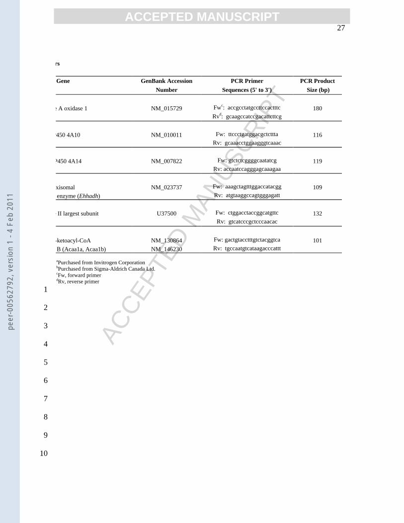
The primers used and their sources are shown in Table 1. Amplification consisted of a 15 10
min hot start (95°C) followed by 35 cycles of denaturation (94°C, 15 sec), annealing (60°C, 11
30 sec) and elongation (72°C, 30 sec). Melting curves followed by separation of PCR 12
products on a 2.5%, 0.5x TAE agarose gel were done to ensure the formation of a single 13
product at the appropriate size. Relative gene expression, normalized to the reference gene 14
RNA polymerase II (RPII), was calculated using the -2∆∆CT method [13]. RPII was used as 15
the housekeeping gene as its expression is constant throughout development, is not gender 16
specific and is not influenced by Tox exposure (not shown). To demonstrate the relative 17
abundance of mRNAs, data for both CTRL and Tox-treated mice were expressed relative to 18
the corresponding levels of RPII mRNA for each group of mice (CD-1, PP(+/+), PP(-/-)). 19
20
Immunoblot analysis For most Western blot analyses, frozen liver samples (~0.1 g) were 21
homogenized in 0.5 mL of ice cold buffer containing 20mM Tris-HCl (pH 7.4), 0.25 M 22
sucrose, 1 mM EDTA, 25 mM KCl, 1 mM dithiothreitol and a complete protease inhibitor 23
peer
-005
6279
2, v
ersi
on 1
- 4
Feb
2011
Page 9
ACC
EPTE
D M
ANU
SCR
IPT
ACCEPTED MANUSCRIPT 8
cocktail (Roche Diagnostics). Triton X-100 (1%, v/v) was added and after 30 min 1
incubation on ice, the homogenates were centrifuged (3000 x g, 10 min, 4°C) and 2
supernatants collected. For PPARα analysis, nuclear extracts were prepared essentially as 3
described by Gebel et al. [14]. Briefly, ~0.1 g of liver were homogenized in 0.3 mL of 4
buffer (above), the nuclei were pelleted and resuspended in fresh buffer containing 0.4 M 5
NaCl. The suspensions were mixed (4°C, 30 min) and centrifuged (2000 x g, 4°C, 30 min), 6
and supernatants (nuclear extracts) were collected. Protein was analysed using a kit from 7
Bio-Rad Laboratories (Mississauga, Canada) with bovine serum albumin as standard. 8
Liver homogenates or nuclear extracts (15 µg protein) were separated on 10% SDS-9
polyacrylamide gels, transferred to PVDF membranes and the membranes were blocked in 10
TBST buffer (20 mM Tris-HCl [pH 7.5], 55 mM NaCl, 0.1% Tween 20) containing 5% 11
non-fat milk powder and incubated with primary antibody for 1-2 hr. Primary antibodies 12
and dilutions used were as follows: goat anti-β-actin (1:500, Santa Cruz Biotechnology, 13
Santa Cruz, CA), rabbit anti-murine PPARα (1:1000, Santa Cruz), rabbit anti-rat CYP4A 14
(1:3000, Affinity BioReagents, Golden, CO), rabbit anti-rat ACOX 1 [15], and rabbit anti-15
rat pTHIO [16]. The last two were generous gifts from Dr. P.P. Van Veldhover, K.U. 16
Leuven, Belgium, and were used at 1:4000 dilutions. An antibody to L-PBE is not 17
commercially available, which precluded analysis of this protein. TBST was used as 18
wash buffer and antibody diluent. After washing 5 X 5 min, blots were incubated (45 min) 19
with horseradish peroxidase-conjugated secondary antibodies, either anti-rabbit IgG 20
(1:100,000, Chemicon, Temecula, CA) or anti-goat IgG (1:8000, Santa Cruz). Blots were 21
given final washes (5 X 10 min) and antibody binding was detected on X-ray film by 22
enhanced chemiluminescence (ECL Plus, Amersham Pharmacia Biotech, Buckinghamshire, 23
peer
-005
6279
2, v
ersi
on 1
- 4
Feb
2011
Page 10
ACC
EPTE
D M
ANU
SCR
IPT
ACCEPTED MANUSCRIPT 9
UK). The relative intensities were quantified using NIH Image software after films were 1
scanned using Umax MagicScan32 software. Relative levels of protein in each group of 2
mice are expressed relative to P13 CTRL = 1. Data for the Tox-treated mice are expressed 3
relative to the CTRL for its corresponding group. 4
5
Lauric acid hydroxylation assay Cytochrome P450 ω-hydroxylase (Cyp4A) activity was 6
determined using liver homogenates (1 mg protein) incubated (37°C, 10 min) in 50 mM 7
Tris-HCl, pH 7.4, containing 50 µM [1-14C]lauric acid (11,000 dpm/nmol, Amersham 8
Biosciences) and 1 mM NAPDH, in a final volume of 0.5 mL. Blank tubes contained all 9
reactants except NAPDH. Reactions were terminated by addition of 0.5 mL acetonitrile and 10
200 mg of each of lauric and 12-hydroxylauric acids. The mixtures were extracted with 11
diethyl ether and organic phases were pooled and dried under nitrogen. The residues were 12
suspended in 25 µL methanol and reaction products were separated on silica gel on 13
polyester plates (Sigma-Aldrich Canada Ltd., Oakville Canada) using the solvent system 14
diethyl ether:petroleum ether:formic acid (70:30:1, vol:vol). Bands corresponding to lauric 15
and 12-hydroxylauric acids were cut and radioactivity was quantitated using a scintillation 16
counter. The data were expressed as nmol lauric acid hydroxylated/min/mg protein. 17
18
Pathology assessment Thin (5 µm) sections were cut from mouse livers that were either 19
fixed in formalin (10%, by vol) and embedded in paraffin, or flash frozen at the time of 20
harvest. The paraffin sections were stained with hematoxylin and eosin, and the frozen 21
sections were stained with Oil Red O. The sections were cut and stained in the Clinical 22
Chemistry Laboratory of the IWK Health Centre. To evaluate liver fat content, images of 23
peer
-005
6279
2, v
ersi
on 1
- 4
Feb
2011
Page 11
ACC
EPTE
D M
ANU
SCR
IPT
ACCEPTED MANUSCRIPT 10
the oil red O stained sections were analysed using Adobe Photoshop CS2 after all non red 1
stained portions were converted to white. The images were converted to grayscale, 2
optimised for contrast, and the number of pixels derived from the red areas were counted 3
(Image J, public domain software from the National Institutes of Health) and expressed as 4
percent total pixel count. 5
6
Statistical analysis The mRNA data for each set of animals are expressed relative to 7
values for RPII (=1) for that set. Control protein levels for the PP(+/+) and PP(-/-) mice are 8
expressed relative to those of the CD-1 controls (=1), and values for the Tox-treated mice 9
are expressed relative to the corresponding controls. Unless indicated otherwise, data are 10
the means ±SEM of values from 4-12 mice. Statistical analyses were done using the two-11
tailed unpaired Student’s t test.12
peer
-005
6279
2, v
ersi
on 1
- 4
Feb
2011
Page 12
ACC
EPTE
D M
ANU
SCR
IPT
ACCEPTED MANUSCRIPT 11
Results and Discussion 1
2
The long-term health effects of prolonged exposure to environmental pollutants, 3
particularly xenobiotic hydrocarbons, is becoming of increasing concern to health care 4
workers as well as the general public. Atypical hydrocarbons that accumulate in the 5
environment or are present in human foods (e.g., phthalates, nonylphenol, 6
perfluorooctanoate, oxidized frying fats) are known to alter hepatic lipid metabolism 7
[9,17,18]. Only recently have several in vivo studies linked these alterations to effects on 8
gene expression [9,19,20]. The objective of this study was to determine whether the 9
widespread metabolic abnormalities that occur in young mice exposed to the pesticide 10
adjuvant, Tox, were due to altered function of the transcription regulator, PPARα, and 11
expression of its target genes involved in pFAO. 12
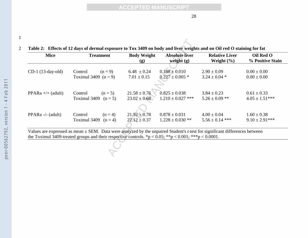
Effects of Tox on liver weights and pathology Chronic dermal exposure to Tox did 13
not have obvious adverse health effects on any of the mice, nor were body weights 14
affected (Table 2). Relative liver weights were significantly increased in Tox treated 15
CD-1 and PP(+/+) mice (12% and 37%, respectively)(Table 2), a response typically seen 16
in wild type rodents exposed to the prototype PPARα agonists, clofibrate and Wy-16,643 17
[12]. This effect of the latter drugs is commonly attributed to their ability to increase 18
hepatocyte number and/or size, and does not occur in PP(-/-) mice. In contrast, relative 19
liver weights in Tox-treated PP(-/-) mice were elevated significantly (Table 2). 20
Pathologic assessment of stained sections of livers from each group of mice showed 21
vacuoles in the PP(+/+) mice, a feature more prominent in the PP(-/-) animals. There were 22
no obvious structural abnormalities in the CD-1 mice, and there was no evidence of 23
peer
-005
6279
2, v
ersi
on 1
- 4
Feb
2011
Page 13
ACC
EPTE
D M
ANU
SCR
IPT
ACCEPTED MANUSCRIPT 12
inflammation or increased numbers of mitotic figures in any Tox-treated mice. The 1
possibility that the vacuoles were due to glycogen storage was ruled out as several 2
sections stained with periodic acid schiff were negative which indicates that the vacuoles 3
did not contain glycogen (data not shown). This was consistent with our earlier findings 4
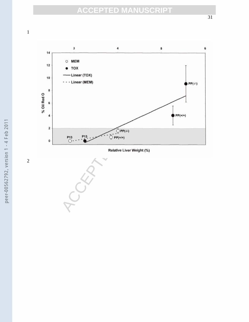
that Tox exposure significantly reduces hepatic glycogen [2]. Oil red O staining showed 5
that vacuoles in the Tox-treated livers were positive for fat (Table 2) in PP(-/-) and 6
PP(+/+), indicating lipid accumulation that could be endogenous fat and/or Tox 7
components. The absence of a statistical difference in oil red O staining of Tox exposed 8
P13 mice may reflect insensitivity of this assay at lower levels of quantification as focal 9
areas of staining were present. There was correlation between increases of relative liver 10
weight and percentage oil red O staining (Figure 3), which supports Tox related liver 11
weight increases are predominantly lipid accumulation. The finding that exposure to 12
petroleum-derived hydrocarbons increases relative liver weights in both PP(+/+) and PP(-/-) 13
mice is not unique, as Yang et al. reported the same result in PP(-/-) mice fed 14
perfluorooctanoic acid for seven days [21]. These authors concluded that the effect on 15
relative liver weights was independent of PPARα. A more likely explanation for the 16
discrepancy between the effects of clofibrates and Tox on relative liver weights in PP(-/-) 17
mice is the marked difference in the substrates being catabolized. Treatment with 18
clofibrate stimulates oxidation of endogenous fatty acids, primarily to ketone bodies that 19
are rapidly cleared from the liver. By contrast, Tox is a complex mixture of linear and 20
branched-chain hydrocarbons, some of which likely have cyclic and/or substituted (e.g., 21
sulfated, methylated) components. As with naturally occurring fatty acids, xenobiotic 22
hydrocarbons that reach the liver have the potential to undergo structural modifications, 23
peer
-005
6279
2, v
ersi
on 1
- 4
Feb
2011
Page 14
ACC
EPTE
D M
ANU
SCR
IPT
ACCEPTED MANUSCRIPT 13
as well as be oxidized and/or incorporated into triglyceride for export to the periphery 1
[22]. There is strong evidence that xenobiotics with abnormal structures are not 2
completely catabolized and fail to be esterified to form triacylglycerols, with resultant 3
hepatic accumulation. We observed this earlier in livers of Tox-treated mice [23]. We 4
propose that in both PP(+/+) and PP(-/-) mice, the Tox components that get into the 5
bloodstream, either via transdermal transport or by ingestion during grooming, are 6
transported to the liver. Their presence in the PP(+/+) mice would activate PPARα, as 7
this is a primary route for at least partial degradation and clearance of xenobiotic 8
hydrocarbons. This pathway is inactive in the PP(-/-) mice, with the result that Tox 9
components accumulate in even higher quantities. The fact that fatty liver also occurred, 10
albeit to a lesser extent, in the CD-1 pups is likely due to age- or strain-dependent 11
differences in the pharmacokinetic properties of the Tox components, a phenomenon 12
observed by others [24]. Of particular interest to this study, several groups have reported 13
that unmetabolized xenobiotic hydrocarbons are more potent in activating PPARα than 14
are endogenous ligands [25,26]. 15
Tox effects on expression of pFAO enzymes In vivo, activation of PPARα by 16
clofibrate and Wy-16,643 increased expression of all three enzymes of the pFAO 17
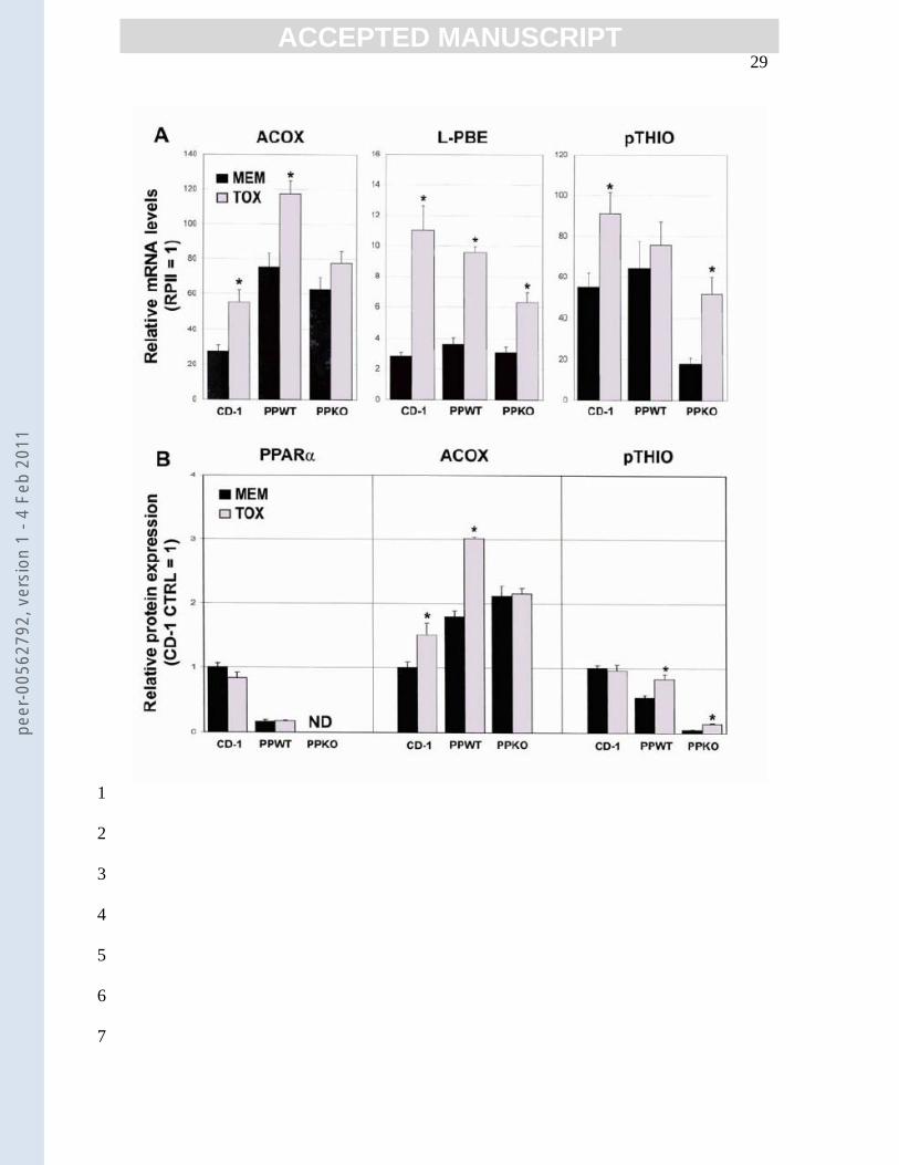
pathway (i.e., ACOX, L-PBE, pTHIO) [12]. Likewise, exposing the CD-1 mice to Tox 18
increased expression of mRNAs coding for ACOX, L-PBE and pTHIO (2-, 4- and 1.7-19
fold, respectively) (Fig. 1A). ACOX protein levels were also elevated (1.6-fold) with 20
Tox treatment, however those of pTHIO were not (Fig. 1B). The reason for the lack of 21
change in pTHIO protein is unclear; perhaps it reflects an adaptive down-regulation of 22
translation and/or rapid protein turnover. Livers of the adult PP(+/+) females exposed to 23
peer
-005
6279
2, v
ersi
on 1
- 4
Feb
2011
Page 15
ACC
EPTE
D M
ANU
SCR
IPT
ACCEPTED MANUSCRIPT 14
Tox exhibited a similar pattern of change in pFAO enzyme expression, with significant 1
increases in ACOX (mRNA and protein) and L-PBE mRNA. Levels of pTHIO mRNA 2
also increased, however values did not reach statistical significance. This is consistent 3
with the reported relative insensitivity of adult female mice to ligand-induced increases in 4
pTHIO [27]. The fact that pTHIO protein was increased (~1.6-fold) in Tox-treated 5
PP(+/+) mice may reflect a stable protein with a long half-life (Fig. 1B). Collectively, 6
these data showing Tox-induced increases in expression of pFAO enzymes (protein 7
and/or mRNA) in CD-1 pups and PP(+/+) mice are consistent with Tox effects being 8
mediated by PPARα. 9
If Tox-mediated increases in pFAO activity [5] and enzyme expression are totally 10
dependent on PPARα, these responses should not occur in PP(-/-) mice. As illustrated in 11
Fig. 1, this was true of ACOX mRNA and protein expression. However, expression of L-12
PBE (mRNA) was increased ~4-fold, and pTHIO mRNA and protein content increased 13
almost 3-fold. This is one of few reports of a putative PPARα agonist increasing 14
expression of L-PBE in PP(-/-) mice [28]. It should be emphasized that Tox-mediated 15
increases in transcription may not translate into increases in protein that have tangible 16
effects on metabolism. For example, pTHIO protein levels in the control PP(-/-) mice 17
were <10 % of their PP(+/+) counterparts and although these levels increased with Tox 18
exposure they did not exceed ~25% of the control PP(+/+) levels. Increases in pTHIO 19
expression also occurred in PP(-/-) mice treated with clofibrate [12] or the branched-chain 20
fatty-acid precursor, phytol [28]. These findings led the authors to conclude that these 21
agonists can mediate responses by both PPARα -dependent and independent pathways. 22
Gloerich et al. [28] did not speculate on the identity of the nuclear receptor(s) that 23
peer
-005
6279
2, v
ersi
on 1
- 4
Feb
2011
Page 16
ACC
EPTE
D M
ANU
SCR
IPT
ACCEPTED MANUSCRIPT 15
mediate(s) PPARα-independent effects of phytol, but two potential candidates are liver X 1
receptor α (LXRα) [29,30] and PPARγ [31]. The LXRα agonist, T0901317, up regulates 2
all three pFAO enzymes in PP(-/-) mice. The natural ligands of LXRα are cholesterol and 3
its derivatives, however other phenolic hydrocarbons are also potent agonists [32], and 4
Tox may contain structurally similar molecules. The second candidate, PPARγ, is not 5
highly expressed in PP(+/+) mouse liver, however feeding PP(-/-) mice high fat diets 6
increased its expression by ~4-fold [31], with a concomitant up regulation of pFAO 7
enzyme expression. 8
Tox effects on expression of PPARα Western blot analysis of the P13 mouse livers 9
showed that chronic dermal Tox exposure did not alter PPARα protein levels, a finding 10
also observed in PP(+/+) mice (Fig. 1B, left panel). Predictably, PPARα protein was not 11
detected in any PP(-/-) mice (Fig. 1B, left panel). The lack of change in PPARα protein 12
in the pups and PP(+/+) mice was not unexpected, as others also have found its levels 13
unchanged following exposure to PPARα agonists [9,12]. This may reflect its 14
demonstrated rapid turnover rate and the fact that the ability of ligands to stabilize its 15
expression is transitory [33]. 16
Tox effects on Cyp4A expression and activity A hallmark response to activation of 17
several nuclear receptors, including PPARα, by xenobiotic agents is an increase in 18
expression of the P450 ω-hydroxylases, including Cyp4A [34]. In all three groups of 19
mice in our study, mRNA levels of only two isoforms of Cyp4A (Cyp4A10 and 20
Cyp4A14) were sufficiently abundant to reliably detect Tox-related changes. 21
Characteristic of most strains of adult female mice [35], expression of Cyp4A12 was 22
extremely low (≤0.2%) in both the pups and adults. Cyp4A10 and 14 are the isoforms 23
peer
-005
6279
2, v
ersi
on 1
- 4
Feb
2011
Page 17
ACC
EPTE
D M
ANU
SCR
IPT
ACCEPTED MANUSCRIPT 16
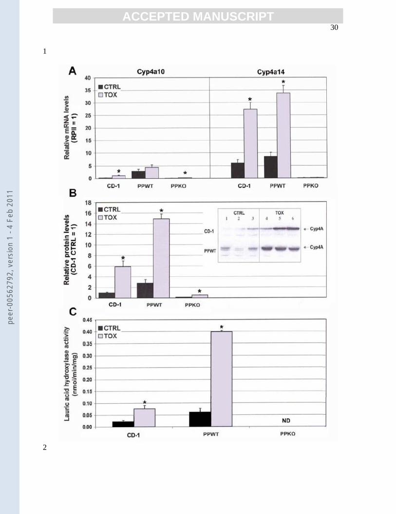
highly inducible by PPARα ligands [12,31,36,37]. In this study, CD-1 pups chronically 1
exposed to Tox had ≥4-fold elevations in mRNA coding for both Cyp4A10 and 14 (Fig. 2
2A); total Cyp4A protein content (Fig. 2B) and enzyme activity (Fig. 2C) were also 3
increased (~6- and >3-fold, respectively). It should be noted that the Cyp4A antibody 4
does not distinguish between isoforms. Consistent with earlier reports [36], there were no 5
gender-specific differences in the responses of the P13 pups to Tox (not shown). PP(+/+) 6
adult females treated with Tox also had increased levels of Cyp4A14 mRNA, total 7
Cyp4A protein and enzyme activity (4-, 5- and >6-fold, respectively)(Fig. 2). Cyp4A10 8
mRNA was increased, however values did not reach statistical significance. One possible 9
explanation is that up regulation of this isoform does not occur except with very long 10
exposure, as seen with dioxin treatment [34]. PP(-/-) mice had predictably low 11
constitutive levels of Cyp4A10 and 14, and no change in Cyp4A14 mRNA occurred with 12
Tox exposure (Fig. 2A). Unexpectedly, Cyp4A10 content was increased ~4-fold with 13
Tox treatment. However, Anderson et al. recently determined that Cyp4A10 was the one 14
Cyp4A isoform whose expression was regulated independent of PPARα [30]. They 15
provided evidence that this could occur by binding of the ligand (e.g., a component of 16
Tox) to one of the retinoid X receptors that are the mandatory heterodimeric partners for 17
activation of most nuclear receptors, including PPARα, LXR and PPARγ. 18
Relevance Pesticides are ubiquitous in the environment, and concern that exposure 19
can pose adverse health risks is growing, particularly as these xenobiotics are stored and 20
accumulate for a very long time in humans [38]. Very few active pesticide ingredients 21
appear to exert health risks in vivo by activation of PPARα [37]. The ‘other’ ingredients 22
in pesticide formulations, the xenobiotic hydrocarbons, are often more toxic than the 23
peer
-005
6279
2, v
ersi
on 1
- 4
Feb
2011
Page 18
ACC
EPTE
D M
ANU
SCR
IPT
ACCEPTED MANUSCRIPT 17
active ingredients [11], yet have received very little attention. This is the first study to 1
obtain evidence that subclinical dyslipidemia in young mice dermally exposed to a 2
pesticide adjuvant involves up regulation of select lipid metabolizing enzymes. These 3
effects occur despite several factors that could attenuate the effective dose. Since Tox is 4
a complex mixture of structurally heterogeneous components, some components may not 5
penetrate skin, and those that do likely have variable metabolic fates and liver clearance. 6
As well, only select components or metabolites of Tox may be responsible for the 7
changes in gene expression. From our data with the PP(-/-) mice it appears that Tox 8
components may serve as ligands for more than one nuclear receptor. Whether effects on 9
enzyme expression are mediated through direct or indirect activation of a nuclear 10
receptor(s) remains to be determined. Nevertheless, our results suggest that exposing 11
high-risk populations, particularly those in the petrochemical and agricultural industries, 12
to this class of persistent organic chemicals has the potential to predispose them to 13
significant perturbations in energy metabolism and development of fatty liver. We 14
believe that this study has far broader implications than exposure only to pesticides, as 15
similar effects are elicited by the wide range of structurally diverse industrial chemicals 16
to which humans have been exposed since the onset of industrialization. 17
In summary, this study has demonstrated that chronic dermal exposure to the 18
currently used pesticide adjuvant, Tox, significantly increases expression of select lipid 19
metabolizing enzymes in livers of young and adult mice. The changes in expression most 20
likely to result in meaningful effects on hydrocarbon β-oxidation (e.g., ACOX) or ω-21
oxidation (Cyp4A) did not occur in PP(-/-) mice, suggesting involvement of PPARα. A 22
significant consequence of Tox exposure that occurred despite the absence of PPARα 23
peer
-005
6279
2, v
ersi
on 1
- 4
Feb
2011
Page 19
ACC
EPTE
D M
ANU
SCR
IPT
ACCEPTED MANUSCRIPT 18
was increased relative liver weights and development of fatty liver. Evidence suggests 1
that other xenobiotic hydrocarbons (e.g., perfluorooctanoic acid) elicit a similar effect 2
[21]. This is a very important distinction between effects of the classic PPARα agonists 3
and environmental pollutants, and the reason we should be even more fearful of these 4
chemicals. 5
6
Acknowledgement: This study was supported by the Natural Sciences and Engineering 7
Research Council of Canada. 8
9
peer
-005
6279
2, v
ersi
on 1
- 4
Feb
2011
Page 20
ACC
EPTE
D M
ANU
SCR
IPT
ACCEPTED MANUSCRIPT 19
References 1
2
[1] E.Liwarska-Bizukojc, K. Miksch, A. Malachowska-Jutsz, J. Kalka, Acute 3
toxicity and genotoxicity of five selected anionic and nonionic surfactants, 4
Chemosphere 58 (2005) 1249-1253. 5
[2] M.G.Murphy, M. Al-Khalidi, J.F.S. Crocker, S.H.S.Lee, P. O’Regan, P.D. Acott, 6
Two formulations of the industrial surfactant, Toximul, differentially reduce 7
mouse weight gain and hepatic glycogen in vivo during early development: 8
effects of exposure to influenza B virus, Chemosphere 59 (2005) 235-246. 9
[3] R. White, S. Jobling, S.A. Hoare, J.P. Sumpter, M.G. Parker, Environmentally 10
persistent alkylphenolic compounds are estrogenic, Endocrinology 135 (1994) 11
175-182. 12
[4] J.F.S. Crocker, R.L. Ozere, S.H. Safe, S.C. Digout, K.R. Rozee, O. Hutzinger, 13
Lethal interaction of ubiquitous insecticide carriers with virus, Science 192 14
(1976) 1351-1353. 15
[5] M.G. Murphy, J.F.S. Crocker, H. Her, Abnormalities in hepatic fatty-acid 16
metabolism in a surfactant/influenza B virus mouse model for acute 17
encephalopathy, Biochim. Biophys. Acta 1315 (1996) 208-216. 18
[6] M.G. Murphy, J.F.S. Crocker, S.H.S. Lee, P. Acott, H. Her, Sequestration of 19
coenzyme A by the industrial surfactant, Toximul MP8. A possible role in the 20
inhibition of fatty-acid β-oxidation in a surfactant/influenza B virus mouse model 21
for acute hepatic encephalopathy, Biochim. Biophys. Acta 1361 (1997) 103-113. 22
23
peer
-005
6279
2, v
ersi
on 1
- 4
Feb
2011
Page 21
ACC
EPTE
D M
ANU
SCR
IPT
ACCEPTED MANUSCRIPT 20
[7] M.G. Murphy, J.F.S. Crocker, P. O’Regan, S.H.S. Lee, L. Geldenhuys, K. 1
Dooley, M. Al-Khalidi, P. Acott, Carnitine, acylcarnitine and amino acid profiles 2
analyzed by tandem mass spectrometry in a surfactant/virus mouse model of acute 3
hepatic encephalopathy, Submitted to Chemosphere, October, 2006. 4
[8] P. Lefebvre, G. Chinetti, J.-C. Fruchart, B. Staels, Sorting out the roles of PPARα 5
in energy metabolism and vascular homeostasis, J. Clin. Invest. 116, 571-580. 6
[9] P.M. Chao, C.-Y. Chao, F.-J. Lin, C.-J. Huang, Oxidized frying oil up-regulates 7
hepatic acyl-CoA oxidase and cytochrome P4504A1 genes in rats and activates 8
PPARα, J. Nutr. 131 (2001) 3166-3174. 9
[10] J.M. Shipley, C.H. Hurst, S.S. Tanaka, F.L. DeRoos, J.L. Butenhoff, A.M. Seacat, 10
D.J. Waxman, trans-Activation of PPARα and induction of PPARα target genes 11
by perfluorooctane-based chemicals, Toxicol. Sci. 80 (2004) 151-160. 12
[11] B. Burroughs, R. Tarone, J.S. Kesner, V.F. Garry, Herbicides and adjuvants: an 13
evolving view, Toxicol. Ind. Health 15 (1999) 160-168. 14
[12] S.T.S. Lee, T. Pineau, J. Drago, E.J. Lee, J.W. Owens, D.L. Kroetz, P.M. 15
Fernandez-Salguero, H. Westphal, F.J. Gonzales, Targeted disruption of the α 16
isoform of the peroxisome-proliferator-activated receptor gene in mice results in 17
abolishment of the pleitrophic effects of peroxisome-proliferators, Mol. Cell. 18
Biol. 15 (1995) 3012-3022. 19
[13] K.J. Livak, T.D. Schmittgen, Analysis of relative gene expression data using real-20
time quantitative PCR and the 2(-delta delta C(T)) method, Methods 25 (2001) 21
402-408. 22
[14] T. Gebel, M. Arand, F. Oesch, Induction of the peroxisome-proliferator activated 23
peer
-005
6279
2, v
ersi
on 1
- 4
Feb
2011
Page 22
ACC
EPTE
D M
ANU
SCR
IPT
ACCEPTED MANUSCRIPT 21
receptor by fenofibrate in rat liver, FEBS Lett. 309 (1992) 37-40. 1
[15] P.P. Van Veldhoven, P. Van Rompuy, M. Fransen, B. De Bethune, G.P. 2
Mannaerts, Large scale purification and further characterization of rat pristanoyl-3
CoA oxidase, Eur. J. Biochem. 222 (1994) 795-801. 4
[16] V.D. Antonenkov, P. Van Veldhoven, E. Waelkens, G.P. Mannaerts, Substrate 5
specificities of 3-oxoacyl-CoA thiolase A and sterol carrier protein 2/3-oxoacyl-6
CoA thiolase purified from normal rat liver peroxisomes. Sterol carrier protein 7
2/3-oxoacyl-CoA thiolase is involved in the metabolism of 2-methyl-branched 8
fatty acids and bile acid intermediates, J. Biol. Chem. 272 (1997) 26023-26031. 9
[17] L.D. Winberg, M.Z. Badr, Mechanism of phthalate-induced inhibition of hepatic 10
mitochondrial beta-oxidation, Toxicol. Lett. 76 (1995) 63-69. 11
[18] S. Khan, M. Codner, J.F. Payne, A.D. Rahimtula, Effect of a Prudehoe Bay 12
crude oil on hepatic and renal peroxisomal β-oxidation and mixed function 13
oxidase activities in rats, Carcinogenesis 10 (1989) 269-272. 14
[19] P.J. Lapinskas, S. Brown, L.M. Leesnitzer, S. Blanchard, C. Swanson, R.C. 15
Cattley, J.C. Corton, Role of PPARα in mediating the effects of phthalates and 16
metabolites in the liver, Toxicology 207 (2005) 149-163. 17
[20] K.S. Guruge, L.W.Y. Yeung, N. Yamanaka, S. Miyazaki, P.K.S. Lam, J.P. Giesy, 18
P.D. Jones, N. Yamashita, Gene expression profiles in rat liver treated with 19
perfluorooctanoic acid (PFOA), Toxicol. Sci. 89 (2006) 93-107. 20
[21] Q. Yang, Y. Xie, S.E.H. Alexson, B.D. Nelson, J.W. DePierre, Involvement of 21
the peroxisome proliferator-activated receptor alpha in the immunomodulation 22
peer
-005
6279
2, v
ersi
on 1
- 4
Feb
2011
Page 23
ACC
EPTE
D M
ANU
SCR
IPT
ACCEPTED MANUSCRIPT 22
caused by peroxisome proliferators in mice, Biochem. Pharmacol. 63 (2002) 1
1893-1900. 2
[22] P.F. Dodds, Xenobiotic lipids: The inclusion of xenobiotic compounds in 3
pathways of lipid biosynthesis, Prog. Lipid Res. 34 (1995) 219-247. 4
[23] J.F.S. Crocker, D. Fung, R. Hudson, S. Safe, Examination of the role of 5
surfactants in Reye’s Syndrome, in: J.D. Pollack (Ed.), Reye’s Syndrome IV, The 6
National Reye’s Syndrome Foundation, Bryan, OH, 1985, pp. 135-140. 7
[24] U. Intrasuksri, S.M. Rangwala, M. O’Brien, D.J. Noonan, D.R. Feller, 8
Mechanisms of peroxisome proliferation by perfluorooctanoic acid and 9
endogenous fatty acids, Gen. Pharmacol. 31 (1998) 187-197. 10
[25] L.N. Larsen, L. Granlund, A.K. Holmeide, L. Skattebøl, H.I. Nebb, J. Bremer, 11
Sulfer-substituted and α-methylated fatty acids as peroxisome-proliferator-12
activated receptor activators, Lipids 40 (2005) 49-57. 13
[26] P.M. Hinderliter, X. Han, G.L. Kennedy, J.L. Butenhoff, Age effect on 14
perfluorooctanoate (PFOA) plasma concentration in post-weanling rats following 15
oral gavage with ammonium perfluorooctanoate (APFO), Toxicology 225 (2006) 16
195-203. 17
[27] Y.-C. Zhou, H.W. Davey, M.J. McLachlan, T. Xie, D.J. Waxman, Elevated basal 18
expression of liver peroxisomal β-oxidation enzymes and CYP4A microsomal 19
fatty acid ω-hydroxylase in STAT5b-/- mice: Cross-talk in vivo between 20
peroxisome-proliferator-activated receptor and signal transducer and activator of 21
transcription signaling pathways, Tox. Appl. Pharmacol. 182 (2002) 1-10. 22
peer
-005
6279
2, v
ersi
on 1
- 4
Feb
2011
Page 24
ACC
EPTE
D M
ANU
SCR
IPT
ACCEPTED MANUSCRIPT 23
[28] J. Gloerich, N. van Vlies, G.A. Jansen, S. Denis, J.P.N. Ruiter, M.A. van 1
Werkhoven, M. Duran, F.M. Vaz, R.J.A. Wanders, S. Ferdinandusse, A phytol-2
enriched diet induces changes in fatty acid metabolism in mice both via PPARα-3
dependent and independent pathways, J. Lipid Res. 46 (2005) 716-726. 4
[29] T. Hu, P. Foxworthy, A. Siesky, J.V. Ficorilli, H. Gao, S. Li, M. Christe, T. Ryan, 5
G. Cao, P. Eacho, M.D. Michael, L.F. Michael, Hepatic peroxisomal fatty acid β-6
oxidation is regulated by Liver X Receptor α, Endocrinology 146 (2005) 5380-7
5387. 8
[30] S.P. Anderson, C. Dunn, A. Laughter, L. Yoon, C. Swanson, T.M. Stulnig, K.R. 9
Steffensen, R.A.S. Chandraratna, J.Å. Gustafsson, J.C. Corton, Overlapping 10
transcription programs regulated by the nuclear receptors peroxisome-11
proliferator-activated receptor α, retinoid X receptor, and liver X receptor in 12
mouse liver, Mol. Pharmacol. 66 (2004) 1440-1452. 13
[31] D. Patsouris, J.K. Reddy, M. Müller, S. Kersten, Peroxisome-proliferator-14
activated receptor α mediates the effects of a high-fat diet on hepatic gene 15
expression, Endocrinol. 147 (2006) 1508-1516. 16
[32] L. Li, J. Liu, L Zhu, S. Cutler, H. Hasegawa, B. Shan, J.C. Medina, Discovery 17
and optimization of a novel series of liver X receptor-α agonists, Bioorg. Med. 18
Chem. Lett. 16 (2006) 1638-1642. 19
[33] C. Blanquart, O. Barbier, J.C. Fruchart, B. Staels, C. Glineur, Peroxisome-20
proliferator-activated receptor-α (PPARα) turnover by the ubiquitin-proteasome 21
system controls the ligand-induced expression level of its target genes, J. Biol. 22
Chem. 277 (2002) 37254-37259. 23
peer
-005
6279
2, v
ersi
on 1
- 4
Feb
2011
Page 25
ACC
EPTE
D M
ANU
SCR
IPT
ACCEPTED MANUSCRIPT 24
[34] M.B. Genter, C.D. Clay, T.P. Dalton, H. Dong, D.W. Nebert, H.G. Shertzer, 1
Comparison of mouse hepatic mitochondrial versus microsomal cytochromes 2
P450 following TCDD treatment, Biochem. Biophys. Res. Comm. 342 (2006) 3
1375-1381. 4
[35] B. Jeffery, A.E. Choudhury, N. Horley, M. Bruce, S.R. Tomlinson, R.A. Roberts, 5
T.J.B. Gray, D.A. Barrett, P.N. Shaw, D. Kendall, D.R. Bell, Peroxisome-6
proliferator activated receptor α regulates a male-specific cytochrome P450 in 7
mouse liver, Arch. Biochm. Biophys. 429 (2004) 231-236. 8
[36] Y.M. Heng, C.-W. Kuo, P.S. Jones, R. Savory, R.M. Schulz, S.R. Tomlinson, 9
T.J.B. Gray, D.R. Bell, A novel murine P-450 gene, Cyp4a14, is part of a cluster 10
of Cyp4a and Cyp4b, but not Cyp4f, genes in mouse and humans, Biochem. J. 11
325 (1997) 741-749. 12
[37] S. Takeuchi, T. Matsuda, S. Kobayashi, T. Takahashi, H. Kojima, In vitro 13
screening of 200 pesticides for agonistic activity via mouse peroxisome-14
proliferator-activated receptor (PPARα) and PPARγ and quantitative analysis of 15
in vivo induction pathway, Toxicol. Appl. Pharmacol. (2006), 16
doi:10,1016/j.taap.2006.08.011. 17
[38] D.E. Duncan, The pollution within, National Geographic 210 (2006) 116-133. 18
19
20
21
22
peer
-005
6279
2, v
ersi
on 1
- 4
Feb
2011
Page 26
ACC
EPTE
D M
ANU
SCR
IPT
ACCEPTED MANUSCRIPT 25
Figure Legends 1
2
Figure 1: Effect of Tox on expression of PPARα and peroxisomal fatty-acid β-3
oxidation enzymes. A. mRNA expression Total RNA was extracted from 4
mouse livers, reverse transcribed and analyzed by qPCR for expression of ACOX, 5
L-PBE and pTHIO as described in Methods. The data for each enzyme in each 6
mouse group (CD-1, PP(+/+), PP(-/-)) are expressed relative to mean RPII levels in 7
that group (RPII=1). B. Protein expression Levels of PPARα, ACOX and 8
pTHIO protein were quantitated by Western blot analysis as described in 9
Methods. Raw data were standardized to β-actin and all values were standardized 10
to those for the control CD-1 pups (CTRL=1). Values represent the mean ± SEM 11
(n = 4-12 [CD-1, PP(+/+)]; n = 2 for PP(-/-) [PPARα, Cyp4A only]). *p<0.05-12
<0.0001, relative to values for corresponding CTRL in that group of mice. ND, 13
not detected. 14
15
Figure 2: Effect of Tox on hepatic Cyp4A expression and enzyme activity. 16
A. mRNA expression. Methods were as described in the legend to Fig. 1, using 17
primers for Cyp4A 10 and 14; values were expressed relative to RPII values 18
for the control group of corresponding mice. B. Cyp4A protein content was 19
quantitated as described in Fig. 1, using an antibody nonspecific for Cyp4A 20
isoform. Data are expressed as described in Fig. 1B. The insert shows 21
representative blots of CTRL and Tox-treated livers from CD-1 pups and 22
PP(+/+) mice probed for total Cyp4A protein. C. Lauric acid ω-hydroxylase 23
peer
-005
6279
2, v
ersi
on 1
- 4
Feb
2011
Page 27
ACC
EPTE
D M
ANU
SCR
IPT
ACCEPTED MANUSCRIPT 26
activity was measured in livers of CD-1 and PP(+/+) mice as described in 1
Methods. The data are expressed as nmol lauric acid ω-hydroxylated/min/mg 2
protein (mean ± SEM, n=3). Statistical analysis and data significance are as 3
described in Fig. 1. 4
5
6
7
8
9
10
11
12
13
14
15
16
17
18
19
20
21
22
23
peer
-005
6279
2, v
ersi
on 1
- 4
Feb
2011
Page 28
ACC
EPTE
D M
ANU
SCR
IPT
ACCEPTED MANUSCRIPT 27
1
2
3
4
5
6
7
8
9
10
rs
Gene GenBank Accession PCR Primer PCR Product Number Sequences (5' to 3') Size (bp)
e A oxidase 1 NM_015729 Fwc: accgcctatgccttccactttc 180
Rvd: gcaagccatccgacattcttcg
P450 4A10 NM_010011 Fw: ttccctgatggacgctcttta 116 Rv: gcaaacctggaagggtcaaac
P450 4A14 NM_007822 Fw: gtctctcggggcaatatcg 119 Rv: accaatccagggagcaaagaa
xisomal NM_023737 Fw: aaagctagtttggaccatacgg 109 enzyme (Ehhadh) Rv: atgtaaggccagtgggagatt
e II largest subunit U37500 Fw: ctggacctaccggcatgttc 132
Rv: gtcatcccgctcccaacac
-ketoacyl-CoA NM_130864 Fw: gactgtacctttgtctacggtca 101 B (Acaa1a, Acaa1b) NM_146230 Rv: tgccaatgtcataagacccattt
aPurchased from Invitrogen CorporationbPurchased from Sigma-Aldrich Canada Ltd.cFw, forward primerdRv, reverse primer
peer
-005
6279
2, v
ersi
on 1
- 4
Feb
2011
Page 29
ACC
EPTE
D M
ANU
SCR
IPT
ACCEPTED MANUSCRIPT 28
1
Table 2: Effects of 12 days of dermal exposure to Tox 3409 on body and liver weights and on Oil red O staining for fat 2 Mice Treatment Body Weight Absolute liver Relative Liver Oil Red O
(g) weight (g) Weight (%) % Positive Stain CD-1 (13-day-old) Control (n = 9) 6.48 ± 0.24 0.188 ± 0.010 2.90 ± 0.09 0.00 ± 0.00 Toximul 3409 (n = 9) 7.01 ± 0.15 0.227 ± 0.005 * 3.24 ± 0.04 * 0.00 ± 0.00 PPARa +/+ (adult) Control (n = 5) 21.58 ± 0.76 0.825 ± 0.038 3.84 ± 0.23 0.61 ± 0.33 Toximul 3409 (n = 5) 23.02 ± 0.68 1.210 ± 0.027 *** 5.26 ± 0.09 ** 4.05 ± 1.51*** PPARa -/- (adult) Control (n = 4) 21.92 ± 0.78 0.878 ± 0.031 4.00 ± 0.04 1.60 ± 0.38 Toximul 3409 (n = 4) 22.12 ± 0.37 1.228 ± 0.030 ** 5.56 ± 0.14 *** 9.10 ± 2.91*** Values are expressed as mean ± SEM. Data were analyzed by the unpaired Student's t-test for significant differences between the Toximul 3409-treated groups and their respective controls. *p < 0.05; **p < 0.001; ***p < 0.0001.
peer
-005
6279
2, v
ersi
on 1
- 4
Feb
2011
Page 30
ACC
EPTE
D M
ANU
SCR
IPT
ACCEPTED MANUSCRIPT 29
1
2
3
4
5
6
7
peer
-005
6279
2, v
ersi
on 1
- 4
Feb
2011
Page 31
ACC
EPTE
D M
ANU
SCR
IPT
ACCEPTED MANUSCRIPT 30
1
2
peer
-005
6279
2, v
ersi
on 1
- 4
Feb
2011
Page 32
ACC
EPTE
D M
ANU
SCR
IPT
ACCEPTED MANUSCRIPT 31
1
2
peer
-005
6279
2, v
ersi
on 1
- 4
Feb
2011