Page 1
The Security of Deep Learning Defences for MedicalImaging
Moshe Levya, Guy Amita, Yuval Elovicia, Yisroel Mirskya,∗
aDepartment of Software and Information Systems Engineering Ben-Gurion University ofthe Negev, Beer-Sheva 8410501, Israel
Abstract
Deep learning has shown great promise in the domain of medical image anal-
ysis. Medical professionals and healthcare providers have been adopting the
technology to speed up and enhance their work. These systems use deep neu-
ral networks (DNN) which are vulnerable to adversarial samples; images with
imperceivable changes that can alter the model’s prediction. Researchers have
proposed defences which either make a DNN more robust or detect the adver-
sarial samples before they do harm. However, none of these works consider an
informed attacker which can adapt to the defence mechanism. We show that an
informed attacker can evade five of the current state of the art defences while
successfully fooling the victim’s deep learning model, rendering these defences
useless. We then suggest better alternatives for securing healthcare DNNs from
such attacks: (1) harden the system’s security and (2) use digital signatures.
Keywords: Medical imaging security, Deep learning, Adversarial samples
1. Introduction
Deep learning is a data driven machine learning technique which provides
state-of-the-art performance in image analysis tasks. The technique uses a
model called a deep neural networks (DNN) which makes predictions by learning
from historic data. Over the past ten years, deep learning technology has proven
itself as an efficient and highly accurate tool for image analysis, solving tasks
∗Corresponding author: [email protected]
Preprint submitted to Elsevier January 24, 2022
arX
iv:2
201.
0866
1v1
[cs
.CR
] 2
1 Ja
n 20
22
Page 2
0.01
Clean Scan Adversarial ScanAdversarial Noise
Prediction: HealthyConfidence: 99.6%
Prediction: PneumoniaConfidence: 99.9%
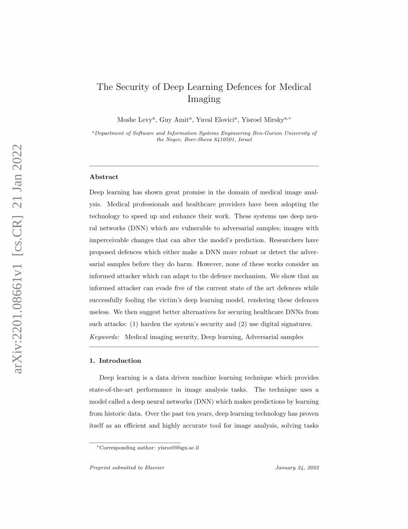
Figure 1: How adversarial noise can shift the prediction of a DNN based pneumothorax
detector.
that require a wide variety of skills: from detecting cancer to spatial alignment
and even content based image retrieval [1]. DNNs are expected to have an even
more substantial role in the near future [2, 3]. Some deep learning solutions
are already approved and deployed [4] with many more major companies in the
medical field working on DNN based products, planned to be deployed soon
[5, 6, 7].
One of the main drawbacks of DNNs is that they are vulnerable to an attack
called adversarial samples [8, 9]. An adversarial sample is a seemingly benign
input (e.g., x-ray image) which contains a crafted noise pattern, such that the
pattern is imperceivable to humans but can dramatically alter the prediction of
the model. For example, to change the classification of a benign x-ray to malign
and vice versa (see Figure 1).
Since DNNs are becoming mainstream in radiology and other medical imag-
ing analysis domains, the existence of adversarial samples now threatens the
healthcare community. There are number of reasons why an attacker would
want to trick a medical imaging DNN.
Earn Money. The attacker may try to earn money. For example, quality
of life insurance fraud can be achieved by inserting a lesion to a brain
MRI as irrefutable evidence as to why the patient can no longer taste
food. Moreover, medical scans can be held hostage in a ransomware attack
2
Page 3
where an unknown number of scans will remain tampered unless payment
is made.
Cause Harm. The goal might be to cause harm for revenge, fame, terrorism,
or to cause political turmoil. For example, the attacker can cause the
model to miss a lesion in an MRI or suggest the wrong diagnosis to a
radiologist.
Get Priority. An attacker may try to get medical attention faster. For exam-
ple, a slight increase in the size of a bone fracture or spinal disk herniation,
may lead to the patient receiving treatment sooner (over more deserving
patients).
Attacks on medical imagery is achievable. This is demonstrated through the
million of medical records stolen in data breaches only this year1. Researchers
have shown how Picture Archiving and Communication System (PACS) can be
hacked into from the Internet [10]. Researchers have also demonstrated how
easy it is to gain physical access to the PACS and plant a backdoor device in
the network [11].
Since these attacks are tangible, the threat of adversarial samples has gained
attention in the medical imaging community over the last few years [12, 13, 14].
As such, researchers have proposed a wide variety of detection and mitigation
techniques to protect DNN-based medical imaging applications.
1.1. Contribution
In this article, we warn the medical imaging community that these state-of-
the-art defences provide no security. The reason is that all of these defences
were designed assuming that the attacker will not consider the defence while
crafting the attack. In reality, adversaries are adaptive and can easily evade
these defences. To support our claim, we attack five state-of-the-art medical
imaging defences and show that all of them fail to protect the victim’s DNN
1https://www.healthcareitnews.com/news/biggest-healthcare-data-breaches-2021
3
Page 4
from adversarial samples. We then suggest better alternatives for securing med-
ical imaging DNNs from such attacks: (1) harden the system’s security and (2)
enable digital signatures for image integrity validation (a technology already
supported in the DICOM standard). This article also provides an introduc-
tion to DNNs and the basic concepts of adversaries samples, at a level which
approachable for individuals who are new to the domain.
By raising awareness to this issue, and by suggesting stronger countermea-
sures, we hope this paper will provide healthcare professionals the ability to
protect their systems and patients before these attacks become more main-
stream.
2. Attack Model
In this section we describe how an attacker can gain access to medical im-
agery, and detail an attacker’s limitations in crafting adversarial samples for
these images.
2.1. Gaining Access
Medical imagery is stored in data files typically using the DICOM format.
Currently, the most common way for healthcare organizations to store, manage
and analyze these files is through a PACS. The PACS provides medical personal
secure access to these files from within the organization, and in some cases, from
anywhere around the world. Although the PACS network was thought to be
secure, in recent years hackers have demonstrated how it can be breached both
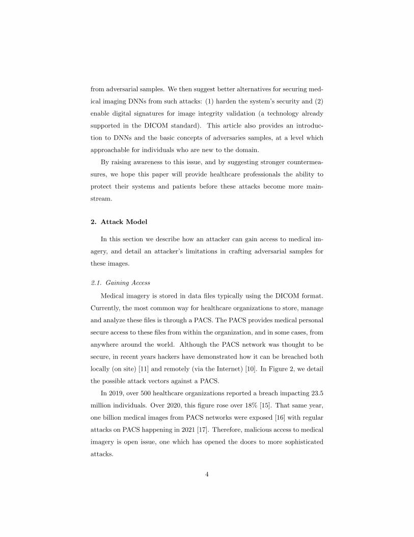
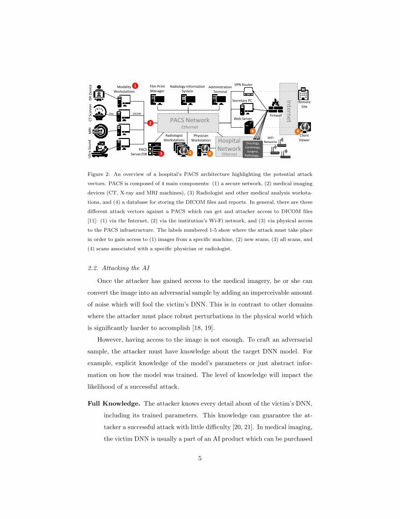
locally (on site) [11] and remotely (via the Internet) [10]. In Figure 2, we detail
the possible attack vectors against a PACS.
In 2019, over 500 healthcare organizations reported a breach impacting 23.5
million individuals. Over 2020, this figure rose over 18% [15]. That same year,
one billion medical images from PACS networks were exposed [16] with regular
attacks on PACS happening in 2021 [17]. Therefore, malicious access to medical
imagery is open issue, one which has opened the doors to more sophisticated
attacks.
4
Page 5
Attack Vectors
Physician Workstation
DR
Dev
ice
CT
Scan
ner
MR
IU
ltra
So
un
d
Web Server
Film Print Manager
Radiology Information System
Administration Terminal
Radiologist Workstations
Intern
et
Remote Site
Client Viewer
Modality Workstations
1
4 4
PACS Server/DB 3 4
Hospital Network
4
2 PACS NetworkEthernet
VPN Router
Secretary PC
Oncology, Cardiology,
Surgery, Pathology…Ethernet
DICOM Firewall
WiFiNetworks
Typical PACs Network of a Hospital:
Figure 2: An overview of a hospital’s PACS architecture highlighting the potential attack
vectors. PACS is composed of 4 main components: (1) a secure network, (2) medical imaging
devices (CT, X-ray and MRI machines), (3) Radiologist and other medical analysis worksta-
tions, and (4) a database for storing the DICOM files and reports. In general, there are three
different attack vectors against a PACS which can get and attacker access to DICOM files
[11]: (1) via the Internet, (2) via the institution’s Wi-Fi network, and (3) via physical access
to the PACS infrastructure. The labels numbered 1-5 show where the attack must take place
in order to gain access to (1) images from a specific machine, (2) new scans, (3) all scans, and
(4) scans associated with a specific physician or radiologist.
2.2. Attacking the AI
Once the attacker has gained access to the medical imagery, he or she can
convert the image into an adversarial sample by adding an imperceivable amount
of noise which will fool the victim’s DNN. This is in contrast to other domains
where the attacker must place robust perturbations in the physical world which
is significantly harder to accomplish [18, 19].
However, having access to the image is not enough. To craft an adversarial
sample, the attacker must have knowledge about the target DNN model. For
example, explicit knowledge of the model’s parameters or just abstract infor-
mation on how the model was trained. The level of knowledge will impact the
likelihood of a successful attack.
Full Knowledge. The attacker knows every detail about of the victim’s DNN,
including its trained parameters. This knowledge can guarantee the at-
tacker a successful attack with little difficulty [20, 21]. In medical imaging,
the victim DNN is usually a part of an AI product which can be purchased
5
Page 6
by the attacker and then extracted, for example using the techniques of
[22, 23, 24, 25, 26, 27].
Limited Knowledge. The attacker knows how the DNN was trained (i.e.,
what the datasets were) but doesn’t know anything about the DNN model
itself. In this scenario, an attacker can train his/her own DNN model and
then use this model to craft the adversarial sample. This approach is
called a transfer attack [28]. However, here, the attacker will not have a
guarantee that the attack will work.
3. Background
In this section, we explain of how adversarial samples are created. We open
with a brief introduction to DNNs and then describe their vulnerability.
3.1. Deep Learning
In general, there are several approaches for training a DNN to perform an
image analysis task. The most common approach in medical imaging is the
‘supervised’ approach where the DNN is given pairs (x, y) where x is an input
image (e.g., a CT scan) and y is the desired output of the DNN –the ground
truth of x for the given task. For example, for classification of lung nodules, x
may be an image of a lung nodule and y may be a label that is either benign
or malign. Moreover, for the task of segmentation (localization) of tumors MRI
scans, x may be an axial slice of the patient’s head and y would be a mask
or probability map which indicates where the tumor is located. The collection
of images and their labels used to train the DNN is referred to as the model’s
training set.
To train a DNN we perform the back-propagation algorithm: (1) pass one
or more examples of x through the model, (2) calculate the errors between the
predicted y and the actual y, (3) propagate the errors backwards through the
model to identify the erroneous parameters, (4) update (correct) the parame-
ters using an optimization algorithm, and (5) repeat this process until model
6
Page 7
converges. The error between the actual and predicted labels is calculated us-
ing a differentiable ‘loss function’. The optimizer uses the gradient of the loss
function to make a small step towards the optimal solution at each iteration.
This optimization algorithm is called ‘gradient descent‘.
After training, the DNN can predict a label y given an input x. If the training
set is large and diverse, then the DNN can generalize unseen observations which
were not seen in the training set.
3.2. Adversarial Samples
An adversarial sample x′ is a modified version of x which includes a small
imperceivable signal δ, such that x′ = x+ δ. The signal δ is crafted such that x′
convinces the DNN to predict the wrong label or segmentation mask for x and
with high confidence. The attacker who crafts x′ can perform a targeted attack
(choose which label or mask should be predicted) [29, 30] or an untargeted
attack (cause a general misclassification) [31, 32]. What makes x′ dangerous is
that it looks like the original x to a human (see figure 1).
The adversarial noise δ is crafted using a gradient-based technique, similar
to how a DNN is trained using back-propagation.
In general, the approach to performing an untargeted attack is to (1) pass
x through the trained DNN, (2) calculate the errors between the predicted y
and ground-truth y using the model’s loss function, (3) propagate the errors
backwards through the model to x, and (4) use the loss function to update the
perturbation (δ) to increase the error of the victim’s model. In the end, the
adversarial sample is x′ = x + δ. Researchers have shown how the attack can
be improved by limiting the magnitude of δ to make it more covert, making
δ universal to different images [33], and making δ robust to interference and
transformations [34]. In parallel, researchers have also proposed methods for
detecting δ in x′ as well as decreasing its effectiveness on DNNs:
Detection. Defender can use an external mechanism to identify and flag poten-
tial adversarial samples. These detectors often take the form of anomaly
7
Page 8
detectors. For example, in [35], adversarial samples are detected by ob-
serving abnormal behaviors within the DNN when executed on x′.
Mitigation. In this approach, the defender either ‘cleans’ all input images
before passing them to the DNN or changes the DNN to make it harder
for the attacker to find an effective δ. For example, in [36] the authors
aimed to increase the robustness of a DNN performing a classification
task. They train the DNN while forcing it to output prediction with
high confidence which makes it very hard for the attacker to compute the
correct gradients.
It has been shown that medical images are much easier to attack than other
domains [37]. Therefore, researchers have proposed and evaluated new defence
solutions for protecting DNNs used for medical image analysis. However, in
2017 researchers found that defences against adversarial attacks can be evaded
by adaptive adversaries; attackers which craft δ to fool both the DNN and the
defence at the same time [20].
4. Breaking the Defences
In this section we describe how we collected and exploited the state-of-the-art
defences in medical image analysis. To accomplish this, we took five defences for
protecting medical imaging DNNs published in reputable conferences (CVPR,
ISBI, MICCAI). After implementing them, we were able to break the defences
by following these strategies:
1. Expand the loss function. When creating δ via back-propagation, we in-
clude both the DNN and the defence mechanism in the loss function. This
approach work well with most defences which are differentiable [21].
2. Simplify the loss function. Sometimes, when including the defence in the
loss function, it can be challenging to find the defence’s mathematical
derivative or to obtain a stable result. However, we can avoid these issues
8
Page 9
by substituting the defence with a simplified version and still obtain good
results against the original defence [21].
3. Calculate δ incrementally. Although the basic approach to calculating δ
involves a single pass through the DNN [31], attack performance can be
improved by (1) performing multiple passes with small update steps [31]
and (2) by dynamically adjusting the step size [38].
4. When you fail, try again. There are adversarial attacks that are random
in nature because their initial δ is selected randomly. Therefore, when the
generated x′ fails to fool the DNN, it is worth trying again until the best
sample is created [30].
5. Employ wisdom of the crowd. When defence is employing a ensemble
of DNNs or when there is limited knowledge of the victim DNN it can be
useful to use a set of DNNs to craft the attack [39].
We now detail each of the five medical imaging DNN defence methods and
how we successfully crafted adversarial samples which evade their protection.
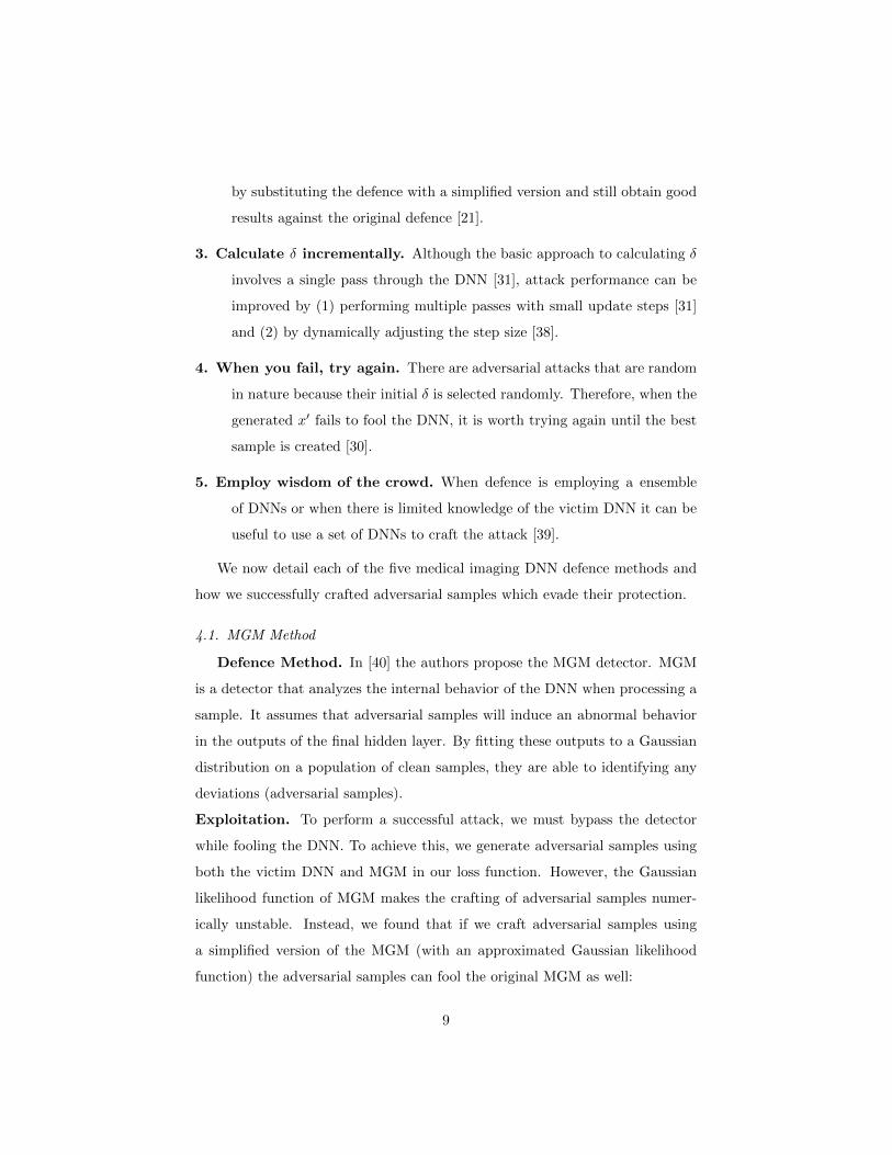
4.1. MGM Method
Defence Method. In [40] the authors propose the MGM detector. MGM
is a detector that analyzes the internal behavior of the DNN when processing a
sample. It assumes that adversarial samples will induce an abnormal behavior
in the outputs of the final hidden layer. By fitting these outputs to a Gaussian
distribution on a population of clean samples, they are able to identifying any
deviations (adversarial samples).
Exploitation. To perform a successful attack, we must bypass the detector
while fooling the DNN. To achieve this, we generate adversarial samples using
both the victim DNN and MGM in our loss function. However, the Gaussian
likelihood function of MGM makes the crafting of adversarial samples numer-
ically unstable. Instead, we found that if we craft adversarial samples using
a simplified version of the MGM (with an approximated Gaussian likelihood
function) the adversarial samples can fool the original MGM as well:
9
Page 10
Original MGM function (multivariate Gaussian):
L = log(N (x;µ,Σ)
)= −d
2log(2π)− 1
2log(|Σ|)− 1
2
∥∥x− µ∥∥2
Σ−1
(1)
Our approximated MGM function:
L =∥∥x− µ∥∥2
2(2)
As seen above, our main finding is that one can simply take the L2 distance
from the center of the learned Gaussian distribution to effectively and efficiently
craft adversarial samples that bypass the MGM detector.
4.2. GMM Method
Defence Method. In [41], the authors perform both prevention and detec-
tion in their solution. For prevention, the DNN is trained on both normal and
adversarial samples to make it more robust to attacks, an idea that has been
shown to be effective in the past [8]. For detection, the approach is similar to
that of MGM; the detector observes the outputs of the last hidden layer and
assumes a Gaussian distribution. However, in contrast to MGM, GMM detector
uses a separate Gaussian distribution for each prediction class of the classifier.
Exploitation. Because of the prevention mechanism, we were not able to
bypass the defence like we did for MGM. Instead, we evaded the prevention-
detection system by using a two stage adversarial crafting process: In the first
stage we find the “nearest” class which fools the victim model, and the second
stage we convince both the victim DNN and detector that it is the correct class
and untampered. To accomplish this, we first lower DNN’s confidence of x′
having the true label y. Then, after several training epochs, we update the
perturbation to increase both the detector and DNN’s confidence that x′ the
input is safe belongs to the attacker’s target class.
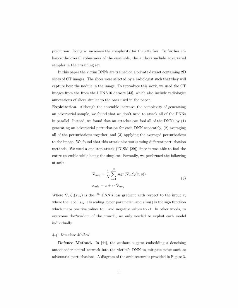
4.3. Ensemble Method
Defence Method. To challenge the attacker, the authors of [42] use “wis-
dom of the crowd“ by using multiple victim DNNs and by taking their average
10
Page 11
prediction. Doing so increases the complexity for the attacker. To further en-
hance the overall robustness of the ensemble, the authors include adversarial
samples in their training set.
In this paper the victim DNNs are trained on a private dataset containing 2D
slices of CT images. The slices were selected by a radiologist such that they will
capture best the nodule in the image. To reproduce this work, we used the CT
images from the from the LUNA16 dataset [43], which also include radiologist
annotations of slices similar to the ones used in the paper.
Exploitation. Although the ensemble increases the complexity of generating
an adversarial sample, we found that we don’t need to attack all of the DNNs
in parallel. Instead, we found that an attacker can fool all of the DNNs by (1)
generating an adversarial perturbation for each DNN separately, (2) averaging
all of the perturbations together, and (3) applying the averaged perturbations
to the image. We found that this attack also works using different perturbation
methods. We used a one step attack (FGSM [29]) since it was able to fool the
entire ensemble while being the simplest. Formally, we performed the following
attack:
∇avg =1
N
N∑i=1
sign(∇xLi(x, y))
xadv = x+ ε · ∇avg
(3)
Where ∇xLi(x, y) is the ith DNN’s loss gradient with respect to the input x,
where the label is y, ε is scaling hyper parameter, and sign() is the sign function
which maps positive values to 1 and negative values to -1. In other words, to
overcome the“wisdom of the crowd”, we only needed to exploit each model
individually.
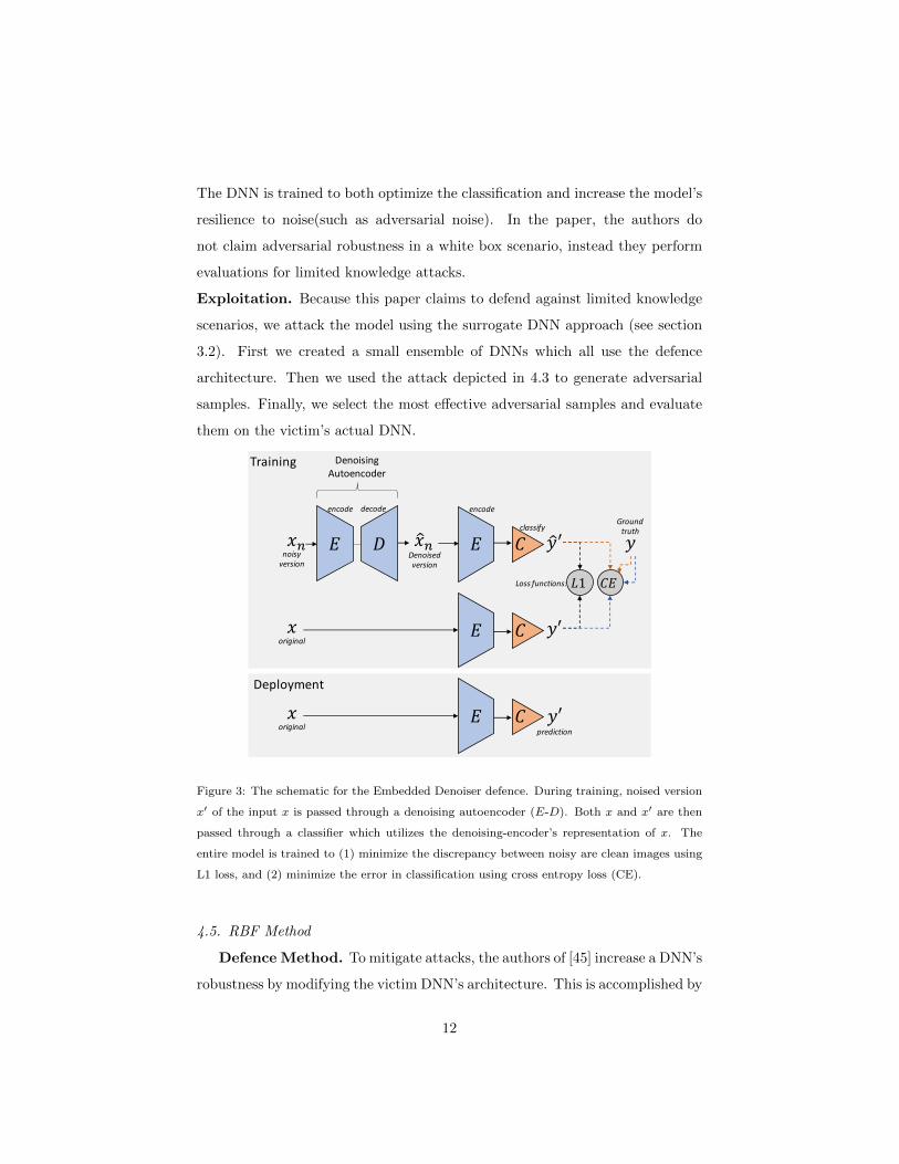
4.4. Denoiser Method
Defence Method. In [44], the authors suggest embedding a denoising
autoencoder neural network into the victim’s DNN to mitigate noise such as
adversarial perturbations. A diagram of the architecture is provided in Figure 3.
11
Page 12
The DNN is trained to both optimize the classification and increase the model’s
resilience to noise(such as adversarial noise). In the paper, the authors do
not claim adversarial robustness in a white box scenario, instead they perform
evaluations for limited knowledge attacks.
Exploitation. Because this paper claims to defend against limited knowledge
scenarios, we attack the model using the surrogate DNN approach (see section
3.2). First we created a small ensemble of DNNs which all use the defence
architecture. Then we used the attack depicted in 4.3 to generate adversarial
samples. Finally, we select the most effective adversarial samples and evaluate
them on the victim’s actual DNN.
𝐸 𝐷 𝐸 𝐶
Denoising Autoencoder
encode decode
𝑥𝑛 ො𝑥𝑛noisy
versionDenoised version
𝑥original
𝐸 𝐶
encode
classify
𝐿1
𝑥original
𝐸 𝐶
Deployment
Training
𝐶𝐸
ො𝑦′
𝑦′
𝑦
Ground truth
Loss functions:
𝑦′prediction
Figure 3: The schematic for the Embedded Denoiser defence. During training, noised version
x′ of the input x is passed through a denoising autoencoder (E-D). Both x and x′ are then
passed through a classifier which utilizes the denoising-encoder’s representation of x. The
entire model is trained to (1) minimize the discrepancy between noisy are clean images using
L1 loss, and (2) minimize the error in classification using cross entropy loss (CE).
4.5. RBF Method
Defence Method. To mitigate attacks, the authors of [45] increase a DNN’s
robustness by modifying the victim DNN’s architecture. This is accomplished by
12
Page 13
adding a layer after each block of convolutions. The layer applies a radial basis
function (RBF) to the concatenation of the block’s input and the convolutional
layer’s output [45]. This approach follows the finding that RBFs have greater
robustness than standard DNNs found in [29].
Exploitation. The RBF layer indeed increases the difficulty of crafting ad-
versarial samples. However, we were able to evade the RBF layers through
patience and diversity: (1) samples were generated using many iterations with
a slow learning rate, and (2) for each x we used different attack algorithms and
selected the best one as suggested in [38].
5. Defence Evaluation
In this section we empirically measure the security of the defences by attack-
ing them with the exploits described in the previous section. First we provide
our experiment setup and then present the impact which our attacks have on
the defences and the respective victim models.
5.1. Datasets
1. CHEST14: a public X-ray dataset gathered by the NIH Clinical Center.
This dataset contains more than 100,000 anonymized X-ray scans from
30,000 patients. The scans are accompanied by annotations for 14 types
of medical conditions. In our paper we use two versions of the dataset.
CHEST14 and CHEST2, where CHEST2 only indicates whether a scan
has some medical condition.
2. RSNA-X-ray: a public dataset published on Kaggle 2 by the Radiologi-
cal Society of North America (RSNA). This dataset contains 30,000 chest
X-rays of patients with and without Pneumonia.
2https://www.kaggle.com/c/rsna-pneumonia-detection-challenge.
13
Page 14
3. CT-Slices: CT images from the from the LUNA16 [43] lung cancer
dataset, which includes annotations from radiologists. We used the anno-
tations to crop benign and malign nodules from the scans.
4. Brain MRI segmentation dataset: a collection of FLAIR MRI scans
from the Cancer Imaging archive (TCIA). The scans consist of 110 pa-
tients included taken from the Cancer Genome Atlas (TCGA) lower-grade
glioma collection. These scans are accompanied with FLAIR abnormality
segmentation masks and genomic cluster data. The dataset can be found
on Kaggle 3.
5. OCT data: a public retinal optical coherence tomography (OCT) database,
which was introduced in [46]. The database contains 84,495 images from
4686 patients, divided into 4 classes according to their medical condi-
tion. For our evaluation, we followed the image sampling process described
in [41].
6. ISIC: the skin lesion dataset from IEEE ISBI International Skin Imaging
Collaboration (ISIC) Challenge described in [47]. The dataset includes
two types of annotations: lesion segmentation masks and disease cate-
gories, we use both of these annotations in our evaluation.
5.2. Metrics
We measure the impact of an attack on a defence by measuring the drop
in the defence’s performance. We used a different performance metric for each
machine learning task:
Classification. For binary classifiers we use accuracy and for multi-class clas-
sifiers we use average-accuracy. We compute GMM’s accuracy using the
inverse of the author’s metric called ‘adversarial risk’ –a combined per-
formance measure of the detector and the victim’s classifier [41]. Addi-
tionally, for MGM[40] and GMM[41], which used an evaluation different
3https://www.kaggle.com/andrewmvd/brain-tumor-segmentation-in-mri-brats-2015
14
Page 15
from accuracy, we performed the exact same evaluation made in the orig-
inal papers. In MGM, we use the AUROC metric that measures the area
under the ROC curve and in GMM we used the adversarial risk (directly).
Segmentation. We used the Dice measure which expresses how much the
ground-truth segmentation map fits (overlaps) the predicted one. The
metric has values on the range of 0-1 where higher is better.
5.3. Experiment Setup
All of our code was written using the Pytorch 1.7.0 framework. Both the
models and attacks were performed using Nvidia 3090 GPUs.
Defences Implementation. For each of the defence methods, we used the
exact same architecture as proposed in the respective paper except for
two cases: (Ensemble Method) The ensemble classifier proposed in [42]
performed poorly on our available medical data (without attacks). There-
fore, we used the popular Resnet20 architecture which is similar in size
and performed better [48]. (RBF Method) The RBF layer method from
[45] achieved low performance on our clean datasets and we could not
obtain authors implementation. Therefore, we used the popular Resnet34
which is similar in size [48] and improved the victim DNN’s performance.
For all of the defences, we measured their performance on clean data (not
attacked) as a baseline for the attack performance.
Attacks Experiments. For each defence method, we first reproduced the
authors’ results by attacking the defence with the original adversarial at-
tack. We obtained a similar defence performance as reported by the au-
thors, indicating that our implementations are correct. We then attacked
the defences using our new attacks. We bounded the adversarial noise
energy to the same level used by the authors (making the attacks invisible
to humans). All boundaries were set by the norm l∞.
15
Page 16
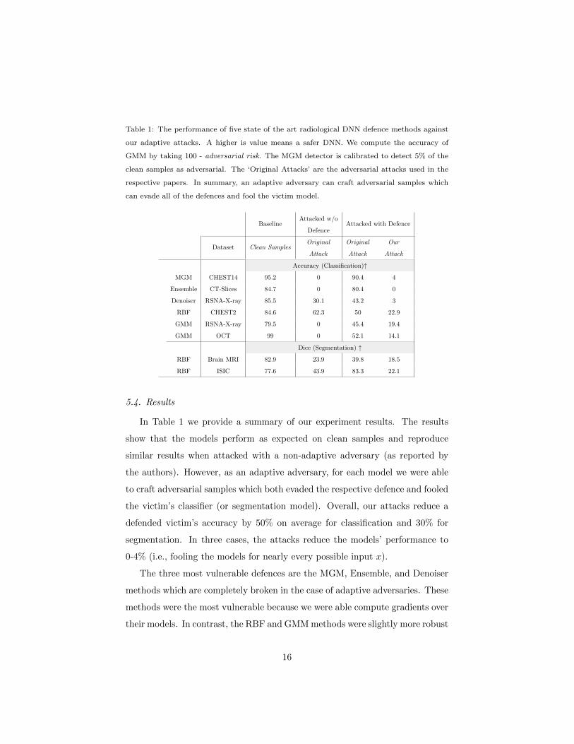
Table 1: The performance of five state of the art radiological DNN defence methods against
our adaptive attacks. A higher is value means a safer DNN. We compute the accuracy of
GMM by taking 100 - adversarial risk. The MGM detector is calibrated to detect 5% of the
clean samples as adversarial. The ‘Original Attacks’ are the adversarial attacks used in the
respective papers. In summary, an adaptive adversary can craft adversarial samples which
can evade all of the defences and fool the victim model.
BaselineAttacked w/o
DefenceAttacked with Defence
Dataset Clean SamplesOriginal
Attack
Original
Attack
Our
Attack
Accuracy (Classification)↑
MGM CHEST14 95.2 0 90.4 4
Ensemble CT-Slices 84.7 0 80.4 0
Denoiser RSNA-X-ray 85.5 30.1 43.2 3
RBF CHEST2 84.6 62.3 50 22.9
GMM RSNA-X-ray 79.5 0 45.4 19.4
GMM OCT 99 0 52.1 14.1
Dice (Segmentation) ↑
RBF Brain MRI 82.9 23.9 39.8 18.5
RBF ISIC 77.6 43.9 83.3 22.1
5.4. Results
In Table 1 we provide a summary of our experiment results. The results
show that the models perform as expected on clean samples and reproduce
similar results when attacked with a non-adaptive adversary (as reported by
the authors). However, as an adaptive adversary, for each model we were able
to craft adversarial samples which both evaded the respective defence and fooled
the victim’s classifier (or segmentation model). Overall, our attacks reduce a
defended victim’s accuracy by 50% on average for classification and 30% for
segmentation. In three cases, the attacks reduce the models’ performance to
0-4% (i.e., fooling the models for nearly every possible input x).
The three most vulnerable defences are the MGM, Ensemble, and Denoiser
methods which are completely broken in the case of adaptive adversaries. These
methods were the most vulnerable because we were able compute gradients over
their models. In contrast, the RBF and GMM methods were slightly more robust
16
Page 17
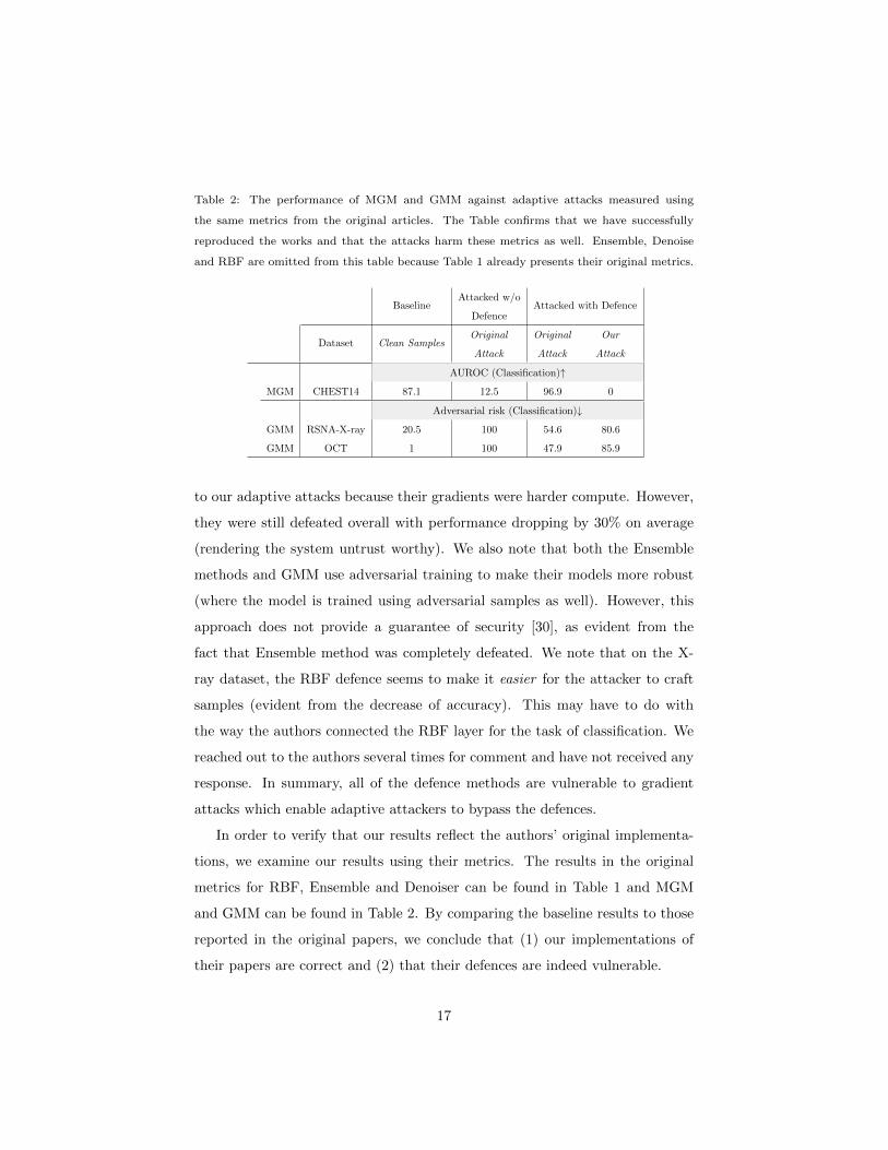
Table 2: The performance of MGM and GMM against adaptive attacks measured using
the same metrics from the original articles. The Table confirms that we have successfully
reproduced the works and that the attacks harm these metrics as well. Ensemble, Denoise
and RBF are omitted from this table because Table 1 already presents their original metrics.
BaselineAttacked w/o
DefenceAttacked with Defence
Dataset Clean SamplesOriginal
Attack
Original
Attack
Our
Attack
AUROC (Classification)↑
MGM CHEST14 87.1 12.5 96.9 0
Adversarial risk (Classification)↓
GMM RSNA-X-ray 20.5 100 54.6 80.6
GMM OCT 1 100 47.9 85.9
to our adaptive attacks because their gradients were harder compute. However,
they were still defeated overall with performance dropping by 30% on average
(rendering the system untrust worthy). We also note that both the Ensemble
methods and GMM use adversarial training to make their models more robust
(where the model is trained using adversarial samples as well). However, this
approach does not provide a guarantee of security [30], as evident from the
fact that Ensemble method was completely defeated. We note that on the X-
ray dataset, the RBF defence seems to make it easier for the attacker to craft
samples (evident from the decrease of accuracy). This may have to do with
the way the authors connected the RBF layer for the task of classification. We
reached out to the authors several times for comment and have not received any
response. In summary, all of the defence methods are vulnerable to gradient
attacks which enable adaptive attackers to bypass the defences.
In order to verify that our results reflect the authors’ original implementa-
tions, we examine our results using their metrics. The results in the original
metrics for RBF, Ensemble and Denoiser can be found in Table 1 and MGM
and GMM can be found in Table 2. By comparing the baseline results to those
reported in the original papers, we conclude that (1) our implementations of
their papers are correct and (2) that their defences are indeed vulnerable.
17
Page 18
There is a general principle that security by obscurity does not provide
protection. Based on our results and analysis, we conclude that these state-of-
the-art defences do not provide protection for the same reason. Attackers with
full-knowledge and even limited-knowledge can exploit vulnerabilities in these
defences to evade detection and prevention.
6. Discussion
The fundamental issue with the current defences is that they do not prevent
an attacker from observing their gradients signals. This is known to be an open
problem in the AI community [21, 49]. Another way of looking at it is that the
machine learning community, including researchers in medical imaging, are stuck
‘fighting fire with fire’, trying to solve machine learning problems with machine
learning tools. As a result, these defences suffer from the same oversight (they
assume a static attacker). To prevent the users of these technologies from being
misled, we recommend that researchers attack their own defences and include
the analysis in their work [21].
In the presence of an adaptive adversary, defenders of DNN models are
indeed at a disadvantage. However, unlike other domains, the medical imaging
community has two advantages which it can use to guarantee the security of its
DNNs:
Closed Environment In general, there are many opportunities for an attacker
to tamper a sample before it reaches a DNN. This is especially since the
attacker usually has full control over the initial sample (e.g., malware de-
tection, autonomous driving, and face recognition). However, unlike other
domains, medical imaging networks (PACS) are closed systems. This
means that most of the attack vectors listed in section 1.1 can be mit-
igated by hardening the system’s security: employing standard security
measures (e.g., network traffic encryption) and by performing regular sys-
tem updates. As a result, the attacker will not be able to access samples
to perform the attack. Therefore, we suggest that PACS administrators
18
Page 19
should focus more on securing their network and end-devices than em-
ploying adversarial attack detection models.
End-to-end Attribution Another advantage medical imaging has over other
domains is that the PACS has access to all medical media over its entire
life-cycle (from creation to analysis). As such, it is possible to deploy a
technology called digital signatures which can provably guarantee that pix-
els or metadata of an image (i.e., a DICOM file) have not been tampered.
Digital signatures work as follows: an entity (e.g., the CT scanner) (1)
takes a hash of the document producing a summary code m, and then (2)
encrypts m using a private one-way encryption key, producing the signa-
ture s. Then, all other entities (radiologist workstations or DNN analysis
tools) can verify that the document has not been tampered by (1) decrypt-
ing s using a public one-way decryption key, and then (2) checking that
the result is identical to the hash of the document. The reason this pro-
cess guarantees the integrity of the signed document is because (1): if the
document were tampered, the signature/hash would not match, and (2)
nobody else can encrypt messages that work with the public key aside from
the scanner (who has the private key). The DICOM standard has a field
which can hold digital signatures, and some modality scanners support
their creation [50]. Therefore, we suggest that all PACS networks enable
digital signatures where possible, and more importantly, verify them at
the end-points before performing any analysis.
In conclusion, we found that all state-of-the-art defences for DNNs in medical
imaging do not provide any security since an adversary can craft attacks which
both evade the defence and fool the DNN model. Our analysis revealed that
the core issue of these defences is that they do not prevent adversaries from
exploiting the victim’s gradient. Since this is an open issue, the defenders
appear to be at a disadvantage. However, medical imaging networks are at
an advantage over other domains: they can mitigate access to medical scans,
and they can employ digital signatures to guarantee image integrity.
19
Page 20
We hope this research will (1) help medical professionals and healthcare
providers understand the threat of adversarial samples in image analysis, (2)
help medical researchers avoid the pitfalls of developing ineffective defences,
and (3) help PACS administrators make informed decisions on how to secure
their networks from these attacks in the future.
Acknowledgements
This material is based upon work supported by the Zuckerman STEM Lead-
ership Program. This project has received funding from the European union’s
Horizon 2020 research and innovation programme under grant agreement 952172.
References
[1] G. Litjens, T. Kooi, B. E. Bejnordi, A. A. A. Setio, F. Ciompi, M. Ghafoo-
rian, J. A. Van Der Laak, B. Van Ginneken, C. I. Sanchez, A survey on
deep learning in medical image analysis, Medical image analysis 42 (2017)
60–88.
[2] T. Ching, D. S. Himmelstein, B. K. Beaulieu-Jones, A. A. Kalinin, B. T.
Do, G. P. Way, E. Ferrero, P.-M. Agapow, M. Zietz, M. M. Hoffman, et al.,
Opportunities and obstacles for deep learning in biology and medicine,
Journal of The Royal Society Interface 15 (141) (2018) 20170387.
[3] D. A. Bluemke, Radiology in 2018: are you working with ai or being re-
placed by ai?, Radiology 287 (2) (2018) 365–366.
[4] K. G. van Leeuwen, S. Schalekamp, M. J. Rutten, B. van Ginneken,
M. de Rooij, Artificial intelligence in radiology: 100 commercially available
products and their scientific evidence, European radiology 31 (6) (2021)
3797–3804.
20
Page 21
[5] M. V. S. de Cea, K. Diedrich, R. Bakalo, L. Ness, D. Richmond, Multi-
task learning for detection and classification of cancer in screening mam-
mography, in: International Conference on Medical Image Computing and
Computer-Assisted Intervention, Springer, 2020, pp. 241–250.
[6] A. Bar, M. M. Havakuk, Y. Turner, M. Safadi, E. Elnekave, Improved ich
classification using task-dependent learning, in: 2019 IEEE 16th Interna-
tional Symposium on Biomedical Imaging (ISBI 2019), IEEE, 2019, pp.
1567–1571.
[7] R. Shadmi, V. Mazo, O. Bregman-Amitai, E. Elnekave, Fully-automatic
deep learning based system for agatston score prediction from any non-
contrast chest ct.
[8] C. Szegedy, W. Zaremba, I. Sutskever, J. Bruna, D. Erhan, I. Goodfel-
low, R. Fergus, Intriguing properties of neural networks, arXiv preprint
arXiv:1312.6199.
[9] N. Papernot, P. McDaniel, S. Jha, M. Fredrikson, Z. B. Celik, A. Swami,
The limitations of deep learning in adversarial settings, in: 2016 IEEE
European Symposium on Security and Privacy (EuroS P), 2016, pp. 372–
387. doi:10.1109/EuroSP.2016.36.
[10] C. Beek, Mcafee researchers find poor security exposes medical data to
cybercriminals — mcafee blogs, https://www.mcafee.com/blogs/other-
blogs/mcafee-labs/mcafee-researchers-find-poor-security-exposes-medical-
data-to-cybercriminals, (Accessed on 11/06/2021) (2018).
[11] Y. Mirsky, T. Mahler, I. Shelef, Y. Elovici, Ct-gan: Malicious tampering
of 3d medical imagery using deep learning, in: 28th {USENIX} Security
Symposium ({USENIX} Security 19), 2019, pp. 461–478.
[12] S. Finlayson, I. Kohane, A. Beam, Adversarial attacks against medical deep
learning systems.
21
Page 22
[13] S. G. Finlayson, J. D. Bowers, J. Ito, J. L. Zittrain, A. L. Beam, I. S. Ko-
hane, Adversarial attacks on medical machine learning, Science 363 (6433)
(2019) 1287–1289. doi:10.1126/science.aaw4399.
URL https://www.science.org/doi/abs/10.1126/science.aaw4399
[14] T. C. Winter, Malicious adversarial attacks on medical image analysis,
American Journal of Roentgenology 215 (5) (2020) W55–W55.
[15] F. H. Security, 2021 horizon report the state of cybersecurity in healthcare
(2021).
[16] Z. Wittaker, A billion medical images are exposed online, as doctors
ignore warnings — techcrunch, https://techcrunch.com/2020/01/10/
medical-images-exposed-pacs/, (Accessed on 11/06/2021) (2020).
[17] J. Davis, Pacs vulnerability of orthopedic specialist ex-
poses data from 28k, https://healthitsecurity.com/news/
pacs-vulnerability-of-orthopedic-specialist-exposes-data-from-28k,
(Accessed on 11/06/2021) (2021).
[18] A. Zolfi, M. Kravchik, Y. Elovici, A. Shabtai, The translucent patch:
A physical and universal attack on object detectors, in: Proceedings of
the IEEE/CVF Conference on Computer Vision and Pattern Recognition,
2021, pp. 15232–15241.
[19] R. Duan, X. Ma, Y. Wang, J. Bailey, A. K. Qin, Y. Yang, Adversarial
camouflage: Hiding physical-world attacks with natural styles, in: Pro-
ceedings of the IEEE/CVF Conference on Computer Vision and Pattern
Recognition (CVPR), 2020.
[20] N. Carlini, D. A. Wagner, Adversarial examples are not easily detected:
Bypassing ten detection methods, Proceedings of the 10th ACM Workshop
on Artificial Intelligence and Security.
[21] F. Tramer, N. Carlini, W. Brendel, A. Madry, On adaptive attacks to
adversarial example defenses, in: H. Larochelle, M. Ranzato, R. Hadsell,
22
Page 23
M. Balcan, H. Lin (Eds.), Advances in Neural Information Processing
Systems 33: Annual Conference on Neural Information Processing Systems
2020, NeurIPS 2020, December 6-12, 2020, virtual, 2020.
URL https://proceedings.neurips.cc/paper/2020/hash/
11f38f8ecd71867b42433548d1078e38-Abstract.html
[22] H. Yu, K. Yang, T. Zhang, Y.-Y. Tsai, T.-Y. Ho, Y. Jin, Cloudleak:
Large-scale deep learning models stealing through adversarial examples.,
in: NDSS, 2020.
[23] J. Wei, Y. Zhang, Z. Zhou, Z. Li, M. A. Al Faruque, Leaky dnn: Steal-
ing deep-learning model secret with gpu context-switching side-channel,
in: 2020 50th Annual IEEE/IFIP International Conference on Dependable
Systems and Networks (DSN), IEEE, 2020, pp. 125–137.
[24] K. Chen, S. Guo, T. Zhang, X. Xie, Y. Liu, Stealing deep reinforcement
learning models for fun and profit, in: Proceedings of the 2021 ACM Asia
Conference on Computer and Communications Security, 2021, pp. 307–319.
[25] R. N. Reith, T. Schneider, O. Tkachenko, Efficiently stealing your machine
learning models, in: Proceedings of the 18th ACM workshop on privacy in
the electronic society, 2019, pp. 198–210.
[26] F. Tramer, F. Zhang, A. Juels, M. K. Reiter, T. Ristenpart, Stealing ma-
chine learning models via prediction apis, in: 25th {USENIX} Security
Symposium ({USENIX} Security 16), 2016, pp. 601–618.
[27] V. Duddu, D. Samanta, D. V. Rao, V. E. Balas, Stealing neural networks
via timing side channels, arXiv preprint arXiv:1812.11720.
[28] Y. Liu, X. Chen, C. Liu, D. Song, Delving into transferable adversarial
examples and black-box attacks, ArXiv abs/1611.02770.
[29] I. J. Goodfellow, J. Shlens, C. Szegedy, Explaining and harnessing adver-
sarial examples, arXiv preprint arXiv:1412.6572.
23
Page 24
[30] A. Madry, A. Makelov, L. Schmidt, D. Tsipras, A. Vladu, Towards deep
learning models resistant to adversarial attacks, in: International Confer-
ence on Learning Representations, 2018.
[31] A. Kurakin, I. Goodfellow, S. Bengio, Adversarial machine learning at scale,
arXiv preprint arXiv:1611.01236.
[32] N. Carlini, D. Wagner, Towards evaluating the robustness of neural net-
works, in: 2017 ieee symposium on security and privacy (sp), IEEE, 2017,
pp. 39–57.
[33] S.-M. Moosavi-Dezfooli, A. Fawzi, O. Fawzi, P. Frossard, Universal adver-
sarial perturbations, in: Proceedings of the IEEE conference on computer
vision and pattern recognition, 2017, pp. 1765–1773.
[34] A. Athalye, N. Carlini, D. A. Wagner, Obfuscated gradients give a false
sense of security: Circumventing defenses to adversarial examples, in:
ICML, 2018.
[35] Z. Katzir, Y. Elovici, Detecting adversarial perturbations through spatial
behavior in activation spaces, in: 2019 International Joint Conference on
Neural Networks (IJCNN), IEEE, 2019, pp. 1–9.
[36] N. Papernot, P. McDaniel, X. Wu, S. Jha, A. Swami, Distillation as a
defense to adversarial perturbations against deep neural networks, in: 2016
IEEE symposium on security and privacy (SP), IEEE, 2016, pp. 582–597.
[37] X. Ma, Y. Niu, L. Gu, Y. Wang, Y. Zhao, J. Bailey, F. Lu, Understanding
adversarial attacks on deep learning based medical image analysis systems,
Pattern Recognition 110 (2021) 107332.
[38] F. Croce, M. Hein, Reliable evaluation of adversarial robustness with an
ensemble of diverse parameter-free attacks, in: International conference on
machine learning, PMLR, 2020, pp. 2206–2216.
24
Page 25
[39] W. He, J. Wei, X. Chen, N. Carlini, D. Song, Adversarial example defense:
Ensembles of weak defenses are not strong, in: 11th {USENIX} workshop
on offensive technologies ({WOOT} 17), 2017.
[40] X. Li, D. Zhu, Robust detection of adversarial attacks on medical im-
ages, in: 2020 IEEE 17th International Symposium on Biomedical Imaging
(ISBI), IEEE, 2020, pp. 1154–1158.
[41] X. Li, D. Pan, D. Zhu, Defending against adversarial attacks on medical
imaging ai system, classification or detection?, in: 2021 IEEE 18th In-
ternational Symposium on Biomedical Imaging (ISBI), IEEE, 2021, pp.
1677–1681.
[42] R. Paul, M. Schabath, R. Gillies, L. Hall, D. Goldgof, Mitigating adversar-
ial attacks on medical image understanding systems, in: 2020 IEEE 17th
International Symposium on Biomedical Imaging (ISBI), IEEE, 2020, pp.
1517–1521.
[43] A. A. A. Setio, A. Traverso, T. De Bel, M. S. Berens, C. Van Den Bogaard,
P. Cerello, H. Chen, Q. Dou, M. E. Fantacci, B. Geurts, et al., Validation,
comparison, and combination of algorithms for automatic detection of pul-
monary nodules in computed tomography images: the luna16 challenge,
Medical image analysis 42 (2017) 1–13.
[44] F.-F. Xue, J. Peng, R. Wang, Q. Zhang, W.-S. Zheng, Improving robustness
of medical image diagnosis with denoising convolutional neural networks,
in: International Conference on Medical Image Computing and Computer-
Assisted Intervention, Springer, 2019, pp. 846–854.
[45] S. A. Taghanaki, K. Abhishek, S. Azizi, G. Hamarneh, A kernelized man-
ifold mapping to diminish the effect of adversarial perturbations, in: Pro-
ceedings of the IEEE/CVF Conference on Computer Vision and Pattern
Recognition, 2019, pp. 11340–11349.
25
Page 26
[46] D. S. Kermany, M. Goldbaum, W. Cai, C. C. Valentim, H. Liang, S. L.
Baxter, A. McKeown, G. Yang, X. Wu, F. Yan, et al., Identifying medical
diagnoses and treatable diseases by image-based deep learning, Cell 172 (5)
(2018) 1122–1131.
[47] N. C. Codella, D. Gutman, M. E. Celebi, B. Helba, M. A. Marchetti, S. W.
Dusza, A. Kalloo, K. Liopyris, N. Mishra, H. Kittler, et al., Skin lesion
analysis toward melanoma detection: A challenge at the 2017 international
symposium on biomedical imaging (isbi), hosted by the international skin
imaging collaboration (isic), in: 2018 IEEE 15th international symposium
on biomedical imaging (ISBI 2018), IEEE, 2018, pp. 168–172.
[48] K. He, X. Zhang, S. Ren, J. Sun, Deep residual learning for image recog-
nition, in: Proceedings of the IEEE conference on computer vision and
pattern recognition, 2016, pp. 770–778.
[49] A. Shamir, O. Melamed, O. BenShmuel, The dimpled manifold model of
adversarial examples in machine learning, arXiv preprint arXiv:2106.10151.
[50] NEMA, Nema standards publication ps 3 supplement 41 — digi-
tal imaging and communications in medicine (dicom) digital signa-
tures, https://www.dicomstandard.org/News-dir/ftsup/docs/sups/
sup41.pdf (September 2001).
26