

222.
A R E V I E V OF U L T R A S O N ~ C I ~ O R I Y I N B I O L O G Y AND MEDICINE^
ROBERT s. T E H P L E ~
The in t e re s t i n the use of ultrasonics a s a t o o l in evaluating l ivestock by animal s c i en t i s t s prompts us t o take a closer look a t research along the same lines in other fields. employs t h e principle of piezoelectricity; t h a t is, the f ac t t ha t a vibrat- ing c rys t a l wiU. develop an e l e c t r i c a l charge, and a lso tha t an e l e c t r i c a l charge w i l l s e t a c rys t a l i n to mechanical vibrations. The sound waves so emitted by a quartz c rys t a l a r e above the frequency which can be heard by the human ear, i.e. 18,OciO t o 20,000 cycles per second. A look a t research i n the medical and biological f i e l d s may stimulate sme new ideas and approaches tha t can be used i n animal research.
Ultrasonics, or hi&-frequency sound,
Research work i n the f i e l d of ultrasonics in biology and medicine i s relat ively new. i n medicine goes back on ly t o the l a t e 1940's and early 1950's (Ludwig, 1950; Wild, 1951; and French, 1951). f i e ld of biology was demonstrated a s early a s 1927 by Wood and Loomis, who reported that f i sh , frogs, and other small animals could be k i l l ed by being placed i n a continuous ultrasonic radiation f ie ld . Schmitt, Olson, and Johnson (1928) reported tha t free-swimming larvae, spirostema, and para- mecium could likewise be destroyed, Williams and Gaines (1960), using fre- quencies of 880 cycles per second, found t h a t a microorganism, E_. coli, could be ki l led, and t ha t the l e t h a l e f fec t was probably due t o pressures within the c e l l s . of l iv ing c e l l s subjected t o ultrasonic vibrations. These references give an indication of the in t e re s t t ha t developed for the use of ultrasonics i n c e l l destruction, k i l l i ng microorganisms, and other destructive uses t h a t l ed t o the development of ultrasonics a s a t o o l i n cleaning techniques, Gne of the in te res t ing future uses of ultresonics might be a greater develop- ment of dishwashers, instrument cleaners, and so for th . Indeed, maybe i n the future we will even take a bath by subjecting ourselves t o high-frequency sound! Even though there i s a considerable amount of l i t e r a t u r e on the use of ultrasonics i n cleaning techniques, we w i l l not go in to t h a t area here. Also, the use of ultrasonics i n physiotherapy and medical treatments w i l l not be reviewed, even though t h i s has been me of the areas in ultrasonic research which the medical field has accepted most readily,
Work i n the f i e l d of ultrasonics a s a diagnostic t o o l
However, i n t e re s t in ultrasonics i n the
Harvey and Loomis (1931) made high-speed photomicrographs
'Presented a t the 16th Reciprocal Meat Conference, St i l lwater , Oklahoma,
2Beef Cat t le Research Branch, Animal Husbandry Research Division. ARS,
June 11-13, 1S63.
Knoxville, Tennessee.

The successful application of the ultrasonic-pulse technique and the echo-ranging pr inciple t o underwater detection and ranging, and t o the local izat ion of flaws i n metals (Firestone, 1946) prompted investigations by the rJaval Research Ins t i tu te , 33ethesda, Maryland (Ludwig, 1950) concerning the use of an analogous technique for diagnostic purposes i n medicine and surgery. (1928) had investigated the frequencies of sound waves t h a t cause damage t o t issues . In the i r work, they indicated tha t continuous exposure t o vibrations of 8,000 t o 20,000 cycles per second produced t i s sue damage. They point out t ha t t h i s damage was produced a s a r e su l t of continuous exposure rather than pulsed exposure, which i s the type used i n diagnostic work. Porter (1939) and Gregg (1950) fe l t t ha t t he damage produced may be due t o the heat produced or t o a chemical factor. who was exposed t o 16,000 cycles per second of an ultrasonic source fo r an hour. chamber. in the neighborhood of 15 mc, per second, modified t o pulse one-half millionth of a second, no damage m s caused, The vibrations were interrupted i n the apparatus so t h a t the experimental animal (rabbit) received 7 1/2 vibrations, skipped 1298 vibrations, and so on. This had the double e f fec t of (1) greatly reducing the power of the apparatus, and (2) permitting the heat produced by the vibrations t o diss ipate i t s e l f p r ior t o the next series of vibrations ,
Prior t o t h i s time, Schmidt -I e t a l ,
Porter reported diminished mental capabi l i t i es i n a wcman
The e f f ec t s were reversible when she was removed from the ultrasonic French, Wild, and Neal (1951) reported t h a t when frequencies were
Wild and Neal (1951) reported no harmful e f fec ts when t i ssues were subjected t o ultrasonic waves of the energy used i n ultrasonography. They noted t h a t t i s sues of different texture gave different ultrasonograms. Their work, as was t rue f o r most of the other early work in sonography, was of a single dimension,i. e. giving a depth penetration only, indicating echoes a t various depths where materials of d i f fe ren t sound impedance or resistance were encountered. Th i s type of technique has been referred t o a s the “A” scan, were calculated by Ludwig in 1950. These data were used t o calculate re- f lec t ion coefficients a t interfaces, such a s those between dissimilar t i s - sues and between foreign bodies and tissues, Sound velocity was measured i n cer ta in human and animal t issues . Specific gravity of each t i s sue was measured, and the character is t ic acoustical impedance calculated frcm the velocity intensi ty data. Sound velocity through t i s sue has been measured a t frequencies of 1.25 and 2.5 mc, by Ludwig, using a pulsed method. The e f fec t of fiber direct ion of the t i s sues on the sound velocity was invest i - gated with beef muscle. muscle was traversed perpendicular t o the long axis of the muscle bundle, as compared t o when it was transversed p a r a l l e l with the muscle bundles. This work indicated tha t there was variat ion in the velocity of sound waves between brain, l i ve r , kidney, and spleen of the dog and hog, and f o r beef muscle. Wild (1950), working on an immediate post-mortem sample of small in tes t ine of a dog, could determine the layers of t issue, when the material vas folded so t h a t there were no a i r interfaces between the layers. He concluded t h a t the method can be applied t o the detection of changes i n t i s sue density, and discussed application f o r the detection of accessible tumors. poss ib i l i ty of locating sub-cortical neoplasms, a t the time of surgery, without the use of brain needles. The ultrasonic principle upon which the
Values of the character is t ic impedance of various t i s sues
No difference was found i n energy values when the
French (1951) and French, Wild, and Neal (1951) examined the
224.
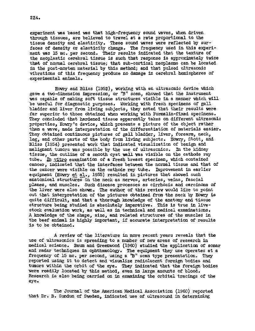
experiment was based was t ha t high-frequency sound waves, when driven through t issues , a r e believed t o t r a v e l a t a r a t e proportional t o the tissue density and e l a s t i c i ty . These sound waves were ref lected by sur- faces of density or e l a s t i c i t y change, The frequency used i n t h i s experi- ment was 15 mc, per second. Their results indicated t h a t the texture of the neoplastic cerebral t i s sue is such that response is approximately twice tbt of normal cerebral t issue; t h a t sub-cortical neoplasms can be located i n the post-mortem material by this method; and t h a t pulsed ultrasonic vibrations of t h i s frequency produce no damage in cerebral hemispheres of experimental animals ,
H o ~ r y and B l i s s (1952), worhing with an ultrasonic device which gave a two-dimension impression, or 'B" scan, showed t h a t the instrument was capable of making sof't t i s sue s t ructures v is ib le i n a manner which will be useful f o r diagnostic purposes, Working with fresh specimens of g a l l bladder and l i v e r from l iv ing subjects, they noted t h a t their results were f a r superior t o those obtained when working with Formalin-fixed specimens. They concluded t h a t hardened tissue apparently takes on difPerent ultrasonic properties, Howry's device, which presents a picture of t he object ra ther than a wave, made interpretat ion of the d i f fe ren t ia t ion of materials easier , They obtained continuous pictures of ga l lb l adde r , l iver , forearm, neck, leg, and other par t s of the body from l iv ing subjects, Howry, Stot t , and Bliss (1954) presented work t h a t indicated visualization of benign and malignant tumors was possible by the use of ultrasonics. In the kidney tissue, the outline of a smooth cyst wall vas vis ib le on the cathode ray tube. Ep vitrQ examination of a f resh breast specimen, which contained cancer, Indicated that the interfaces between the normal tissue and t h a t of the cancer were v is ib le on the cathode ray tube. Bnprovement i n e a r l i e r equipment ( H a m y et a l a , 1955) resulted i n pictures tha t shared such anatomical s t ructures in the neck a s nerves, a r t e r i e s , veins, f a s c i a l planes, and muscles, Such disease processes a s c i r rhos is and carcinoma of the l i v e r were a l so shuwn. !The author of t h i s review would 1W.e t o point out that; interpretat ion of the pictures obtained f rc jm the neck by Howry is qui te d i f f i c u l t , and t h a t a thorough knowledge of t h e anatorny and t i s sue s t ruc ture being studied is absolutely imperative. This i s t r u e in l ive - stock evaluation Work, as well a s in technical and medical examinations. A hawledge of the shape, size, and related structures of the muscles i n the beef animal is highly important, i f accurate interpretat ion of r e su l t s i s t o be obtained.
A review of the l i t e r a t u r e in more recent years reveals t h a t the use of ultrasonics is spreading t o a number of new areas of research in medical science. and radar techniques i n ophthamology, The equipment they use operates a t a frequency of 15 mc, per second, using a '93" scan type presentation. reported using it t o detect and visualize radiolucent foreign bodies and t m o r s within the o rb i t of the eye. They indicated t h a t the foreign bodies were readily located by this method, even in large amounts of blood. Research is a l so being carr ied on in examining the o r b i t a l tracings of the eye
Baum and Greenwood (1960) studied the application of sonar
They
The Journal of the American Medical Association (1960) reported t h a t D r , B. Sundun of Sweden, indicated use of ultrasound in determining
225
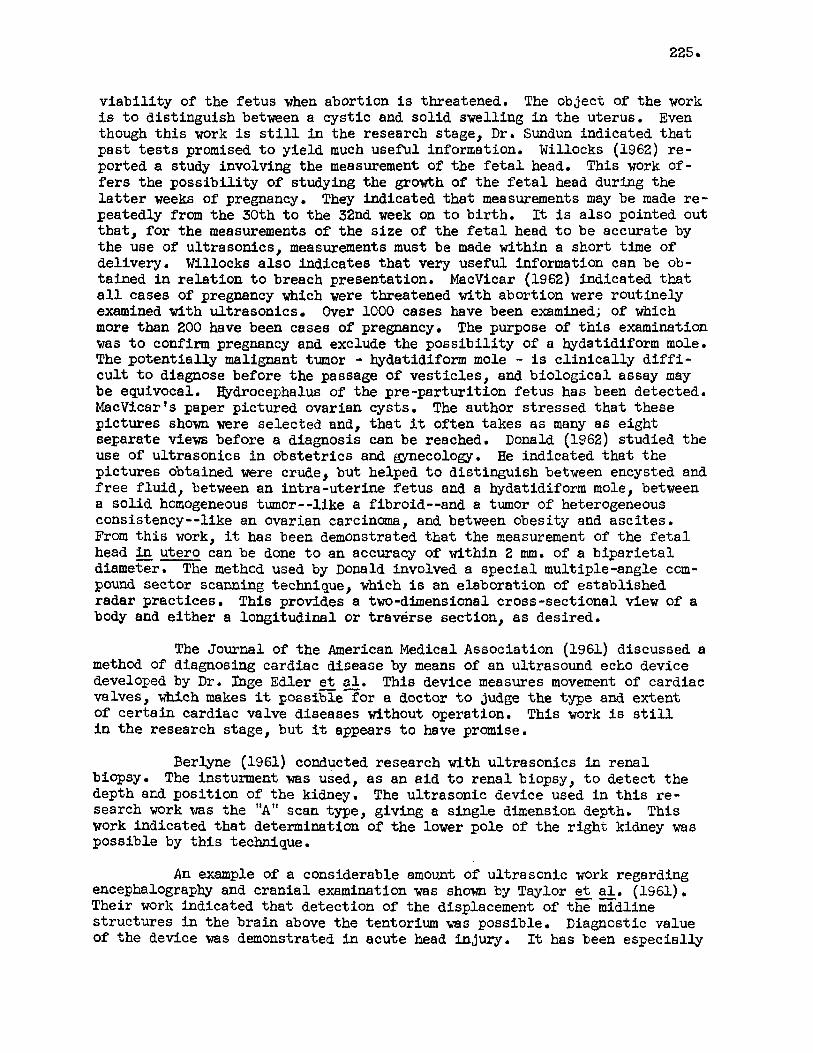
v iab i l i t y of the fe tus when abortion i s threatened. i s t o distinguish between a cyst ic and sol id swelling i n the uterus. Even though t h i s work i s s t i l l i n the research stage, Dr. Sundun indicated tha t past tests promised t o yield much useful information. Willocks (1962) r e - ported a study involving the measurement of the f e t a l head. This work of- fers the poss ib i l i ty of studying the growth of the f e t a l head during the l a t t e r weeks of pregnancy. peatedly from the 30th t o the 32nd week on t o bir th . that , fo r the measurements of t h e s i ze of the f e t a l head t o be accurate by the use of ultrasonics, measurements must be made within a short time of delivery. Willocks a l s o indicates t ha t very useful information can be ob- tained i n re la t ion t o breach presentation. MacVicar (1962) indicated tha t a l l cases of pregnancy which were threatened with abortion were routinely examined with ultrasonics. Over 1000 cases have been examined; of which more than 200 have been cases of pregnancy. The purpose of t h i s examination vas t o confirm pregnancy and exclude the poss ib i l i ty of a hydatidiform mole. The potent ia l ly malignant tumor - hydatidiform mole - i s c l in ica l ly d i f f i - c u l t t o diagnose before the passage of vest ic les , and biological assay may be equivocal. Bydrocephalus of the pre-parturit ion fe tus has been detected. MacVicarIs paper pictured ovarian cysts. pictures shown were selected and, t h a t it often takes as many a s eight separate views before a diagnosis can be reached. Donald (1962) studied t h e use of ultrasonics i n obstetr ics and gynecology. pictures obtained were crude, but helped t o distinguish between encysted and f r ee f luid, between an int ra-uter ine f e tus and a hydatidiform mole, between a sol id honiogeneous tumor--like a fibroid--and a tumor of heterogeneous consistency--like an ovarian carcinoma, and between obesity and asci tes . From t h i s work, it has been demonstrated tha t the measurement of the f e t a l head -- in utero can be done t o an accuracy of within 2 m a . of a b ipa r i e t a l diameter. The method used by Donald involved a special multiple-angle ccm- pound sector scanning technique, which is an elaboration of established radar practices. This provides a two-dimensional cross-sectional view of a body and either a longitudinal or traverse section, a s desired.
The object of the work
They indicated tha t measurements may be made re- It i s a l so pointed out
The author stressed t h a t these
He indicated tha t the
The Journal of the American Medical Association (1961) discussed a
This device measures movement of cardiac method of diagnosing cardiac disease by means of an ultrasound echo device developed by Dr. lhge Edler e t $1. valves, which makes it possible for a doctor t o judge the type and extent of cer ta in cardiac valve diseases without operation. This work i s s t i l l i n t he research stage, but it appears t o have promise.
-I
Berlyne (1961) conducted research with ultrasonics i n renal biopsy. The insturment was used, as an a i d t o renal biopsy, t o detect the depth and posit ion of the kidney. The ultrasonic device used i n t h i s re- search work was the "A" scan type, giving a s ingle dimension depth. This work indicated tha t determination of the lower pole of the r igh t kidney was possible by t h i s technique.
An example of a considerable amount of ultrasonic work regarding encephalography and cran ia l examination vas shown by Taylor e t a l . (1961) . Their work indicated tha t detection of the displacement of the midline s t ructures i n the brain above the tentorium was possible. of the device was demonstrated i n acute head injury.
-- Diagnostic value
It has been especially

226.
noteworthy i n the d i f f e ren t i a l diagnosis of acute cerebral vascular acci- dents, major a r t e r i a l occlusions being regularly distinguished from space- occupying hematomas .
A rather complete review of the work with ultrasonics fn biolbg- i c a l and medical research by Frye (1958) indicated that ultrasound may con- s t i t u t e a t o o l of considerable power fo r select ively disrupting in t ra - ce l lu la r structures. The forces involved, even i n an intense ultrasonic f ield, a r e not suff ic ient t o disrupt small molecules, but they may exert suff ic ient force on large molecular species t o break re la t ive ly weak bonds. This disruption of bonds can be expected t o dras t ica l ly a f f ec t t he subse- quent behavior of the ce l l . the use of intense ultrasound, which destroyed a t l e a s t some types of malignant neoplasms in humans. vest igators have been developing methods of visualizing soft t i s sue s t ruc- tu res by ref lected ultrasonic pulses, and indicates t h a t no other method, a t present, can yield t h i s type of information. Even though many problems remain f o r study i n t h i s field, in part icular , t he mechanics of re f lec t ion of sound as re lated t o the type of t i s sue structure, Frye indicates t h a t t h i s area of investigation contains poss ib i l i t i e s f o r tremendous advances in the near future. Another area of ultrasonic research in biomedicine, indicated by Frye, is that of elucidating micro-structures of cells. Measurements of ultrasonic absorption coeff ic ients of proteins in solution indicate t h a t absorption by these components may account f o r a large f rac t ion of the ab- sorption in soft t i s sue structures. tunately, research in medicine involving ultrasonics has not yet caught the imagination o f many investigators.
Fry@ gives an example of Russian reports On
F'rye a l so indicates t h a t a number of in-
However, Frye concludes tha t , unfor-
A review of l i t e r a t u r e of t h i s kind stimulates one t o v i suauze how some of the work in the medical f ield might be applied t o animal sciences. The work by animal s c i en t i s t s in trying t o visualize f a t thickness, l o in eye size, and other muscles through t h e use of ultrasonics i s f a i r l y w e l l known. It is encouraging t o read tha t medical Workers feel t h a t they can measure the s i ze of the f e t a l head in utero t o an accuracy of within 2 mm. give us some indication of the poten t ia l accuracy with which we might be able t o measure various t i s sue depths in l i v e animals.
Th i s would I-
Reviewing the works of Willocks (1962) and MacVicar (1962) in- volved in determining pregnancy, stages of pregnancy, types of presentation, and the ident i f icat ion of malignant tumors, presents a vast opportunity for the use of th i s device in livestock. Feasibly, this could be a very helpful t o o l t o the reproduction physiologist, not only in determining pregnancy, but a l so i n determining causes fo r non-retention of the fetus, as well as non-pregnancy itself.
The use of ultrasonics by veterinarians in determining locations of tumors, cysts, ulcerated t issues , and so forth, a s well as the very p rac t i ca l idea of locating hardware in the l i v e animal, may have some r e a l potent ia l , The use of ultrasonics in identifying bydrocephalus in the fetus , a s well as post-parturition, might indicate a poss ib i l i ty f o r i t s use a s a diagnostic t o o l in genetic studies.

227
From the standpoint of meats research, there may be an addi t ional use of ultrasonics i n the estimation of kidney knob size, marbling of cer ta in muscles, and i n the study of the s ize of bones. concerning the use of ultrasound, a s a t o o l f o r selectively disrupting in t r a - ce l lu la r structures, might have some poss ib i l i ty i n tenderness studies. Since it has been shown i n some of the research reviewed i n t h i s paper t ha t cer ta in t i s sues have different responses t o ultrasound, it might be feasible t o determine i f connective t i s sue could be broken down with a cer ta in fre- quency of ultrasound without disturbing any other body t issues . Indeed, it might be possible t o break down the f a t molecules so tha t they could be r e - absorbed by the body. layer of f a t of the animal's body, fo r instance, leaving the marbling i n the meat--the consuming housewife would cer tainly be pleased!
The ideas by Frye (1958)
If t h i s were done selectively--just on the outside
A t any rate , regardless of some of the exotic ideas tha t I have presented here f o r the use of ultrasonics i n animal science, I believe it i s obvious from the research work already accomplished tha t ultrasonics does have a potential .
LITERATURE: CITED
Baum, G. and I. Greenwoad. 1960. Ultrasound in ophthalmology. American Journal of Ophthalmology, 49:249.
Berlyne, G. M, 1961. Ultrasonics i n renalbiopsy: An a id t o determfnation of kidney position. Lancet, 2:750.
Brown, T. G. 1962. An explanation of the principles of ultrasonic echo sounding. Proceedings, Royal Society of Medicine, 55:637.
Donald, Ian. 1962. Sonar: A new diagnostic echo-sounding technique i n Proceedings, Royal Society of Medicine, obstetr ics and gynecology.
55: 637 . Echo-sounder. 1961. Journal of the American Medical Association, 178:1198.
French, L. A. 1951. The experimental application of ultrasonics t o the local izat ion of brain tumors. Journal of Neurosurgery, 8:198.
French, L. A,, J. J. Wild, and D. Neal. 1951. Detection of cerebral tumors by ultrasonic pulses . Cancer, 4:342 .
Frye, W. J. 1958, Biological and Medical Acoustics, 30:387.
Frye, W. J., V. J. Wulff, D, Tucker, and F. J. me. 1950. Physiological factors involved i n ultrasonically-induced changes i n l iv ing systems: I. Ident i f icat ion of non-temperature effects. Journal of the Acoustical Society of America, 22:867.
Gregg, E. C., Jr. 1950. Ultrasonics: Biological e f fec ts . Medical Physics, 2 : 1132.

228 . Earvey, E. N. and A. L. Loomis. 1931. High-speed photo-micrography of
l iving ce l l s subjected t o supersonic vibrations. Physiology, l5:147.
Journal of General
Hany, D. H. and W. R. B l i s s . 1952. Ultrasonic visualization of sof t tissue structures of the body. Medicine, 40:579.
1955. Ultrasonic visualization of l iving organ6 and t issues. Geriatrics,
Journal of Laboratory and Clinical
Howry, D. H., 3. H. Holmes, C. R. Cushman, and G. J. Posakony.
10 : 123
H o w , D. HI, D. A. Stot t , and W. R, Bliss. 1954. The ultrasonic visualization of carcinoma of the breast and other sof t t issue structures. Cancer, 7:354.
Ludwig, G. D. 1950, The velocity of sound through t issues and the acoustical impedance of t issues, America, 22 : 862 . Proceedings, Royal Society of Medicine, 55:638.
Journal of the Acoustical Society of
MacVicar, John. 1962. I l lus t ra t ive examples of ultrasonic echograms.
Porter, C , W. 1939. Curious effects of ultrasound. California Engineering. (cited by Gregg) .
Schmidt, F. C., A. R . Olson, and C. H. Johnson. frequency sound waves on protoplasm.
1928. Effects of high- Proceedings, Society of Experimental
Biology, 25 718
Taylor, J. C., J. A. Newell, and Peter Karvounis. 1961. Ultrasonics i n the diagnosis of intra-cranial space occupying lesions. Lancet, 1:1197.
Ultrasound tes t ing i n obstetrics and gynecoloGy. 1960. Journal of the American Medical Association, 173:708,
Wild, J. J. 1950. The use of ultrasonic pulses for the measurement of biological t issues and the detection of t issue density changes. Surgery, 27:183.
Wild, J. J. and D. Neal. 1951. detecting changes of texture i n l iving tissues.
Use of high-frequency ultrasonic waves fo r Lancet, 1:655.
Willocks, James. 1962. The use of ultrasonics i n cephalometry. Proceedings, Royal Society of Medicine, 55:640.
Williams, 0. B. and N. Gaines. 1930. The bacter ic idial effect of high- frequency sound waves . Journal of Infectious Diseases, 47: 485 .
Wood, R. W. and A, L. Loomis. 1927. Physical and biological effects of high-frequency sound waves. Phil. Mag., 4: 417 .
229
DR, RAIGEY: Thank you, Bob.
You can see tha t Bob has expended a very large e f fo r t t o assemble a l l of t h i s material, and we certainly appreciate it, Bob.
Next, we would l i k e t o learn something about the instrumentation used in ultrasonic work. To present a paper on t h i s topic, we a re fortunate t o have with us Jim Davis of the University of Georgia, Jim has worked with D r . Stouffer of Cornell and with the Branson Company i n helping test and improve some of the instruments which a re being used i n ultrasonic work. Jim i s concentrating h i s e f fo r t s a t Georgia on l i v e animal-carcass evaluation,
# 7% # #Si: 7% # S# # # # #


