

التيم عبارة عن ساليدات المحاضرات باإلضافة إلى شرح الدكاترة
حاولنا تقديم المادة بأفضل ما نستطيع
لذا إن وجدتم أي خلل أو خطأ فاعذرونا و بلغونا حتى يتم تصحيحه
نتمنى لكم التوفيق و السداد
ال تنسونا من دعواتكم
429 lecture + Dr`s notes
F1

Common Adult fractures
DR. Khalid A Bakarman
Assistant Prof, Pediatric, trauma orthopedic Consultant
F1 lecture notes added by : Reham Al-Henaki
objectives
At the end of this lecture the students should be able to:
know most of mechanisms of fracture injury
make the diagnosis of common adult fractures
request and interpret the appropriate x-rays
initiate the proper management of fractures
know which fractures can be treated by conservative or operative method
know the possible complications of different fractures and how to avoid them.
Upper limbs fractures
•Clavicle
•Humeral
( Proximal , shaft )
•Both Bone forearm
( Radius, ulna )
•Distal Radius
Mechanism of Injuries of the Upper Limb •Mostly Indirect
•Commonly described as “ a fall on outstretched hand “
•Typeof injury depends on –position of the upper limb at the time of impact
–force of injury
–age
Fracture of the clavicle : •Common fracture
•Commonest site is the middle one third
•Mainly due to indirect injury
•Direct injury leads to comminuted fracture
Pt present wit pain , swelling
If the fracture is grossly displaced
the swelling will be obvious

CLINICAL EVALUATION •splinting of the affected extremity, with the arm adducted
•neurovascular examination is necessary
•Assessment of skin integrity
•The chest should be auscultated to rule out chest trauma.
RADIOGRAPHIC EVALUATION anteroposterior radiographs
Treatment
Conservativearm sling or figure of 8
Operative fixation indicated if there is:
tenting of the skin
open fracture
neurovascular injury
nonunion
Plate and screws
COMPLICATIONS •Neurovascular compromise
•Malunion
•Nonunion
0.1% to 13.0%, with 85% of all nonunion occurring in the middle third.
•Posttraumatic arthritis at AC joint ,SC joint .
Humerus Fractures •Proximal Humerus ( includes surgical and anatomical neck )
comprise 4% to 5% of all fractures and represent the most common humerus fracture (45%)
CLINICAL EVALUATION •pain, swelling, tenderness, painful range of motion, and variable crepitus. Ecchymosis
•A careful neurovascular examination is essential, axillary nerve function
RADIOGRAPHIC EVALUATION •AP and lateral views
•Computed tomography
•Magnetic resonance imaging to assist the soft tissue
Spiral fracture that means the
MO fracture is indirect
Motor: movement of the Deltoid muscle – abduct the shoulder
Sensory: to deltoid Muscle- lateral aspect

CLASSIFICATION (Neer’s) •Four parts: These are the greater and lesser tuberosities, humeral shaft, and humeral head
•A part is defined as displaced if >1 cm of fracture displacement or >45 degrees of angulation
TREATMENT
•Minimally displaced fractures
–85% of proximal humerus fractures are minimally displaced or non displaced.
–Sling immobilization for comfort.
–Early shoulder motion may be instituted at 7 to 10 days.
–Pendulum exercises and passive range-of-motion exercises.
–At 6 weeks, active range-of-motion exercises are started.
Surgical indication •Anatomic neck fracture.
•Surgical neck fracture.
•Greater tuberosity fractures: If they are displaced more than 5 to 10 mm.
•Lesser tuberosity fractures displaced fragment blocks internal rotation or associated posterior dislocation.
•Three-and part fractures
•Four-part fractures
–osteonecrosis ranges from 13% to 35%.
–ORIF may be attempted in young patients
–Primary prosthetic replacement of the humeral head for senile patient
•Fracture-dislocation
COMPLICATIONS
•Vascular injury:
(5% to 6%) the axillary artery
The worse
the
fracture
,the more
(high)
incidence
of AVN

•Neural injury
–Brachial plexus injury: (6%).
–Axillary nerve injury
•Chest injury: Intrathoracic dislocation; pneumothorax
and hemothorax. rule out clinically by breath sound and
auscultation and radiological by X-ray
•Myositisossificans “ calcification within the ms will decrease ROM , pain”:
•Shoulder stiffness
•Osteonecrosis: 3% to 14% of three-part proximal humeral fractures, 13% to 34% of
four-part fractures, and a high rate of anatomic neck fractures.
•Nonunion
•Malunion
Fractures Shaft of the Humerus •Commonly Indirect injury
•3% to 5% of all fractures
•Indirect injury results in Spiral or Oblique fractures more soft tissue injury
•Direct injuries results in transverse or comminuted fracture
•May be associated with Radial Nerve injury
CLINICAL EVALUATION •pain, swelling, deformity, and shortening of the affected arm ,crepitus.
•Soft tissue abrasions and minor lacerations must be differentiated from open fractures
•careful neurovascular examination is essential, with particular attention to radial nerve function
Some pt present with radial N palsy “ wrist drop” unable to dorsiflex the rest .
RADIOGRAPHIC EVALUATION AP and lateral radiographs of the humerus should be obtained,
including the shoulder and elbow joints on each view
Dorsal aspect of the hand in the web space “loss of sensation “
CLASSIFICATION(Descriptive) •Open vs. closed.
•Location: proximal third, middle third, distal third.
•Degree: nondisplaced, displaced.
•Direction and character: transverse, oblique, spiral, segmental, comminuted
•Articular extension.
Radial injury common with comminuted F
Management of Fracture Shaft of the Humerus
•Most of the time is Conservative •Closed Reduction in upright position followed by application of U shaped Slab of POP or Cylinder cast
•Few weeks later or initially in stable fractures Functional Brace may be used
Indications for ORIF Fracture Shaft of Humerus •Multiple trauma
•Inadequate closed reduction or unacceptable malunion
•Pathologic fracture
•Associated vascular injury
•Floating elbow
•Segmental fracture
•Intraarticular extension
•Bilateral humeral fractures
•Open fracture
•Neurologic loss following penetrating trauma
•Radial nerve palsy after fracture manipulation (controversial)
•Nonunion
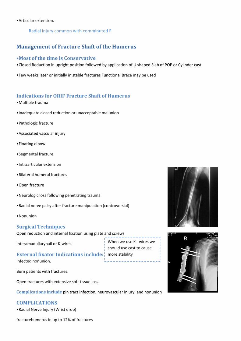
Surgical Techniques Open reduction and internal fixation using plate and screws
Interamadullarynail or K-wires
External fixator Indications include: Infected nonunion.
Burn patients with fractures.
Open fractures with extensive soft tissue loss.
Complications include pin tract infection, neurovascular injury, and nonunion
COMPLICATIONS •Radial Nerve Injury (Wrist drop)
fracturehumerus in up to 12% of fractures
When we use K –wires we
should use cast to cause
more stability
2/3 ( 8%) of Radial injury are Neuropraxia Most -the axion intact with nerve compression
1/3 ( 4%) are nerve lacerations or transection
Management of Radial Nerve injury •open fractures ; immediate exploration and ±repair
•closed injuries treated conservatively
- If No spontaneous recovery occurs in 6weeks confirmed by NCS and EMG go for nerve exploration after 12
weeks
- Recovery usually starts after few days but may take up to 9 months for full recovery
Both Bone forearm ( Radius, ulna ) •Forearm fractures are more common in men than women;
•motor vehicle accidents, contact athletic participation, altercations, and falls from a height
Clinical Evaluation •gross deformity of the involved forearm, pain, swelling, and loss of hand and forearm function.
•A careful neurovascular
•open wound
•compartment syndrome
Radiographic Evaluation Anteroposterior(AP) and lateral views
Radiographic evaluation should include the two joints.
Classification Descriptive •Closed versus open
•Location
•Comminuted, segmental, multifragmented
•Displacement
•Angulation
•Rotational alignment
Treatment( Nonoperative) •a well-molded, long arm cast in neutral rotation with the elbow flexed to 90 degrees.
•follow-up to evaluate for possible loss of fracture reduction.

Operative A. Open reduction and internal fixation
B. External fixation indication:
1.severe bone
2.soft tissue loss
3.gross contamination
4.infected nonunion
5.open elbow fracture-dislocations with soft tissue loss.
Complications a. Nonunion and malunion
b. Infection:
c. Neurovascular injury
d. Volkman ischemia follow CS
e. Posttraumatic radioulnarsynostosis(3% to 9% )
Distal Radius •Distal radius fractures are among the most common fractures of the upper extremity.
•one-sixth of all fractures treated in emergency departments
CLINICAL EVALUATION •The wrist is typically swollen with ecchymosis, tenderness, and painful range of motion.
•neurovascular assessment
median nerve function. Carpal tunnel compression symptoms are
common (13% to 23%)
•Look for ?open fracture.
RADIOGRAPHIC EVALUATION Posteroanteriorand lateral views
Normal radiographic relationships
a.Radial inclination: averages 23 degrees (range, 13 to 30 degrees)
b.Radial length: averages 11 mm (range, 8 to 18 mm).
c.Palmar(volar) tilt: averages 11 to 12 degrees (range, 0 to 28 degrees).
CLASSIFICATION (Descriptive) •Open versus closed
If the fracture displace we should check the skin if intact or not
b/c of high risk of open fracture

•Displacement
•Angulation
•Comminution
•Loss of radial length
Colles’ fracture •extraarticularfractures.
•90% of distal radius fractures
•dorsal angulation(apex volar), dorsal displacement, radial shift, and radial shortening.
•Clinically .dinner forkadeformity.
•a fall onto a hyperextended, radiallydeviated wrist with the forearm in pronation.
Usually don’t Need operative Treatment
Barton fracture intraarticular fractures. •a fracture-dislocation or subluxationof the wrist in which the dorsal or volarrim of the distal radius is displaced
with the hand and carpus. Volarinvolvement is more common --ORIF
•a fall onto a dorsiflexedwrist with the forearm fixed in pronation
Usually don’t Need operative Treatment
Smith fracture( reverse Colles fracture) •A volarangulation(apex dorsal) of the distal radius with an garden spades deformity or volardisplacement of the
hand and distal radius ----ORIF
•a fall onto a flexed wrist with the forearm fixed in supination
TREATMENT •Acceptable radiographic parameters for a healed radius in an active, healthy patient include:
- Radial length: within 2 to 3 mm of the contralateral wrist.
- Palmartilt: neutral tilt (0 degrees).
- Intraarticularstep-off: <2 mm.
- Radial inclination: <5-degree loss.
Nonoperative
Closed reduction and below elbow colle’s cast
Operative : indications
- High-energy injury
- Secondary loss of reduction
Normal

- Articular comminution, step-off, or gap
- Metaphysealcomminutionor bone loss“ unstable”
- Loss of volarbuttress with displacement
- DRUJ incongruity“distal radioulnar joint”
Operative Techniques
•Percutaneouspinning
ORIF
External fixation
COMPLICATIONS •Median nerve dysfunction
•Malunion or nonunion
•Complications of external fixation include reflex sympathetic dystrophy, pin tract infection, wrist and finger
stiffness, fracture through a pin site, and radial sensory neuritis
•Tendon rupture, most commonly extensor pollicislongus with K- wires
•Midcarpalinstability
•Posttraumatic osteoarthritis
•Finger, wrist, and elbow stiffness
Lower limbs Fractures
•Pelvic
•Hip fractures
( Neck , intertrochantric )
•Femoral shaft
•Tibia shaft
•Ankle( Medial malleolus ,Lateral malleolus, B.M)
Mechanism of fractures •Lower limb fracture is a result of a high energy trauma like MVA, fall , except in elderly people or diseased bones
•Types of fracture are depend on position of limb during impaction and magnitude of forces applied.
Management •The proper way to treat a patient with high energy trauma is to look at the patient as whole ,not to injured limb
alone!

•So the aim to treat such patient is to save life first, then save limb ,finally to save function.
•A.B.C.D in high energy trauma
Pelvic fractures in ER we use a towel to prevent the expand hematoma or external fixation
•Classifications. ( Tile)
Type A. Stable bed rest and analgesia
Type B. Rotationally Unstable ,Vertically Stable. plate ,screw. External fixation
Type C. Rotationally and Vertically Unstable Need to close anterior ,posterior
Type A
Type A stable Fracture of superior & inferior pubic remi& no diasthesis of SP
- in the pic there is a gap that means there is injury to the joint
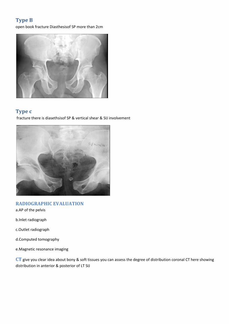
Type B open book fracture Diasthesisof SP more than 2cm
Type c fracture there is diasethsisof SP & vertical shear & SIJ involvement
RADIOGRAPHIC EVALUATION a.AP of the pelvis
b.Inlet radiograph
c.Outlet radiograph
d.Computed tomography
e.Magnetic resonance imaging
CT give you clear idea about bony & soft tissues you can assess the degree of distribution coronal CT here showing
distribution in anterior & posterior of LT SIJ

MANEGEMENT •Aggressive treatment . By A.B.C. D
•Think in systemic approach.
•Specific treatment:
type A . Bed rest& symptomatic treatment
type B .ORIF with plates& screws ,External Fix.
Type C . ORIF with plates & screws. Both AP.
pelvic banding external fixation

Operative treatment Surgical correction of type B open book fracture by anterior plating [ ORIF ]
Surgical correction of type C fracture by percutanous screw & plating of SP anterior[ ORIF
Complications •Infection up to 25%
•Thromboembolism
•Malunion
•Nonunion
•Hemorrhage –life threatening –hypovolemic shock– most common cause of death
•Bladder (15% )/bowel injuries
•Neurological damage ( L5-S1)
•Obstetrical difficulties
•Persistent sacro-iliac joint pain
Antalgic gate: the stance gate will be shorter b/c of the pain

AVN –depend on the degree of displacement
.
Any fracture affecting the blood supply will cause AVN
Pain swelling decrease ROM type 2” high risk of AVN.

Treatment NO rule for non operative
For neck and intertrochantric
Femur fractures
Treatment neck of femur
Nondisplaced fracture of neck of femur can be treat with canulated screws
Displaced fracture ----------DHS “ dynamic hip screw” in patient less than 60 years.
Age > than 65 years look for.
. Level of activities.
. Status of the acetabulum.
then chose THR(if acetabulum is disease! ) vs. hemi arthoplasty. if the diseased acetabulum “ OA, fracture”is not
replaced will cause pain
COMPLICATIONS •Nonunion
5% of nondisplaced fractures and up to 25% of displaced fractures
12 months as groin or buttock pain
•Osteonecrosis
10% of nondisplaced fractures and up to 27% of displaced fractures.
•Fixation failure
osteoporotic bone or technical problems

Femoral shaft
The best treatment of is I.M.N (intramedullary femoral nail) Mid shaft femur fracture
Open reduction and plate fixation for femur fracture
If the fracture near to the joint we
use plate Not a Wight
Bearing

Tibia shaft fracture
Tibia is covered by ms in the posterior
aspect but in the anterior there is skin and
soft tissue

Classification •Open versus closed
•Anatomic location: proximal, middle, or distal third
•Fragment number and position: comminution, butterfly fragments
•Configuration: transverse, spiral, oblique
•Angulation: varus/valgus, anterior/posterior
•Shortening
•Displacement: percentage of cortical contact
•Rotation
•Associated injuries
Clinical examination Look to injured limb for.
a.Soft tissue condition
b.R/O open fracture
c.Deformity
Feel for
Open fracture : In the ER : Realignment Analgesia Antibiotic Back slap To prevent extend and minimize the pain Obtain X ray
OR : No gross contamination Wash wound by NS Swab culture Type if fixation
a. Tenderness , pain .
Move
a. ROM
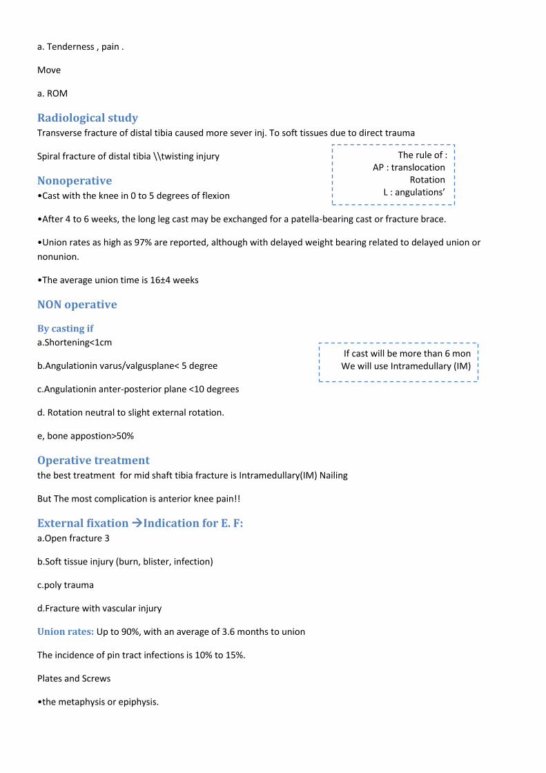
Radiological study Transverse fracture of distal tibia caused more sever inj. To soft tissues due to direct trauma
Spiral fracture of distal tibia \\twisting injury
Nonoperative •Cast with the knee in 0 to 5 degrees of flexion
•After 4 to 6 weeks, the long leg cast may be exchanged for a patella-bearing cast or fracture brace.
•Union rates as high as 97% are reported, although with delayed weight bearing related to delayed union or
nonunion.
•The average union time is 16±4 weeks
NON operative
By casting if
a.Shortening<1cm
b.Angulationin varus/valgusplane< 5 degree
c.Angulationin anter-posterior plane <10 degrees
d. Rotation neutral to slight external rotation.
e, bone appostion>50%
Operative treatment the best treatment for mid shaft tibia fracture is Intramedullary(IM) Nailing
But The most complication is anterior knee pain!!
External fixation Indication for E. F: a.Open fracture 3
b.Soft tissue injury (burn, blister, infection)
c.poly trauma
d.Fracture with vascular injury
Union rates: Up to 90%, with an average of 3.6 months to union
The incidence of pin tract infections is 10% to 15%.
Plates and Screws
•the metaphysis or epiphysis.
The rule of : AP : translocation
Rotation L : angulations’
L : angulations’
If cast will be more than 6 mon We will use Intramedullary (IM)

•success rates as high as 97%. Complication rates of infection, wound breakdown, and malunion or nonunion
increase with higher-energy injury patterns.
Ankle Fractures •the incidence has increased
•an elderly women
•Most ankle fractures are isolated malleolarfractures
•Open fractures are rare < 2%.
•MOI: position of the foot at time of injury,
the magnitude, direction, and rate of loading
CLINICAL EVALUATION •pain and discomfort, with swelling, tenderness, and variable deformity
•Neurovascular status
•The extent of soft tissue injury possible open injuries and blistering
•A dislocated ankle should be reduced and splinted immediately (before radiographs if clinically evident)
RADIOGRAPHIC EVALUATION
AP view
-Tibiofibulaoverlap of <10 mm is abnormal and implies syndesmoticinjury.
-Tibiofibulaclear space of >5 mm is abnormal and implies syndesmoticinjury
-Talartilt
Lateral view
-The dome of the talus should be centered under the tibia and congruous with the tibialplafond
-Posterior tibialtuberosity fractures can be identified
Mortise view the foot in 15 to 20 degrees of internal rotation
A medial clear space >4 to 5 mm is abnormal and indicates lateral talarshift
Tibiofibularoverlap <1 cm indicates syndesmoticdisruption
Talarshift >1 mm is abnormal.
Denis –weberclassification A.infra-syndesmotic
B.Trans-syndesmotic
C.supra-syndesmotic

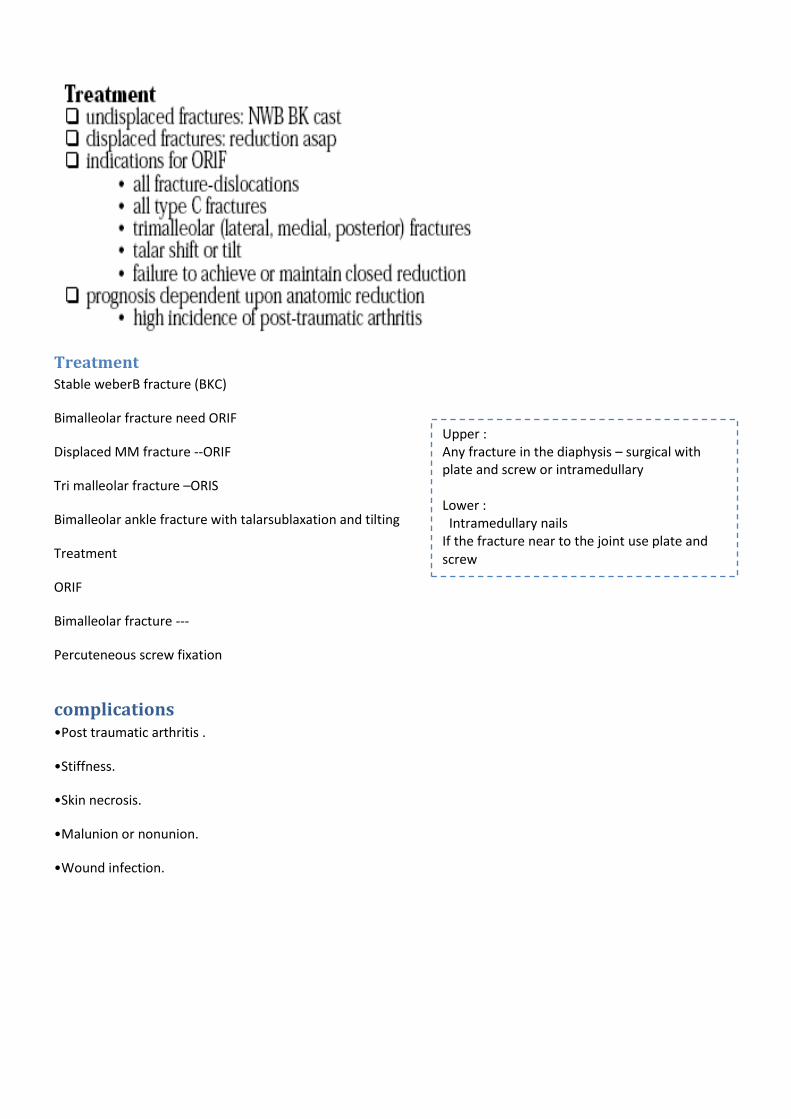
Treatment Stable weberB fracture (BKC)
Bimalleolar fracture need ORIF
Displaced MM fracture --ORIF
Tri malleolar fracture –ORIS
Bimalleolar ankle fracture with talarsublaxation and tilting
Treatment
ORIF
Bimalleolar fracture ---
Percuteneous screw fixation
complications •Post traumatic arthritis .
•Stiffness.
•Skin necrosis.
•Malunion or nonunion.
•Wound infection.
Upper : Any fracture in the diaphysis – surgical with plate and screw or intramedullary
Lower : Intramedullary nails If the fracture near to the joint use plate and screw


