

1
Macrophage Migration Inhibitory Factor: A Key Mediator of Inflammation
Aaron Kithcart, B.S.
Introduction
Jean-Martin Charcot first distinguished multiple sclerosis (MS) from other neurological
diseases in 1868 as a pattern of tremors and paralysis in young adults, differing from James
Parkinson’s description of paralysis agitans in elderly patients1. Mediated by an autoimmune
attack against the myelin sheath surrounding axons, the course of MS is both chronic and
progressive2. The autoimmune nature of MS was first suggested by early animal studies in
which self-myelin antigens were used to immunize rodents causing an MS-like disease, later
termed experimental autoimmune encephalomyelitis (EAE). EAE is viewed as the major animal
model for MS, since the two diseases have in common loss of the myelin sheath, accumulation of
autoreactive T lymphocytes, and production of inflammatory cytokines and chemokines3,4.
In healthy individuals, the blood brain barrier (BBB) plays a key role in regulating
leukocyte infiltration into the CNS. Migration of leukocytes across the vascular endothelium
during MS and EAE is largely determined by the expression of adhesion molecules and their
ligands. Multiple studies have shown that the interaction between two adhesion molecules, α4-
integrin and VCAM-1, is required for the recruitment of inflammatory cells into the CNS 5-7.
Additional adhesion molecules, including ICAM-1, likely also have significant roles during
migration.
The expression of adhesion molecules on the endothelium of the BBB is strongly
influenced by the presence of inflammatory cytokines. Tumor necrosis factor α (TNF-α) is one
cytokine present in MS lesions and is associated with the expression of ICAM-18. Much
2
attention has also been focused on a second inflammatory cytokine, IL-17. Like TNF-α, IL-17
can be detected in inflammatory lesions of MS patients9. IL-17 is produced by TH17 CD4+ T
lymphocytes, which are expanded by two other cytokines, IL-6 and transforming growth factor-β
(TGF-β)10,11. Co-expression of IL-6 with IL-17 was shown to increase VCAM-1 expression
during EAE12. Another well known cytokine, IL-10, is protective during EAE and produced by a
regulatory population of T lymphocytes. Regulatory lymphocytes express CD4, CD25, and a
transcription factor Foxp313, and depletion of this regulatory population of T cells increased the
severity of EAE14. These cells are expanded following exposure to TGF-β.
Modulation of cytokines can dictate the development of or recovery from MS. A study
of MS patients showed that an additional cytokine, macrophage migration inhibitory factor
(MIF), was elevated in the cerebrospinal fluid (CSF) of patients undergoing a relapse, as well as
in the CNS of mice following the induction of EAE15,16. Several MIF knockout studies have
shown MIF is upstream of the production of TNF-α, IL-1β, and IL-617. As we have already
discussed, TNF-α is a critical cytokine for adhesion molecule expression, and IL-6 has an
important function during the differentiation of TH17 lymphocytes.
From these studies, we predict the expression of MIF plays a significant role in the
pathogenesis of EAE and MS. Part of the difficulty of elucidating new MS therapies has been
that no single cytokine has been identified which alone can either explain or prevent ongoing
MS. Thus, the focus of this project has been to better understand the broader underlying
mechanisms that lead to neuroinflammation. The objectives of this project are to determine the
role of MIF in EAE using genetically deficient mice and to identify potential mechanisms of
protection. We will also describe a novel therapeutic inhibitor of MIF.
3
Results
MIF is required for susceptibility to EAE
We immunized MIF knockout and wild type control mice with MOG35-55 peptide.
Knockout mice showed less severe EAE relative to wild type controls (Figure 1). Furthermore,
MIF knockout mice also had a lower incidence of EAE, reduced cumulative disease index, and a
lower peak clinical score relative to controls (Table 1). Interestingly, the absence of MIF did not
affect the day of onset, which was similar between groups, or the peripheral response to MOG
antigen (data not shown). Previous studies have shown MIF knockout mice are
immunosuppressed18, so we investigated other mechanisms by which MIF knockout mice could
be protected.
MIF Knockout Mice Have Reduced Mononuclear Infiltration
To evaluate whether MIF was required for leukocyte migration, we measured the
presence of inflammatory infiltrates in the CNS. Migration of inflammatory leukocytes into the
brain and spinal cord is a key factor during EAE and MS. In wild type mice, there was a
considerable presence of mononuclear cells surrounding blood vessels in the brain (Figure 2A).
We found reduced perivascular infiltration in MIF knockout mice and reduced CNS
inflammation as scored by a pathologist (Figure 2C). These observations strongly suggested that
MIF has a role during migration.
We also examined damage in the brain following immunization. Thirty-five days
following immunization, damage was assessed by luxol fast blue staining with a silver
counterstain. Luxol fast blue measures the presence of myelin, which was reduced in wild type
mice (Figure 2B). Wild type mice also developed enhanced axonal severing, appearing as

4
numerous retraction bulbs along axons. Knockout mice, on the other hand, had much less
demyelination and axonal severing. This correlated with reduced clinical severity observed in
knockout mice. These data suggest MIF not only facilitates migration into the CNS but also
subsequent neuronal damage. Previous reports have shown MIF increases the expression of
adhesion molecules19. In MIF knockout mice, reduced expression of adhesion molecules would
have a profound effect on migration and progression of disease. However, given the reduced
damage in the CNS, we predicted other mechanisms of MIF might also mediate inflammation.
MIF Knockout Mice have a Larger Population of Regulatory Cells
We measured the lymphocyte and antigen presenting cell populations of wild type and
MIF knockout mice following immunization. We found no differences in CD4+ or CD8+ T
lymphocytes and CD19+ B lymphocytes. Populations of other antigen presenting cells were also
comparable between groups (data not shown). A striking difference was the elevation of
CD25+Foxp3+ lymphocytes in MIF knockout mice (Figure 3A and B). This population of cells
can exert regulatory activity through the transcription of Foxp3 and secretion of IL-10. We
measured a greater numbers of these cells, plus enhanced production of IL-10 in MIF-deficient
mice (data not shown). We concluded from these studies that this population of regulatory cells
was a mechanism of protection in MIF knockout mice.
A Small Molecule Inhibitor of MIF Reduces Ongoing EAE
All studies thus far explored EAE in mice genetically lacking MIF with wild type
controls. We investigated whether administration of an inhibitor of MIF after onset of acute
disease could reduce ongoing EAE. Through collaboration with Cytokine PharmaSciences, we
5
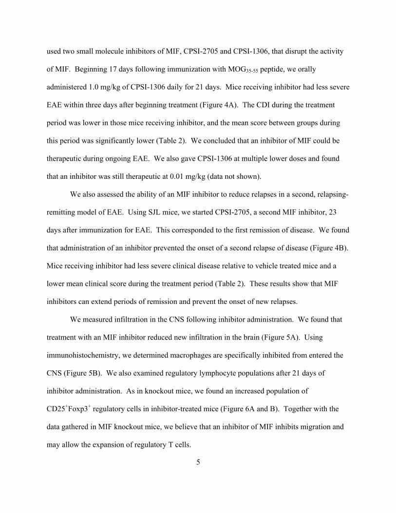
used two small molecule inhibitors of MIF, CPSI-2705 and CPSI-1306, that disrupt the activity
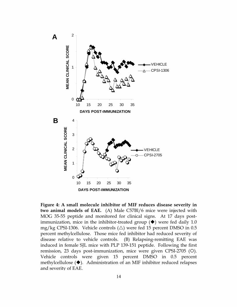
of MIF. Beginning 17 days following immunization with MOG35-55 peptide, we orally
administered 1.0 mg/kg of CPSI-1306 daily for 21 days. Mice receiving inhibitor had less severe
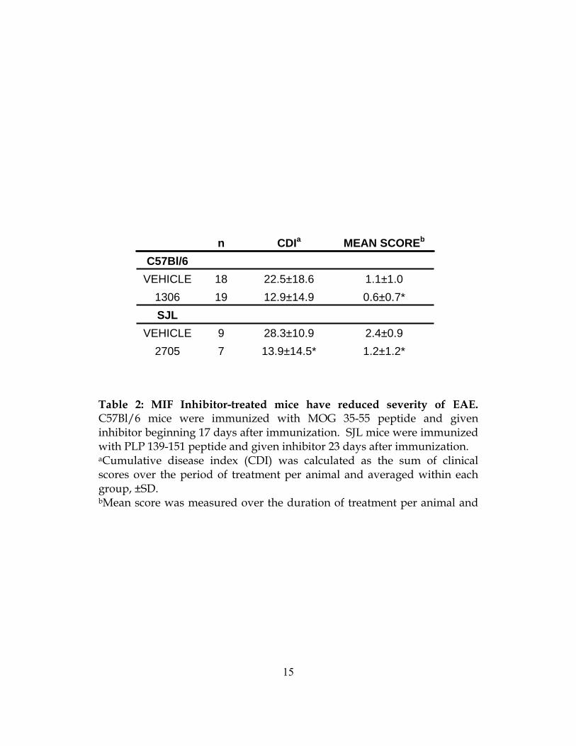
EAE within three days after beginning treatment (Figure 4A). The CDI during the treatment
period was lower in those mice receiving inhibitor, and the mean score between groups during
this period was significantly lower (Table 2). We concluded that an inhibitor of MIF could be
therapeutic during ongoing EAE. We also gave CPSI-1306 at multiple lower doses and found
that an inhibitor was still therapeutic at 0.01 mg/kg (data not shown).
We also assessed the ability of an MIF inhibitor to reduce relapses in a second, relapsing-
remitting model of EAE. Using SJL mice, we started CPSI-2705, a second MIF inhibitor, 23
days after immunization for EAE. This corresponded to the first remission of disease. We found
that administration of an inhibitor prevented the onset of a second relapse of disease (Figure 4B).
Mice receiving inhibitor had less severe clinical disease relative to vehicle treated mice and a
lower mean clinical score during the treatment period (Table 2). These results show that MIF
inhibitors can extend periods of remission and prevent the onset of new relapses.
We measured infiltration in the CNS following inhibitor administration. We found that
treatment with an MIF inhibitor reduced new infiltration in the brain (Figure 5A). Using
immunohistochemistry, we determined macrophages are specifically inhibited from entered the
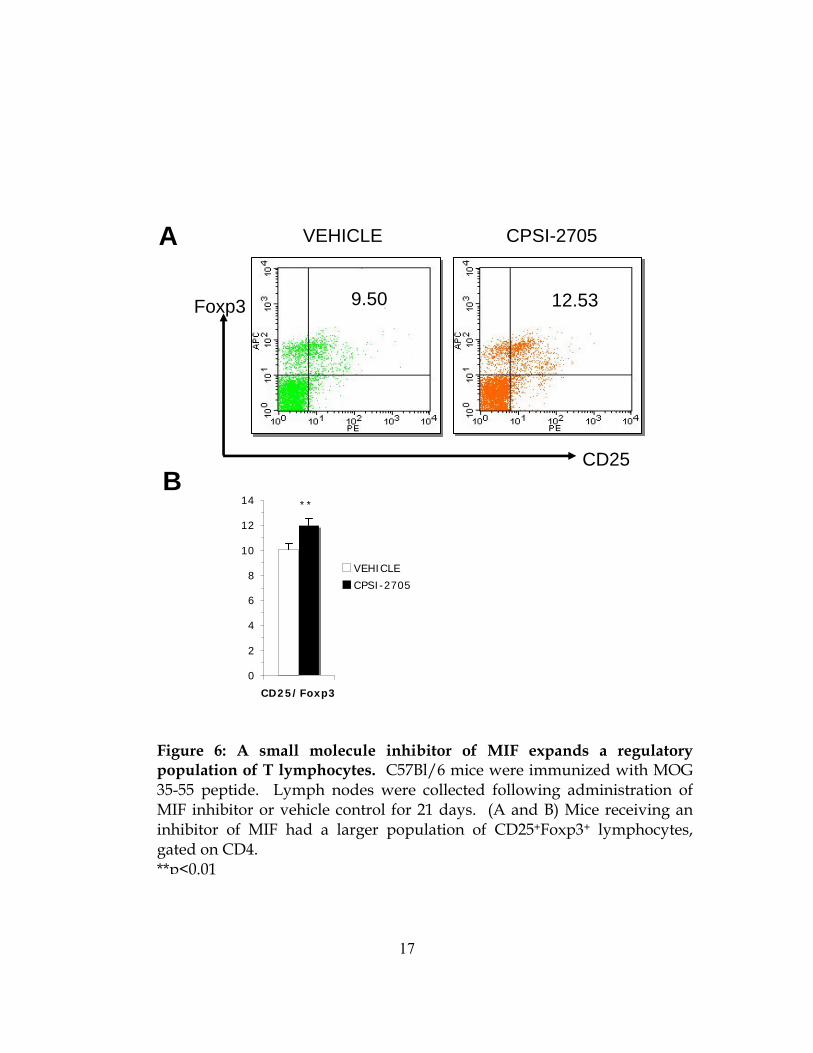
CNS (Figure 5B). We also examined regulatory lymphocyte populations after 21 days of
inhibitor administration. As in knockout mice, we found an increased population of
CD25+Foxp3+ regulatory cells in inhibitor-treated mice (Figure 6A and B). Together with the
data gathered in MIF knockout mice, we believe that an inhibitor of MIF inhibits migration and
may allow the expansion of regulatory T cells.

6
An Inhibitor of MIF is Available in the CNS
The efficacy of any pharmaceutical intervention depends not only on the mechanism of
action of the drug but also its availability at the site of disease. We evaluated the presence of
CPSI-2705 in the serum and brain after five days of oral administration. We found significant
amounts of inhibitor in both tissues (Figure 7). However, there was more inhibitor present in the
brain of mice with EAE than those mice that were healthy controls. This strongly suggests
patency of the BBB improves availability of MIF inhibitor in the CNS tissues. This would
greatly increase the therapeutic benefit for MS patients, since most drugs cannot typically cross
the BBB. Our data shows an MIF inhibitor would be best available at sites of inflammation.
Discussion
Autoimmune diseases result from complex processes in which the immune system
attacks the body, confusing the distinction between self and non-self. MS is a debilitating
autoimmune disease that affects nearly 2.5 million individuals worldwide. Many of the key
mediators of MS pathogenesis have been actively investigated as therapeutic targets but, at
present, no single factor such as an individual cytokine or chemokine has been identified which
alone can slow progression of disease. Targeting cytokines that mediate multiple mechanisms
during the inflammatory response represent more focused therapies.
One such specific cytokine is MIF, a cytokine widely conserved across many species that
plays a role in balancing inflammation with suppression and regulation of an immune response.
In order to better understand the role of MIF in the pathogenesis of MS and EAE, we utilized
mice lacking MIF on a C57Bl/6 background, a mouse strain that is susceptible to EAE and

7
commonly used to model MS. We found mice lacking MIF are significantly less susceptible to
EAE induction. This suggests that MIF plays an important role in influencing susceptibility to
inflammation. To better understand how MIF knockout mice were protected, we evaluated the
degree of migration into the CNS. We found MIF knockout and inhibitor-treated mice had
significantly less infiltration. We also noted MIF knockout mice had reduced neuronal damage.
We predicted the expression of MIF acted through another mechanism, in addition to migration,
that facilitated inflammation. We noted both wild type and MIF knockout mice had
CD4+CD25+Foxp3+ regulatory cells following immunization, but in knockout and inhibitor-
treated mice, these cells were significantly expanded.
The combination of reduced migration into the CNS and an increased number of Foxp3+
regulatory cells in MIF knockout and inhibitor-treated mice are powerful factors mediating
protection. We believe the absence of MIF allowed the expansion of regulatory lymphocytes
and inhibited expression of adhesion molecules, which slowed the progression of EAE. Past
studies show MIF increases expression of IL-620. Co-expression of IL-6 with TGF-β expands
the TH17 population of cells, a subtype of lymphocytes known to mediate several inflammatory
diseases. While driving the expansion of TH17 cells, large production of IL-6 inhibits the
differentiation of CD4+CD25+Foxp3+ regulatory T cells. The absence of MIF in knockout mice
protects this population of regulatory cells. MIF is also upstream of the production of TNF-α20.
Both IL-6 and TNF-α activate vascular endothelial cells and cause the surface expression of
ICAM-1, VCAM-1, and other adhesion molecules. Without these markers, the incidence and
severity of EAE is severely reduced. We believe that the expression of MIF increases production
of a host of cytokines, each mediating an important aspect of pathogenesis.

8
We have shown that an inhibitor of MIF is therapeutic during EAE. Two different
inhibitors, CPSI-1306 and CPSI-2705, reduced ongoing disease severity. This was marked by
increases in the number of regulatory T lymphocytes and reduced CNS leukocyte infiltration.
Any of these could be primary mechanisms in which inhibition of MIF is therapeutic, and all
could be successful for the management of MS. Importantly, none of the mechanisms of MIF
inhibition appear to suppress the immune system. Rather, MIF is an important mediator of
several components of the immune response, including autoregulation through regulatory T cells
and trafficking into the peripheral tissues. Targeting these multiple components could prove
most successful for the management of a complicated autoimmune disease like MS.
Materials and Methods
Mice. Age-matched C57Bl/6 and SJL mice were purchased from Jackson Laboratories.
Mouse strains lacking the MIF gene (B6;129S4-Miftm1Dvd) were developed as previously
described20 and extensively backcrossed onto C57Bl/6 background.
Induction of Experimental Autoimmune Encephalomyelitis. For the induction of EAE in
C57Bl/6 mice, animals were immunized with 200 μg MOG35-55 peptide (Princeton
Biomolecules) emulsified in complete Freund’s adjuvant (containing 200 μg Mycobacterium
tuberculosis Jamaica strain), injected intradermally in each of four flanks. Pertussis toxin (List
Biological Labs) was injected as an additional adjuvant intraperitoneally (i.p.) on the day of
immunization and 48 hours later (200 ng in 0.2 ml PBS). Female SJL mice were immunized
with 150 μg PLP139-151 peptide (Sigma-Genosys) emulsified in complete Freund’s adjuvant.
Pertussis toxin was not used for the induction of EAE in SJL mice.
9
All animals were observed daily for EAE clinical signs and scored according to degree of
paralysis; 0 = no paralysis, 1 = limp tail or ataxia, 2 = limp tail with ataxia, 3 = partial hind limb
paralysis, 4 = complete hind limb paralysis, and 5 = death. Cumulative disease index (CDI) was
calculated as the sum of daily clinical scores from each animal during the course of observation
and reported as an average within each group. Peak score was reported as the average maximum
clinical score within each group over the observed period. Additional outcome measures
included EAE incidence and mortality and were calculated based on individual animals reported
as a mean within each group.
Flow Cytometry. Single cell suspensions derived from draining lymph nodes at the sites
of injection and spleens were stained with anti-CD4 or -CD25 FITC-, PE-, or APC-conjugated
fluorescent antibodies (BD Biosciences). Isotype control monoclonal antibodies (BD
Biosciences) were matched for each fluorochrome. Cells were labeled at 1 x 10⁶ cells per tube,
incubated for 30 minutes at 4°C, and processed for intracellular staining. Cells were
permeabilized with fix/perm working solution (eBioscience) and stained intracellularly for the
transcription factor Foxp3 for 30 minutes at 4°C. Cells were washed twice with permeabilization
buffer and analyzed using a FACSCalibur flow cytometer.
Histopathologic Assessment. Immunohistochemical, hematoxylin and eosin (H&E),
luxol fast blue with silver contrast, and Bielschowsky silver staining were carried out by The
Ohio State University Veterinary Sciences core facility. Brains and spinal cords were removed
at various time points following immunization and frozen at -80°C in OCT media. H&E
sections were graded by a blinded pathologist for mononuclear cell infiltration on a scale of zero
(no inflammation) to three (diffuse parenchymal infiltration). Immunohistochemical stains were
labeled with anti-F4/80 antibodies and evaluated by a blinded pathologist.

10
Drug Administration. Several small molecule inhibitors of MIF (gift from Cytokine
PharmaSciences) were administered to mice prior to or following induction of EAE. Inhibitors
were given either i.p. or orally (p.o.) for 10 to 21 days. CPSI-2705 and CPSI-1306 inhibited
tautomerase assay as previously described21 at concentrations ranging from 1 to 10 μM. For i.p.
administration of inhibitors, drug was dissolved in sterile DMSO, and then diluted in PBS for an
overall ratio of 1:3 (DMSO to PBS). Orally administered inhibitor was dissolved in 15 percent
DMSO in 0.1 percent methycellulose in water. Mice were fed by gavage a total of 50 μl at a
concentration of inhibitor. Vehicle controls received 15 percent DMSO in 0.1 percent
methylcellulose and were included in all experiments. The time of day of inhibitor
administration was kept constant, between 1000 h and 1200 h.
Inhibitor Assays. Brains and serum were collected from mice receiving MIF inhibitors
orally for five days. To collect serum, mice were anesthetized and blood drawn from the retro-
orbital sinus using heparinized Natelson blood collecting tubes. Samples were centrifuged at
10,000 g for 15 minutes and collected serum was stored at -20°C. Analysis of brain
homogenates and serum was performed by Cytokine PharmaSciences.
Statistical Analysis. All statistical references were made based on appropriate methods as
outlined in the literature22. Statistical significance between groups for cumulative disease index,
mean clinical score, and day of onset was calculated using the Students’ t test. Measures of
lymphocyte populations, cytokine production, and proliferation also used the t test. Significance
for incidence was calculated using a Chi-square.

0
1
2
10 20 30
DAYS POST-IMMUNIZATION
MEA
N C
LIN
ICA
L S
CO
RE
C57Bl/6-/-MIF
Figure 1: The genetic deletion of MIF is protective against EAE. Wild type ( ) and MIF knockout ( ) mice were immunized for EAE with 200 μg MOG 35-55 peptide in adjuvant. MIF knockout mice had less severe EAE relative to wild-type controls. Data are representative of four separate experiments.
14.6±2.3
15.9±4.6
ONSETa
0.9±1.0** 6.4±8.1**7/13 (54%)**MIF-/-
2.2±1.3 24.8±18.115/18 (83%)C57Bl/6
PEAK SCOREcCDIbINCIDENCE
Table 1: MIF knockout mice have reduced incidence and severity of EAE. Wild type and MIF knockout mice were immunized with MOG 35-55 peptide in adjuvant. aDay of onset was calculated as the mean of the first day of clinical scores among mice that developed EAE, ±SD. bCumulative disease index (CDI) was calculated as the sum of clinical scores over the duration of disease per animal and averaged within each group, ±SD. cPeak score was measured over the duration of disease per animal and averaged, ±SD. **p<0.01
11

0
1
2
INFL
AM
MA
TIO
N
C57Bl/6MIF-/-*
Figure 2: The absence of MIF prevents infiltration into the CNS. At 17 days following immunization with MOG 35-55 peptide, brains were taken from wild type and MIF-deficient animals. (A) Hematoxylin and eosin (H&E) staining of brain sections showed less perivascular infiltration in MIF knockout mice versus wild type controls. (B) At 35 days post-immunization, luxol fast blue with silver counterstain revealed more axonal degeneration in wild type mice versus MIF knockouts. (C) Inflammation was significantly reduced in MIF-deficient mice, as graded by a blinded pathologist (n=5 per group). Results are representative of three separate experiments.
B
A C57Bl/6 MIF-/-
C
12

A
16.12Foxp3
CD25
Foxp3
CD25
C57Bl/6 MIF-/-
21.35
0
5
10
15
20
25
CD25/Foxp3
C57Bl/6MIF-/-
**B
Figure 3: Mice lacking MIF have a larger population of regulatory cells that are functional. Lymph nodes from wild type and MIF knockout animals were collected 10 days post-immunization. (A and B) Knockout mice had an elevation in regulatory CD25+Foxp3+ population of T lymphocytes, gated on CD4. **p<0.01
13
0
1
2
10 15 20 25 30 35
DAYS POST-IMMUNIZATION
MEA
N C
LIN
ICA
L SC
ORE
VEHICLECPSI-1306
0
1
2
3
4
10 15 20 25 30 35
DAYS POST-IMMUNIZATION
ME
AN C
LINI
CAL
SCO
RE
VEHICLECPSI-2705
A
B
Figure 4: A small molecule inhibitor of MIF reduces disease severity in two animal models of EAE. (A) Male C57Bl/6 mice were injected with MOG 35-55 peptide and monitored for clinical signs. At 17 days post-immunization, mice in the inhibitor-treated group ( ) were fed daily 1.0 mg/kg CPSI-1306. Vehicle controls ( ) were fed 15 percent DMSO in 0.5 percent methylcellulose. Those mice fed inhibitor had reduced severity of disease relative to vehicle controls. (B) Relapsing-remitting EAE was induced in female SJL mice with PLP 139-151 peptide. Following the first remission, 23 days post-immunization, mice were given CPSI-2705 ( ). Vehicle controls were given 15 percent DMSO in 0.5 percent methylcellulose ( ). Administration of an MIF inhibitor reduced relapses and severity of EAE.
14
1.2±1.2* 13.9±14.5*7 2705 2.4±0.928.3±10.99 VEHICLE
SJL 0.6±0.7* 12.9±14.919 1306 1.1±1.022.5±18.618 VEHICLE
C57Bl/6 MEAN SCOREbCDIan
Table 2: MIF Inhibitor-treated mice have reduced severity of EAE. C57Bl/6 mice were immunized with MOG 35-55 peptide and given inhibitor beginning 17 days after immunization. SJL mice were immunized with PLP 139-151 peptide and given inhibitor 23 days after immunization. aCumulative disease index (CDI) was calculated as the sum of clinical scores over the period of treatment per animal and averaged within each group, ±SD. bMean score was measured over the duration of treatment per animal and
15
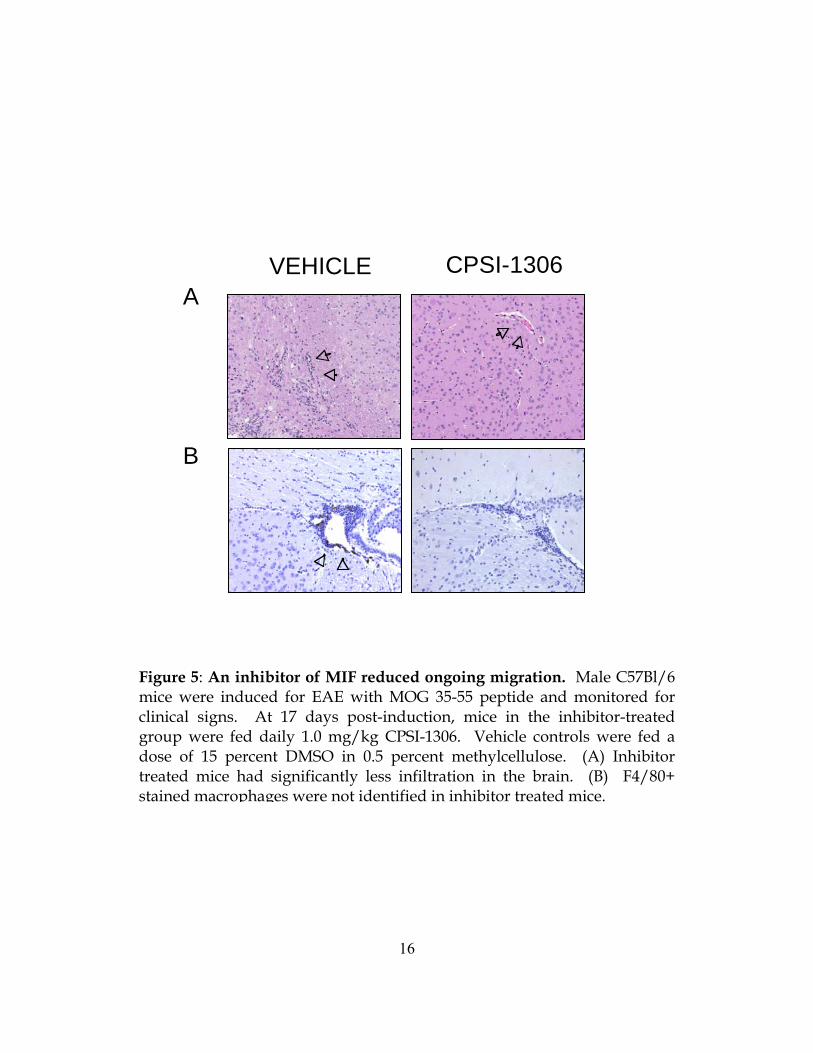
Figure 5: An inhibitor of MIF reduced ongoing migration. Male C57Bl/6 mice were induced for EAE with MOG 35-55 peptide and monitored for clinical signs. At 17 days post-induction, mice in the inhibitor-treated group were fed daily 1.0 mg/kg CPSI-1306. Vehicle controls were fed a dose of 15 percent DMSO in 0.5 percent methylcellulose. (A) Inhibitor treated mice had significantly less infiltration in the brain. (B) F4/80+ stained macrophages were not identified in inhibitor treated mice.
VEHICLE CPSI-1306
B
A
16
9.50Foxp3
CD25
VEHICLE CPSI-2705
12.53
A
B
0
2
4
6
8
10
12
14
CD25/Foxp3
VEHICLE
CPSI-2705
**
Figure 6: A small molecule inhibitor of MIF expands a regulatory population of T lymphocytes. C57Bl/6 mice were immunized with MOG 35-55 peptide. Lymph nodes were collected following administration of MIF inhibitor or vehicle control for 21 days. (A and B) Mice receiving an inhibitor of MIF had a larger population of CD25+Foxp3+ lymphocytes, gated on CD4. **p<0.01
17
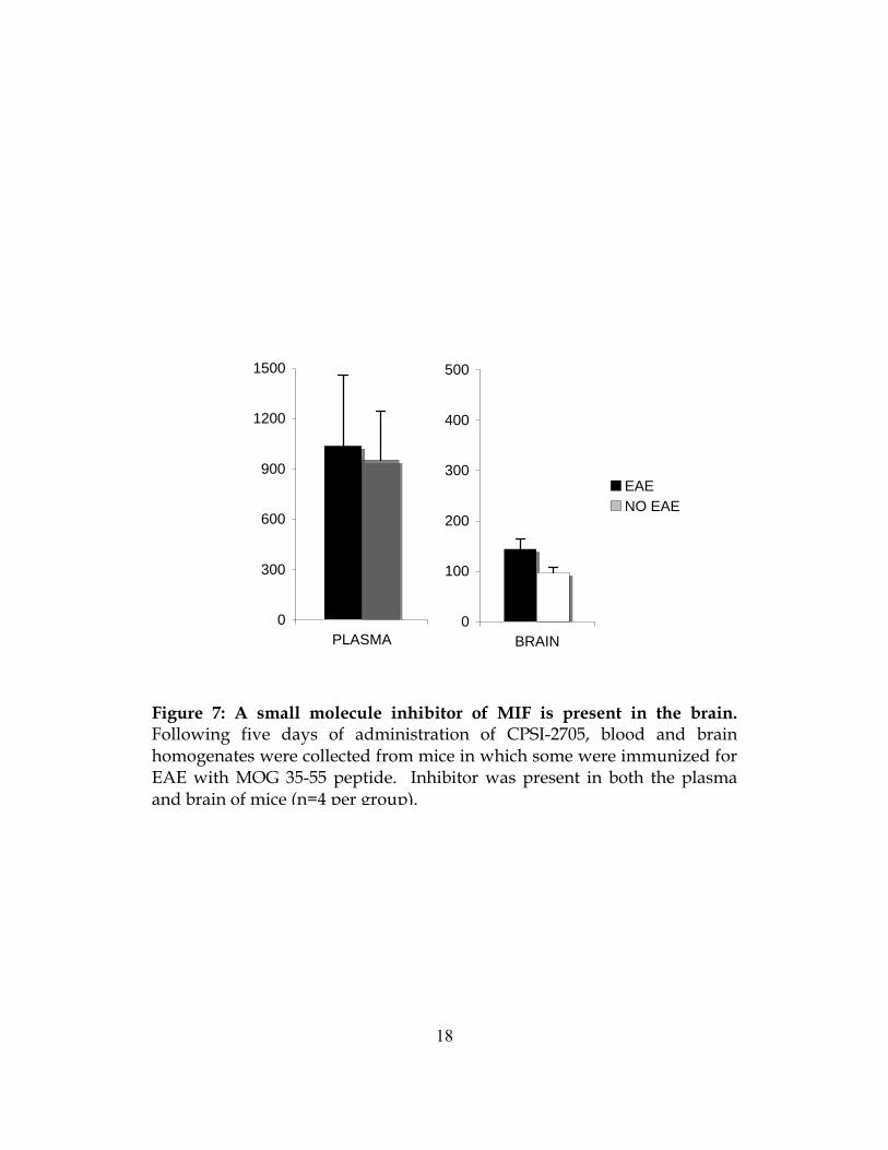
Figure 7: A small molecule inhibitor of MIF is present in the brain. Following five days of administration of CPSI-2705, blood and brain homogenates were collected from mice in which some were immunized for EAE with MOG 35-55 peptide. Inhibitor was present in both the plasma and brain of mice (n=4 per group).
0
300
600
900
1200
1500
PLASMA0
100
200
300
400
500
BRAIN
EAENO EAE
18
19
Bibliography
1. Murray, T.J. Multiple sclerosis : the history of a disease, (Demos Medical Pub., New York, 2005).
2. Peterson, J.W. & Trapp, B.D. Neuropathobiology of multiple sclerosis. Neurol Clin 23, 107-129, vi-vii (2005).
3. Gold, R., Linington, C. & Lassmann, H. Understanding pathogenesis and therapy of multiple sclerosis via animal models: 70 years of merits and culprits in experimental autoimmune encephalomyelitis research. Brain 129, 1953-1971 (2006).
4. Gold, R., Hartung, H.P. & Toyka, K.V. Animal models for autoimmune demyelinating disorders of the nervous system. Mol Med Today 6, 88-91 (2000).
5. Baron, J.L., Madri, J.A., Ruddle, N.H., Hashim, G. & Janeway, C.A., Jr. Surface expression of alpha 4 integrin by CD4 T cells is required for their entry into brain parenchyma. J Exp Med 177, 57-68 (1993).
6. Steffen, B.J., Butcher, E.C. & Engelhardt, B. Evidence for involvement of ICAM-1 and VCAM-1 in lymphocyte interaction with endothelium in experimental autoimmune encephalomyelitis in the central nervous system in the SJL/J mouse. Am J Pathol 145, 189-201 (1994).
7. Kent, S.J., et al. A monoclonal antibody to alpha 4 integrin suppresses and reverses active experimental allergic encephalomyelitis. J Neuroimmunol 58, 1-10 (1995).
8. Sharief, M.K. & Thompson, E.J. In vivo relationship of tumor necrosis factor-alpha to blood-brain barrier damage in patients with active multiple sclerosis. J Neuroimmunol 38, 27-33 (1992).
9. Tzartos, J.S., et al. Interleukin-17 production in central nervous system-infiltrating T cells and glial cells is associated with active disease in multiple sclerosis. Am J Pathol 172, 146-155 (2008).
10. Harrington, L.E., et al. Interleukin 17-producing CD4+ effector T cells develop via a lineage distinct from the T helper type 1 and 2 lineages. Nat Immunol 6, 1123-1132 (2005).
11. Park, H., et al. A distinct lineage of CD4 T cells regulates tissue inflammation by producing interleukin 17. Nat Immunol 6, 1133-1141 (2005).
12. Linker, R.A., et al. IL-6 transsignalling modulates the early effector phase of EAE and targets the blood-brain barrier. J Neuroimmunol 205, 64-72 (2008).

20
13. Segal, B.M., Dwyer, B.K. & Shevach, E.M. An interleukin (IL)-10/IL-12 immunoregulatory circuit controls susceptibility to autoimmune disease. J Exp Med 187, 537-546 (1998).
14. Kohm, A.P., Carpentier, P.A., Anger, H.A. & Miller, S.D. Cutting edge: CD4+CD25+ regulatory T cells suppress antigen-specific autoreactive immune responses and central nervous system inflammation during active experimental autoimmune encephalomyelitis. J Immunol 169, 4712-4716 (2002).
15. Niino, M., Ogata, A., Kikuchi, S., Tashiro, K. & Nishihira, J. Macrophage migration inhibitory factor in the cerebrospinal fluid of patients with conventional and optic-spinal forms of multiple sclerosis and neuro-Behcet's disease. J Neurol Sci 179, 127-131 (2000).
16. Gao, Y.C., Wang, Y.Z., Wang, R., Yan, J.J. & Zhou, W.B. [Mouse model of experimental antoimmune encephalomyelitisin C57BL/6J and expression of macrophage migration inhibitory factor]. Zhong Nan Da Xue Xue Bao Yi Xue Ban 33, 931-936 (2008).
17. Riedemann, N.C., Guo, R.F. & Ward, P.A. Novel strategies for the treatment of sepsis. Nat Med 9, 517-524 (2003).
18. Powell, N.D., et al. Cutting edge: macrophage migration inhibitory factor is necessary for progression of experimental autoimmune encephalomyelitis. J Immunol 175, 5611-5614 (2005).
19. Denkinger, C.M., Denkinger, M., Kort, J.J., Metz, C. & Forsthuber, T.G. In vivo blockade of macrophage migration inhibitory factor ameliorates acute experimental autoimmune encephalomyelitis by impairing the homing of encephalitogenic T cells to the central nervous system. J Immunol 170, 1274-1282 (2003).
20. Bozza, M., et al. Targeted disruption of migration inhibitory factor gene reveals its critical role in sepsis. J Exp Med 189, 341-346 (1999).
21. Aroca, P., Solano, F., Garcia-Borron, J.C. & Lozano, J.A. Specificity of dopachrome tautomerase and inhibition by carboxylated indoles. Considerations on the enzyme active site. Biochem J 277 ( Pt 2), 393-397 (1991).
22. Fleming, K.K., et al. Statistical analysis of data from studies on experimental autoimmune encephalomyelitis. J Neuroimmunol 170, 71-84 (2005).


