

ABMA, a small molecule that inhibits intracellular toxins and pathogens by interfering
with late endosomal compartments
Yu Wu1, Valérie Pons2, Amélie Goudet1, Laetitia Panigai3, Annette Fischer4, Jo-Ana Herweg4,
Sabrina Kali5, Robert A. Davey6, Jérôme Laporte3, Céline Bouclier1, Rahima Yousfi7, Céline
Aubenque7, Goulven Merer2, Emilie Gobbo1, Roman Lopez2, Cynthia Gillet8, Sandrine Cojean9,
Michel R. Popoff10, Pascal Clayette7, Roger Le Grand11, Claire Boulogne8, Noël Tordo5,
Emmanuel Lemichez12, Philippe M. Loiseau9, Thomas Rudel4, Didier Sauvaire3, Jean-Christophe
Cintrat2, Daniel Gillet1*, Julien Barbier1*
1 Service d’Ingénierie Moléculaire des Protéines (SIMOPRO), CEA, Université Paris-Saclay, LabEx LERMIT, 91191, Gif-sur-Yvette, France 2 Service de Chimie Bio-organique et Marquage (SCBM), CEA, Université Paris-Saclay, LabEx LERMIT, 91191, Gif-sur-Yvette, France 3 Agence Nationale de Sécurité du Médicament et des Produits de santé (ANSM), CTROL/TOMIC, 34740, Vendargues, France
4 Biocenter, Department of Microbiology, University of Würzburg, 97074, Würzburg, Germany 5 Antiviral Strategies Unit, Virology Department, Institut Pasteur, 75015, Paris, France 6 Department of Virology and Immunology, Texas Biomedical Research Institute, San Antonio, 78227, TX, USA 7ImmunoPharmacology and Biosafety Laboratory, BERTIN Pharma, CEA, 92260, Fontenay-aux-Roses, France 8 IMAGERIE GIF, Institute for Integrative Biology of the Cell (I2BC), CEA, CNRS, Université Paris-Sud, Université Paris-Saclay, 91190, Gif-sur-Yvette, France 9Antiparasitic Chemotherapy, Faculty of Pharmacy, BioCIS, UMR 8076 CNRS, University Paris-Sud, 92296, Chatenay-Malabry, France 10 Bactéries anaérobies et Toxines, Institut Pasteur, 75015, Paris, France 11 U1184, Immunology of Viral Infections and Autoimmune Diseases, IMETI, IDMIT, CEA, 92260, Fontenay-aux-Roses, France 12 INSERM U1065, Equipe Labellisée Ligue Contre le Cancer, Centre Méditerranéen de Médecine Moléculaire (C3M), Université de Nice Sophia-Antipolis, 06204, Nice, France Corresponding author Correspondence to Daniel Gillet, [email protected] and Julien Barbier [email protected]

Fig. S1 Stability in culture medium. Aliquots of complete culture medium supplemented with
ABMA were prepared, breathable sealed and placed in the incubator, 1, 2, 4 and 6 days before
assessing cell protection activity against ricin by protein biosynthesis determination. The
biological activity corresponds to the EC50 ratio at each time point in comparison to the direct
protection assay against ricin-induced cytotoxicity as shown in Fig 1B (100% activity for aliquot
prepared 1 day before protein biosynthesis measurement).

Fig. S2 ABMA does not affect cell protein synthesis.
A549 cells were incubated with ABMA at the indicated concentrations in complete culture
medium for 20 h. Protein biosynthesis was determined as in Fig 2A and normalized to cells
treated with DMSO alone. Values were from mean ± S.E.M of 4 independent experiments.

Fig. S3 AlamarBlue® testing of ABMA cytotoxicity on HUVEC and HeLa cells. HUVEC
and HeLa cells seeded in 96-well assay plates (black plates, clear bottom with lid) were
incubated with increasing concentrations of ABMA in complete culture medium for 20 h.
AlamarBlue® reagent was then added as 10% of the culture medium volume and the fluorescence
was measured 2 h later with a Cytation 5 cell imaging multi-mode reader (BioTek). Results are
from a single experiment.
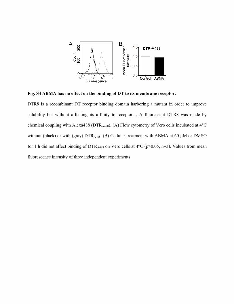
Fig. S4 ABMA has no effect on the binding of DT to its membrane receptor.
DTR8 is a recombinant DT receptor binding domain harboring a mutant in order to improve
solubility but without affecting its affinity to receptors1. A fluorescent DTR8 was made by
chemical coupling with Alexa488 (DTRA488). (A) Flow cytometry of Vero cells incubated at 4°C
without (black) or with (gray) DTRA488. (B) Cellular treatment with ABMA at 60 µM or DMSO
for 1 h did not affect binding of DTRA488 on Vero cells at 4°C (p>0.05, n=3). Values from mean
fluorescence intensity of three independent experiments.

Fig. S5 ABMA does not inhibit infection by CHIKV.
HEK293 cells were pre-treated with ABMA solubilized in DMSO for 1 h and then challenged
with CHIKV for 1 h. Cells were washed and then incubated again with same concentrations of
ABMA for 72 h. Virus induced-cytopathic effect (CPE) was measured by MTT assay.
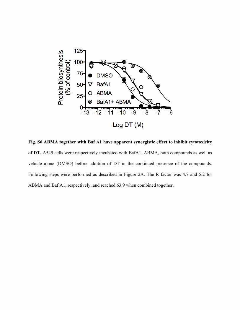
Fig. S6 ABMA together with Baf A1 have apparent synergistic effect to inhibit cytotoxicity
of DT. A549 cells were respectively incubated with BafA1, ABMA, both compounds as well as
vehicle alone (DMSO) before addition of DT in the continued presence of the compounds.
Following steps were performed as described in Figure 2A. The R factor was 4.7 and 5.2 for
ABMA and Baf A1, respectively, and reached 63.9 when combined together.

Fig. S7 ABMA does not affect the morphology of the Golgi apparatus or the endoplasmic
reticulum.
PC3 cells were incubated with vehicle alone (DMSO) or ABMA (120 µM) for 2 h at 37°C. Cells
were fixed, permeabilized and labeled for the indicated compartment markers: PDI (Endoplasmic
reticulum), TGN46 (Golgi apparatus). Images were obtained with confocal microscope (SP8X,
Leica). Nuclei were stained with Hoechst 33342 (blue). Scale bar, 10 µm.

Fig. S8 Full-length blots for Figure 2.

Fig. S9 Full-length blots for Figure 4.

Fig. S10 Full-length blots for Figure 5.
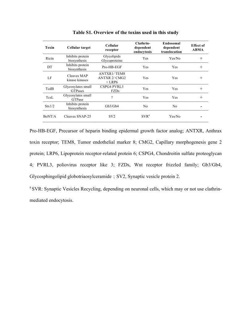
Table S1. Overview of the toxins used in this study
Toxin Cellular target Cellular receptor
Clathrin- dependent endocytosis
Endosomal dependent
translocation
Effect of ABMA
Ricin Inhibits protein biosynthesis
Glycolipids Glycoproteins Yes Yes/No +
DT Inhibits protein biosynthesis Pro-HB-EGF Yes Yes +
LF Cleaves MAP kinase kinases
ANTXR1/ TEM8 ANTXR 2/ CMG2
+ LRP6 Yes Yes +
TcdB Glycosylates small GTPases
CSPG4 PVRL3 FZDs Yes Yes +
TcsL Glycosylates small GTPase ? Yes Yes +
Stx1/2 Inhibits protein biosynthesis Gb3/Gb4 No No -
BoNT/A Cleaves SNAP-25 SV2 SVRa Yes/No -
Pro-HB-EGF, Precursor of heparin binding epidermal growth factor analog; ANTXR, Anthrax
toxin receptor; TEM8, Tumor endothelial marker 8; CMG2, Capillary morphogenesis gene 2
protein; LRP6, Lipoprotein receptor-related protein 6; CSPG4, Chondroitin sulfate proteoglycan
4; PVRL3, poliovirus receptor like 3; FZDs, Wnt receptor frizzled family; Gb3/Gb4,
Glycosphingolipid globotriaosylceramide;SV2, Synaptic vesicle protein 2.
a SVR: Synaptic Vesicles Recycling, depending on neuronal cells, which may or not use clathrin-
mediated endocytosis.
Table S2. Overview of the viruses used in this study
Virus Family Genus
Genome
Envelope Main cellular receptor
Endosomal -dependent
translocation
Effect of ABMA
Ebola Zaire EBOV Filoviridae Ebolavirus negative single-
stranded RNA
Yes NPC1 (in endosome)
TIM-1 Yes +
Rabies RABV Rhabdoviridae Lyssavirus negative single-
stranded RNA
Yes
nAChR NCAM LNGFR
Yes +
Dengue-4 DENV4 Flaviviridae Flavivirus positive single-
stranded RNA
Yes
DC-SIGN (CD209) Mannose receptor
(CD206) Yes +
Chikungunya CHIKV Togaviridae Alphavirus positive single-
stranded RNA
Yes PHB, PVEERs,
GAGs
Yes(95% within early
endosome) -
NPC1, Niemann-Pick C1; TIM-1, T cell immunoglobulin mucin domain 1; nAChR, Nicotinic
acetylcholine receptors; NCAM, neural cell adhesion molecule; LNGFR, low-affinity nerve
growth factor receptor; DC-SIGN (CD209), dendritic cell-specific adhesion receptor; PHB,
Prohibitin; PVEERs, Phosphatidylserine (PtdSer)-mediated virus entry-enhancing receptors;
GAGs, Glycosaminoglycans.

SUPPLEMENTAL MATERIALS AND METHODS
Evaluating efficacy on toxins by protein synthesis assay
Assay. Human HeLa cells (ATCC) or A549 cells were maintained at 37°C under 5% CO2 in
DMEM (Dulbecco’s modified Eagle’s medium, Invitrogen), supplemented with 10% fetal
bovine serum (12476-016, Gibco), 100U/mL penicillin, 100 µg/mL streptomycin (15140-122,
Gibco), 0.1 mM non-essential amino acids (11140-035, Gibco). HUVECs (PromoCell,
Heidelberg, Germany) were maintained at same conditions in human endothelial SFM (11111-
044, Gibco), supplemented with 20% fetal bovine serum, 20 ng/mL bFGF (13256-029, Gibco),
10 ng/mL EGF (13247-051, Gibco), 1 µg/mL Heparin (H3149, Sigma) and 100 U/mL penicillin
- 100 µg/mL streptomycin (15140-122, Gibco).
The cells were plated in 96-well Cytostar-TTM scintillating microplates (Perkin-Elmer) with
scintillator incorporated into the polystyrene plastic one day before. After incubation with
various concentration of compounds (or carrier, DMSO) for indicated time at 37 °C, cells were
challenged with increasing doses of ricin, diphtheria toxin or Stx2 for at least 16 h in continued
presence of compounds. Subsequently, the medium was removed and replaced with DMEM
without leucine (Gibco) containing 10% fetal bovine serum, 2 mM L-glutamine, 0.1 mM non-
essential amino acids, 100 U/mL penicillin - 100 µg/mL streptomycin supplemented by 0.5
µCi/mL [14C]-leucine. The cells were grown for an additional 3-6 hours at 37 °C in an
atmosphere of 5% CO2 and 95% air. Protein biosynthesis was then determined by measuring the
incorporation of radiolabeled leucine into cells using a Wallac 1450 MicroBeta liquid
scintillation counter (Perkin Elmer).
The mean percentage of protein biosynthesis was determined and normalized from duplicate
wells. All values are expressed as means ± SEM. Data were fitted with Prism v5 software
(Graphpad Inc., San Diego, CA) to obtain the 50% inhibitory toxin concentration (IC50) i.e. the
concentration of toxin that is required to inhibit 50% protein synthesis of cells. IC50 values and
protection factor R (R = IC50 drug/IC50 DMSO) were determined by the software’s nonlinear
regression “dose-response EC50 shift equation”. The goodness of fit for toxin alone (carrier) or
with drug was assessed by r2 and confidence intervals.
Determination of EC50 values of ABMA against ricin, DT, Stx2. EC50 was used to evaluate
ABMA efficacy because it is more precise than R values and associated % protection. This is
due to the fact that R values may fluctuate between cell experiments from different 96-wells
plates corresponding to compound tested on different days. In contrast EC50 value for a single
compound is calculated from experimental data obtained on a single 96-well plate. Cell assays
were performed with various concentrations of the compound. For each concentration, a
percentage of protection was determined from R values calculated with Prism software with
Rmax corresponding to the higher value of R of the series:
% protection = R− 1
Rmax− 1 × 100
Drug concentration was plotted against the corresponding percentage of protection of cells and
the 50% efficacy concentration (EC50) was calculated by non-linear regression using the Prism
software package.
Cell rounding induced by large clostridial glucosylating toxins on cultured cells
Cytotoxic effects of the TcdB and TcsL toxins on Vero cells (Sigma-Aldrich) were assessed by
cell rounding assays. Vero cells seeded in 96-well plates at a density of 10,000 cells per well one
day before exposuring to increasing doses of TcdB (4 hours) or TcsL (16 hours) in the continued

presence of compounds at various concentrations. Cell rounding was visualized by phase-
contrast microscopy using an inverted microscope (Ti-U, Nikon) with a CFI Achro Ph ADL-
10X/0.25 objective. Approximately 100-200 cells, counterstained with Hoechst 33342 dye, were
counted and the percentage of rounded cells was determined. EC50 was calculated by non-linear
regression using the Prism v5 software.
Anthrax toxin MEK cleavage Assay
Cell lysates from HUVECs intoxicated with LT (3 µg/ml protective antigen (PA) and 1µg/ml
Lethal factor (LF)) in the absence or presence of ABMA were resolved on 12% SDS-PAGE gels
using standard conditions and transferred to Immobilon-P PVDF membranes (Millipore). The
antibodies used were as follows: an antibody directed toward the amino-terminal part of MEK2
(MEK2-N20, Santa Cruz), anti-beta-actin (Clone AC74, Sigma). The primary antibodies were
revealed using horseradish peroxidase-conjugated goat anti-mouse or anti-rabbit secondary
antibodies (DAKO) followed by chemiluminescence using Immobilon Western (Millipore). The
chemiluminescent signals were recorded on a FUJIFILM LAS-3000, and the data were
quantified using the MultiGauge V3.0 software.
BoNT/A SNAP25 cleavage assay
Primary cultures of rat cerebellar granule neurons (CGNs) were prepared from 6-days-old rats as
previously described 2. Briefly, cerebella were isolated, mechanically disrupted and then
trypsinized. Cells were then collected and plated into 24 well plates, pre-coated with poly-L-
lysine (50 µg/ml), at a cell density of 5 × 10⁵ cells per well. Cultures were maintained at 37 °C,
5% CO2, 95% humidity in neurobasal medium supplemented with B27, to which 20 mM KCl,

0.5 mM glutamine 100 U/ml penicillin and 100 U/ml streptomycin were added. To inhibit
proliferation of non-neuronal cells, cytosine β-D-arabinofuranoside (5 µM) was added to the
medium 24 h after plating. ABMA (30 µM) was added to cell medium 1 h prior to BoNT/A
exposure (500 pM, 24h). Cell lysate were collected and SNAP-25 protein was detected by
immune blot with antibody (SMI-81, Biolegend).
EBOV experiments
All experiments with replication competent EBOV were performed in the biosafety level 4
(BSL-4) laboratory at the Texas Biomedical Research Institute (San Antonio, TX). The
recombinant Zaire EBOV strain Mayinga expressing enhanced GFP (EBOV-eGFP) was kindly
provided by Heinz Feldmann (NIH, Hamilton, MT). The viruses were amplified in Vero-E6 cells
(CDC, Atlanta, GA) for 7 days in DMEM containing 2% FBS. The culture supernatants
containing the viruses were clarified of cell debris, overlaid over a 20% sucrose cushion in PBS,
and centrifuged at 4°C at 28,000 rpms for 2 h. The pellets were resuspended in PBS. The titer of
EBOV-eGFP stock was determined by incubating serial dilutions of the stock on Vero cells for
24 h. Infected cells were fixed with 10% buffered formalin (Sigma, St. Louis, MO) for 24 h, and
then stained with Hoechst 33342 dye (Life Technologies, Carlsbad, CA) to identify cell nuclei.
Cells were photographed using a Nikon Ti-Eclipse microscope running high content analysis
software (Nikon, Tokyo, Japan). The numbers of cell nuclei and infected cells were counted
using CellProfiler software (Broad Institute, Boston, MA) with pipelines developed by the
authors (available upon request).
EBOV infection. To test impact of ABMA on virus infection, HeLa cells grown in 96-well
plates in MEM medium supplemented with 10% fetal bovine serum and incubated at 37°C, 5%

CO2. Cells were either left untreated or pretreated in triplicate with the compound or DMSO in
2-fold serial dilutions for 1 h and then challenged with EBOV-eGFP, at a multiplicity of
infection (MOI) of 0.1 for 24 h. This virus is replication competent and shows normal
pathogenesis in animals but expresses GFP 24 hours after infection has taken place. For this
reason, the experiment was halted at 24 hours after virus challenge, which was sufficient time for
a single round of virus infection and expression of the GFP infection marker. Subsequently,
infected cells were fixed in 10% formalin and stained with DAPI. The infection was then
analyzed by photographing each well using an epifluorescent microscope. Total and infected
cells were counted by counting DAPI stained cells and green fluorescent cells respectively using
CellProfiler software. The infection rate was calculated as the ratio of infected cells to cell
nuclei.
fraction infection = number of green fluorescent cellsnumber of DAPI stained cell nuclei
inhibition % = 100 × 1−fraction infection after drug treatmentfraction infection without treatment
Three parameter dose response curves were fitted to data by non-linear regression and EC50
values determined using Graphpad Prism 5 software. The equation used is below:
Y = lower plateau+ upper− lower plateau ÷ 1+ 10 !!!"# !"#$
In vitro RABV infection experiments
Assay. BSR cells (a clone of BHK cells, gift of Monique Lafon) were incubated in DMEM with
8% FBS and 40 µg/mL of gentamycin for 24 h at 37°C and 5% CO2 in 96-well plate (35,000
cells/well). After washing with DMEM, cells were pre-incubated for 4 h at 37°C in DMEM with
2% SVF and 40 µg/mL of gentamycin in presence of ABMA or ribavirin. After 4 h of pre-
incubation with compounds, cells were infected with the PV strain of RABV (MOI=14, seed lot
also grown on BSR cells) for 1 h at 37°C and 5% CO2. Cells were then washed once with
DMEM, then incubated again in DMEM with 2 % FBS and 40 µg/mL of gentamycin in presence
of compound at the same concentration. After 24h, the supernatant was removed and kept at -80
°C for titration, cells were washed with PBS Ca2+, Mg2+, then 80% acetone was added for fixation
for 30 min at 4°C. Cells were then incubated with a mouse monoclonal anti-RNP
(ribonucleoprotein) conjugated with FITC diluted in PBS for 1 h to detect the cytoplasmic
accumulation of intracellular RABV RNP. Hoechst in PBS (1/2000) was used for nucleus
staining.
Image acquisition and analysis. Images were acquired on an automated spinning disk confocal
microscope (Opera QEHS, Perkin Elmer Technologies) at the Imagopole (Institut Pasteur
imaging platform). Briefly, 15 fields were acquired per well with a 10× air objective (NA 0.4) to
reveal the nuclei stained with Hoechst (405 nm/450 nm) and to detect the RABV-RNP revealed
by a fluorescein-coupled antibodies (488 nm/540 nm). The image analysis was performed using
AcapellaTM (Perkin Elmer Technologies) as follows: (1) the nuclei were segmented in the
Hoechst channel to estimate the number of cells imaged in each well (above 10,000 total nuclei –
TN - in the non-infected control wells); (2) the segmentation mask was then increased to the
limit of each individual cytoplasm, generating a “cell mask” in which the viral RNP (green dots)
were segmented using a spot detection module of the software, allowing the reporting of the

number of infected cells – InfC – i.e. containing at least 1 green spot. Finally, we calculated the
ratio [InfC/TN*100], which reports the percentage of infection of each well.
DENV4 infection experiments
Vero cells (ATCC) were plated into 96-wells microplates for 30 min pre-treated with 6
concentrations of ABMA and one reference compound ribavirine, and infected with 125 TCID50
per well of a DEN-4 serotype virus. Seven days later, cells were lysed using Igepal 2.5% in
phosphate buffer (PBS) before quantifying viral replication using specific ELISA conditions (a
specific serum from DENV-4-infected non-human primate and peroxidase-conjugated goat anti-
monkey IgG diluted in PBS, 5% milk, 0.5% Tween 20). Absorbance was measured using a
spectrophotometer at 492 nm.
Previously, this viral strain was amplified and titrated by limit-dilution; the 50% tissue culture
infectious dose (TCID50) was calculated using Kärber’s formula.
CHIKV infection experiments
HEK 293 (ATCC) were plated into 96-wells microplates 24 h before. Cells were pre-treated
with different concentrations of ABMA for 1 h and then infected with CHIKV for 1 h. Cells
were washed and incubated again with same concentration of ABMA for 72 h. Virus induced-
cytopathic effect (CPE) was measured by MTT assay.
Sn infectivity assay
40,000 HeLa229 cells (ATCC) were seeded in 12-well cluster plates, compound-treated and
infected as indicated in the respective experiment. Cells were either fixed and stained or

harvested for immunoblotting at indicated time points or bacteria were released via one freeze
thaw cycle (-70°C/37°C) followed by mechanical release through pipetting and transfer to fresh
HeLa229 cells (1:25-1:50, progeny/infectivity). Cells were centrifuged for 1 h at 35°C and
medium exchanged to infection medium. Progeny was fixed at day 3 post infection and
processed for staining or harvested for immunoblotting. Infectivity assays were imaged on an
automated fluorescence microscope Leica DMIR. Numbers and average sizes of the SnCV as
well as host cell numbers were determined via snHsp60 and DAPI staining and images were
analysed and quantified using FIJI (Image J) and Excel (Microsoft).
In this assay, bacteria are first grown in cells treated with compound (ABMA at a concentration
of 25 and 75 µM) and the infectious particles from this primary infection are applied to fresh
cells in the absence of compounds to measure the bacterial load (snHSP60 immunoblotting) and
inclusion formation (immunofluorescence microscopy).
Immunoblotting and immunofluorescence staining. Western blotting was performed
according to standard procedures and signals were detected with a Chemo Cam Imager (Intas).
Quantification was performed using FIJI (ImageJ) and Excel (Microsoft). For
immunofluorescence, cells were seeded in 12-well cluster plates with or without coverslips, and
were infected with MOIs indicated in the respective experiments in a humidified incubator at
35°C and 5% CO2. Cells were fixed at indicated time points with 4% PFA-sucrose for 25 min,
washed once with PBS. Subsequently cells were permeabilized with 0.2% Triton X-100 in PBS
for 45 min and washed 3 times with PBS for 15 min at RT. Cells were blocked with 2% goat
serum in PBS for 60 min and were stained with primary antibodies diluted in 2% goat serum in
PBS for 24 h at 4°C. After 3x washing with PBS for 15 min, samples were incubated with
secondary antibodies for 1 h in blocking solution at RT in the dark. Cells were washed 3 times

with PBS and counterstained with DAPI (1 µg/mL) in PBS for 15 min. Coverslips were mounted
with Mowiol 4–88 (Carl Roth) after short wash with H2O. 12-well cluster plates without
coverslips were washed 3 times with PBS for 15 min and visualized at RT. Samples were
subsequently viewed on a Leica DMIR or TCS SP5 microscope, respectively. Data were
processed using FIJI (Image J). Antibodies against snHsp60 were prepared as previously
described 3.
Ctr infectivity assays
HeLa229 were grown in RPMI1640 medium (Glutamax, 10% heat-inactivated FBS, w/o
HEPES) (Invitrogen). Cells were pretreated with ABMA in concentrations of 25, 50 and 75 µM
for 1 h until Ctr (MOI=1) were added to the cells. ABMA was present during primary infection.
To obtain progeny infection, compound-treated cells were lysed with glass beads 48 h post
infection (hpi) and lysate was used to infect fresh HeLa229 cells. Progeny infection in absence of
ABMA was lysed 24 hpi and analyzed together with primary infection samples by
immunoblotting. Chlamydial growth was detected with antibodies against ctrHSP60 (clone A57-
B9, Santa Cruz Biotechnology) and β-actin (clone AC-15, Sigma-Aldrich) was used as loading
control. Immunofluorescence analysis of Progeny infection was also performed 24 hpi, cells
were fixed and stained for DAPI. Ctr inclusions were detected by their GFP-expression signal.
In vitro antileishmanial evaluation
The experiments were performed by using the Leishmania infantum strain
(MHOM/FR/2008/LEM5700). Antileishmanial activity on L. infantum axenic amastigotes was
carried out as previously described 4.

Antileishmanial activity on the L. infantum/RAW 264.7 macrophage model. As previously
described 5, mouse macrophage cells RAW 264.7 (ATCC) were seeded into a 96-well microplate
and maintained in DMEM medium (Applied Biosystems, France) supplemented with 10% heat-
inactivated fetal bovine serum in a 5% CO2 incubator at 37 ºC for 24 h. Then, the medium was
replaced with 100 µL fresh medium containing a suspension of 105 L. infantum amastigotes and
the incubation time was 16 h. Then, extracellular parasites were eliminated from the medium and
100 µL fresh medium containing compound in various concentrations were added for a 48 h-
incubation. Parasite growth was determined by using SYBR® Green I (Life technology, Fisher
Scientific, France), a dye with marked fluorescence enhancement upon contact with DNA. The
cell lysis suspension was diluted 1:1 in lysis buffer (10 mM NaCl, 1 mM Tris HCl pH8, 2.5 mM
EDTA pH 8, 0.05% SDS, 0.01 mg/mL proteinase K and 10 × SYBR Green I). Incorporation of
SYBR Green I in DNA amplification was measured using the Master epRealplex cycler®
(Eppendorf, France) according to the following program to increase the SYBR green
incorporation: 90°C for 1 min, decrease in temperature from 90°C to 10°C for 5 min with
reading the fluorescence at 10°C for 1 min and a new reading at 10°C for 2 min. The EC50 as the
concentration inhibiting the parasite growth by 50% was calculated by nonlinear regression using
icestimator website 1.2 version: http://www.antimalarial-icestimator.net/MethodIntro.htm.
Amphotericin B, miltefosine were used as reference drugs. The concentrations used for drug
evaluation were in a range from 0.05 to 100 µM, obtained by serial dilution with a factor of two,
except for amphotericin B, for which the range was from 0.005 to 10 µM.
Cytotoxicity evaluation on RAW 264.7 macrophages. RAW 264.7 cells were seeded into a 96-
well microtiter plate at a density of 105 cells/well in 100 µL of DMEM. After incubation in a 5%
CO2 incubator at 37°C for 24 h, the culture medium was replaced with 100 µL of fresh DMEM

containing the compounds for a new incubation of 48 h. The final compound concentrations used
were from 0.05 µM to 100 µM obtained by serial dilution. Triplicates were used for each
concentration. The viability of the macrophages was then assessed using the SYBR® Green I
(Invitrogen, France) incorporation method and the results were expressed as cytotoxic
concentration inhibiting the cell growth by 50% (CC50) in µM.
Flow cytometry
Mutant receptor binding domain of diphtheria toxin (DTR8) was produced in house 1 and
fluorescent DTR8 (DTRA488) was obtained using Alexa Fluor labeling kit (A20181, Thermo
Fisher). Cell surface toxin receptors (proHB-EGF) were incubated and labeled with DTRA488
(1:50 dilution, on trypsinized Vero cells) in PBS/BSA 1% at 4°C prior to incubate with
compound for 1 h. After rinsing with the same buffer, single cell suspension was subjected to
FACS analysis. Analysis was performed by flow cytometry (Accuri C6 cytometer, BD
Biosciences) and CFlow Plus software (BD Biosciences).
SUPPLEMENTAL REFERENCES
1 Gillet, D., VILLIERS, B., Pichard, S., Maillere, B. & Sanson, A. Hb-egf inhibitor derived from the R domain of diphtheria toxin for the treatment of diseases associated with the activation of the HB-EGF/EGFR pathway (US Patent 9,758,552, 2017).
2 Wong, J. K., Kennedy, P. R. & Belcher, S. M. Simplified serum- and steroid-free culture conditions for high-throughput viability analysis of primary cultures of cerebellar granule neurons. Journal of neuroscience methods 110, 45-55 (2001).
3 Mehlitz, A. et al. The chlamydial organism Simkania negevensis forms ER vacuole contact sites and inhibits ER-stress. Cell Microbiol 16, 1224-1243, doi:10.1111/cmi.12278 (2014).
4 Audisio, D. et al. Synthesis and antikinetoplastid activities of 3-substituted quinolinones derivatives. European journal of medicinal chemistry 52, 44-50, doi:10.1016/j.ejmech.2012.03.003 (2012).
5 Balaraman, K. et al. In vitro and in vivo antileishmanial properties of a 2-n-propylquinoline hydroxypropyl beta-cyclodextrin formulation and pharmacokinetics via

intravenous route. Biomedicine & pharmacotherapy = Biomedecine & pharmacotherapie 76, 127-133, doi:10.1016/j.biopha.2015.10.028 (2015).


![4: Zootoxins (toxins of animals) [Biological-origin toxins]](https://static.documents.pub/doc/80x56/61cddf54f2b98d6a6b5b05e1/4-zootoxins-toxins-of-animals-biological-origin-toxins.jpg)

