
Pulmonary
Pulmonary
Adult CCRN/CCRN‐E/CCRN‐K Certification Review Course:
Pulmonary
Carol RauenRN‐BC, MS, PCCN, CCRN, CEN
Pulmonary
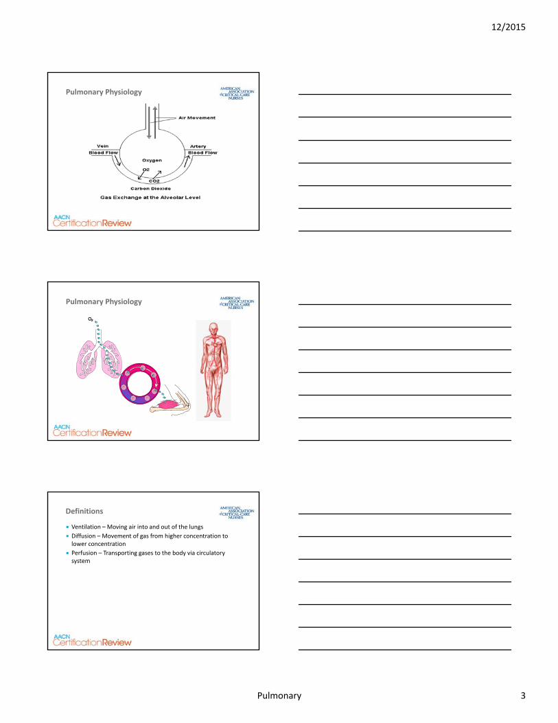
Syndrome
Direct or indirect injury
Significant inflammatory insult
Mediator release
Increased capillary permeability
Acute Lung Injury (ALI)/ARDS
Illustration Copyright ©2011 Nucleus Medical Media. All rights reserved. www.nucleusinc.com
12/2015
Pulmonary 1
Pulmonary
Nothing to disclose
Disclosures
Pulmonary
Acute respiratory failure and acute
respiratory distress syndrome (ARDS)
Acute pulmonary embolism (PE)
Acute respiratory infections (eg, pneumonia)
Air‐leak syndromes
Aspiration and pulmonary fibrosis
Chronic conditions (eg, COPD, asthma)
Failure to wean
Pulmonary hypertension
Thoracic surgery and trauma
Status asthmaticus
Pulmonary
80%
20%
17%
Pulmonary
Anatomy Review
Air Sacs
Alveoli
Bronchiole
Illustration Copyright ©2011 Nucleus Medical Media. All rights reserved
12/2015
Pulmonary 2
Pulmonary
Pulmonary Physiology
Pulmonary
Pulmonary Physiology
Pulmonary
Ventilation – Moving air into and out of the lungs
Diffusion – Movement of gas from higher concentration to lower concentration
Perfusion – Transporting gases to the body via circulatory system
Definitions
12/2015
Pulmonary 3
Pulmonary
Definitions
Dead space ventilation – Alveolar ventilation with no perfusion
Pulmonary embolism (PE)
Intrapulmonary shunting –Perfusion with no ventilation Atelectasis
Pulmonary
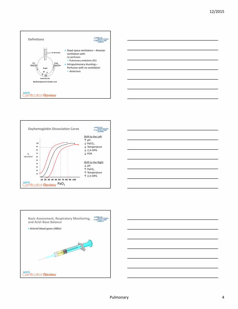
10 20 30 40 50 60 70 80 90 100
100
90
80
70
60
50
40
30
20
10
PaO2
%saturation
Shift to the Left↑ pH↓ PaCO2
↓ Temperature↓ 2,3–DPG↓ PO4
Shift to the Right↓ pH↑ PaCO2
↑ Temperature↑ 2,3–DPG
Oxyhemoglobin Dissociation Curve
Pulmonary
Basic Assessment, Respiratory Monitoring, and Acid–Base Balance
Arterial blood gases (ABGs)
12/2015
Pulmonary 4
Pulmonary
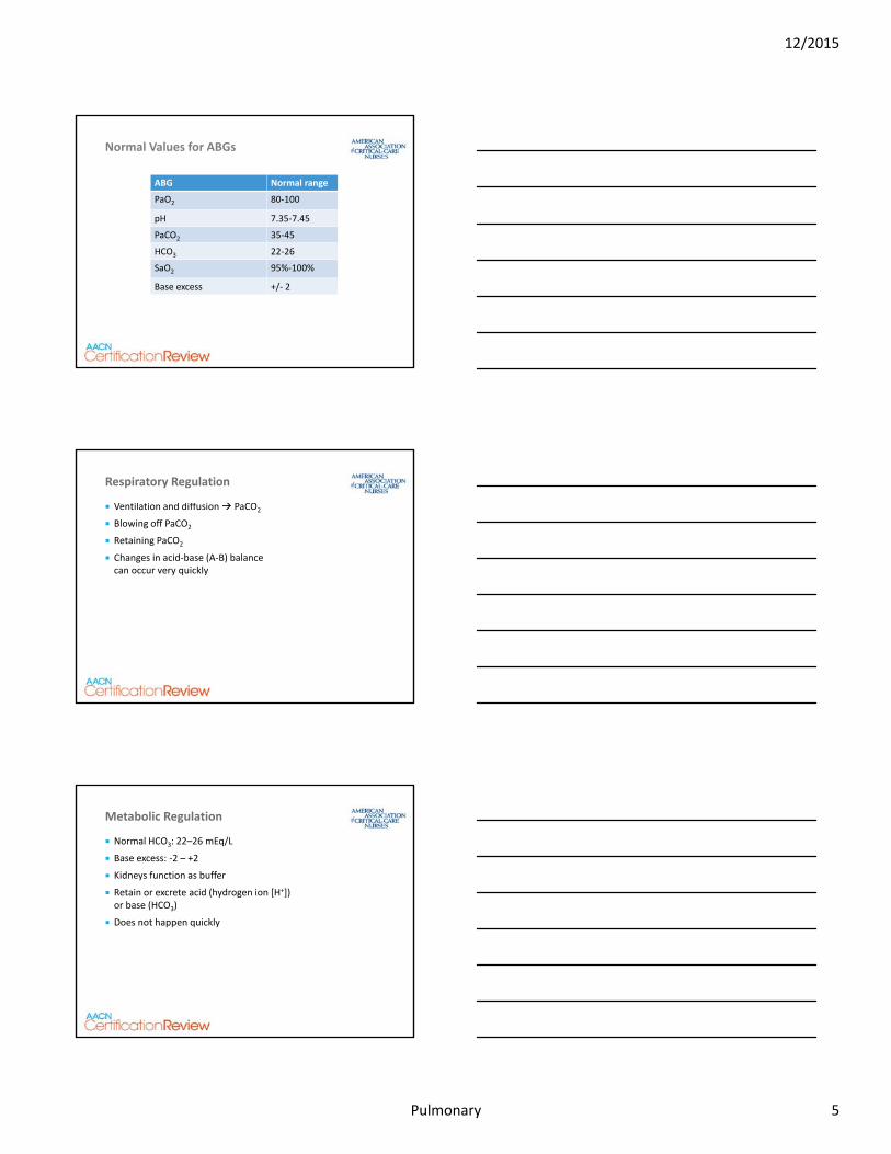
ABG Normal range
PaO2 80‐100
pH 7.35‐7.45
PaCO2 35‐45
HCO3 22‐26
SaO2 95%‐100%
Base excess +/‐ 2
Normal Values for ABGs
Pulmonary
Ventilation and diffusion PaCO2
Blowing off PaCO2
Retaining PaCO2
Changes in acid‐base (A‐B) balance can occur very quickly
Respiratory Regulation
Pulmonary
Normal HCO3: 22–26 mEq/L
Base excess: ‐2 – +2
Kidneys function as buffer
Retain or excrete acid (hydrogen ion [H+]) or base (HCO3)
Does not happen quickly
Metabolic Regulation
12/2015
Pulmonary 5
Pulmonary
Review Questions
Pulmonary
A. pH 7.51, PaCO2 32, HCO3 23
B. pH 7.31, PaCO2 49, HCO3 28
C. pH 7.29, PaCO2 37, HCO3 17
D. pH 7.55, PaCO2 40, HCO3 29
Question 1
Renal failure presents most commonly with which of the following acid‐base imbalance patterns?
Pulmonary
Question 1—Rationale
C. pH 7.29, PaCO2 37, HCO3 17
pH 7.51, PaCO2 32, HCO3 23—Respiratory alkalosis
pH 7.31, PaCO2 49, HCO3 28—Respiratory acidosis with partial met compensation
pH 7.55, PaCO2 40, HCO3 29—Metabolic alkalosis
Renal failure presents most commonly with which of the following acid‐base imbalance patterns?
12/2015
Pulmonary 6
Pulmonary
A. Acute tracheal obstruction
B. Anxiety‐induced hyperventilation
C. Chronic obstructive pulmonary disease
D. Diarrhea for 36 hours in a debilitated patient
Question 2
Which of the following clinical situations correlates with ABG results of pH 7.22, HCO3
23 mEq/L, PaCO2 65 mmHg, PaO2 56 mmHg?
Pulmonary
Question 2—Rationale
A. Acute tracheal obstruction—Hypoxia respiratory acidosis correlates because the O2 cannot get in or CO2 out
Anxiety‐induced hyperventilation—Respiratory alkalosis
Chronic obstructive pulmonary disease—Respiratory acidosis and hypoxia with partial metabolic compensation
Diarrhea for 36 hours in a debilitated patient—Metabolic acidosis
Which of the following clinical situations correlates with ABG results of pH 7.22, HCO323 mEq/L, PaCO2 65 mmHg, and PaO2 56 mmHg?
Pulmonary
Acute Respiratory Failure
Failure of the pulmonary system to provide adequate oxygenation or ventilation
Sudden drop in PaO2 or elevation in PaCO2
12/2015
Pulmonary 7
Pulmonary
Modes are classified by inspiratory trigger
Mechanical Ventilation
Pulmonary
Volume modes
Volume is set and pressure is variable
Mechanical Ventilation
Compliance alveoli and chest wellResistance airways
Pulmonary
Pressure modes
Pressure is set and volume is variable
Mechanical Ventilation
Compliance → alveoli and chest wellResistance → airways
12/2015
Pulmonary 8
Pulmonary
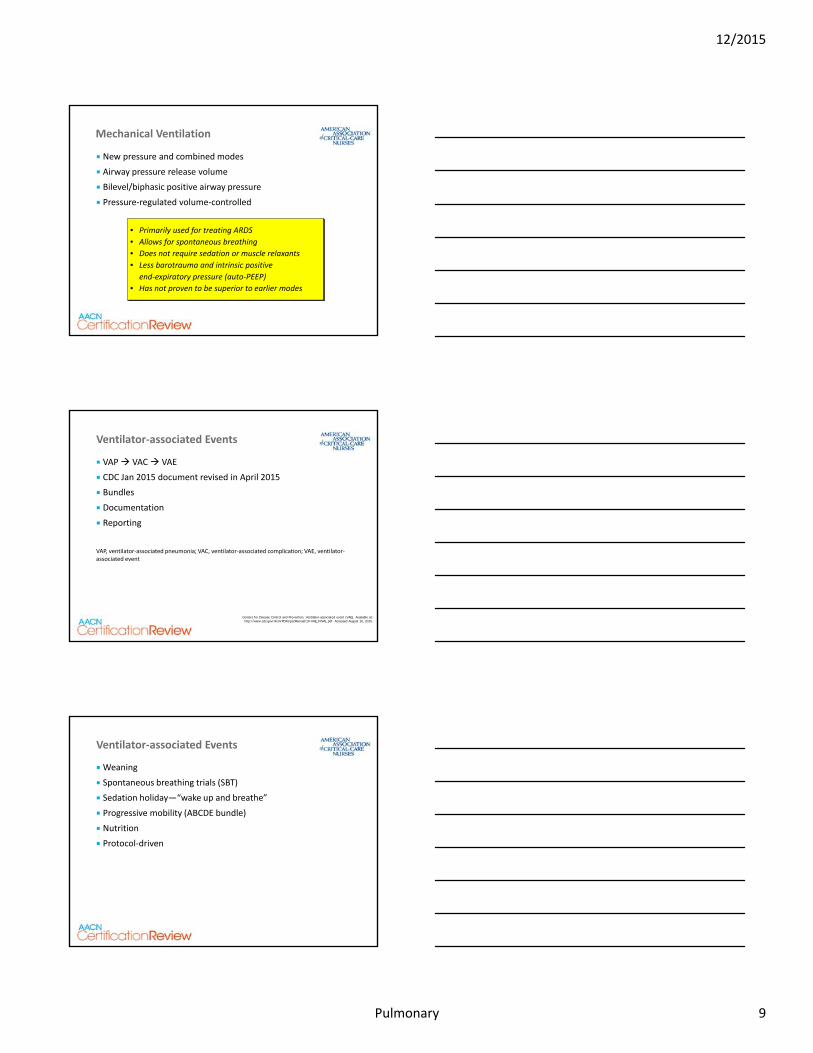
New pressure and combined modes
Airway pressure release volume
Bilevel/biphasic positive airway pressure
Pressure‐regulated volume‐controlled
Mechanical Ventilation
• Primarily used for treating ARDS
• Allows for spontaneous breathing
• Does not require sedation or muscle relaxants
• Less barotrauma and intrinsic positive
end‐expiratory pressure (auto‐PEEP)
• Has not proven to be superior to earlier modes
Pulmonary
VAP VAC VAE
CDC Jan 2015 document revised in April 2015
Bundles
Documentation
Reporting
VAP, ventilator‐associated pneumonia; VAC, ventilator‐associated complication; VAE, ventilator‐associated event
Ventilator‐associated Events
Centers for Disease Control and Prevention. Ventilator-associated event (VAE). Available at: http://www.cdc.gov/nhsn/PDFs/pscManual/10-VAE_FINAL.pdf. Accessed August 16, 2015.
Pulmonary
Weaning
Spontaneous breathing trials (SBT)
Sedation holiday—“wake up and breathe”
Progressive mobility (ABCDE bundle)
Nutrition
Protocol‐driven
Ventilator‐associated Events
12/2015
Pulmonary 9
Pulmonary
Fewer intubations
Continuous positive airway pressure (CPAP)
Bilevel positive airway pressure
High‐flow nasal cannula
Noninvasive Ventilation
Pulmonary
Review Questions
Pulmonary
A. Prophylactic antibiotics
B. Keeping the head of the bed elevated >30⁰
C. Decontaminating the room with a bleach mixture
D. Changing the ventilator circuit every 4 hours
Question 3
Which of the following is the most appropriate means of preventing ventilator‐associated pneumonia?
12/2015
Pulmonary 10
Pulmonary
Question 3—Rationale
B. Keep the head of the bed elevated >30⁰—Shown to help decrease risk of VAP
Give prophylactic antibiotics—Could increase incidence of resistance development
Decontaminate the room with a bleach mixture—Room cleanliness is important, but bleach is not required
Change the ventilator circuit every 4 hours—The circuit should not be changed that frequently
Which of the following is the most appropriate means of preventing ventilator‐associated pneumonia?
Pulmonary
A. Report the colleague to the charge nurse or manager
B. Note the practice on the patient’s chart to ensure consistency of suctioning techniques
C. Ask the attending physician to review the suctioning policy
D. Collaborate with the colleague to review the evidence about this practice
Question 4
A patient with a tracheostomy requires frequent suctioning for thick sputum. A nurse finds a colleague instilling saline in the endotracheal tube prior to suctioning. The most appropriate response by the nurse would be to:
Pulmonary
Question 4—Rationale
D. Collaborate with the colleague to review the evidence about this practice—Best practice is not to instill NS; it does not loosen secretions and does harms the patient. The practice should be stopped and communication and education given to the colleague
Report the colleague to the charge nurse or manager—Direct communication is more professional and appropriate
Note the practice on the patient’s chart to ensure consistency of suctioning techniques—NS instillation is not recommended
Ask the attending physician to review the suctioning policy—The policy should reflect current evidence‐based practice. The most immediate concern is patient safety
A patient with a tracheostomy requires frequent suctioning for thick sputum. A nurse finds a colleague instilling saline in the endotracheal tube prior to suctioning. The most appropriate response by the nurse would be to:
12/2015
Pulmonary 11
Pulmonary
Restrictive Lung Disorders
• ARDS
• Infections
• Occupational lung disease
• Sarcoidosis
• Atelectasis
Pulmonary disorders that restrict the lung from expanding
Lung compliance and volumes are decreased
Pulmonary
Syndrome
Direct or indirect injury
Significant inflammatory insult
Mediator release
Increased capillary permeability
Acute Lung Injury (ALI)/ARDS
Illustration Copyright ©2011 Nucleus Medical Media. All rights reserved. www.nucleusinc.com
Pulmonary
Pulmonary edema
Alveolar collapse
Lung damage
Lung failure
Subsequent death
ARDS
12/2015
Pulmonary 12
Pulmonary
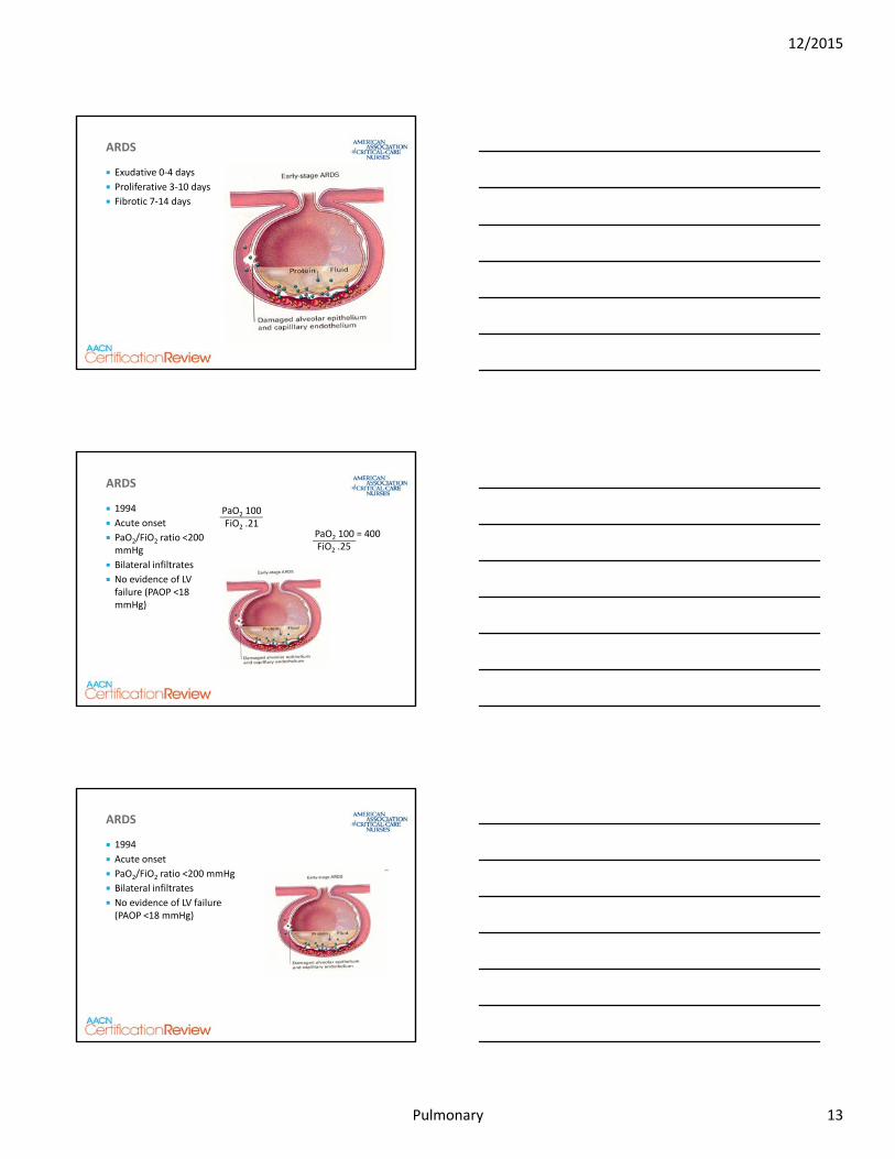
Exudative 0‐4 days
Proliferative 3‐10 days
Fibrotic 7‐14 days
ARDS
Pulmonary
PaO2 100FiO2 .21
PaO2 100 = 400FiO2 .25
1994
Acute onset
PaO2/FiO2 ratio <200 mmHg
Bilateral infiltrates
No evidence of LV failure (PAOP <18 mmHg)
ARDS
Pulmonary
1994
Acute onset
PaO2/FiO2 ratio <200 mmHg
Bilateral infiltrates
No evidence of LV failure (PAOP <18 mmHg)
ARDS
12/2015
Pulmonary 13
Pulmonary
ARDS
ARDS Definition Task Force, et al. JAMA. 2012;307(23):2526–2533.
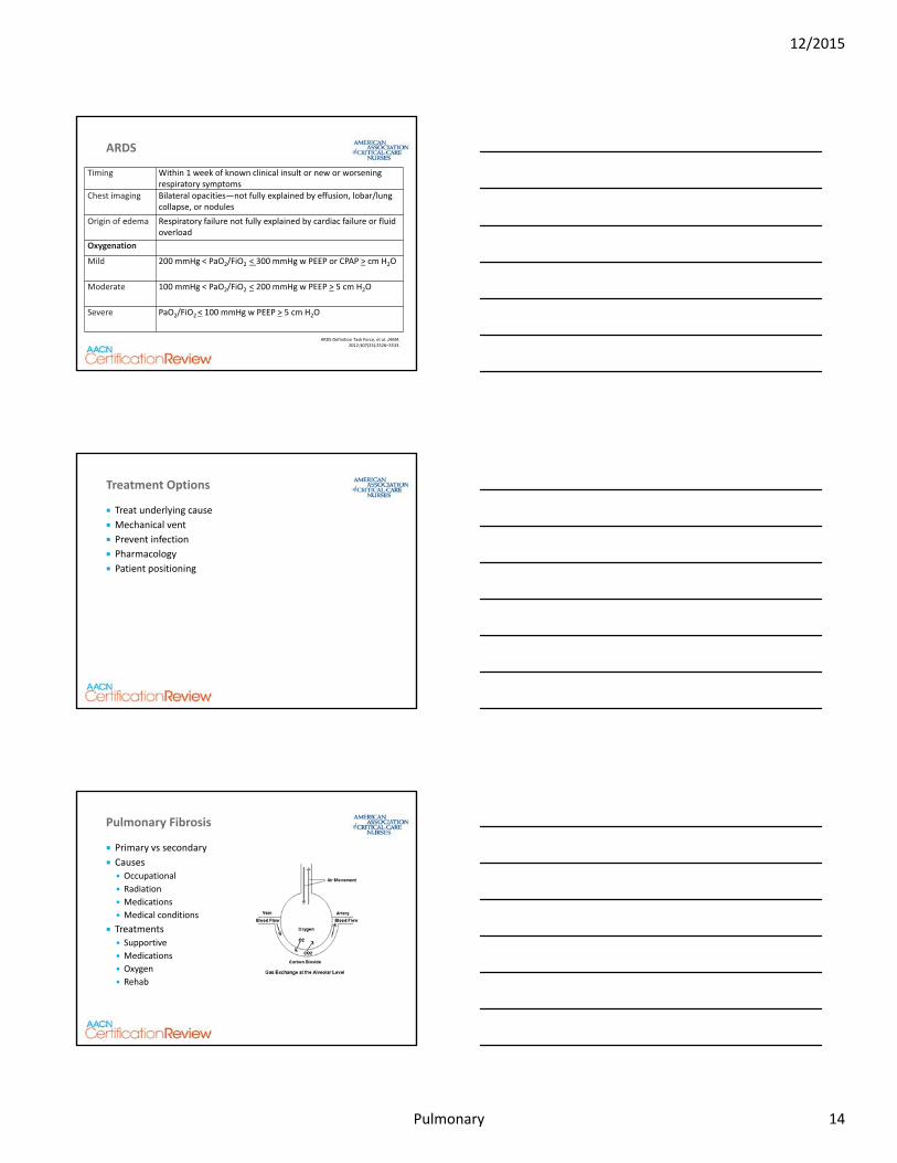
Timing Within 1 week of known clinical insult or new or worsening respiratory symptoms
Chest imaging Bilateral opacities—not fully explained by effusion, lobar/lung collapse, or nodules
Origin of edema Respiratory failure not fully explained by cardiac failure or fluid overload
Oxygenation
Mild 200 mmHg < PaO2/FiO2 < 300 mmHg w PEEP or CPAP > cm H2O
Moderate 100 mmHg < PaO2/FiO2 < 200 mmHg w PEEP > 5 cm H2O
Severe PaO2/FiO2 < 100 mmHg w PEEP > 5 cm H2O
Pulmonary
Treat underlying cause
Mechanical vent
Prevent infection
Pharmacology
Patient positioning
Treatment Options
Pulmonary
Pulmonary Fibrosis
Primary vs secondary
Causes Occupational
Radiation
Medications
Medical conditions
Treatments Supportive
Medications
Oxygen
Rehab
12/2015
Pulmonary 14
Pulmonary
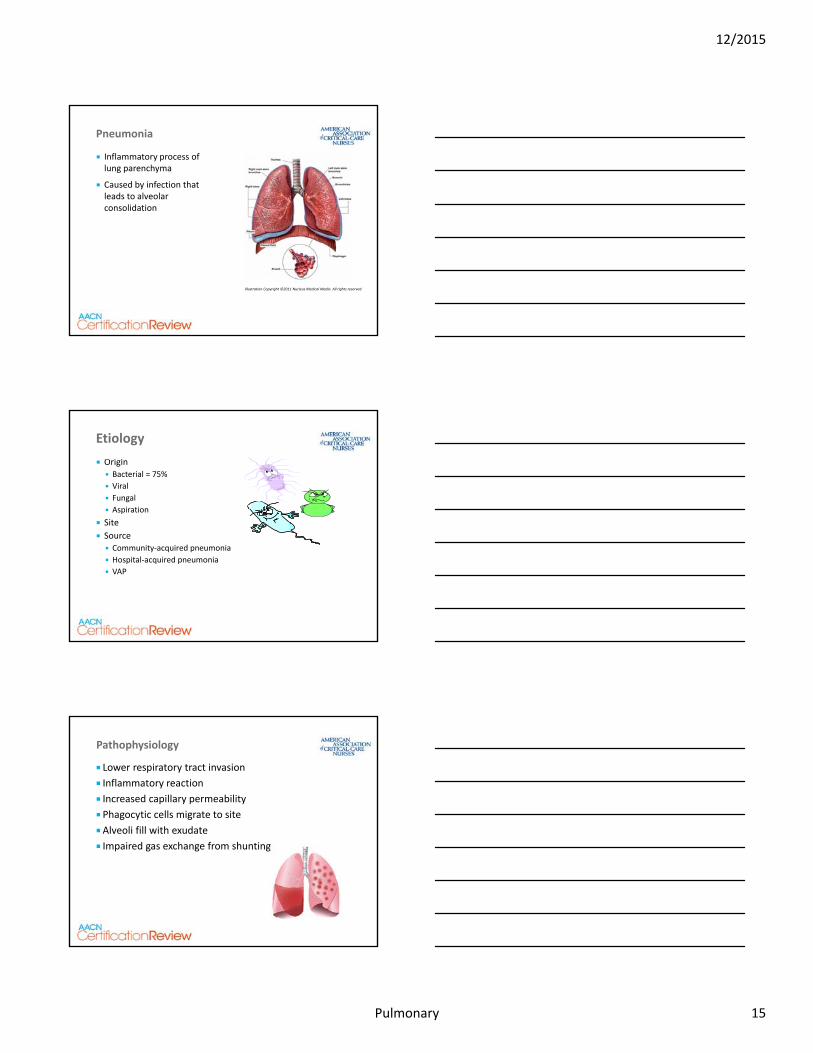
Inflammatory process of lung parenchyma
Caused by infection that leads to alveolar consolidation
Pneumonia
Illustration Copyright ©2011 Nucleus Medical Media. All rights reserved.
Pulmonary
Origin Bacterial = 75%
Viral
Fungal
Aspiration
Site
Source Community‐acquired pneumonia
Hospital‐acquired pneumonia
VAP
Etiology
Pulmonary
Pathophysiology
Lower respiratory tract invasion
Inflammatory reaction
Increased capillary permeability
Phagocytic cells migrate to site
Alveoli fill with exudate
Impaired gas exchange from shunting
12/2015
Pulmonary 15
Pulmonary
Clinical presentation
Diagnosis
Treatment
Pneumonia
Pulmonary
Review Questions
Pulmonary
A. Decreased compliance, hypoxemia, rapid shallow breathing
B. Increased compliance, hypercarbia, slow deep respirations
C. Decreased compliance, normal PaO2, shunting
D. Hypoxemia, dead space ventilation, low pH
Question 5
A restrictive lung disease is one that is characterized by:
12/2015
Pulmonary 16
Pulmonary
Question 5—Rationale
A. Decreased compliance, hypoxemia, rapid shallow breathing—ARDS and pneumonia are classic restrictive disorders
Increased compliance, hypercarbia, slow deep respirations—Decreased compliance occurs in restrictive diseases
Decreased compliance, normal PaO2, shunting—The PaO2 is typically low
Hypoxemia, dead space ventilation, low pH—Shunting is the ventilation/perfusion (V/Q) mismatch in restrictive disease
A restrictive lung disease is one that is characterized by:
Pulmonary
A. Aspiration pneumonia
B. Pulmonary embolism
C. Interstitial pneumonitis
D. ARDS
Question 6
Two days after a near‐drowning, a patient is dyspneic, using accessory muscles, expectorating large amounts of secretions, and reports feelings of “impending death.” Changes to the assessment data include:
Which of the following do these changes most likely represent?
Admission Day 2
RR 24 36
Chest x‐
ray
Clear Bilateral diffuse infiltrates
ABG 40% face mask 100% non‐rebreather mask
pO2 120 mm Hg 56 mmHg
pCO2 33 mm Hg 50 mmHg
pH 7.42 7.35
HCO3 24 mEq/L 27 mEq/L
Pulmonary
Question 6—Rationale
D. ARDS—Meets ARDS criteria and restrictive lung dis
Aspiration pneumonia—Could lead to ARDS
Pulmonary embolism—Presents with respiratory alkalosis
Interstitial pneumonitis—Could lead to ARDS
Which of the following do these changes most likely represent?
12/2015
Pulmonary 17
Pulmonary
A. Purulent sputum
B. Mediastinal shift to the right
C. Bradypnea
D. Intermittent apneic periods
Question 7
A patient is admitted with acute respiratory failure, left lobar pneumonia, and COPD. Physical exam reveals severe fatigue, coarse inspiratory crackles, and expiratory wheezing. Data also include:
Based on this info, the nurse should anticipate which of the following additional clinical findings?
HR 132 RR 36 T 102.6°F (38.9°C) pH 7.28 pCO2 72 pO2 48 HCO3 36
Pulmonary
Question 7—Rationale
A. Purulent sputum—Pneumonia, crackles, temp, and wheezing
Mediastinal shift to the right—Common with pneumothorax
Bradypnea—Hypoxia would cause tachycardia
Intermittent apneic periods—Hypoxia and acidosis causes hyperventilation
Which additional clinical findings might the nurse anticipate?
Pulmonary
A. He has been on the ventilator for 1 week
B. He is breathing over the set ventilator rate
C. The vasopressor was discontinued yesterday
D. He has been trached for 3 days
Question 8Which of the following assessment data would indicate that a patient who is receiving mechanical ventilation is ready for an SBT?
12/2015
Pulmonary 18
Pulmonary
Question 8—Rationale
C. The vasopressor was discontinued yesterday—Hemodynamic stability is an important criteria for weaning
He has been on the ventilator for 1 week—Pulmonary and hemodynamic stability are criteria, not time on vent
He is breathing over the set ventilator rate—This could be a sign of hypoxia, not always readiness
He has been trached for days—Being trached is a positive sign for weaning ability, but not a criteria
Which of the following assessment data would indicate that a patient who is receiving mechanical ventilation is ready for an SBT?
Pulmonary
A. Hypothermia, drowning, and acidosis will increase the O2 unloading at the cellular level
B. Hypothermia and alkalosis will decrease the O2 unloading at the cellular level
C. Hypothermia and alkalosis will help protect the heart from going in to Vfib
D. The hypothermia and hypoxia will need to be resolved before she can be declared dead
Question 9
A freshwater drowning victim is hypothermic and intubated upon arrival. Her ABGs are: PaO2 80, PaCO2 30, pH 7.51, HCO3 24. Which of the following factors are most important to consider when directing her care?
Pulmonary
Question 9—Rationale
B. “Hypothermia and alkalosis will decrease the O2 unloading at the cellular level”—Hypothermia and alkalosis cause of a shift of the oxyhemoglobin dissociation curve to the left and less unloading of O2
Hypothermia, drowning, and acidosis will increase the O2 unloading at the cellular level—Hypothermia causes a shift to the left and decreased unloading of O2
Hypothermia and alkalosis will help protect the heart from going in to Vfib—Not protective abnormalities
The hypothermia and hypoxia will need to be resolved before she can be declared dead—Attempts will be made to warm the patient, but this does not answer the question that was asked
A freshwater drowning victim is hypothermic and intubated upon arrival. Her ABGs are: PaO2 80, PaCO2 30, pH 7.51, HCO3 24. Which of the following factors are most important to consider when directing her care?
12/2015
Pulmonary 19
Pulmonary
A. No changes to the vent settings; administer an antianxiety agent
B. Decrease the FiO2 and consider pain medication
C. Decrease the tidal volume (TV) and increase the PEEP
D. Change the mode to PC and decrease the FiO2
Question 10
An intubated post‐op patient is beginning to wake up. Vent settings are AC, with a rate of 14, TV 450, FiO2 60%, 5 cm PEEP. Other assessments: RR 36; ABG: PaO2 150, PaCO2 28, pH 7.52, HCo3 24. What changes (if any) should the nurse anticipate to the vent settings?
Pulmonary
Question 10—Rationale
B. Decrease the FiO2 and consider pain medication—Hyperoxygenation and hyperventilation must be treated
No changes to the vent settings; administer an antianxiety agent—The Hyperoxygenation must be treated
Decrease the TV and increase the PEEP—Increasing PEEP would increase oxygenation
Change the mode to PC and decrease the FiO2—Changing to PC from AC will not treat the hyperoxygenation or hyperventilation
An intubated post‐op patient is beginning to wake up. Vent settings are AC, with a rate of 14, TV 450, FiO2 60%, 5 cm PEEP. Other assessments: RR 36; ABG: PaO2 150, PaCO2 28, pH 7.52, HCO3 24. What changes (if any) should the nurse anticipate to the vent settings?
Pulmonary
Obstructive Lung Disorders
• COPD
• Emphysema
• Bronchitis
• Asthma
Pulmonary disorders in which airway obstruction and gas trapping are the primary problem
Illustration Copyright ©2011 Nucleus Medical Media. All rights reserved.
12/2015
Pulmonary 20
Pulmonary
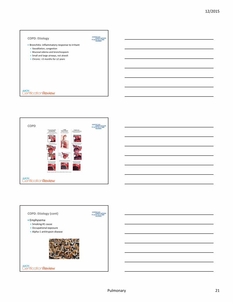
Bronchitis: inflammatory response to irritant
Vasodilation, congestion
Mucosal edema and bronchospasm
Small and large airways, not alveoli
Chronic: >3 months for ≥2 years
COPD: Etiology
Pulmonary
COPD
Illustration Copyright ©2011 Nucleus Medical Media. All rights reserved.
Pulmonary
Emphysema Smoking #1 cause
Occupational exposure
Alpha‐1 antitrypsin disease
COPD: Etiology (cont)
12/2015
Pulmonary 21
Pulmonary
COPD: Pathophysiology
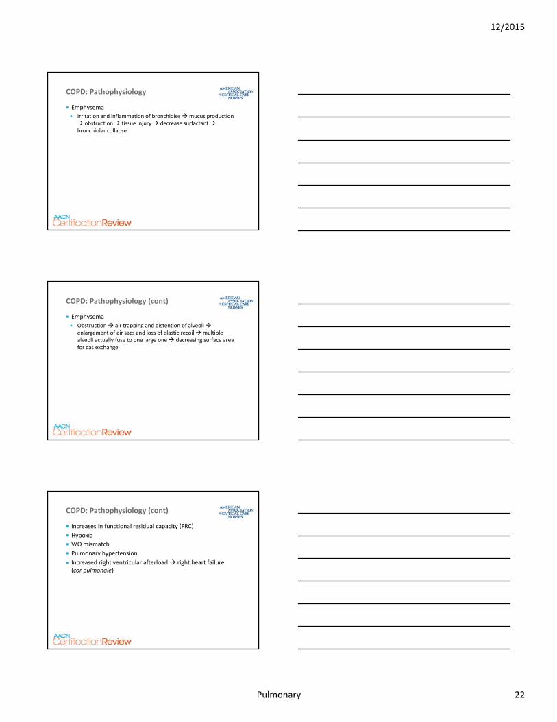
Emphysema
Irritation and inflammation of bronchioles mucus production obstruction tissue injury decrease surfactant bronchiolar collapse
Pulmonary
Emphysema
Obstruction air trapping and distention of alveoli enlargement of air sacs and loss of elastic recoil multiple alveoli actually fuse to one large one decreasing surface area for gas exchange
COPD: Pathophysiology (cont)
Pulmonary
Increases in functional residual capacity (FRC)
Hypoxia
V/Q mismatch
Pulmonary hypertension
Increased right ventricular afterload right heart failure (cor pulmonale)
COPD: Pathophysiology (cont)
12/2015
Pulmonary 22
Pulmonary
Dyspnea on exertion dyspnea at rest
Productive cough nonproductive cough
Tachypnea with small TV
Dropping FEV1
COPD: Clinical Presentation
Pulmonary
Malnutrition/muscle wasting (including diaphragm)
Increase in anterior‐posterior diameter
Diminished breath sounds in bases
COPD: Clinical Presentation (cont)
Pulmonary
Pulmonary function tests: Increased: FRC, residual volume (RV), total lung capacity
Decreased: FEV1, TV
ABGs: Hypoxia with respiratory acidosis over time will develop a degree of metabolic compensation Example: PaO2 71; PaCO2 52; pH 7.29; HCO3 34; SaO2 72
COPD: Clinical Presentation (cont)
12/2015
Pulmonary 23
Pulmonary
Chest x‐ray Flattened diaphragm
Decreased vascular markings
Bullae
Right heart failure
Chronic multisystem dysfunction related to chronic hypoxemia and hypercapnia
COPD: Clinical Presentation (cont)
Pulmonary
Chronic Illness Pneumonia
Heart failure
Pulmonary emboli
Respiratory failure
Bronchospasm
Spontaneous pneumothorax
Noncompliance with pulmonary medical therapies
COPD
Illustration Copyright ©2011 Nucleus Medical Media. All rights reserved. www.nucleusinc.com
Pulmonary
Treat primary cause of admission
O2 administration (with caution)
Hydration and humidification
Removal of secretions
COPD: Treatment Options
12/2015
Pulmonary 24
Pulmonary
Pharmacology Antibiotics
Steroids
Beta 2 agonists
Anticholinergics
Methylxanthines
Mucolytics
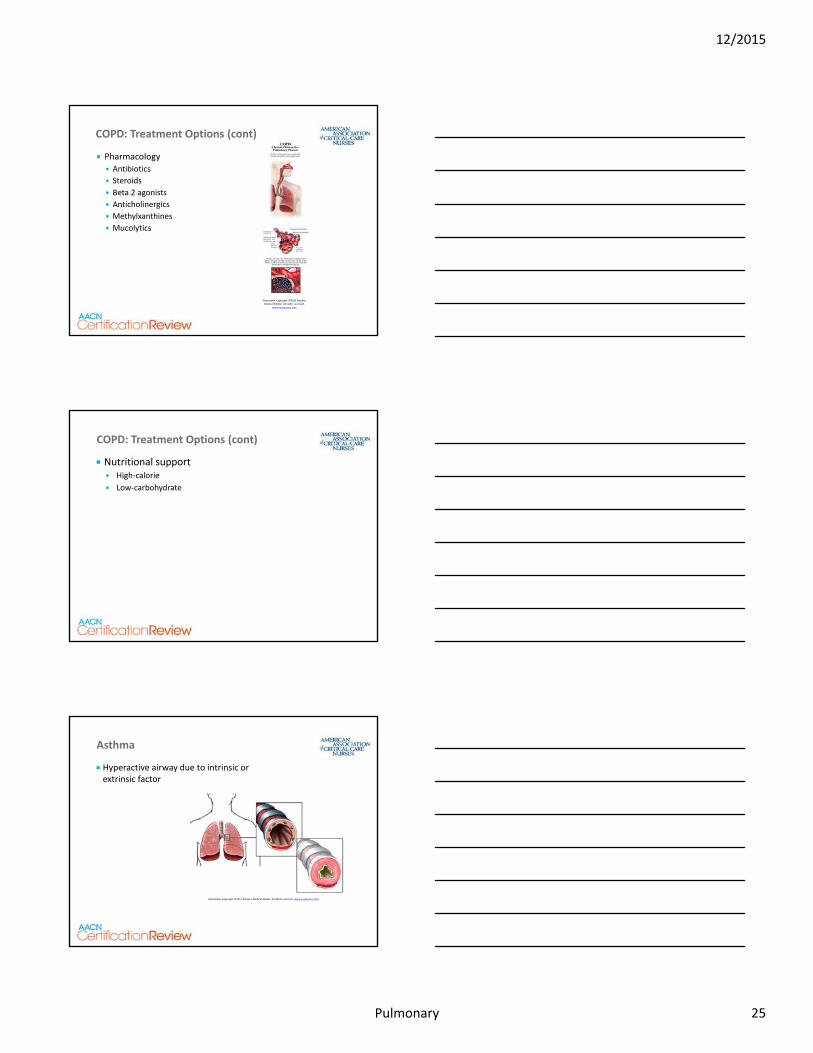
COPD: Treatment Options (cont)
Illustration Copyright ©2011 Nucleus Medical Media. All rights reserved.
www.nucleusinc.com
Pulmonary
Nutritional support High‐calorie
Low‐carbohydrate
COPD: Treatment Options (cont)
Pulmonary
Asthma
Hyperactive airway due to intrinsic or extrinsic factor
Illustration Copyright ©2011 Nucleus Medical Media. All rights reserved. www.nucleusinc.com
12/2015
Pulmonary 25
Pulmonary
“Typical” asthma therapies don’t work
Bronchospasm, mucus production, and air trapping continue, potentially to the point where there is no air movement
Hyperinflation increases intrathoracic pressures, which decreases venous return and increases RV afterload
Status Asthmaticus
Illustration Copyright ©2011 Nucleus Medical Media. All rights reserved. www.nucleusinc.com
Pulmonary
Review Questions
Pulmonary
A. Call anesthesia to intubate the patient and begin mechanical ventilation
B. Administer the antibiotic for the pneumonia as soon as possible
C. Increase the patient’s O2 to 4 L
D. Continue to monitor the patient for any respiratory distress
Question 11
A patient arrives from the ED with COPD and pneumonia. Assessment includes unlabored RR 28; HR 112; Afib; BP 168/82; T 37.9◦C; coarse breath sounds—diminished in bases. Patient denies SOB or chest pain and is on 1 L O2 via NC. ABGs: PaO2 71; PaCO2 55; pH 7.28; HCO3 35. The nurse contacts the physician with the ABG results and anticipates the order to be:
12/2015
Pulmonary 26
Pulmonary
Question 11—Rationale
D. Continue to monitor the patient for any respiratory distress—The assessment and ABG are consistent for a patient with these diagnoses. Patient denies SOB and chest pain
Call anesthesia to intubate the patient and begin mechanical ventilation—Patient is stable; no need to intubate
Administer the antibiotic for the PNA as soon as possible—Patient does need antibiotics; this is not answering the question asked
Increase the patient’s O2 to 4 L—Oxygen administration to a COPD patient should be increased slowly
A patient arrives from the ED with COPD and pneumonia. Assessment includes unlabored RR 28; HR 112; Afib; BP 168/82; T 37.9◦C; coarse breath sounds—diminished in bases. Patient denies SOB or chest pain and is on 1 L O2 via NC. ABGs: PaO2 71; PaCO2 55; pH 7.28; HCO3 35. The nurse contacts the physician with the ABG results and anticipates the order to be:
Pulmonary
Pulmonary Emboli
Occlusion in the pulmonary arterial circulation, blocking flow to a region(s) of the lung, and creating dead space ventilation
Pulmonary
Fat
Air
Amniotic fluid
Pulmonary Emboli: Etiology
12/2015
Pulmonary 27
Pulmonary
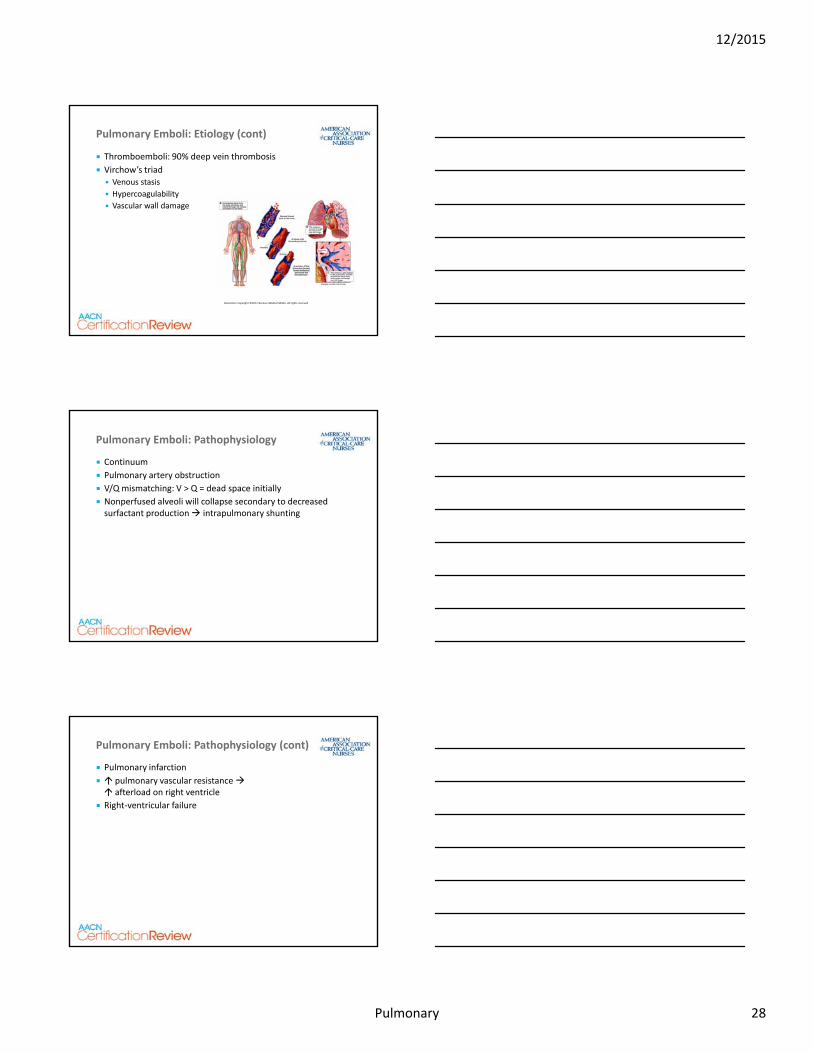
Thromboemboli: 90% deep vein thrombosis
Virchow’s triad Venous stasis
Hypercoagulability
Vascular wall damage
Pulmonary Emboli: Etiology (cont)
Illustration Copyright ©2011 Nucleus Medical Media. All rights reserved
Pulmonary
Continuum
Pulmonary artery obstruction
V/Q mismatching: V > Q = dead space initially
Nonperfused alveoli will collapse secondary to decreased surfactant production intrapulmonary shunting
Pulmonary Emboli: Pathophysiology
Pulmonary
Pulmonary infarction
pulmonary vascular resistance afterload on right ventricle
Right‐ventricular failure
Pulmonary Emboli: Pathophysiology (cont)
12/2015
Pulmonary 28
Pulmonary
Dyspnea and pleuritic chest pain
Tachypnea
Refractory hypoxemia
ABGs: hypoxemia with respiratory alkalosis Example: PaO2 71; PaCO2 28; pH 7.59; HCO3 25; SaO2 72
Fat emboli: petechiae on thorax, upper extremities
Pulmonary Emboli: Clinical Presentation
Pulmonary
Chest x‐ray
V/Q scan
CT
Pulmonary angiogram
MRI
D‐dimer
Lower extremity Doppler studies (not emergent)
Pulmonary Emboli: Diagnostic Tests
Pulmonary
ABCs Airway
Breathing
Circulation
Administer 100% O2
Intubate if necessary
Thrombolytics
Pulmonary Emboli: Treatment Options
12/2015
Pulmonary 29
Pulmonary
Embolectomy
Inferior vena cava filter
Pain management
Treat cause
Future prevention
Pulmonary Emboli: Treatment (cont)
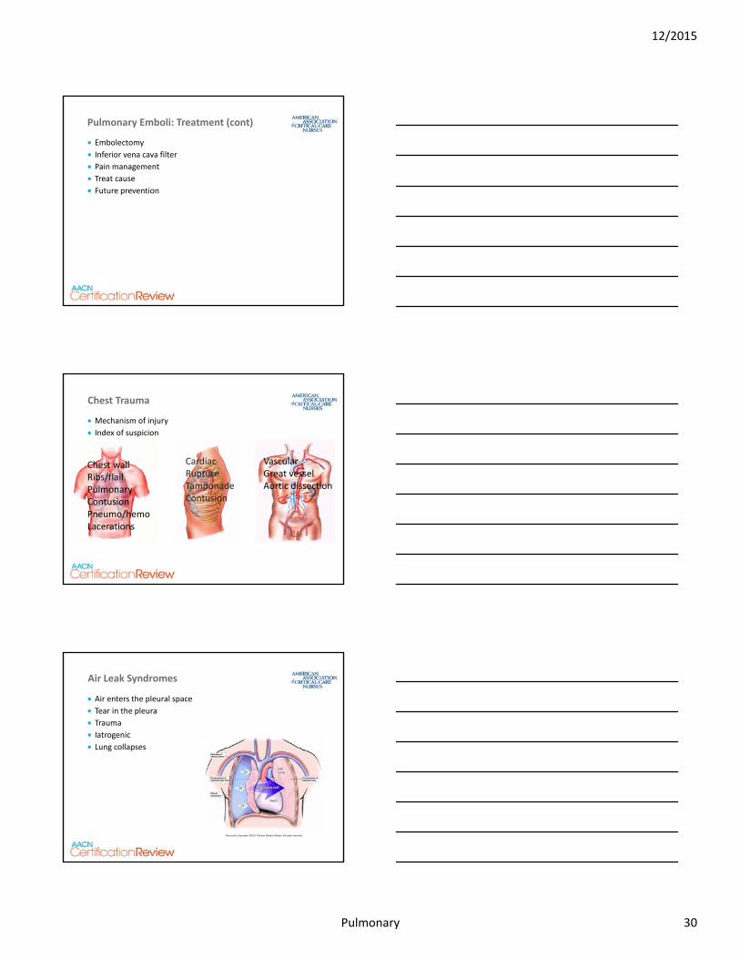
Pulmonary
Mechanism of injury
Index of suspicion
Chest Trauma
Chest wallRibs/flailPulmonaryContusionPneumo/hemoLacerations
Cardiac RuptureTamponadeContusion
VascularGreat vesselAortic dissection
Pulmonary
Air enters the pleural space
Tear in the pleura
Trauma
Iatrogenic
Lung collapses
Air Leak Syndromes
Illustration Copyright ©2011 Nucleus Medical Media. All rights reserved.
12/2015
Pulmonary 30
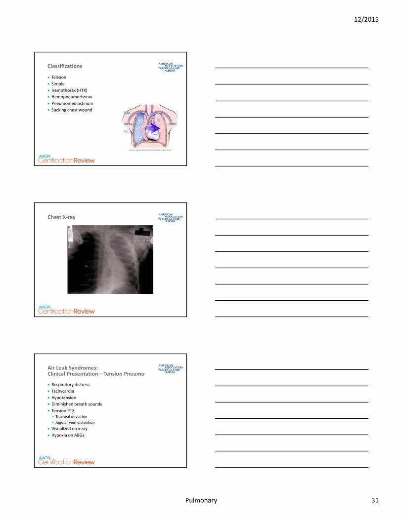
Pulmonary
Tension
Simple
Hemothorax (HTX)
Hemopneumothorax
Pneumomediastinum
Sucking chest wound
Classifications
Illustration Copyright ©2011 Nucleus Medical Media. All rights reserved.
Pulmonary
Chest X‐ray
Pulmonary
Respiratory distress
Tachycardia
Hypotension
Diminished breath sounds
Tension PTX Tracheal deviation
Jugular vein distention
Visualized on x‐ray
Hypoxia on ABGs
Air Leak Syndromes: Clinical Presentation—Tension Pneumo
12/2015
Pulmonary 31
Pulmonary
Emergent needle decompression
Chest tube placement
Insert high for PTX
Insert low for HTX
Air Leak Syndromes: Treatment Options
Pulmonary
Potential for air leak
No striping or milking routinely
O2 and, potentially, intubation
Sucking chest wound
Air embolism Trendelenburg position
Left side to trap air in heart (right ventricle)
Surgery may be required
Air Leak Syndromes: Treatment Options (cont)
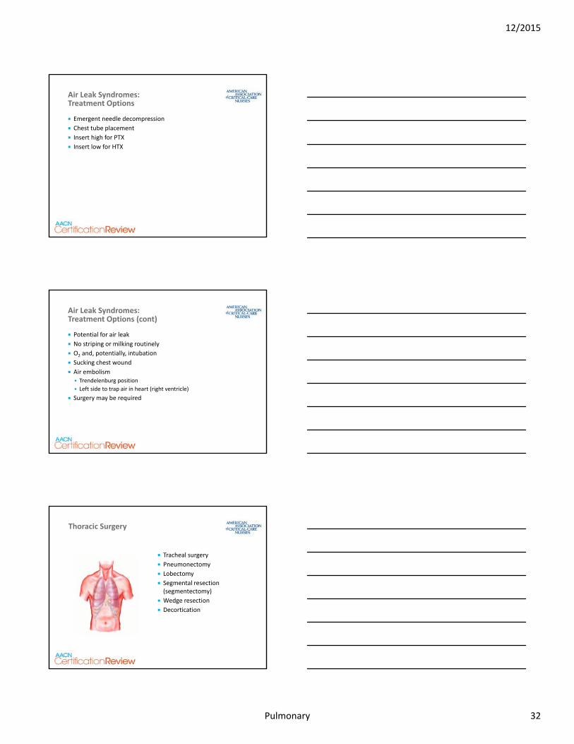
Pulmonary
Tracheal surgery
Pneumonectomy
Lobectomy
Segmental resection (segmentectomy)
Wedge resection
Decortication
Thoracic Surgery
12/2015
Pulmonary 32
Pulmonary
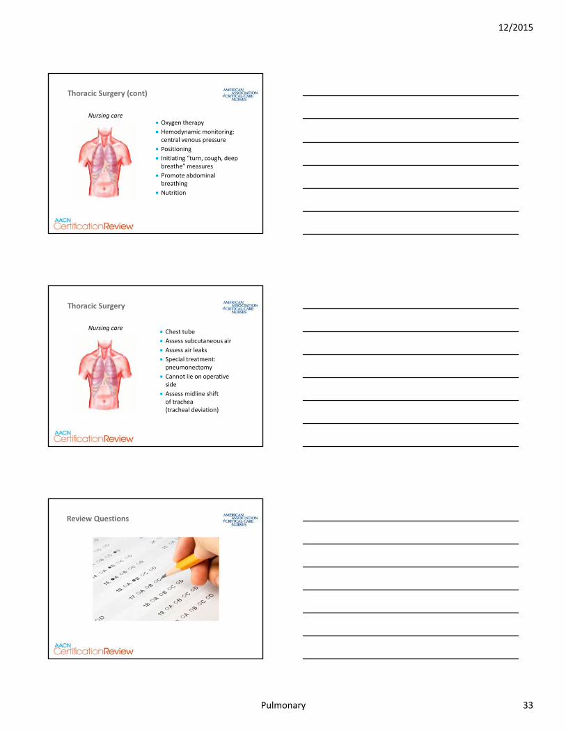
Oxygen therapy
Hemodynamic monitoring: central venous pressure
Positioning
Initiating “turn, cough, deep breathe” measures
Promote abdominal breathing
Nutrition
Thoracic Surgery (cont)
Nursing care
Pulmonary
Chest tube
Assess subcutaneous air
Assess air leaks
Special treatment: pneumonectomy
Cannot lie on operative side
Assess midline shift of trachea (tracheal deviation)
Thoracic Surgery
Nursing care
Pulmonary
Review Questions
12/2015
Pulmonary 33
Pulmonary
A. Connect a drainage system to the catheter used for the needle decompression
B. Contact anesthesia to intubate the patient
C. Set up for a pulmonary artery catheter (PAC) insertion
D. Set up the chest tube insertion
Ten minutes after having a central line placed, a patient complains of SOB and chest pain. The SpO2 is falling, and a chest x‐ray reveals a tension PTX. A needle decompression is successfully performed. The next action by the nurse should be which of the following?
Question 12
Pulmonary
Question 12—Rationale
D. Set up the chest tube insertion—A needle decompression is followed by a chest tube insertion. The “real” answer to this question would be to monitor the patient, but that is not an option
Connect a drainage system to the catheter used for the needle decompression—The needle is removed and a CT catheter would be inserted
Contact anesthesia to intubate the patient—Not treating problem
Set up for a PAC insertion—No indication for PAC
Ten minutes after having a central line placed, a patient complains of SOB and chest pain. The SpO2 is falling, and a chest x‐ray reveals a tension PTX. A needle decompression is successfully performed. The next action by the nurse should be which of the following?
Pulmonary
A. A tension pneumothorax
B. A pulmonary embolism
C. Post extubation
D. Respiratory failure
Question 13
Twelve hours after sustaining a pelvic fracture, a patient reports chest pain, hemoptysis, and severe shortness of breath. RR is 34. ABGs on O2 at 4 L/min via NC are: pH 7.48, pCO2 28, pO2 68. The nurse should suspect that the patient has developed:
12/2015
Pulmonary 34
Pulmonary
Question 13—Rationale
B. A pulmonary embolism—12‐hour post long bone fracture, hypoxia, chest pain and air hunger classic for PE (from fat)
A tension pneumothorax—Would have tracheal deviation, absent breath sounds
Post extubation laryngeal edema—Would present with strider
Respiratory failure—True, but a nonspecific answer
The nurse should suspect that the patient has developed:
Pulmonary
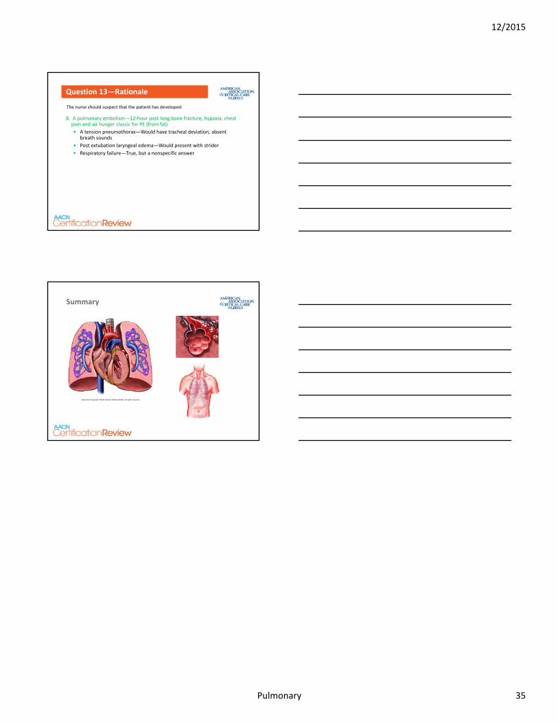
Summary
Illustration Copyright ©2011 Nucleus Medical Media. All rights reserved.
12/2015
Pulmonary 35


