

Anatomic and FunctionalGuidance of Neurosurgery.
Robert L. Galloway Jr.Associate Professor of Biomedical
Engineering and NeurosurgeryDirector, Center for Technology-
Guided Therapy
TGT

Technology-Guided Therapy
The use of technology to improve the spatial and temporal specificity of the
delivery of therapy
TGT

Technology -Guided Therapy• Pre-therapeutic data (images, population
data, therapy plan…)
• Registration methodology (matching pre-therapeutic data to specific patient in specific position)
• Intraoperative guidance and data collection.
• Display of position, plan, anatomy, function and disease.

TGT
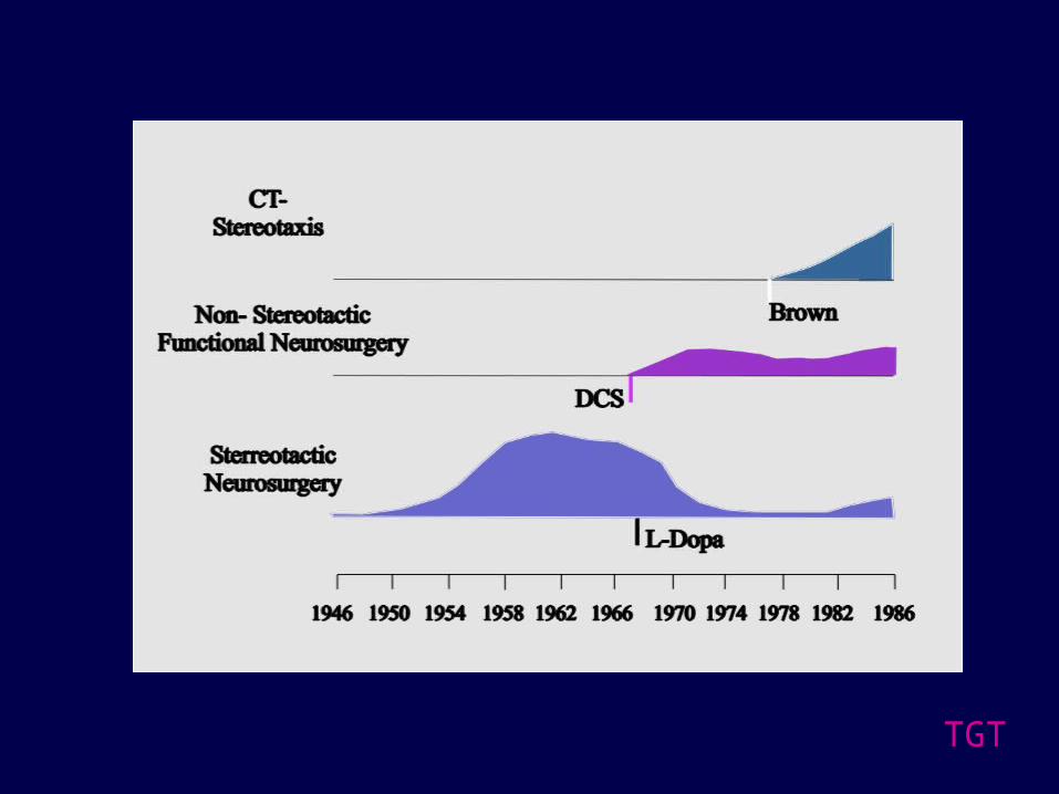
History of Image-Guided Therapy
• 1896 J.H. Clayton. X-Ray use in surgery
• 1904 Horsley and Clarke. Stereotactic frame
• 1946 Spiegel and Wycis. Stereotactic frame using xrays.
• 1940’s~1950s: Leksell, Riechert-Mundinger, Talairach, Cooper...

TGT

TGT
TGT
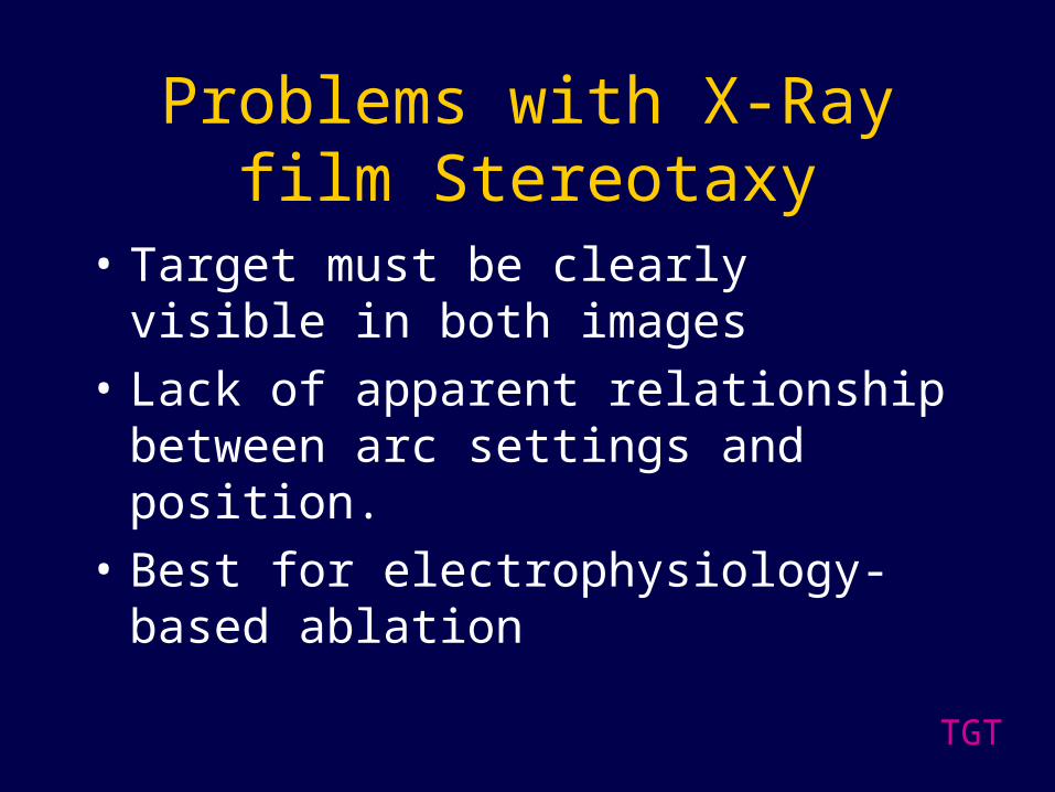
Problems with X-Ray film Stereotaxy
• Target must be clearly visible in both images
• Lack of apparent relationship between arc settings and position.
• Best for electrophysiology-based ablation
TGT

TGT

TGT
TGT

TGT
Problems with Frame-Based Stereotaxy
• Still point-based
• Frame obstructs surgical field
• Arc-based approach obscures relationship between target points and arc settings.
TGT

TGT

TGT
Registration
When the mathematical relationship between a point in one space and the homologous point in another space is known, the spaces are considered registered. If that relationship can be reduced to a single common translation and rotation, the registration is considered rigid,

TGT
Translate
Rotate
Rigid Registration
TGT
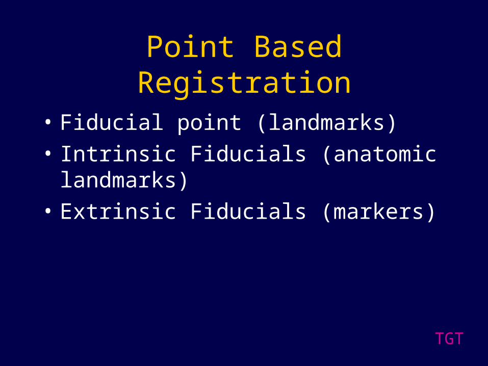
Point Based Registration
• Fiducial point (landmarks)
• Intrinsic Fiducials (anatomic landmarks)
• Extrinsic Fiducials (markers)

TGT

TGT

TGT

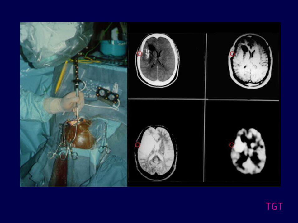
Marker -Based Registration
TGT

New Methods of Registration
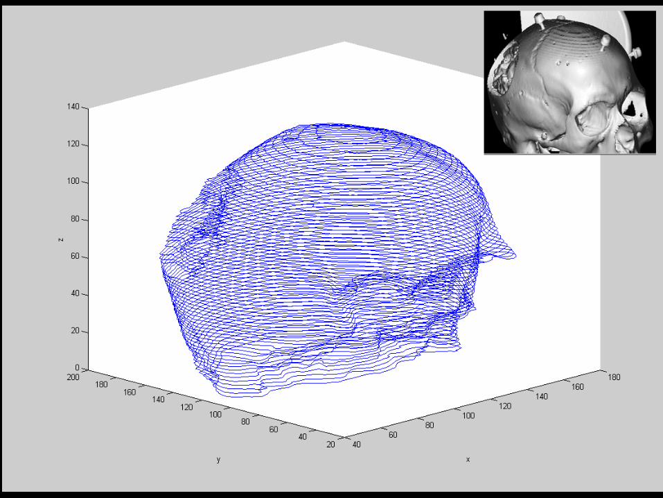
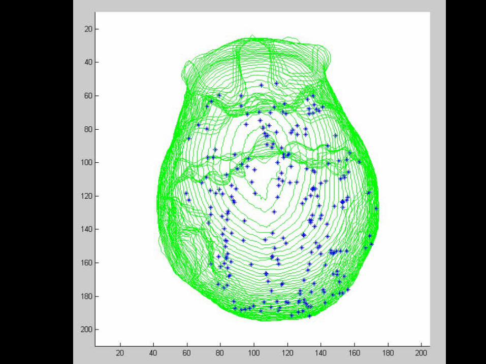
Surface-Based Registration
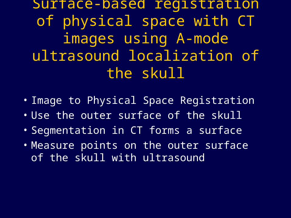
• Image to Physical Space Registration
• Use the outer surface of the skull
• Segmentation in CT forms a surface
• Measure points on the outer surface of the skull with ultrasound
Surface-based registration of physical space with CT images using A-mode ultrasound localization of the skull
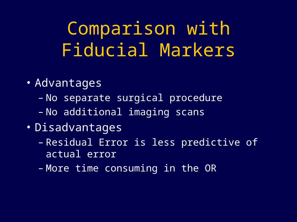
• Advantages – No separate surgical procedure – No additional imaging scans
• Disadvantages– Residual Error is less predictive of actual error– More time consuming in the OR
Comparison with Fiducial Markers

OptotrakS ys temC ontro lle r
OptotrakInterfac e Card
Serial Port
P C
M ulti-s c anner RCV out
CL K in
Ga ge A /D

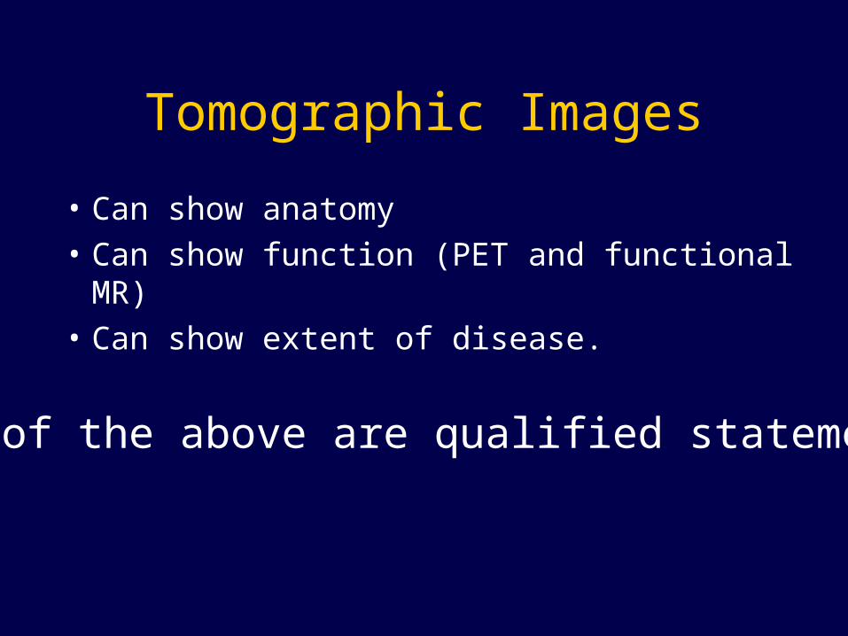
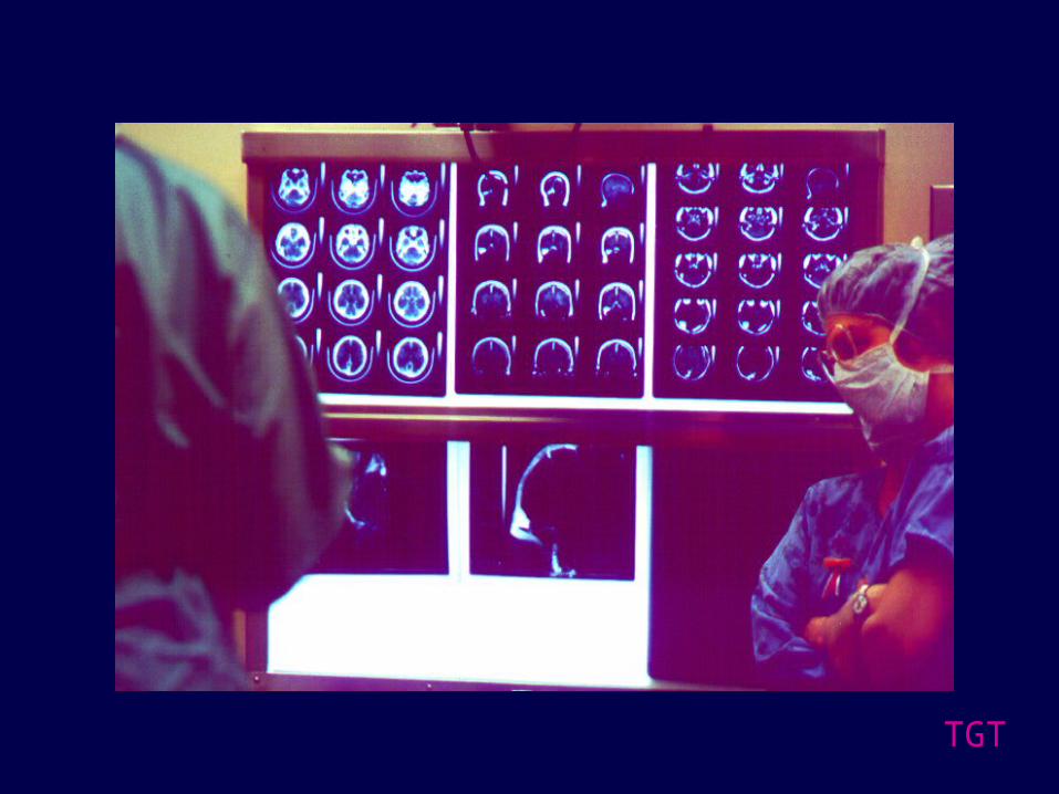
Tomographic Images
• Can show anatomy
• Can show function (PET and functional MR)
• Can show extent of disease.
All of the above are qualified statements

TGT

TGT
TGT

TGT
TGT
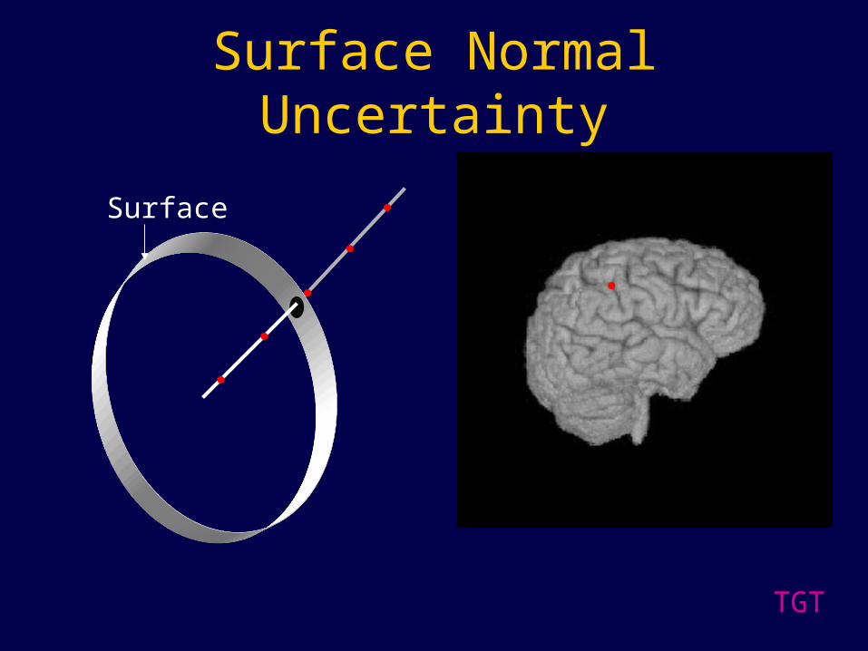
Surface Normal Uncertainty
Surface
TGT

TGT

TGT
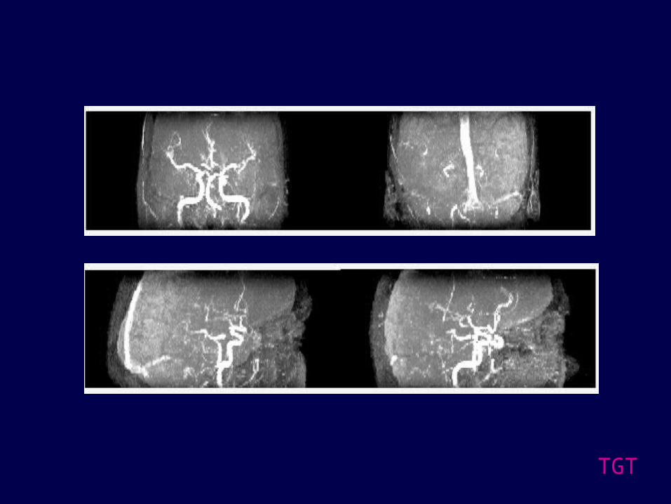
Tomographic Angiography

Surgically Appropriate MIPs
1) Threshold image, make binary image2) Calculate center of mass from binary image3) Fit center of mass points from each slice to a line4) Extend plane from the line perpendicular to MIP trajectory
TGT
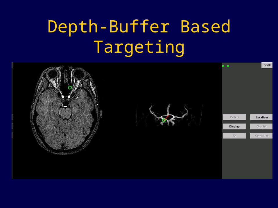
Depth-Buffer Based Targeting

Depth-Buffer Based Targeting
TGT
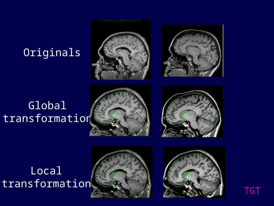
Originals
Globaltransformation
Localtransformation

TGT
Implantation of Thalamic Stimulators

Population Data
• Goal - to provide information from multiple subjects during surgery
• Create a functional Atlas (MR or CT)

Tglobal
Tlocal
Tlocal + Tglobal

Optical Flow
• Differences between images considered as motion from one time frame to the next
• This motion is observed as a change in intensity, represented as a field of velocity vectors called the optical flow
constant]),(),(),([ ttztytxE
tzyx EvE.
,,

Atlas
Atlas Patient
Patient

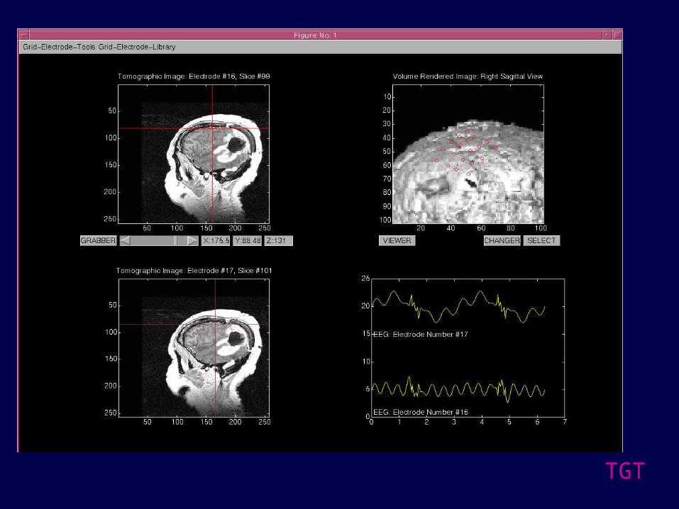
3D Visualization Display
Intraoperative Tracking and Data Collection

Interactive, Image Guided Endoscopic Surgery
Tomographic Volume Endoscopic View
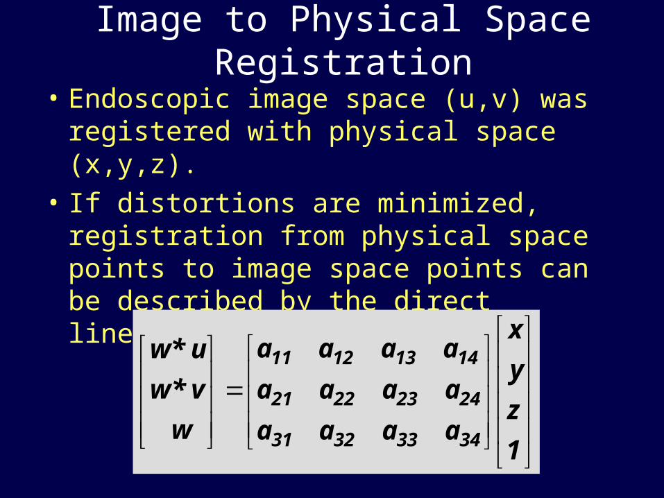
Image to Physical Space Registration
• Endoscopic image space (u,v) was registered with physical space (x,y,z).
• If distortions are minimized, registration from physical space points to image space points can be described by the direct linear transformation (DLT):
1
z
y
x
aaaa
aaaa
aaaa
w
v *w
u *w
34333231
24232221
14131211

TGT
Integration of Intraoperative Imaging
TGT

Tracked Ultrasound

Preliminary Experiments• Vertebral surface point extraction using tracked
U/S on phantom spine
• Spinal CT surface extraction• Point-to-surface registration• Qualitative assessment of registration accuracy

Visual Results: Phantom Ultrasound Points

New Software Design
Video
Tomogram
Rotational
2-D Images
DisplayOptotrak
Polaris
Art. Arms
Magnetic
Localizer
Render
Biopsy
Preop Plan
Transparency
GraphicA-Mode US
Electrophys.
Biomechanical
Optical Biopsy
IO
Quaternion
SVD
HTM
ICP
Registration
Deformation
Functional
Correction
ORION

TGT
What’s Next?• Functional Mapping
• Deformable Models
• In-Vivo Tissue Identification
• Image-Guided Delivery of FEL Beam
• Direct Injection Chemotherapy
• Guided Gene Therapy Delivery
• Brachytherapy seed placement

The Center For Technology-Guided Therapy
• Robert L. Galloway BME&Neurosurgery• Cynthia B. Paschal BME &Radiology• Anita Mahadevan-Jansen BME• E. Duco Jansen BME• J. Michael Fitzpatrick EECS&Radiology
&Neurosurgery• Benoit M. Dawant EECS
TGT
The Center For Technology-Guided Therapy
• William C. Chapman, MD Surgery
• Anthony Cmelak, MD Radiation Oncology
• Charles Coffey, PhD Radiation Oncology
• Dennis Duggan, PhD Radiation Oncology
• Dennis Hallahan, MD Radiation Oncology & BME
• Robert M. Kessler, MD Radiology
• Peter E. Konrad, MD, PhD Neurosurgery & BME
• Steven Toms, MD, MPH Neurosurgery TGT
The Center For Technology-Guided Therapy
Andy BassJim Stefansic
Diane MuratoreSteve Hartmann
David CashTuhin Sinha
Steve Gebhart
BME
Jeannette HerringJay West
Duane YoderRui Li
Yi Quiao
EECS
Alan Herline, MD
Surgery
Blake Arrington Gwen Banks Ryan Beasley Caryl Brzymialkiewicz Calley Hardin Elaine Isom Tammy McCreary Kristjan Onu
Danielle Pinson Stacey Scheib Chee Xiong
REU


