

Anatomy of the Brain and Cranial
Nerves
1
The Nervous System can be divided in:
Central Nervous System (CNS)Brain and Spinal Cord
Peripheral Nervous System (PNS)Cranial and spinal nerves, ganglia, sensory
receptors
2
Division of the Peripheral Nervous System
Sensory or afferentSomaticVisceral
Motor or efferentSomatic - voluntaryVisceral or Autonomic Nervous System
(ANS) - involuntarySympatheticParasympathetic
3

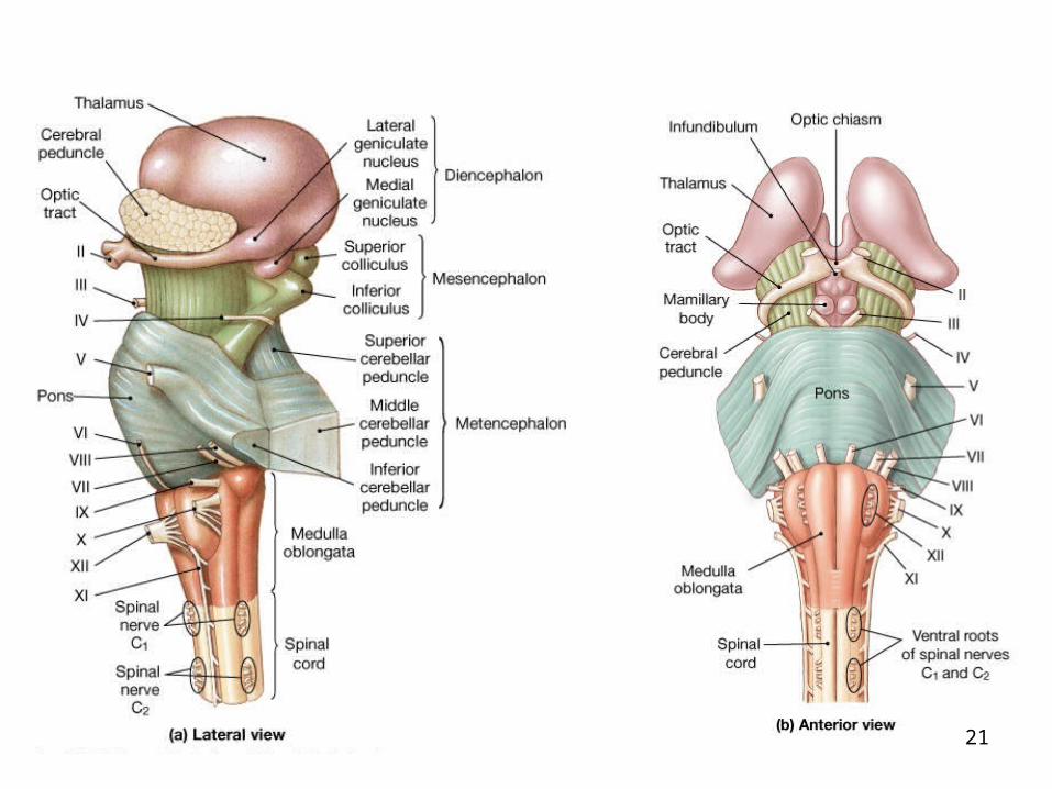
Six regions in the adult brainCerebrumDiencephalonMesencephalonPonsCerebellumMedulla oblongata
Brain contains extensive areas of neural cortexLayer of gray matter on the surface of the
cerebellum and cerebrum
Major regions and landmarks
4
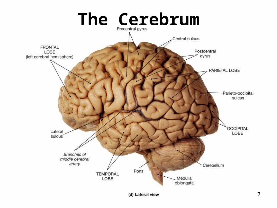
The Cerebrum
5
Surface contains gyri, sulci, fissuresFissures
Longitudinal fissure separates two cerebral hemispheres
Transverse fissure separates cerebellum from cerebrum
The cerebral hemispheres
6
The Cerebrum
7
The cerebral hemispheres
SulciParieto-occipital sulcus separates parietal
from occipital lobeLateral sulcus separates temporal from
parietal lobeCentral sulcus separates frontal and parietal
lobe
8
The cerebral hemispheres
GyriPrecentral gyrusPoscentral gyrus
9

The cerebral lobes Frontal
Precentral gyrusPrimary motor area – conscious
control of voluntary movements. Premotor cortex – memory bank for
skilled motor activities or of patterned and repetitious nature.
Broca’s areaLocated on the left hemisphere.
Controls speech.
10

The cerebral lobesPrefrontal cortex – responsible for
personality, cognition, intellect. Lesion cause mental and personality disorder
ParietalPrimary Somatosensory Area – touch,
pressure, temperature, vibration, and pain from body wall
Somatosensory association area – interprets stimulus sent by the above area. Ex: recognizes objects by touch.
11
The cerebral lobesTemporal
Primary Auditory area – temporal lobe. Primary association auditory area – interprets
the sound heard by above areaWernicke’s area – only on left hemisphere,
between parietal and temporal lobes. Area responsible for understanding spoken language
Olfactory area –uncus. Smell area.
12
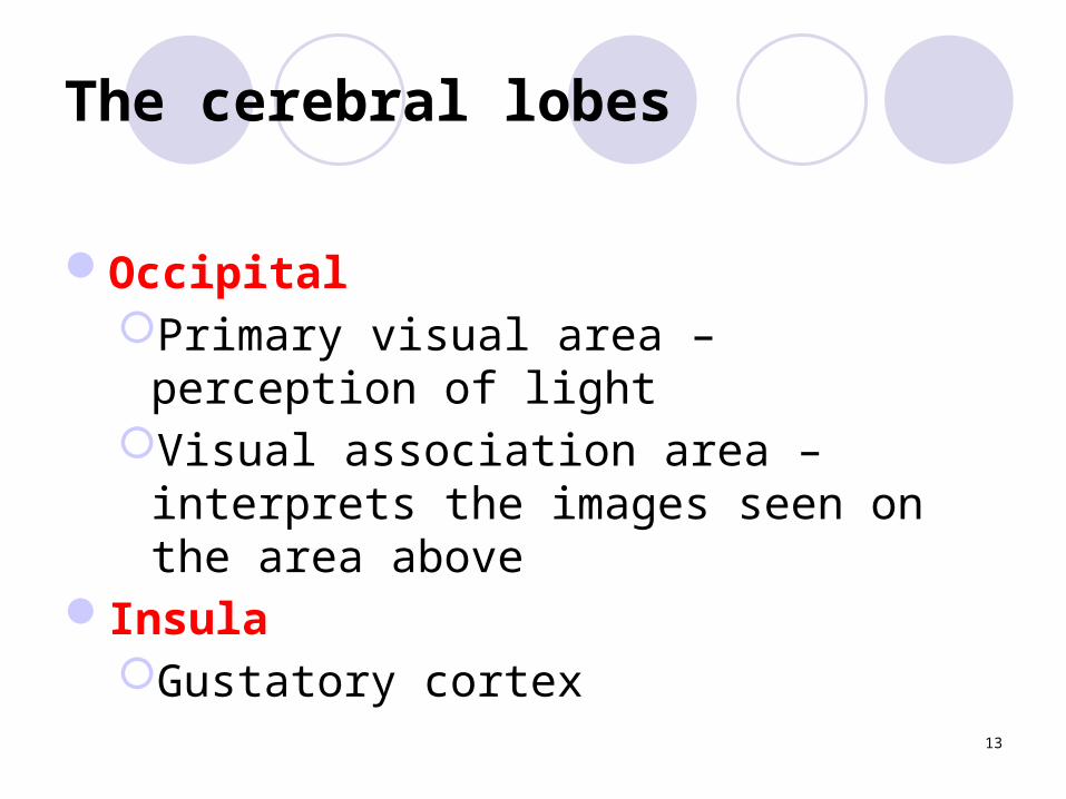
The cerebral lobes
OccipitalPrimary visual area – perception of lightVisual association area – interprets the
images seen on the area aboveInsula
Gustatory cortex
13

Cerebral hemispheres - internal structures
Gray matterCell bodies of the neuronsDendrites Small unmyelinated axonsNeuroglias
14
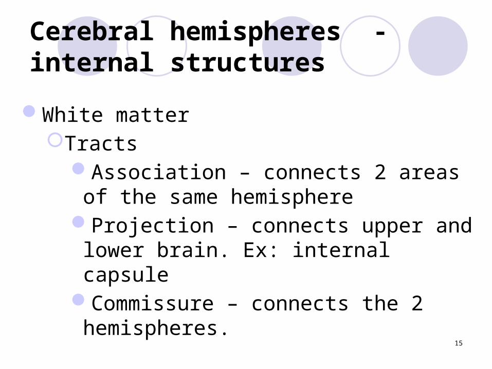
Cerebral hemispheres - internal structures
White matterTracts
Association – connects 2 areas of the same hemisphere
Projection – connects upper and lower brain. Ex: internal capsule
Commissure – connects the 2 hemispheres.
15

Tracts
16
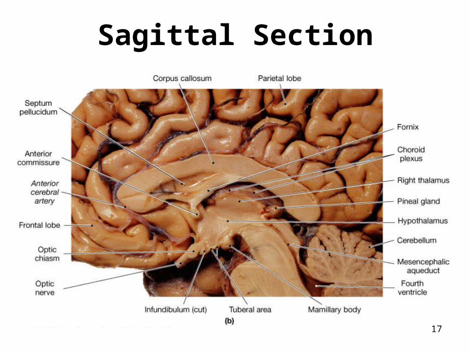
Sagittal Section
17

Cerebral hemispheres - internal structures
Corpus callosumConnects the 2 hemispheres
FornixConnects limbic system areas
Septum pellucidumSeparates the 2 lateral ventricles
18
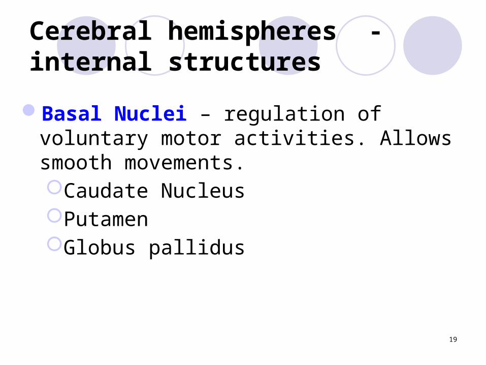
Cerebral hemispheres - internal structures
Basal Nuclei – regulation of voluntary motor activities. Allows smooth movements.Caudate NucleusPutamen Globus pallidus
19
The Diencephalon
20
21

22
Diencephalon: external view
Olfactory tractOlfactory bulbOptic nerveChiasma opticPituitary gland or hypophysis Mammilary bodies – relay for olfaction
23

EpithalamusHypothalamusThalamus
The diencephalon is composed of
24

Figure 14.12b
Sagittal Section
25
The Epithalamus
Roof of the third ventricleContains choroid plexus Contains pineal gland
Regulates sleep-awake cycle
26
Relay area for impulses Two large lobes of gray matterInterthalamic adhesion or intermediate mass
The thalamus
27

Autonomic center for regulation of body temperature, water balance, etc
Secretes hormonesMammilary bodies – relay station for olfactionPituitary glands – secretes hormonesOptic chiasm
The hypothalamus
28
The Brain Stem
MidbrainCerebral Aqueduct – connects third and forth
ventriclesCerebral peduncles – connects pons to
cerebrumCorpora quadrigemina
Superior colliculi – visual reflex centerInferior colliculi – auditory reflex center
29
The Brain Stem Pons
Consists of tracts and nucleiConnects brain to lower CNS
• Medulla Oblongata• Tracts• Decussation of the pyramids• Autonomic reflex centers – heart rate, blood
pressure, vomiting, swallowing, respiratory rhythm
• Olives30

PART 2
31
Cerebellum
Two hemispheres connected by the vermisArbor vitae – white matterCortex of gray matter
32
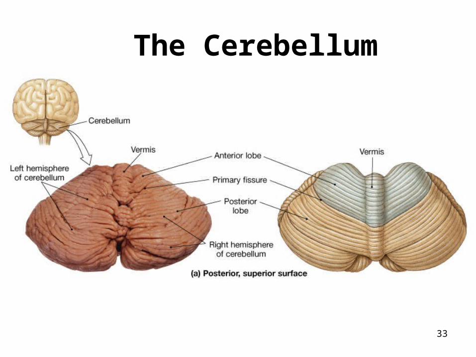
The Cerebellum
33

The Cerebellum
34
Dura mater Falx cerebri-formed by dura mater that
dips into the longitudinal fissure and separates the 2 hemispheres
Falx cerebelli – separate the two cerebellar hemispheres
The cranial meninges
35

The cranial meninges – dura mater
Superior sagittal Sinus – collects blood from the brain
Tentorium cerebelli – separates the cerebrum from the cerebellum
36
The cranial meninges
Arachnoid Subarachnoid space
Filled with CSF Arachnoid villi – projections of the mater
that protrude through the dura For the CSF to drain back to the venous
circulation
37

The cranial meninges
Pia mater Highly vascular Covers the entire brain
Meningites
38
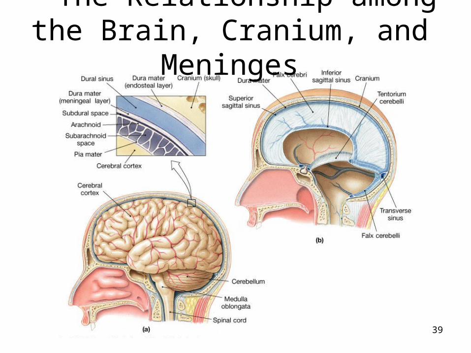
The Relationship among the Brain, Cranium, and Meninges
39
Filled with cerebrospinal fluid (CSF)Lateral ventricles
Septum pellucidumInterventricular foramina or foramen of
Monro
Ventricles of the brain
40

Ventricles of the brain
Third ventricleCerebral aqueduct
Forth ventricle3 Apertures
41
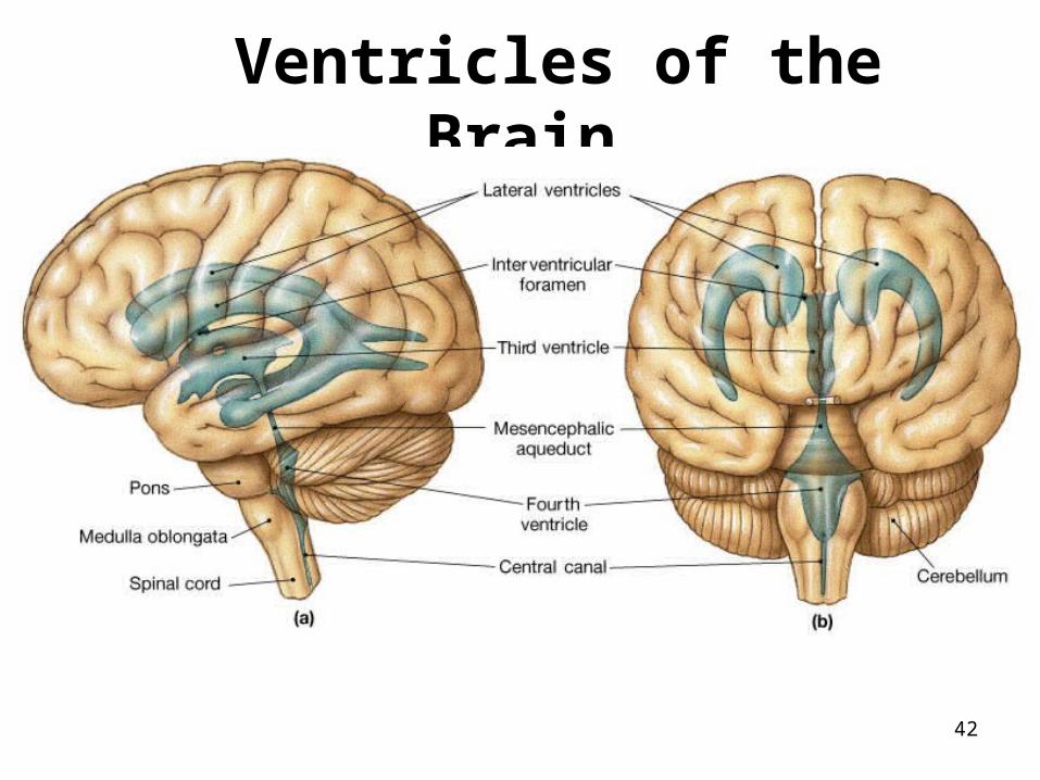
Ventricles of the Brain
42
CSF cushions delicate neural structuresSupports the brainPathway of CSF
Produced at the Choroid plexusTravels through the apertures on the 4th
ventricle to the subarachnoid space Diffuses across the arachnoid villus
(granulation) into the superior sagittal sinus
Cerebrospinal fluid (CSF)
43
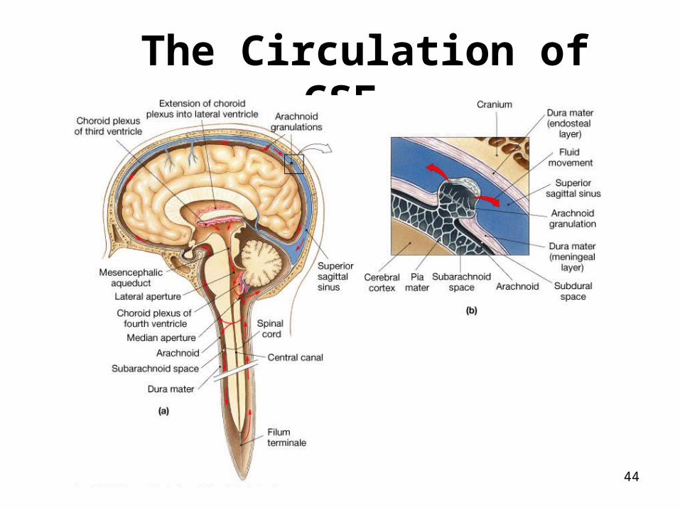
The Circulation of CSF
44
12 pairs of cranial nervesTo help to remember“Old Opie Occasionally Tries Trigonometry And
Fells Very Gloomy Vague And Hypoactive”
Cranial Nerves
45
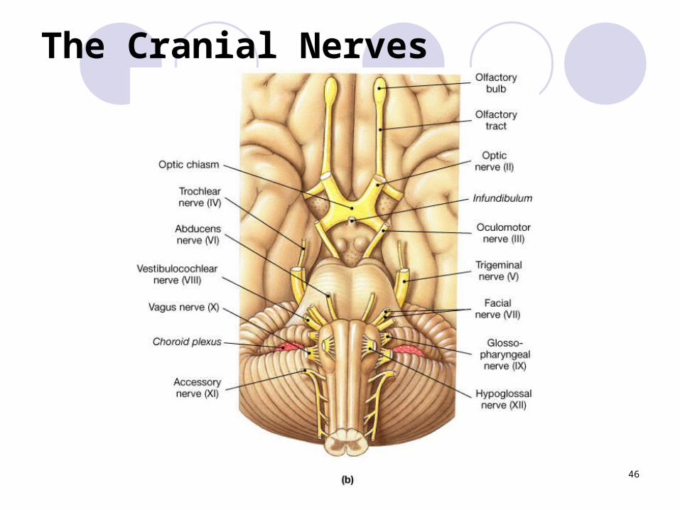
The Cranial Nerves
PLAY
46
Cranial Nerves
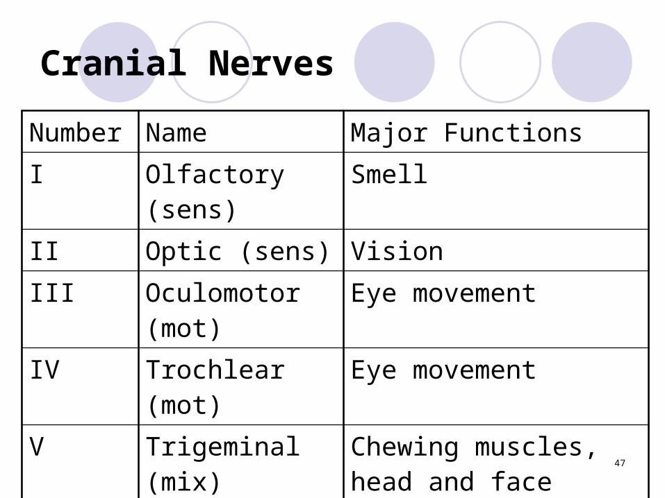
Number Name Major FunctionsI Olfactory (sens) SmellII Optic (sens) VisionIII Oculomotor
(mot)Eye movement
IV Trochlear (mot) Eye movement
V Trigeminal (mix) Chewing muscles, head and face sensation
47
Cranial nerves
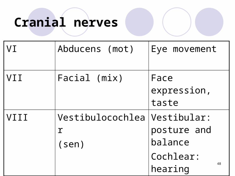
VI Abducens (mot) Eye movement
VII Facial (mix) Face expression, taste
VIII Vestibulocochlear(sen)
Vestibular: posture and balanceCochlear: hearing
48
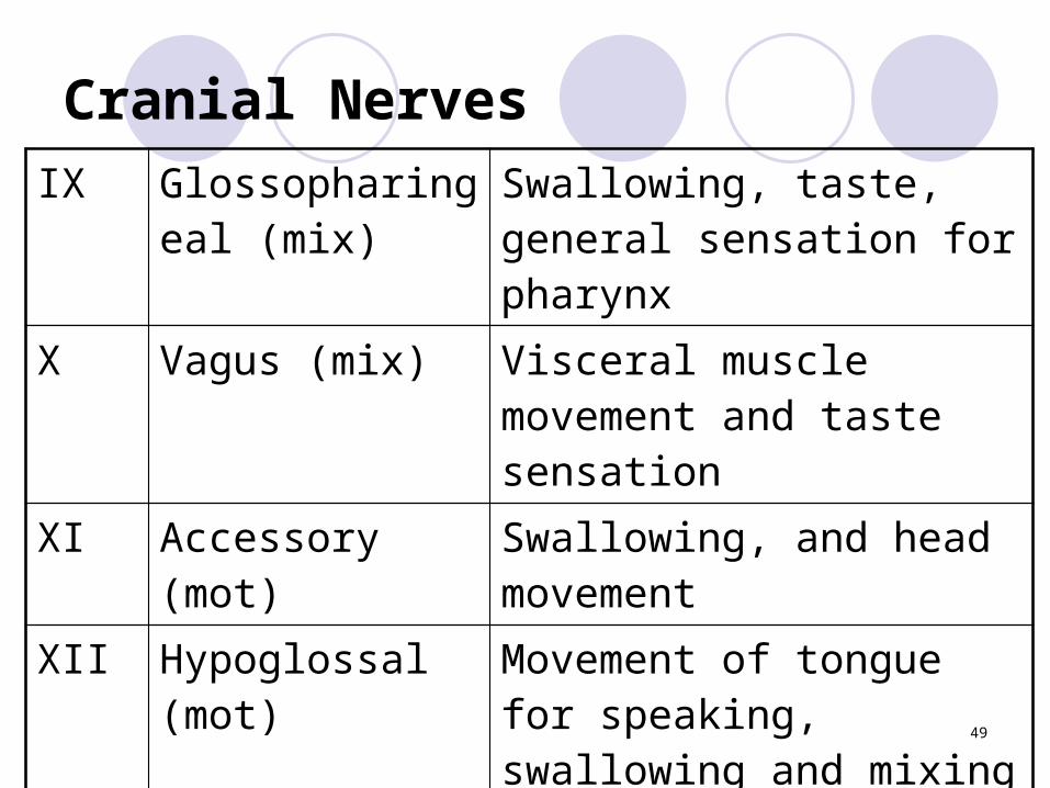
Cranial NervesIX Glossopharingeal
(mix)Swallowing, taste, general sensation for pharynx
X Vagus (mix) Visceral muscle movement and taste sensation
XI Accessory (mot) Swallowing, and head movement
XII Hypoglossal (mot)
Movement of tongue for speaking, swallowing and mixing food
49

Brain Dissection
Whole BrainPia-Arachnoid GyrusSulcusFissure
TransverseLongitudinal
Cerebrum50

Brain Dissection
PonsMedulla OblongataCerebellumCranial nerves: I (bulb, tract) II (nerve, chiasma) III
51
Brain Dissection
ColliculiSuperiorInferior
Pineal Gland
52
Brain Dissection
Sagittal CutDiencephalon
EpithalamusThalamusHypothalamus
VentriclesLateral, third, forth
53
Brain Dissection
Septum pellucidumCorpus callosumFornixArbor vitae (cerebellum)
54


