
In vitro and in vivo oncolytic properties of four
new Ad3-hTERT-E1A-viruses
Joonas Lehikoinen, BM
University of Helsinki
In Helsinki, 29.12.2013
Supervisor:
Akseli Hemminki, MD, PhD, Research director
Otto Hemminki, MD
Cancer Gene Therapy Group
University of Helsinki

i
HELSINGIN YLIOPISTO HELSINGFORS UNIVERSITET
Tiedekunta/Osasto Fakultet/Sektion – Faculty
Faculty of Medicine
Laitos Institution – Department
Haartman Institue
TekijäFörfattare – Author
Joonas Lehikoinen
Työn nimi Arbetets titel – Title
In vitro and in vivo oncolytic properties of four new Ad3-hTERT-E1A-viruses
Oppiaine Läroämne – Subject
Työn laji Arbetets art – Level
Aika Datum – Month and year
21.08.2014
Sivumäärä-Sidoantal - Number of pages
20 + 3
Tiivistelmä Referat – Abstract
The objective of this project was to determine whether the new Ad3-hTERT-E1A based
viruses armed with immunological genes are functional and have efficacy for further
research aiming at clinical experiments. The hypothesis was that they would be
oncolytically as potential as earlier studied Ad3-hTERT-E1A virus in vitro and in vivo.
The encouraging results from previous studies had shown the potential of serotype 3
adenoviruses (Ad3). (1, 2) Unlike the more extensively researched Ad5 and Ad5/3
viruses, the Ad3 viruses open epithelial junctions while infecting cells and use other
receptors to enter cells, which might enable them to spread more easily in tumours.(3)
The new viruses were compared with the E1A first in vitro (progressive TCID50, MTS)
and then in vivo in a SKOV3-luc intra peritoneal tumour animal experiment with SCID
mice (immunodeficient).
We found that the new viruses have the same oncolytic potential as the old E1A virus
both in vitro and in vivo, which confirmed the basic hypothesis. This provides a starting
point for further research on the immunologically armed Ad3 viruses.
Avainsanat – Nyckelord – Keywords
Adenovirus, Ad3, hTERT, E1A, E2F, CMV, CD40L, GMCSF, oncolytic
Säilytyspaikka – Förvaringställe – Where deposited
Muita tietoja – Övriga uppgifter – Additional information

ii
Table of Contents 1 Introduction............................................................................................................................ 1
2 Results .................................................................................................................................... 2
2.1 The in vitro functionality of the viruses........................................................................... 2
2.3 The in vivo properties of the viruses ............................................................................... 4
3 Discussion ............................................................................................................................. 11
4 Conclusion ............................................................................................................................ 13
5 Materials and methods ........................................................................................................ 14
6 Acknowledgements and report ............................................................................................ 19
References ............................................................................................................................... 20

1
1 Introduction
Cancer is becoming increasingly common as people live longer and die ever more
seldom from other forms of disease. Research has shown that cancer is among the
most adaptable and wide raging diseases in the world. After all, every genome on the
planet is unique, so every cancer genome is also unique. The problem is even more
difficult to solve due to the nature of cancer, because it is not only a genomic disease,
but also characterized by a failure of the immune system and the wide range of other
tissue-specific effects the tumour cells induce in the body.
New research findings provide a basis for new possibilities in treating cancer. As cancer
consists of a variety of pathological events, the treatment of cancer should combine
the best therapies available and target these different aspects. Probably the best
treatment in the near future will combine multiple areas of cancer research.
Furthermore, the individualisation of treatments is bound to increase considerably and
to a greater extent than before when the genomic screening of the tumour cells is
utilized.
Adenoviruses’ genome is double stranded DNA. They are nonenveloped and their
nucleocapsid is icosahedral. There are 51 known serotypes which are divided into six
subcategories. Serotype 3 adenoviruses (here forth Ad3) belong to the B subcategory
and they use CD46 and desmoglein-2 as their primary receptor in infecting the human
cell. (2) The wild Ad3 viruses usually cause respiratory infections such as common cold.
Adenoviruses are commonly used as vectors in gene therapy and oncolytic
adenoviruses have been researched for some time now. These viruses are designed to
specifically target tumour cells and the immunostimulatory armament of these viruses
enhances the immune response towards the tumours. Viruses can also be armed so
that they make tumours sensitive to the drugs about to be used before the treatment.
Furthermore because the wild types of adenoviruses are relatively harmless to humans
the side effects of these modified viruses are usually mild (common cold symptoms),
therefore making them more interesting in a clinical perspective.

2
The oncolytic adenovirus Ad3-hTERT-E1A (hereafter E1A) showed some signs of
efficacy and seemed safe in a clinical experiment with the group of twenty-five
patients. In this case, the virus treatments did not provide a cure, however, and further
efficacy was needed. Thus, the idea of further arming this virus was considered. (1) -
The result was four new viruses built on the basis of the old E1A: Ad3-hTERT-E1A-CMV-
CD40L (hereafter CMV-CD40L), Ad3-hTERT-E1A-CMV-GMCSF (CMV-GMCSF), Ad3-
hTERT-E1A-E2F-CD40L (E2F-CD40L), and Ad3-hTERT-E1A-E2F-GMCSF (E2F-GMCSF).
These new viruses were thought to form the next generation of Ad3 virus research and
so became a continuation project for the two previous projects.(1, 2)
Like E1A, the new viruses have the human telomerase reverse transcriptase (hTERT) to
make the viruses specific towards cancer cells with telomerase activity leaving healthy
cells alone. (1, 2) However, the new viruses have either a CMV or an E2F promoter
locus to control the production of CD40L or GMCSF.
The in vitro and in vivo oncolytic properties of these new Ad3-viruses were researched
during the project. The basic hypothesis was that these new viruses would be as
potent in vitro as E1A and that when introduced into a system with a functional
immune system, the new viruses should be more potent than E1A. To determine this,
we aimed to compare the earlier results acquired in vitro and in vivo with E1A.(2)
2 Results
2.1 The in vitro functionality of the viruses
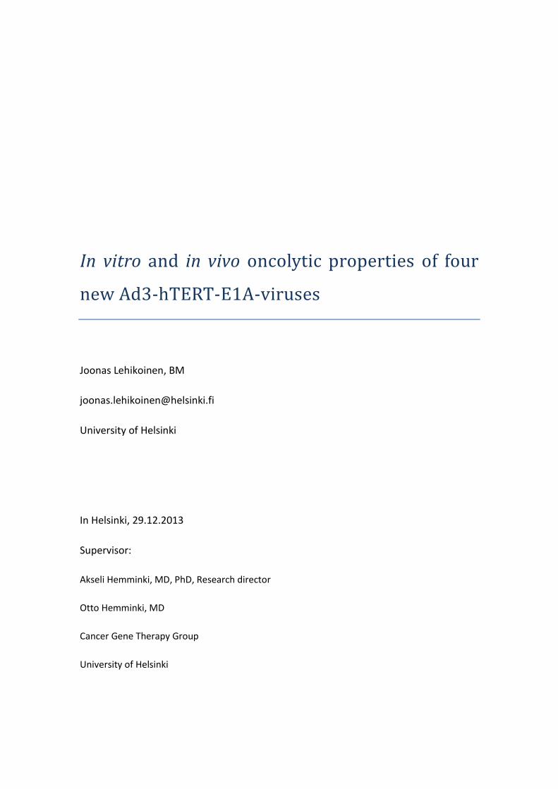
Progressive TCID50.
The newly produced viruses were first tested with progressive TCID50 to determine
whether they have oncolytic properties. After nine (9) days of incubation, the
infections became visible in all culture plates of A549 cells, which indicated that all the
new viruses were functional. During the following days, the infections continued
3
spreading according to the amount of virus pipetted per cell. Slight differences were
detected in the amount and speed of cell lysis. (Figure 1)
Figure 1 A graph of the relative visual titre yielded by the progressive TCID50 (PFU/ml,
logarithmic scale) plotted against days post-infection. (d). This shows that the viruses were
functional and capable of infecting at least some tumour cell lines. The dilutions of virus were
not made according to the VP titres.
The viruses were also tested on CHO-K7, but they showed no effect on the viability of
these cells during the TCID50. This was probably due to the lack of human-like
desmoglein-2 on the surface of these hamster cells.
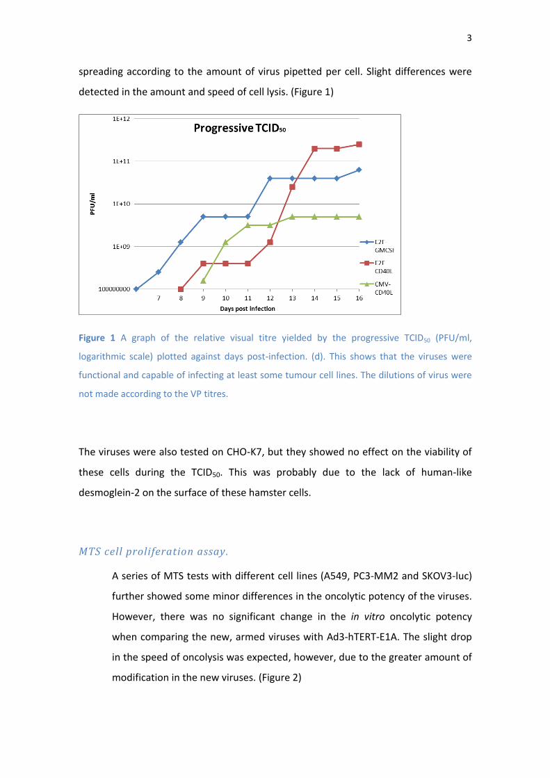
MTS cell proliferation assay.
A series of MTS tests with different cell lines (A549, PC3-MM2 and SKOV3-luc)
further showed some minor differences in the oncolytic potency of the viruses.
However, there was no significant change in the in vitro oncolytic potency
when comparing the new, armed viruses with Ad3-hTERT-E1A. The slight drop
in the speed of oncolysis was expected, however, due to the greater amount of
modification in the new viruses. (Figure 2)
4
Figure 2 Result graphs from MTS assays performed with different cell lines: A549 in the top left
and PC3-MM2 in the top right graph. The two bottom graphs are SKOV3-luc. Complete
oncolysis was achieved with the highest concentration of virus. As hypothesized, the rate of
the infection of the new viruses was almost the same as the previous Ad3-hTERT-E1A virus. By
comparison, the unarmed Ad5/3-Δ24 seems somewhat faster.
2.3 The in vivo properties of the viruses
Animal experiment: SCID-mice with intra peritoneal SKOV3-luc cell
tumours. (Picture 1)
The start of the experiment was promising; the growth of relatively large tumours was
clearly reduced by all the viruses and with E2F promoter groups even some drop in the
size of the tumours was detected during the early stages of the experiment. However,
as anticipated the tumours quickly formed resistance towards the viruses and their
growth speed reached the level of the mock group between the day 14 and 20. (Figure
3 and Figure 4) Still the treatments were able to slow down the advancement of the
cancer and not a single mouse had to be put to sleep due to the tumours. One mouse

5
of the E1A group had to be put to sleep because of a joint infection on the second
week of the experiment.
Picture 1 The tumour cells were injected i.p. and imaged with a fluorescent camera. The image
on the left shows the tumours during the early stage of the experiment, when the tumours are
rather small and concentrated on a limited area while the image on the right shows the same
group later in the experiment. Tumours have grown in size and the tumour of mouse number
four (4) seems to have metastasized to the whole lover peritoneum and the areas of pancreas
and spleen.

6
Figure 3 The viruses slowed down the tumour growth and E1A, E2F-CD40L and E2F-GMCSF
groups even showed signs of tumour shrinkage. Unfortunately all the tumours seemed to
develop an increasing resistance towards the treatment and after day 14 the growth of the
tumours is similar to the mock (PBS) group. (Logarithmic scale, error bars in SD)

7
Figure 4 The virus treatments show to slow down the tumour development on a relatively long
time scale, considering the starting size of the tumours was big. Furthermore, the results of the
E2F-CD40L group were statistically relevant (p-value ≤ 0.05) throughout the whole experiment.
This virus treatment seems to perform a little better than others in this animal experiment
model. Stars show the point on the time scale, where there was statistical relevance detected
(p-value ≤ 0. 05 when comparing the time points to the corresponding mock results (the PBS
group)). (Error bars in SD) *) Every group, except for CMV-GMCSF, was found to be statistically
relevant. **) The data of every group, except for CMV-CD40L, was statistically relevant. ***)
The data of the E1A, the E2F-CD40L and the E2F-GMCSF groups where statistically valid at this
time point. ****) There was a statistical relevancy with the results of the E2F-CD40L group
even at the final data point (day 25) when comparing to the PBS.
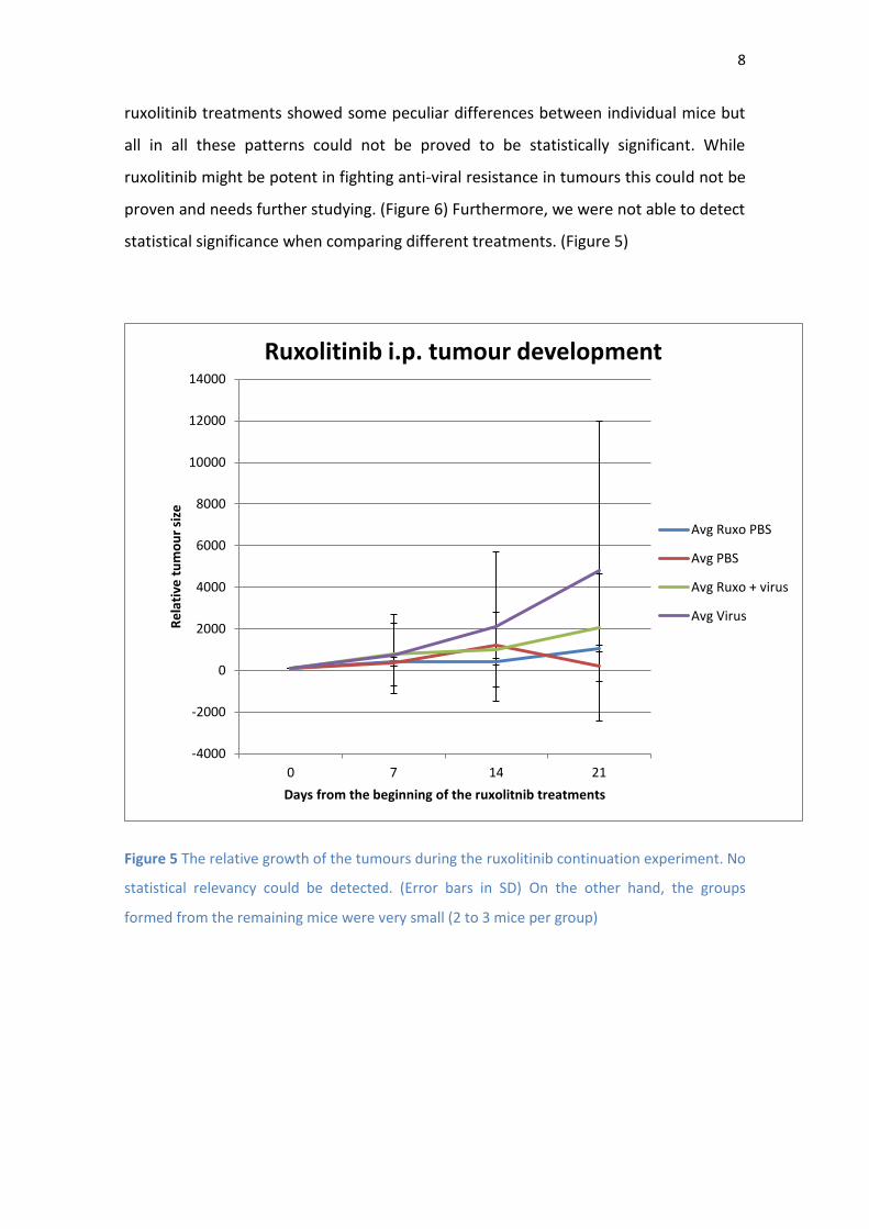
Ruxolitinib continuation experiment.
We hypothesised that using ruxolitinib (JAK inhibitor used in treating for instance
myelofibrosis) would overcome the resistance the tumours had formed towards the
viruses. Thus ruxolitinib treatment was started at day 25 of the animal experiment. The
8
ruxolitinib treatments showed some peculiar differences between individual mice but
all in all these patterns could not be proved to be statistically significant. While
ruxolitinib might be potent in fighting anti-viral resistance in tumours this could not be
proven and needs further studying. (Figure 6) Furthermore, we were not able to detect
statistical significance when comparing different treatments. (Figure 5)
Figure 5 The relative growth of the tumours during the ruxolitinib continuation experiment. No
statistical relevancy could be detected. (Error bars in SD) On the other hand, the groups
formed from the remaining mice were very small (2 to 3 mice per group)
-4000
-2000
0
2000
4000
6000
8000
10000
12000
14000
0 7 14 21
Re
lati
ve t
um
ou
r si
ze
Days from the beginning of the ruxolitnib treatments
Ruxolitinib i.p. tumour development
Avg Ruxo PBS
Avg PBS
Avg Ruxo + virus
Avg Virus

9
Figure 6 Graphs show the relative growth (vertical axis [%]) of the tumours during the
ruxolitinib experiment (horizontal axis [d]). The effect of the ruxolitinib treatment seems to be
opposite with different viruses (no statistical difference). Promoter being the same the only
difference is in the immunostimulatory armament. With immunodeficient mice and human
transgenes this should not make any difference in the results. Results are inconclusive. (C-C =
CMV-CD40L; C-G = CMV-GMCSF) N=3 (mice with ruxolitinib and virus) n=2 (only virus). The
shape of the graph on the right is much due to the overly fast growth of one of the ruxolitinib
treated mice.
Analysing samples from mice.
The blood and tumour samples were analysed with ELISA and they showed that the
viruses produce the immunostimulatory substances, GMCSF and CD40L, which were
also released into the blood stream in low quantities.

10
Figure 7 Charts showing the tumour development of the individual mice during the ruxolitinib
continuation experiment. In some groups there is a chance of a statistically relevant pattern
(for example CMV-CD40L). This should be further studied. The mouse number 4 of the PBS
group had to be put to sleep on day 7 of the ruxolitinib experiment due to the poor condition.
The number of the mouse with the received treatment indicated on the right. On vertical axis
the relative size of the tumours (%), on the horizontal axis time from the day the ruxolitinib
treatments started (d). (The ‘missing’ mouse from the E1A group was put to sleep before the
start of the ruxolitinib experiment due to a joint infection)

11
3 Discussion
The fact that the new Ad3 viruses were able to slow down the growth of relatively
large tumours in SCID mice is encouraging. However, the rapidly formed resistance
towards the treatment during the animal experiment may require further
consideration and adjustments to the treatment. For instance, the dose and treatment
frequency could be changed. There is also a chance of combining the serotype 3
viruses with other viruses (for example Ad5/3) due to their effect on the epithelial
junctions and use of a different receptor through which the virus enters the cell
(desmoglein-2) (3). This might make the Ad3 viruses useful in ensuring that drugs or
other viruses used during the treatment can spread more effectively in the tumour
stroma if they can cause the tight epithelial cell junctions to open.
Compared to the previous results from animal experiments with the virus E1A, this
time the oncolytic results seemed not as potent as before. We believe that this was at
least partly due to the relatively large size of the tumours in the beginning of the
experiment. The already formed necrotic areas in the tumour can form a physiological
barrier that can protect the cancer cells from viruses which rely on passive diffusion to
get to the target. This could be overcome by injecting the viruses straight into the
tumour or developing some other sort of vector for the virus.
During this project we did not research the effects of the four new adenoviruses on
tumours in functioning immune systems. The immunological activating effect of
adenoviruses is considered one of the key factors using these viruses in treating
cancer.(1) However, the finding that these Ad3 viruses cannot infect CHO-K7 cells
makes it more difficult to find experimental animal models that would be capable of
demonstrating the effects of these viruses on the immune system. Nevertheless, the
fact that these viruses produce functional GMCSF and CD40L in detectable quantities is
encouraging. Furthermore, the detected release of these substances to the blood flow
could provide a more systemic response to the treatment in a functional immune
system thus helping to inhibit the formation of metastases and to destroy them.

12
One of the objectives of this preclinical study was to determine whether there are any
significant differences between the new viruses. There appears to be little difference
between the function of E2F and the CMV promoters. Moreover, on the basis of the
animal experiment, it appears that all the viruses have potential for further
development and research. However, at this point, we believe that E2F-CD40L and
CMV-CD40L are the most promising viruses from the clinical perspective.
The inconclusive results with ruxolitinib treatment should be thoroughly researched
with another experiment that would introduce ruxolitinib to the tumours right from
the beginning of the treatments. We believe that the differences in the effects of
ruxolitinib between individual animals were due to the different resistance
mechanisms the tumours used towards the viruses in the first place. It appeared that
in some cases, new viruses were able to be reproduced in infected cells, while in
others we hypothesised that the reactivation occurred in the promotion regions
forming GMCSF and CD40L. (4) However, we could not conclusively confirm this
reactivation. Also, there is the possibility that some new tumour cell lines that were
not immune to the viruses had grown after virus injections were stopped, and were
destroyed with the new treatment. This would explain the notches in some of the
curves (virus-killing tumour cells causing shrinkage, while resistant tumour cells
continue thriving.)
However, these results have a relatively high possibility of error caused by the large
size of the tumours at the initiation of ruxolitinib treatment, so that the imaging data
might not be entirely reliable since the bio luminance measuring IVIS camera system
has its limitations. Nevertheless, some groups can be considered promising for further
research when noting that the restarted treatments with conjoined ruxolitinib
injections were able to slow down the growth and even reduce the size (with individual
mice; no confirmed statistical relevancy) of large, final stage tumours. (Figure 7)
Another animal experiment is needed to determine whether the relevant curative
influence occurs with ruxolitinib treatment. If the treatment is capable of reactivating
some of the dormant adenoviruses in tumours to replicate, we could be able to
prolong the time before the onset of tumour derived resistance against the viruses

13
occurs. This also has the potential to enhance the immune response towards the
tumour cells if we can cause a chronic infection in them.
The future challenges for the oncolytic viral treatments include for instance the proper
activation of the immune system in a patient. The virus used during any treatment
should direct the immune response towards the tumour and simultaneously avoid the
antibodies meant to neutralize the virus itself. It would also seem that the tumour cells
can form an interferon response towards viruses as one part of the resistance
mechanisms against viral treatments and also that the ability to form resistance would
exist before the treatments (5). However, trying to overcome this resistance simply by
inhibiting the interferon production might repress some of the immunological affects
towards tumour cells initiated by the interferon system. (6) This is one reason why it is
probably best to keep modifying the oncolytic Ad3 viruses more and more towards the
role of a ‘silent helper’ and to be used in co-operation with other forms of treatment.
4 Conclusion
We found that the new immunologically armed Ad3 viruses were as oncolytically
potent as their precursor thus confirming the basic hypothesis. The results gained
during this project form a basis for further research and development on the modified
Ad3 viruses. Especially the suggested immunostimulatory component provided by
these viruses on their own together with their armament should be considered for
further research in patients.(1)
14
5 Materials and methods
Progressive TCID50
105 A549 cells per well were plated on a 96-well plate and incubated for a day. The
following day a dilution of virus was added to the wells (100 µl/well; dilutions of 10-7;
10-8 … 10-14 virus; mock rows on the plate received no virus). The progression of the
infections was monitored under a light microscope and the results mathematically
changed into relative PFU titres. The TCID50 was stopped after the cells in the mock
rows showed CPE.
MTS cell proliferation assay
On day one, 105 cells per well (A549, PC3-MM2 or SKOV3-luc) were seeded into 96-
well plates in 100µl of growth medium (GM), which contained 5% of FBS. On day two,
the monolayer was washed once with GM containing 5% of FBS. Then the cells were
infected with different viruses at doses of 100, 10, 1, 0.1 and 0 virus particles per cell.
Thereafter the cells were incubated for one hour on a rocking machine and then
washed with GM. After adding new 5% GM the cells were left to the incubator and the
GM was replaced every fourth day. The test was terminated by adding mts reagent
(Promega) after the cytopathic effect of one of the tested viruses reached 100% with
the highest concentration. After two hours of incubation the absorbance was
measured at 490 nm filter. The background was then subtracted and results analysed.

15
Animal experiment
48 mice with SCID (Severe combined immunodeficiency); 6 groups of 8 mice.
Groups:
1) Ad3-hTERT-CMV-CD40L
2) Ad3-hTERT-CMV-GMCSF
3) Ad3-hTERT-E2F-CD40L
4) Ad3-hTERT-E2F-GMCSF
5) Ad3-hTERT-E1A
6) PBS
Tumor model:
Each mouse was injected with 5∙10⁶ SKOV3-luc cells in 300 μl of pure DMEM on day 0.
(=> 24∙10⁷ cells in total) Due to error from using syringes, the calculated cell number
was valuated to 6,25∙10⁶ in 375 μl DMEM. (Total amount of cells was 30∙10⁷ in 18ml of
DMEM) (see Table 1 and 2 for further details)
Table 1 The virus titres of the viruses used during the animal experiment.
Ad3 viruses
Virus Titer VP/ml
Ad3-hTERT-E2F-CD40L 2,266x10e11
Ad3-hTERT-CMV-CD40L 1,298x10e11
Ad3-hTERT-E2F-GMCSF 9,889x10e11
Ad3-hTERT-E1A 9,6x10e12VP/ml
Ad3-hTERT-CMV-GMCSF 6,16x10e12

16
Group
numb
er
name
of
virus
pure
virus
(µl)/mou
se
Diluti
on
(µl)
pure
PBS/mou
se (µl)
virus d or
not for 8
(µl)
virus
for 12
(µl)
PBS
for 8
(µl)
PBS
for 12
(µl)
1 CMV-
CD40L
7,704160
247
no 92,29583
975
61,63328
198
92,449
923
738,3
667
1107,
55
2 CMV-
GMCS
F
0,162337
662
16,23
3766
23
83,76233
77
129,8701
298
194,80
5195
670,0
987
1005,
148
3 E2F-
CD40L
4,413062
665
no 95,58693
734
35,30450
132
52,956
752
764,6
955
1147,
043
4 E2F-
GMCS
F
1,011224
593
10,11
2245
93
89,88775
407
80,89796
744
121,34
6951
719,1
02
1078,
653
5 E1A 0,104166
66
10,41
6666
67
89,58333
334
83,33333
333
125 716,6
667
1075
6 PBS
(no
virus)
0 no 100 0 0 800 1200
Table 2 The table with calculated amounts of virus for making dilutions to treat the mice
during the animal experiment and the ruxolitinib continuation experiment. The calculations
were made according to the VP titres. (Table 1)
Treatment:
Mice were treated on days 3, 7 and 14 with virus injections after the tumour
implantation. Injections included 10⁹ VP in PBS or only PBS. (Altogether this meant
24∙10⁹ VP per group [8 mice*3 injections]) Because of the error due to syringes etc.
(residual volume of fluid that stays in the syringe even after the injection) the

17
calculations were performed to 12 mice (group of 8 in real life) and 8 mice (group of 5
mice).
Imaging:
5 mice (the mice “numbered” accordingly: 1= no ear piercings; 2= hole in the right ear;
3= hole in the left ear; 4= both ears pierced; 5= 2 holes in the right ear) per group were
imaged on days 3, 7, 14, 21, 28 after the cell injection. 150 mg/kg D-luciferin was
injected during each imaging and captured 10 min later with 10 s exposure time,
1f/stop, medium binning and open filter. Images were overlaid with Living Image 2.50
(Xenogen). Total flux (photons/s) was measured by drawing regions of interest (ROI)
around the peritoneal area of the mice. Background was subtracted. (In vivo luciferace:
Diluted to 3 mg/100 µl (1 g to 33.3 ml) -> 100 µl/mouse, 1 ml and 300 µl aliquots
estimated minimum requirement: 3 mg x 5 x 6 x 6 = 540 mg)
Samples:
The remaining 3 mice per group, which were only treated with virus (were not
imaged), were put to sleep on day 13 after the cell injections. Both blood and tumor
samples from these mice were analyzed for viruses’ qPCR and GMCSF or CD40L. Blood
samples by heart puncture and the tumors were collected and stored appropriately.
Ruxolitinib experiment
The virus treatments were restarted to the remaining mice straight after the last
imaging of the animal experiment (described above). The restarted virus treatment
was conjoined with a ruxolitinib treatment for half of the mice. These mice were
treated with 100 µl of diluted ruxolitinib every Monday, Wednesday and Friday. Virus
was injected every Monday two hours after the ruxolitinib injection. The mice were
imaged on every Monday and data was then analysed. This experiment lasted 21 days
starting from the end of the animal experiment. Ruxolitinib experiment was then
stopped due to the overly sized tumours and rapidly forming metastases.

18
ELISA
Biotinylated antibody reagent was added to each well on a 96-well plate. After this
samples (in this case venous blood) were added and the plate was covered and
incubated at room temperature for 3 hours. The plate was washed trice and
Streptavidin-HRP solution was added, after which the plate was covered and incubated
for 30 minutes. Then the plate was again washed trice with washing buffer and TMB
Substrate solution was added. The plate was then developed in the dark for 30
minutes. The reaction was stopped and the absorbances measured at 450 nm. The
results were analysed using a standard curve.

19
6 Acknowledgements and report
The project was led by M.D. Otto Hemminki.
Viruses were produced from the plasmids by lab technician Saila Pesonen, who also
participated in the carrying out the animal experiment.
The in vitro testing was conducted by BM Joonas Lehikoinen who also mainly carried
out the animal experiment according the instructions and in the observation of M.D.
Otto Hemminki. He also took part in the planning of the animal experiment and
analysed the data as instructed and participated in the discussions considering the
continuation of the project.
The data for this report paper was acquired during the timeline from August 2012 to
July 2013 and the research project was conducted in the facilities of University of
Helsinki at Biomedicum 1 and Haartman Institute.
The final article about the project will be published later, this paper being a report for
the Medical Faculty of the University of Helsinki written by Joonas Lehikoinen as a
form of completing his advanced studies essay.
I thank M.D. Otto Hemminki for mentoring and supervising me and research professor,
M.D. PhD Akseli Hemminki for scientific support and mentorship.
I declare no conflict of interest.
20
References
1. Hemminki O, Diaconu I, Cerullo V, Pesonen SK, Kanerva A, Joensuu T, et al. Ad3-hTERT-E1A, a fully serotype 3 oncolytic adenovirus, in patients with chemotherapy refractory cancer. Mol Ther. 2012 Sep;20(9):1821-30.
2. Hemminki O, Bauerschmitz G, Hemmi S, Lavilla-Alonso S, Diaconu I, Guse K, et al. Oncolytic adenovirus based on serotype 3. Cancer Gene Ther. 2011 Apr;18(4):288-96.
3. Wang H, Li ZY, Liu Y, Persson J, Beyer I, Moller T, et al. Desmoglein 2 is a receptor for adenovirus serotypes 3, 7, 11 and 14. Nat Med. 2011 Jan;17(1):96-104.
4. Gallagher NJ, Eliopoulos AG, Agathangelo A, Oates J, Crocker J, Young LS. CD40 activation in epithelial ovarian carcinoma cells modulates growth, apoptosis, and cytokine secretion. Molecular Pathology. 2002 Apr;55(2):110-20.
5. Liikanen I, Monsurro V, Ahtiainen L, Raki M, Hakkarainen T, Diaconu I, et al. Induction of interferon pathways mediates in vivo resistance to oncolytic adenovirus. Molecular Therapy: the Journal of the American Society of Gene Therapy. 2011 Oct;19(10):1858-66.
6. Donia M, Hansen M, Sendrup SL, Iversen TZ, Ellebaek E, Andersen MH, et al. Methods to improve adoptive T-cell therapy for melanoma: IFN- enhances anticancer responses of cell products for infusion. J Invest Dermatol. 2013 Feb;133(2):545-52.


