
veterinarians and private practitioners should
NEW
SOUT
H WA
LES
January - March 2007 • Number 2007/1
DISTRICT VETERINARIANS PRESERVE EXPORT MARKET
Have you ever wondered how a veterinarian from the government, a Rural Lands Protection Board or the private sector can make a genuine contribution to Australia’s eligibility to export livestock and livestock products? Submitting samples for transmissible spongiform encephalopathy (TSE) exclusion is one way. The European Union (EU) says we do the job well enough, but it could be better.
Four inspectors from the Food and Veterinary Office of the European Commission evaluated Australia’s bovine spongiform encephalopathy (BSE) controls in October 2006, and their report, together with Australia’s response, makes interesting reading.
The key findings were that certifi cation of beef exports to the EU are done in accordance with EU requirements and that the current level of surveillance for BSE could detect BSE if it were present in Australia. This is good news for us.
However, some of their other fi ndings were contentious, and their suggestions had the aura of wanting Australia (a BSE-free country) to adopt surveillance and identifi cation regimes more like the EU’s (where BSE cases occur but are gradually being eradicated). They suggested improvements to cattle identification, testing more stock and removing fallen stock from the rendering chain.
Australian governments and relevant industries believe that many of the recommendations are not warranted, because Australia is recognised by the World
ANIMAL HEALTH SURVEILLANCE
In this issue!
District veterinarians preserve export markets 1
QUARTERLY HIGHLIGHTS 2 Bacterial rhinitis in Merino ewes 2 Brassica poisoning in cattle 3 Possible Ward’s weed toxicity 4 Cattle mortalities 4 Blackleg infection in lambs 4 Suspected salmonellosis in cattle 5 Bovine ephemeral fever 5 Buff alo fl y 5 Metabolic disease in lambs 5 Salmonellosis in sheep 6 Salmonella typhimurium infection in lambs: from sewage? 7
NOTIFIABLE DISEASES 8 Anthrax 8 Strangles in horses 8 Chlamydiosis in chickens 8 Footrot 9 Improving sheep fertility in Western NSW 9
DISEASE SURVEILLANCE AND CONTROL PROGRAMS 9 Cattle tick program 9 Bovine Johne’s disease (BJD)infected herds in NSW as at 31 March 2007 10 Johne’s disease Market Assurance Programs (MAPs) as at 31 March 2007 10 Transmissible spongiform encephalopathy (TSE) survelliance submissions by RLPB, 1 Jan 2007– 31 March 2007 11 Enzootic Bovine Leukosis (EBL) 11 New staff 12
Information contributed by staff of the Rural Lands Protection Boards and the NSW Department of Primary Industries
Organisation for Animal Health (OIE) as meeting all relevant requirements for a BSE-free country.
Two useful points emerge. First, trade to the EU without substantiation of our BSE status and without proper audits is not a given. Substantiation does not happen without district veterinarians and private practitioners submitting samples from stock with eligible clinical signs. NSW DPI gratefully acknowledges all of those veterinarians who contribute to the TSE surveillance program. Secondly, all official veterinarians and private practitioners should read the report and Australia’s response; these documents are case studies of why Competent Authorities (as the report calls us) are essential for market access.
The European Commission’s report can be found at http://ec.europa.eu/food/ fvo/ir_search_en.cfm?stype=insp_ nbr&showResults=Y&REP_INSPECTION_ REF=8081/2006.
A reference to Australia’s response report is contained at the end of the report, and the response is presented in full on the website as Annexes 1 and 2.
For further information contact Rory Arthur, NSW DPI, on (02) 6391 3823 or Sally Spence, NSW DPI, on (02) 6391 3630.
PAGE 2
QUARTERLY HIGHLIGHTS
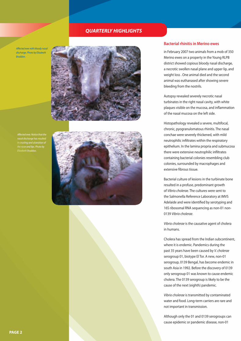
Affected ewe with bloody nasal discharge. Photo by Elizabeth Braddon.
Affected ewe. Notice that the nasal discharge has resulted
crusting and ulceration of nose and lips. Photo by
Elizabeth Braddon.
in cru the noseElizabeth BraE
Bacterial rhinitis in Merino ewes
in February 2007 two animals from a mob of 350 Merino ewes on a property in the Young RLPB district showed copious bloody nasal discharge, a necrotic swollen nasal plane and upper lip, and weight loss . One animal died and the second animal was euthanased after showing severe bleeding from the nostrils.
Autopsy revealed severely necrotic nasal turbinates in the right nasal cavity, with white plaques visible on the mucosa, and inflammation of the nasal mucosa on the left side.
Histopathology revealed a severe, multifocal, chronic, pyogranulomatous rhinitis. The nasal conchae were severely thickened, with mild neutrophilic infiltrates within the respiratory epithelium. In the lamina propria and submucosa there were extensive neutrophilic infiltrates containing bacterial colonies resembling club colonies, surrounded by macrophages and extensive fibrous tissue.
Bacterial culture of lesions in the turbinate bone resulted in a profuse, predominant growth of Vibrio cholerae. The cultures were sent to the Salmonella Reference Laboratory at IMVS Adelaide and were identified by serotyping and 16S ribosomal RNA sequencing as non-01 non0139 Vibrio cholerae.
Vibrio cholerae is the causative agent of cholera in humans.
Cholera has spread from the Indian subcontinent, where it is endemic. Pandemics during the past 35 years have been caused by V. cholerae serogroup 01, biotype El Tor. A new, non-01 serogroup, 0139 Bengal, has become endemic in south Asia in 1992. Before the discovery of 0139 only serogroup 01 was known to cause endemic cholera. The 0139 serogroup is likely to be the cause of the next (eighth) pandemic.
Vibrio cholerae is transmitted by contaminated water and food. Long-term carriers are rare and not important in transmission.
Although only the 01 and 0139 serogroups can cause epidemic or pandemic disease, non-01
and non-0139 serotypes can be pathogenic and associated with small outbreaks of diarrhoeal disease. Occasionally they cause a variety of severe extra-intestinal infections, including wound infection and acute sepsis, especially in people with liver disease and immunosuppression. An example from Australia was a case of facial cellulitis caused by V. cholerae non-01 non 0–139 in an indigenous girl from North Queensland.
Vibrio cholerae, including strains of 01 and 0139, are normal inhabitants of surface waters (particularly brackish waters), and they survive and multiply in association with zooplankton and phytoplankton independently of infected human beings. However, environmental isolates from areas that are distant from areas of human infection do not generally have the cholera toxin genes (cholera toxin is responsible for the excessive secretion of electrolyte rich-water in the intestines).
Vibrio cholerae has been isolated from the Australian aquatic environment since 1977, and periodically cholera cases have occurred following exposure to these environments. Molecular techniques have been used to confirm association between epidemiologically related clinical isolates and the aquatic environment and the persistence of the 01 serovar in the Australian environment over an 8-year period.
Cases of animal diseases caused by V. cholerae have been reported from countries other than Australia. The organism was isolated from the brain of a feedlot heifer with meningoencephalitis and cerebral abscessation in the United States. In the Netherlands V. cholerae non-01 has been associated with enterotoxicosis in a goat, abomasitis and enteritis in a bull, haemorrhagic diarrhoea in a heifer, calf diarrhoea, diarrhoea in lambs and bovine abortion. Outbreaks caused by V. cholerae 01, leading to keratoconjunctivitis and deaths, have occurred in cattle in Argentina.
The property in the Young RLPB district was in a region that had been hit with torrential
rain in February 2007, and a lot of paddock
debris and faeces had been washed into dams
across the area. It is possible that this led to
the exposure of these ewes to V. cholera. There
have been no more cases reported of this kind
from the affected property or any others near
it.
Vibrio cholera was also isolated from a steer
in the NSW Western Division during the
quarter. This case is described in this issue in
‘Suspected salmonellosis in cattle’.
This report was written by Erika Bunker, NSW DPI, with contributions from Elizabeth Braddon and Darryl Lawler. For further information contact Elizabeth Braddon, DV Young RLPB, on (02) 6382 1255.
Brassica poisoning in cattle
Brassica poisoning was suspected to be the
cause of blindness and central nervous system
disease in four out of 35 yearling cattle in
the South Coast RLPB. The cattle had been
grazing forage brassica for 2 weeks. Three of
the affected animals presented with blindness,
depression, inappetence, drooling, aimless
movement and collapse. One heifer was
recumbent and non-responsive. All animals
had elevated temperatures. In addition,
exposure to lead at a low level was found in
one affected steer. (The paddock had a rubbish
tip in one section.)
Glucosinolates, S-methyl-L-cysteine-sulfoxide
(SMCO), tryptophan, sulfur and possibly
an organic nitrile are the potentially toxic
compounds in brassicas. Clinical signs of
toxicity depend upon the toxin involved. The
risk of toxicity increases when brassicas are
flowering and setting seed, or when they have
been affected by drought or frost, or when
they put out fresh growth after rain.
Glucosinolates can cause gastrointestinal
irritation, neonatal goitre, embryonic death or
poor conception rates, reduced birth weights
and an increased risk of induced copper
deficiency. Isothiocyanates (metabolites of
glucosinolate) are believed to cause a rumen
stasis and constipation syndrome.
After several weeks of ingestion, SMCO may
cause haemolysis of red blood cells, resulting
in anaemia, jaundice and haemoglobinuria.
Tryptophan can cause ‘fog fever’, an acute
respiratory disorder characterised by
interstitial pneumonia, pulmonary oedema
and emphysema.
High levels of sulfur may result in a
polioencephalomalacia-like syndrome
characterised by reduced awareness, aimless
wandering, incoordination, recumbency,
seizures and death. Temporary blindness of
unknown cause (possibly an organic nitrile)
can occur in cattle. The pupils are dilated and
the cornea sometimes opaque.
Photosensitisation, bloat, nitrate poisoning
and oxalate poisoning are also associated
with brassicas. More poisoning problems are
encountered with sheep than with cattle. The
blindness syndrome typically occurs in cattle.
In this case toxicity due to a high level of
sulfur was suspected on the basis of clinical
signs. The most severely affected animal was
shot, with the other three making reasonable
recoveries following thiamine treatment
and removal from the crop. In this case it
is uncertain whether the thiamine aided
recovery. Brassicas are valuable, high quality
forages for ruminants; however, they should
form only 30% to 40% of the diet, so as to
avoid sulfur toxicity problems.
For further information contact Ian Lugton,
DV South Coast RLPB, on (02) 6492 1283 or
Chris Bourke, NSW DPI, on (02) 6391 3867.
PAGE 3
P
rst
the
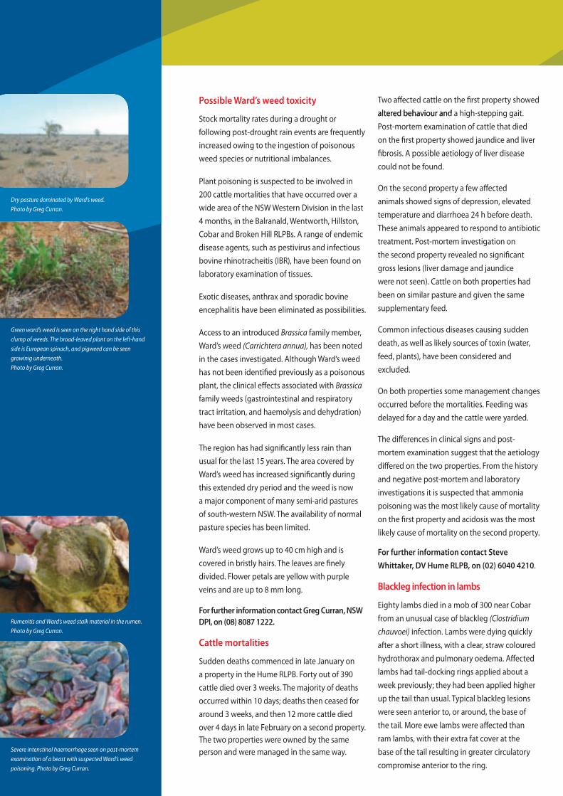
Dry pasture dominated by Ward’s weed. Photo by Greg Curran.
Green ward’s weed is seen on the right hand side of this clump of weeds. The broad-leaved plant on the left-hand side is European spinach, and pigweed can be seen growinig underneath. Photo by Greg Curran.
Rumenitis and Ward’s weed stalk material in the rumen. Photo by Greg Curran.
Severe intenstinal haemorrhage seen on post-mortem examination of a beast with suspected Ward’s weed poisoning. Photo by Greg Curran.
Possible Ward’s weed toxicity
Stock mortality rates during a drought or following post-drought rain events are frequently increased owing to the ingestion of poisonous weed species or nutritional imbalances.
Plant poisoning is suspected to be involved in 200 cattle mortalities that have occurred over a wide area of the NSW Western Division in the last 4 months, in the Balranald, Wentworth, Hillston, Cobar and Broken Hill RLPBs. A range of endemic disease agents, such as pestivirus and infectious bovine rhinotracheitis (IBR), have been found on laboratory examination of tissues.
Exotic diseases, anthrax and sporadic bovine encephalitis have been eliminated as possibilities.
Access to an introduced Brassica family member, Ward’s weed (Carrichtera annua), has been noted in the cases investigated. Although Ward’s weed has not been identified previously as a poisonous plant, the clinical effects associated with Brassica family weeds (gastrointestinal and respiratory tract irritation, and haemolysis and dehydration) have been observed in most cases.
The region has had significantly less rain than usual for the last 15 years. The area covered by Ward’s weed has increased significantly during this extended dry period and the weed is now a major component of many semi-arid pastures of south-western NSW. The availability of normal pasture species has been limited.
Ward’s weed grows up to 40 cm high and is covered in bristly hairs. The leaves are finely divided. Flower petals are yellow with purple veins and are up to 8 mm long.
For further information contact Greg Curran, NSW DPI, on (08) 8087 1222.
Cattle mortalities
Sudden deaths commenced in late January on a property in the Hume RLPB. Forty out of 390 cattle died over 3 weeks. The majority of deaths occurred within 10 days; deaths then ceased for around 3 weeks, and then 12 more cattle died over 4 days in late February on a second property. The two properties were owned by the same person and were managed in the same way.
Two affected cattle on the fi rst property showed
ost-mor
paltered behaviour and aaltered behaviour and a high-stepping gait.
emtPost-mortem examination of cattle that diedex on the first property showed jaundice and liverfi fibrosis. A possible aetiology of liver disease could not be found.
On the second property a few affected animals showed signs of depression, elevated temperature and diarrhoea 24 h before death. These animals appeared to respond to antibiotic treatment. Post-mortem investigation on the second property revealed no significant gross lesions (liver damage and jaundice were not seen). Cattle on both properties had been on similar pasture and given the same supplementary feed.
Common infectious diseases causing sudden death, as well as likely sources of toxin (water, feed, plants), have been considered and excluded.
On both properties some management changes occurred before the mortalities. Feeding was delayed for a day and the cattle were yarded.
The differences in clinical signs and postmortem examination suggest that the aetiology differed on the two properties. From the history and negative post-mortem and laboratory investigations it is suspected that ammonia poisoning was the most likely cause of mortality on the first property and acidosis was the most likely cause of mortality on the second property.
For further information contact Steve Whittaker, DV Hume RLPB, on (02) 6040 4210.
Blackleg infection in lambs
Eighty lambs died in a mob of 300 near Cobar from an unusual case of blackleg (Clostridium chauvoei) infection. Lambs were dying quickly after a short illness, with a clear, straw coloured hydrothorax and pulmonary oedema. Affected lambs had tail-docking rings applied about a week previously; they had been applied higher up the tail than usual. Typical blackleg lesions were seen anterior to, or around, the base of the tail. More ewe lambs were affected than ram lambs, with their extra fat cover at the base of the tail resulting in greater circulatory compromise anterior to the ring.
As part of routine exclusion of exotic diseases, samples were collected for the exclusion of heartwater, which has similar post-mortem characteristics. Heartwater results from infection with Ehrlichia ruminantium and is endemic in Africa and in a few islands in the Caribbean. It is a tick-borne disease transmitted by at least 12 species of Amblyomma ticks. Field reports indicated that some sheep were carrying ornate kangaroo ticks of the Amblyomma genus, the same genus that transmits Ehrlichia ruminantium in Africa and the Caribbean islands. Heartwater was excluded.
For further information contact Greg Curran, NSW DPI, on (08) 8087 1222.
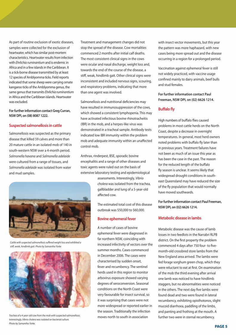
Suspected salmonellosis in cattle
Salmonellosis was suspected as the primary disease that killed 59 calves and more than 20 mature cattle in an isolated mob of 140 in south-western NSW over a 4-month period. Salmonella havana and Salmonella adelaide were cultured from a range of tissues, and Salmonella adelaide was isolated from water and mud samples.
Cattle with suspected salmonellosis suffered weight loss and exhibited a stiff, weak, hindlimb gait. Photo by Samantha Yorke
Trachea of a 4-year-old cow from the mob with suspected salmonellosis. Interestingly, Vibrio cholera was isolated on bacterial culture. Photo by Samantha Yorke.
Treatment and management changes did not stop the spread of the disease. Cow mortalities commenced 2 months after initial calf deaths. The most consistent clinical signs in the cows were ocular and nasal discharge, weight loss and, towards the end of the course of the disease, a stiff, weak, hindlimb gait. Other clinical signs were inconsistent and included nervous signs, scouring, and respiratory problems, indicating that more than one agent was involved.
Salmonellosis and nutritional deficiencies may have resulted in immunosuppression of the cows, which showed a consistent lymphopenia. This may have activated infectious bovine rhinotracheitis (IBR) in the mob, and a herpes-like virus was demonstrated in a tracheal sample. Antibody tests indicated low IBR immunity within the problem mob and adequate immunity within an unaffected control mob.
Anthrax, rinderpest, BSE, sporadic bovine encephalitis and a range of other diseases and toxic agents were ruled out on the basis of extensive laboratory testing and epidemiological
assessments. Interestingly, Vibrio cholera was isolated from the trachea, gallbladder and lung of a 5-year-old affected cow.
The estimated total cost of this disease outbreak was $50,000 to $60,000.
Bovine ephemeral fever
A number of cases of bovine ephemeral fever were diagnosed in far northern NSW, coinciding with increased infectivity of vectors over the summer months. Cases commenced in December 2006. The cases were characterised by sudden onset, fever and recumbency. The sentinel herds used in this region to monitor arbovirus exposure showed varying degrees of seroconversion. Seasonal conditions on the North Coast were very favourable for insect survival, so it was surprising that cases were not more widespread or reported earlier in the season. Traditionally the infection moves north to south in association
with insect vector movements, but this year the pattern was more haphazard, with new cases being more spread out and the disease occurring in a region for a prolonged period.
Vaccination against ephemeral fever is still not widely practiced, with vaccine usage confined mainly to dairy animals, beef bulls and stud females.
For further information contact Paul Freeman, NSW DPI, on (02) 6626 1214.
Buffalo fly
High numbers of buffalo flies caused problems in most cattle herds on the North Coast, despite a decrease in overnight temperatures. In general, most herd owners noted problems with buffalo fly later than in previous years. Treatment failures have not been as much of an issue this year as has been the case in the past. The reason for the reduced length of the buffalo fly season is unclear. It seems likely that widespread drought conditions in southeast Queensland may have reduced the size of the fly population that would normally have moved southwards.
For further information contact Paul Freeman, NSW DPI, on (02) 6626 1214.
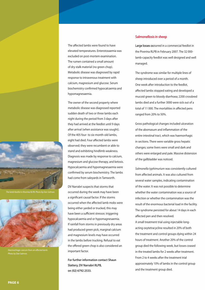
Metabolic disease in lambs
Metabolic disease was the cause of lamb losses in two feedlots in the Narrabri RLPB district. On the first property the problem commenced 4 days after 750 four- to fivemonth-old crossbred store lambs from the New England area arrived. The lambs were fed forage sorghum green chop, which they were reluctant to eat at first. On examination of the mob the third evening after arrival one lamb was noticed to have hindlimb staggers, but no abnormalities were noticed in the others. The next day five lambs were found dead and two were found in lateral recumbency, exhibiting opisthotonos, slight mucoid diarrhoea, paddling of the limbs, and panting and frothing at the mouth. A further two were in sternal recumbency.
PAGE 5PAGE 5
losses
iv
The lamb feedlot in Riverina RLPB. Photo by Dan Salmon.
Haemorrhagic caecum from an affected lamb. Photo by Dan Salmon.
PAGE 6
The affected lambs were found to have
elevated temperatures. Enterotoxaemia was
excluded on post-mortem examination.
The rumen contained a small amount
of dry stalk material (no green chop).
Metabolic disease was diagnosed by rapid
response to intravenous treatment with
calcium, magnesium and glucose. Serum
biochemistry confirmed hypocalcaemia and
hypomagnesaemia.
The owner of the second property where
metabolic disease was diagnosed reported
sudden death of two or three lambs each
night during the period from 3 days after
they had arrived at the feedlot until 9 days
after arrival (when assistance was sought).
Of the 405 four- to six-month-old lambs,
eight had died. Four affected lambs were
observed; they were recumbent or able to
stand and exhibiting hindlimb weakness.
Diagnosis was made by response to calcium,
magnesium and glucose therapy, and ketosis.
Hypocalcaemia and hypomagnesaemia were
confirmed by serum biochemistry. The lambs
had come from saleyards in Tamworth.
DV Narrabri suspects that storms that
occurred during the week may have been
a significant causal factor. If the storms
occurred when the affected lamb mobs were
being either yarded or trucked, this may
have been a sufficient stressor, triggering
hypocalcaemia and or hypomagnesaemia.
If rainfall from storms in previously dry areas
had produced green pick, marginal calcium
and magnesium levels may have occurred
in the lambs before trucking. Refusal to eat
the offered green chop is also considered an
important factor.
For further information contact Shaun
Slattery, DV Narrabri RLPB,
on (02) 6792 2533.
Salmonellosis in sheep
Large occurrLarge losses occurred in a commercial feedlot in
the R iverina RLPB in February 2007. The 32 000e
lamb-capacity feedlot was well designed and well
managed.
The syndrome was similar for multiple lines of
sheep introduced over a period of a month.
One week after introduction to the feedlot,
affected lambs stopped eating and developed a
mucoid green to bloody diarrhoea; 2200 crossbred
lambs died and a further 3000 were sick out of a
total of 11 000. The mortalities in affected pens
ranged from 20% to 50%.
Gross pathological changes included ulceration
of the abomasum and inflammation of the
entire intestinal tract, which was haemorrhagic
in sections. There were variable gross hepatic
changes; some livers were small and dark and
others were enlarged and pale. Massive distension
of the gallbladder was noticed.
Salmonella typhimurium was consistently cultured
from affected animals. It was also cultured from
several water samples, indicating contamination
of the water. It was not possible to determine
whether the water contamination was a source of
infection or whether the contamination was the
result of the enormous bacterial load in the facility.
The syndrome persisted for about 14 days in each
affected pen and then resolved.
A small treatment trial using injectable long-
acting oxytetracycline resulted in 20% of both
the treatment and control groups dying within 24
hours of treatment. Another 20% of the control
group died the following week, but losses ceased
in the treated lambs for 2 weeks after treatment.
From 2 to 4 weeks after the treatment trial
approximately 10% of lambs in the control group
and the treatment group died.

Not all pens were affected, but there was
some apparent spread between adjacent
pens. Lambs from a wide variety of sources
were affected, but there appeared to be a line
effect: some lines of lambs were unaff ected
and others in the same pen had a high attack
rate.
There was no obvious susceptibility-
determining factor in the history of the lambs
before their introduction.
Strategies to control future outbreaks
include the cleaning and disinfection of
pens, changes to induction protocols, water
disinfection, and retrospective investigation
of the differences between lines of lambs that
were affected and lines that were not.
Seventy out of 1300 merino hoggets died and
50 were sick from Salmonella typhimurium
infection on a property in the Hume RLPB
in March. The sheep had been transported
from Walcha a week before the losses. Severe
scouring, weakness, inappetence, excessive
drinking and shade-seeking were the clinical
signs and behaviours noted.
For further information contact Dan Salmon, DV Riverina RLPB, on (03) 5881 1055, or Steve Whittaker, DV Hume RLPB, on (02) 6040 4210.
Salmonella typhimurium infection in a lamb: from sewage?
In early March 2007 a Merino weaner lamb was presented to DV Mudgee-Merriwa for post mortem. It was weak and ataxic, with mild bottle jaw, a green scour and a temperature of 40.2 oC. It was one of 1100 mixed-sex weaners being fed a barley, lupin and faba bean mix (with lime and salt) trailed out in paddocks. Cereal and poor-quality pasture hay in round bales were provided ad lib in the paddocks. Water was supplied by dams and/or troughs.
The lamb was euthanased. The major gross pathology findings were oedema of the small intestines, especially the jejunum, ileum and ileocaecal valve. The mesenteric lymph nodes were swollen and oedematous. There was mild ascites and the heart was very flabby, with white plaques on the atrium.
Histopathology found moderate to severe subacute suppurative enteritis and typhlitis with moderate multifocal acute to subacute necrotising hepatitis consistent with salmonellosis. Salmonella typhimurium phage type 197 was isolated from mesenteric lymph node and intestinal content cultures.
This finding was part of an ongoing investigation into weaner losses on this property that commenced when a number of weaners died following a large storm event on 16 February 2007. Malnutrition was found to be the main cause of losses at this initial investigation; it was thought to have resulted from a missed feeding (the sheep missed one feed on 16 February 2007) combined with the extra energy demands created by the storm event.
Initial investigations conducted on the farm on 19 February 2007 found that rations were carefully calculated and feeding was measured. Weak weaners were drafted off and fed separately. About 130 weaners were in the weak mob and losses were estimated to total 13.
The cause of the losses is considered to be multifactorial, with malnutrition a major component. However, the isolation of S. typhimurium phage type 197 poses the
interesting possibility of a reverse zoonotic contribution, as the water supply was pumped from a creek fed by the Kandos sewerage treatment works. The earlier storm event could have caused an overflow of raw sewage into the creek and contributed to later losses in this mob after nutritional factors had been corrected.
Salmonella typhimurium phage type 197 is one of the most common salmonella isolates in humans.
For further information contact David Gardiner, DV Mudgee-Merriwa RLPB, on (02) 63721866.
PAGE 7
PAGE 8
QUARTERLY HIGHLIGHTS
Anthrax
Eight isolated incidents of anthrax were
reported in the quarter: four in the
Condobolin region, two in the region
bordering Victoria, one at Narrandera and
one at Nyngan. All regions were in drought.
All cases occurred in areas where anthrax
has been reported previously. In each case,
quarantines, tracings, vaccinations and
disposal followed standard procedures and
losses were restricted to a few stock.
For further information contact Barbara
Moloney, NSW DPI, on (02) 6391 3687.
Strangles in horses An outbreak of strangles occurred on a
Thoroughbred stud in January 2007. The
source of infection was an introduced mare.
Approximately 40 out of 200 mares were
affected. Clinical signs ranged from mild nasal
discharge to submandibular abscessation
with associated dysphagia and dyspnoea.
Streptococcus equi subspecies equi was
cultured at a private laboratory. Testing
was also performed at RVL Menangle
microbiology unit as part of a new PCR test
validation.
During March 2007 strangles occurred on
three properties in the Southern Slopes
region. A polocrosse horse developed clinical
disease after being in a polocrosse match. A
number of horses that had been in the same
match later developed strangles.
For further information contact Sarah Robson, NSW DPI, on (02) 6938 1967.
Chlamydiosis in chickens
Chlamydophila psittaci caused respiratory disease in
300 nineteen-day-old broilers at a university research
facility. Mortality was low, with a morbidity of 5%
to 10%. Coughing, foamy conjunctivitis, and slight
gasping in a few chickens were the clinical symptoms.
Post-mortem examination revealed an upper-tracheal
mucoid inflammation, sometimes blood-tinged.
Histology revealed extensive monocytic infi ltration of
the tracheal mucosa in the absence of viral inclusion
bodies. Immunofluorescence antibody (IFA) tests of
impression smears from the spleen of one bird and
the conjunctiva were positive for Chlamydophila
psittaci.
The flock was housed in a room previously
occupied by 3-week-old ducks that had exhibited
a mild conjunctivitis. Initially this was attributed to
mechanical irritation from the hay used as litter, but
it is now suspected that the ducks were the source of
the infection. The experiment was terminated and all
birds were culled because of student health concerns
and the inability to reconcile the experimental
objectives with the long-term therapy required to
eliminate the infection.
In another case, chlamydiosis was diagnosed by
IFA tests in 2000 multi-age, free-range poultry in
southern NSW. Mortality rates of up to 15 birds
per day occurred, especially in newly introduced
pullets. Both Haemophilus paragallinarum and
Chlamydophila psittaci were demonstrated in aff ected
birds. The former is likely to mask the presence of
Chlamydophila unless specific tests are employed.
The flock responded well to treatment with
chlortetracycline.
Chlamydophila is common in psittacines, especially
in aviaries, and has been widely reported in turkeys
and domestic ducks overseas and in Australia, often
in association with human illness. The current cases
demonstrate that chlamydiosis should be included
as a differential diagnosis for respiratory disease in
chickens, even in cases where initial laboratory results
confirm the presence of E. coli, Haemophilus
or Pasteurella.
For further information contact George Arzey, NSW DPI, on (02) 4640 6402.
DISEASE SURVEILLANCE AND CONTROL PROGRAMS
Footrot
The Footrot Strategic Plan is progressing well, with only four RLPBs with significant numbers of sheep in their district with a footrot-infected fl ock prevalence above 1% (Central Tablelands, Hume, Gundagai and Armidale). This was the position at the start of 2006. Depending on seasonal conditions and the outcomes of eradication programs in summer 2007, it is expected that all of NSW will reach Protected Area status by the end of 2007.
Producer support for the eradication of footrot remains high. A review of the 2006 Annual Footrot Returns from Boards revealed over 273 lameness investigations across 18 Boards, showing that producers are actively seeking help to have problems investigated and resolved. Most of the footrot detections were made as a result of owner notifi cation.
Saleyard inspections are regularly undertaken by all Boards with saleyards in their districts. Four cases of footrot were detected out of a total of 871 saleyard inspections in 2006. An additional four cases of footrot were detected from property sales in 2006. The cost versus benefit of saleyard inspections as a means of surveillance is being considered.
Allocation of greater priority to footrot tracing and breach investigations is planned for 2007.
For further information contact John Seaman, NSW DPI, on (02) 6391 3248.
Improving sheep fertility in western NSW
The effect of selecting for increased lamb survival and improved reproductive performance has been demonstrated on a property at Toms Lake, Booligal. Over the past 20 years all ewes that fail to lamb or rear a lamb have been culled. Fertility has improved from about 92% to 98% and fecundity has improved from about 30% to 64%. Maiden ewe reproductive performance has improved more rapidly than that of older ewes, from about 90% fertility and 25% fecundity to 98% fertility and 68% fecundity in 2007. The expected lambing rate, as determined by ultrasound scanning, has increased from 125% to 168% this year. The lamb marking rate is expected to be around 110% (compared with 75% 20 years ago), but will depend largely on conditions at lambing.
For further information contact Greg Curran, NSW DPI, on (08) 8087 1222.
Cattle tick program
Cattle ticks were detected at Casino abattoir on one animal in a consignment from Wauchope saleyard. Only fully engorged ticks (at the 21-day stage) were found on the animal. The cattle were on a Tamworth property 21 days before the tick detection and were the last animals to have been removed from that property. They were held overnight, on the day of removal to the Wauchope sale at Upper Rollands Plains. They were taken to Wauchope sale on 3 February 2007, where they were sold and then held in the surrounding yards and paddocks until being sent to Casino abattoir’s holding paddocks 8 days later.
Further investigations revealed that the Tamworth property had been destocked due to drought. Cattle ticks were detected on the home property at Upper Rollands Plains where the cattle were held for a day or two. The ticks were not numerous and were restricted to the paddocks surrounding the yards where the index cases had been held.
A second parcel of land at Upper Rollands Plains, a forest area leased by the owner of the cattle, had more heavily infested cattle, and the neighbour at that location had even more heavily infested stock. It would appear that the home property had recently become infested by movements of cattle from the forest lease. The origin of the infestation at that location has not been identified. A third property east of the Pacific Highway has been recently infested by the purchase of cattle from the index property. A single tick was detected on one animal that had been purchased less than 21 days prior.
At this stage the tick infestations on the aff ected properties on the Upper Rollands Plains appear to have been contained, but not all herd inspections have been completed. Tracing of cattle movements is under way.
Arrangements have been made to increase cattle tick surveillance at the Wauchope and Kempsey sales from 50% of sales to 100% to maximise the chances of identifying any other infested property.
For further information contact Peter McGregor, NSW DPI, on (02) 6626 1334.
PAGE 9
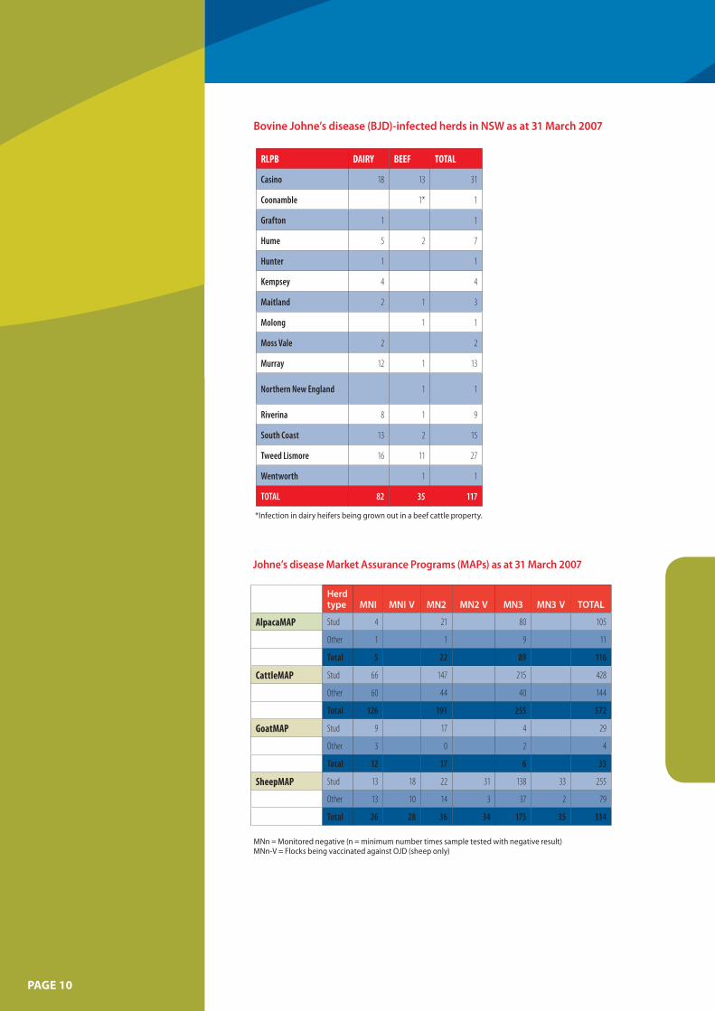
Bovine Johne’s disease (BJD)-infected herds in NSW as at 31 March 2007
PAGE 10
RLPB DAIRY BEEF TOTAL
Casino 18 13 31
Coonamble 1* 1
Grafton 1 1
Hume 5 2 7
Hunter 1 1
Kempsey 4 4
Maitland 2 1 3
Molong 1 1
Moss Vale 2 2
Murray 12 1 13
Northern New England 1 1
Riverina 8 1 9
South Coast 13 2 15
Tweed Lismore 16 11 27
Wentworth 1 1
TOTAL 82 35 117
*Infection in dairy heifers being grown out in a beef cattle property.
Johne’s disease Market Assurance Programs (MAPs) as at 31 March 2007
Herd type MNI
Stud 4
Other 1
Total 5
Stud 66
Other 60
Total 126
Stud 9
Other 3
Total 12
Stud 13
Other 13
Total 26
MNn = Monitored negative (n = minimum number times sample tested with negative result) MNn-V = Flocks being vaccinated against OJD (sheep only)
AlpacaMAP
CattleMAP
GoatMAP
SheepMAP
MNI V MN2 MN2 V MN3 MN3 V TOTAL
21 80 105
1 9 11
22 89 116
147 215 428
44 40 144
191 255 572
17 4 29
0 2 4
17 6 33
18 22 31 138 33 255
10 14 3 37 2 79
28 36 34 175 35 334
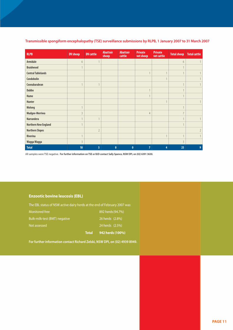
Transmissible spongiform encephalopathy (TSE) surveillance submissions by RLPB, 1 January 2007 to 31 March 2007
RLPB DV sheep DV cattle Abattoir sheep
Abattoir cattle
Private vet sheep
Private vet cattle Total sheep Total cattle
Armidale 6 1 6 1
Braidwood 1 1
Central Tablelands 1 1 1 1
Condobolin 1 1
Coonabarabran 1 1 1 1
Dubbo 1 1
Hume 1 1
Hunter 1 1
Molong 1 1
Mudgee-Merriwa 3 4 7
Narrandera 1 1 1 1
Northern New England 1 1
Northern Slopes 2 2
Riverina 1 1 1 1
Wagga Wagga 3 3
Total 18 5 0 0 7 4 25 9
All samples were TSE negative. For further information on TSE or BJD contact Sally Spence, NSW DPI, on (02) 6391 3630.
Enzootic bovine leucosis (EBL)
The EBL status of NSW active dairy herds at the end of February 2007 was:
Monitored free 892 herds (94.7%)
Bulk-milk-test (BMT) negative 26 herds (2.8%)
Not assessed 24 herds (2.5%)
Total 942 herds (100%)
For further information contact Richard Zelski, NSW DPI, on (02) 4939 8949.
PAGE 11
Getting Information on Animal Diseases This surveillance report can convey
only a very limited amount of
information about the occurrence
and distribution of livestock diseases
in New South Wales. If you would
Regional Health Leader, or Regional
Veterinary Laboratory.
For Statewide information, contact
NSW DPI’s Animal and Plant
Biosecurity Branch in Orange on
(02) 6391 3237 or
fax (02) 6361 9976.
New Staff Samantha Allan commenced duty as the
Regional Animal Health Leader (Central
Slopes) in January 2007. Samantha is
based at Bathurst Agricultural Research
and Advisory Station. Samantha joined
NSW DPI in May 2005 as Veterinary Offi cer
(Policy). Before joining DPI she worked in
mixed practice in NSW and the UK.
like more specific information about For more information on national diseases occurring in your part of disease status, check the National the State, contact your local Rural Animal Health Information System Lands Protection Board District (NAHIS) via the internet at: Veterinarian, Departmental Senior http://www.animalhealthaustralia. Regional Animal Health Manager, com.au/status/nahis.cfm
Prepared by:
Sarah Robson Regional Animal Health Leader, Wagga Wagga Agricultural Institute, Wagga Wagga NSW 2650
Phone (02) 6938 1967 or fax (02) 6938 1995 e-mail: [email protected]
Copies of NSW Animal Health Surveillance reports are available on the internet at: http://www.dpi.nsw.gov.au/reader/ah-surveillance
Disclaimer
The information contained in this publication is based on knowledge and understanding at the time of writing (September 2006 to May 2007). However, because of advances in knowledge, users are reminded of the need to ensure that information upon which they rely is up-to date and to check the currency of the information with the appropriate offi cer of New South Wales Department of Primary Industries or the user s independent adviser.
06/2007 7839


