
JOURNAL OF BACTERIOLOGY, Feb., 1967, p. 544-549 Vol. 93, No. 2Copyright © 1967 American Society for Microbiology Printed in U.S.A.
Antigens of Brucella abortusI. Chemical and Immunoelectrophoretic Characterization
RONALD D. HINSDILL' AND DAVID T. BERMANDepartment of Veterinary Science, University of Wisconsin, Madison, Wisconsin
Received for publication 5 October 1966
ABSTRACTExtracts of Brucella abortus 2308S, prepared either by aqueous extraction of son-
ically ruptured cells or by phenol-water extraction of whole cells, were subjectedto various fractionation procedures and then analyzed to determine their immu-noelectrophoretic patterns and chemical properties. Fraction A, prepared fromsonic extracts, contained at least nine precipitable components when analyzed byimmunoelectrophoresis. Of these, five components gave reactions of nonidentitywith each other and, hence, represented separate antigens having unrelated deter-minant groups. Antigenic component IX, found in both the phenol and sonic ex-tracts, did not form a precipitin line in the presence of serum that had been adsorbedwith whole cells and was therefore tentatively identified as a "surface" antigen.From several lines of evidence, component IX was thought to be a lipopolysac-charide similar to the AP substance described by Miles and Pirie and shown bythem to carry the "abortus" and "melitensis" determinant groups.
There is a striking lack of precise knowledgeconcerning the identity and biological significanceof the antigens of the brucellae. The present in-vestigation was undertaken in an effort to provideinformation about the number and types of anti-gens present in Brucella abortus and, if possible,to bring about their separation by chemical orphysical means so as to allow eventually for astudy of their individual biological properties.The starting material was obtained either by
water extraction of cells ruptured by sonic oscilla-tion or by the Westphal phenol-water extractionprocedure. Only the fractionation procedures em-ployed and the chemical and immunoelectropho-retic characterization of the various antigens iso-lated will be dealt with in this communication.The biological significance of these antigens, asdetermined by their toxicity for monocytes inculture and ability to evoke skin reactions inguinea pigs, will be reported separately.
MATERIALS AND METHODS
Source of microorganisms. All antigenic fractionswere prepared from cultures of B. abortus 2308S.This strain is a subculture of the one originally ob-tained from the Agricultural Research Service, U.S.Department of Agrlculture. Lyophilized stock cul-tures of the organism in skim milk (18) were preparedand used as needed to avoid problems arising from
I Present address: Department of Bacteriology,University of Wisconsin, Madison.
population changes. Evidence of smoothness wasobtained by the acriflavine test (4) and the crystalviolet test (19).
Methods of culture. Subcultures were grown onbrucella agar (Albimi) slants. After incubation for 12hr at 37 C, the growth from each tube was suspendedin sterile saline and was used as the inoculum foreach flask containing 500 ml of brucella broth (Al-bimi). The flasks were placed on a rotary shaker at37 C for 24 hr, after which phenol was added to theflasks to give a final concentration of 0.5% (w/v).The flasks were returned to the shaker for an addi-tional 12 hr at 37 C to insure killing. The cells weresedimented by centrifugation, suspended in saline,and again collected by centrifugation. The washingprocedure was repeated three more times by use ofdistilled water. A sufficient quantity of distilledwater was added to 50 g of cells (wet weight) to givea final volume of 60 ml prior to making the sonic ex-tracts. Cells to be used for the phenol extractionprocedure were harvested in the same manner, sus-pended in a minimal quantity of distilled water, andlyophilized.
Sera for immunological studies. The immune serumused for the immunoelectrophoretic studies camefrom a single pooled harvest, obtained from sixcows which had been artificially infected with B.abortus 2308S. The agglutination titer of the serumwas 1:10,240 when tested by the tube method, with U.S.Department of Agriculture B. abortus standard tubetest antigen. Phenol was used as the preservative in afinal concentration of 0.5% (w/v). Absorbed serumwas prepared by mixing 0.4 g of dry, lyophilized B.abortus 2308S with 25 ml of immune serum, incu-
544
on March 1, 2019 by guest
http://jb.asm.org/
Dow
nloaded from

CHARACTERIZATION OF B. ABORTUS ANTIGENS
bating in a water bath at 37 C for 30 min, and col-lecting the supernatant fluid after centrifugation. Theprocedure was repeated three more times, the drybacteria being mixed with the supernatant fluid ofthe previous tube in each case. The final absorbedserum had an agglutination titer of less than 1:20when tested by the tube method with the standardtube test antigen.
Sonic extracts. Cell breakage was achieved in 1 hrat 3 C by use of a 10-kc, 250-w sonic oscillator. Im-mediately after this procedure, the suspension wascentrifuged at 18,400 X g for 30 min at 4 C. Thesupernatant fluid was set aside, and the cellulardebris was resuspended in 60 ml of distilled waterand centrifuged in the same manner. The two super-natant fluids were combined and filtered throughWhatman no. 1 filter paper. The resulting filtratewas lyophilized and labeled fraction A.
Phenol-water extracts. These were prepared by theWestphal procedure as modified by Redfearn (Ph.D.Thesis, Univ. Wisconsin, Madison, 1960), and weresubsequently described in a paper by Baker andWilson (1). The material recovered from the phenolphase was designated as fraction 5.
Zone electrophoresis in starch. The starch blockwas made of powdered potato starch (technicalgrade) and tris(hydroxymethyl)aminomethane-aceticacid buffer adjusted to pH 8.2 (ionic strength,0.02). The starch trough was 35 cm long with a radiusof 2.75 cm. Continuous-flow buffer chambers similarto those described by Kunkel and Slater (7) wereused. The technique employed was essentially thatdescribed by Bodman (3).
Immunoelectrophoresis. The immunoelectrophoreticstudies were carried out in a buffered gel consisting of1.5% agar (Difco) and sodium diethylbarbiturate-hydrochloric acid buffer (pH 8.2; ionic strength,0.385). The gel was supported on 4 by 5 inch (10.2by 12.7 cm) photographic plates, and the antigenswere incorporated into melted buffered agar beforebeing deposited in the antigen wells.
Chemical determinations. Nitrogen was deter-mined by micro-Kjeldahl analysis (6), and the per-centage of protein was calculated by multiplying thepercentage of nitrogen by a factor of 6.25. The pro-tein content of eluates obtained from starch segmentsafter zone electrophoresis was determined by theFolin-Ciocalteu tyrosine method as modified byLowry et al. (9). Crystalline bovine serum albuminwas used to prepare a standard curve. Carbohydratewas estimated by the method described by Loewus(8), with glucose as a standard. The qualitative testfor the presence of polysaccharide in immunoelectro-phoretic plates was performed as described by Urieland Grabar (17), except that the initial oxidationwith periodic acid was lengthened to 3 hr.
RESULTS
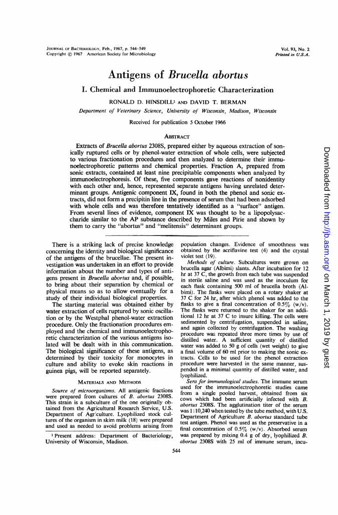
Nine precipitin lines were observed when frac-tion A was subjected to immunoelectrophoresis(Fig. 1). If a shorter period of electrophoresiswas used, lines I and II crossed lines III, IV, andV. Antigenic component IX was immobilized at
the origin and formed a diffuse, heavy precipitinline just inside the antigen well. When fraction Awas subjected to immunoelectrophoresis withabsorbed serum, a precipitin line was not ob-served inside the antigen well, suggesting thatcomponent IX was of a "surface" nature (Fig. 2).All the other lines were formed in the presence ofabsorbed serum, although some of them werebarely visible. In situations where the precipitinlines crossed each other, the determinant groupswere clearly not identical, as was the case forantigenic components III, IV, and V or for com-ponents IV, V, and VII. The antigenic relation-ship among some of the other components, suchas V, VI, and IX, was not clear since the lines didnot cross under any of the experimental condi-tions employed.
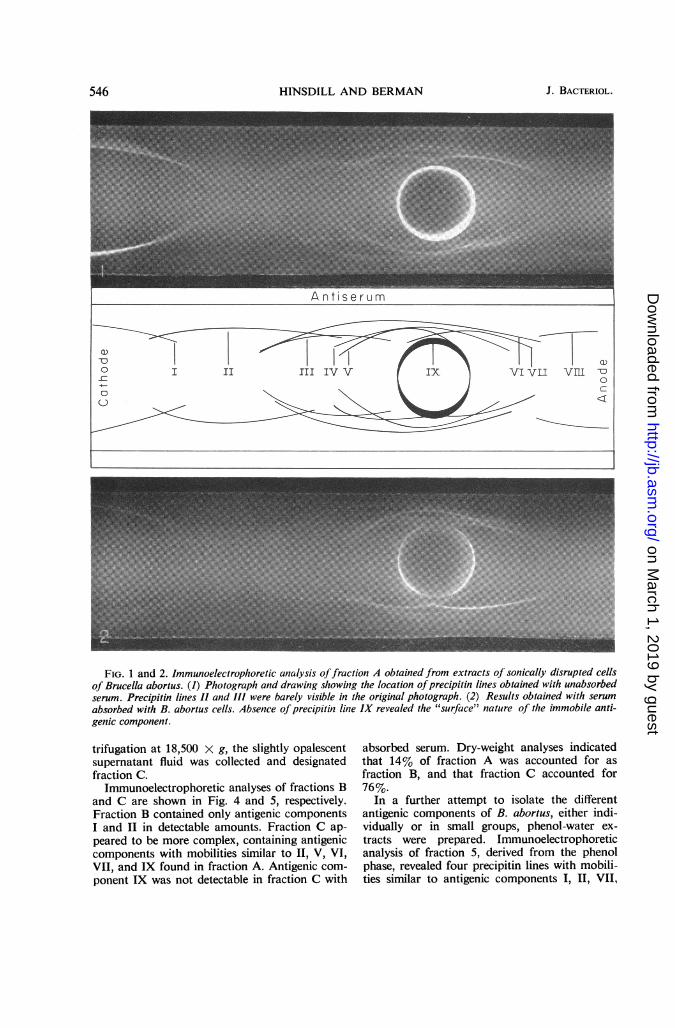
These results suggested that some of the anti-genic components of Brucella could be separatedon the basis of electrophoretic mobility. A 150-mgamount of fraction A was subjected to zone elec-trophoresis in starch. The starch block was thencut into 1-cm segments, and the eluants obtainedfrom these were tested by immunodiffusion andwere also analyzed for protein (Fig. 3). Besidesthe difficulty experienced in trying to correlatethe protein content of the eluants with the ap-pearance of precipitin lines, it was found that thebulk of the protein remained in the vicinity of theorigin. The failure of most of the material to mi-grate, in addition to the poor recoveries obtained,indicated that the antigens were being stronglybound by the starch. This approach was thereforeabandoned, and other methods of separationwere sought.Ammonium sulfate precipitation appeared to
be of limited usefulness, since the precipitatedantigens were extremely difficult to redissolve.Acid precipitation proved to be of some value,however. A solution containing 60 mg/ml offraction A was lowered to pH 3.5 by the slowaddition of glacial acetic acid. The resultingflocculent precipitate was sedimented by cen-trifugation at 8,000 x g. The supernatant fluid,designated fraction B, was filtered through aSeitz filter and was dialyzed until free from acidand yellow color.The acid-insoluble material was suspended in
water, and 1.0 N NaOH was slowly added untilpH 11.0 was reached. At this pH, the precipitatereadily went into solution. Glacial acetic acid wasthen slowly added and the acid precipitation pro-cedure was carried out as before, the processbeing repeated two more times. The supernatantfluids were discarded in each case, and the finalprecipitate was suspended in water. The suspen-sion was adjusted to pH 7.4 with 1.0 N NaOH andwas dialyzed against distilled water. After cen-
545VOL. 93, 1967
on March 1, 2019 by guest
http://jb.asm.org/
Dow
nloaded from
HINSDILL AND BERMAN
FIG. 1 and 2. Immunoelectrophoretic analysis offraction A obtained from extracts of sonically disrupted cellsof Brucella abortus. (1) Photograph and drawing showing the location ofprecipitin lines obtained with unabsorbedserum. Precipitin lines II and III were barely visible in the original photograph. (2) Results obtained with serumabsorbed with B. abortus cells. Absence ofprecipitin line IX revealed the "surface" nature of the immobile anti-genic component.
trifugation at 18,500 x g, the slightly opalescentsupernatant fluid was collected and designatedfraction C.
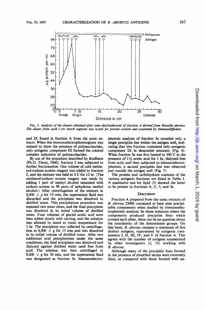
Immunoelectrophoretic analyses of fractions Band C are shown in Fig. 4 and 5, respectively.Fraction B contained only antigenic componentsI and II in detectable amounts. Fraction C ap-peared to be more complex, containing antigeniccomponents with mobilities similar to II, V, VI,VII, and IX found in fraction A. Antigenic com-
ponent IX was not detectable in fraction C with
absorbed serum. Dry-weight analyses indicatedthat 14% of fraction A was accounted for asfraction B, and that fraction C accounted for76%.
In a further attempt to isolate the differentantigenic components of B. abortus, either indi-vidually or in small groups, phenol-water ex-tracts were prepared. Immunoelectrophoreticanalysis of fraction 5, derived from the phenolphase, revealed four precipitin lines with mobili-ties similar to antigenic components 1, I, VII,
546 J. BACTERIOL.
on March 1, 2019 by guest
http://jb.asm.org/
Dow
nloaded from
CHARACTERIZATION OF B. ABORTUS ANTIGENS
_ j= _AntiserumQQ_
I10 15 20Origin
Distance in cm
Antigen
a a a . A
25 30Cathode
FIG. 3. Analysis of the eluants obtained after zone electrophoresis offraction A derivedfrom Brucella abortus.The eluant from each 1-cm starch segment was tested for protein content and examined by immunodiffusion.
and IX found in fraction A from the sonic ex-tracts. When this immunoelectrophoretogram wasstained to show the presence of polysaccharides,only antigenic component IX formed the coloredcomplex indicative of polysaccharides.By use of the procedure described by Redfearn
(Ph.D. Thesis, 1960), fraction 5 was subjected tofurther fractionation. One volume of cold metha-nol-sodium acetate reagent was added to fraction5, and the mixture was held at 4 C for 12 hr. (Themethanol-sodium acetate reagent was made byadding 1 part of methyl alcohol saturated withsodium acetate to 99 parts of anhydrous methylalcohol.) After centrifugation of the mixture at8,000 x g for 15 min, the supernatant fluid wasdiscarded and the precipitate was dissolved indistilled water. This precipitation procedure wasrepeated two more times, and the final precipitatewas dissolved in its initial volume of distilledwater. Four volumes of glacial acetic acid werethen added slowly with stirring, and the solutionwas allowed to stand at room temperature for1 hr. The precipitate was collected by centrifuga-tion at 8,000 X g for 15 min and was dissolvedin its initial volume of distilled water. After twoadditional acid precipitations under the sameconditions, the final precipitate was dissolved anddialyzed against distilled water until free fromacid. The solution was then centrifuged at8,000 X g for 30 min, and the supernatant fluidwas designated as fraction 5a. Immunoelectro-
phoretic analysis of fraction 5a revealed only asingle precipitin line within the antigen well, indi-cating that this fraction contained only antigeniccomponent IX in detectable amounts (Fig. 6).When fraction 5a was first heated to 100 C in thepresence of 1% acetic acid for 1 hr, dialyzed freefrom acid, and then subjected to immunoelectro-phoresis, a second precipitin line was observedjust outside the antigen well (Fig. 7).The protein and carbohydrate contents of the
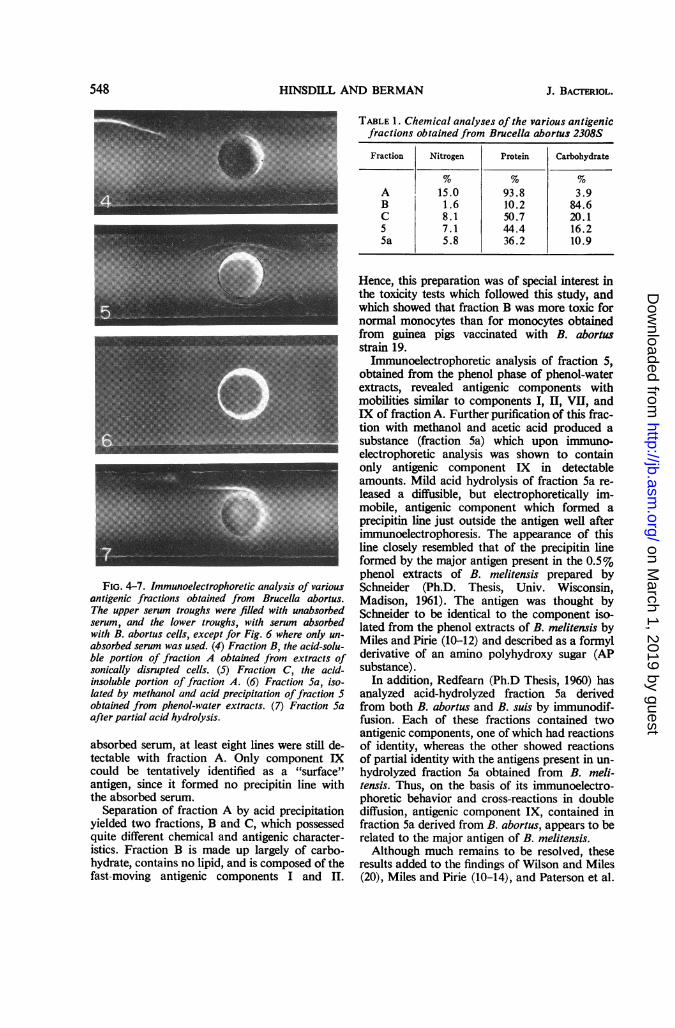
various antigenic fractions are listed in Table 1.A qualitative test for lipid (5) showed the latterto be present in fractions A, C, 5, and 5a.
DISCUSSION
Fraction A prepared from the sonic extracts ofB. abortus 2308S contained at least nine precipi-table components when studied by immunoelec-trophoretic analysis. In those instances where thecomponents produced precipitin lines whichcrossed each other, there can be no question aboutthe nonidentity of the determinant groups. Onthis basis, B. abortus contains a minimum of fivedistinct antigens, represented by antigenic com-
ponents 1, II, III, IV, and V of fraction A. Thisagrees with the number of antigens enumeratedby other investigators (2, 15) working withB. abortus.Although many of the precipitin lines formed
in the presence of absorbed serum were extremelyfaint, as compared with those formed with un-
84
76
a)
ca-
0)0.I..QaCD
68
60
52
44
36
28
20t
O0T-5 'Anode
547VOL. 93, 1967
on March 1, 2019 by guest
http://jb.asm.org/
Dow
nloaded from
HINSDILL AND BERMAN
TABLE 1. Chemical analyses of the various antigenicfractions obtained from Brucella abortus 2308S
Fraction Nitrogen Protein Carbohydrate
A 15.0 93.8 3.9B 1.6 10.2 84.6C 8.1 50.7 20.15 7.1 44.4 16.25a 5.8 36.2 10.9
FIG. 4-7. Immunoelectrophoretic analysis of variousantigenic fractions obtained from Brucella abortus.The upper serum troughs were filled with unabsorbedserum, and the lower troughs, with serum absorbedwith B. abortus cells, except for Fig. 6 where only un-absorbed serum was used. (4) Fraction B, the acid-solu-ble portion of fraction A obtained from extracts ofsonically disrupted cells. (5) Fraction C, the acid-insoluble portion of fraction A. (6) Fraction Sa, iso-lated by methanol and acid precipitation offraction 5obtained from phenol-water extracts. (7) Fraction Saafter partial acid hydrolysis.
absorbed serum, at least eight lines were still de-tectable with fraction A. Only component IXcould be tentatively identified as a "surface"antigen, since it formed no precipitin line withthe absorbed serum.
Separation of fraction A by acid precipitationyielded two fractions, B and C, which possessedquite different chemical and antigenic character-istics. Fraction B is made up largely of carbo-hydrate, contains no lipid, and is composed of thefast-moving antigenic components I and II.
Hence, this preparation was of special interest inthe toxicity tests which followed this study, andwhich showed that fraction B was more toxic fornormal monocytes than for monocytes obtainedfrom guinea pigs vaccinated with B. abortusstrain 19.
Immunoelectrophoretic analysis of fraction 5,obtained from the phenol phase of phenol-waterextracts, revealed antigenic components withmobilities similar to components I, II, VII, andIX of fraction A. Further purification of this frac-tion with methanol and acetic acid produced asubstance (fraction 5a) which upon immuno-electrophoretic analysis was shown to containonly antigenic component IX in detectableamounts. Mild acid hydrolysis of fraction 5a re-leased a diffusible, but electrophoretically im-mobile, antigenic component which formed aprecipitin line just outside the antigen well afterimmunoelectrophoresis. The appearance of thisline closely resembled that of the precipitin lineformed by the major antigen present in the 0.5%phenol extracts of B. melitensis prepared bySchneider (Ph.D. Thesis, Univ. Wisconsin,Madison, 1961). The antigen was thought bySchneider to be identical to the component iso-lated from the phenol extracts of B. melitensis byMiles and Pirie (10-12) and described as a formylderivative of an amino polyhydroxy sugar (APsubstance).
In addition, Redfearn (Ph.D Thesis, 1960) hasanalyzed acid-hydrolyzed fraction 5a derivedfrom both B. abortus and B. suis by immunodif-fusion. Each of these fractions contained twoantigenic components, one of which had reactionsof identity, whereas the other showed reactionsof partial identity with the antigens present in un-hydrolyzed fraction 5a obtained from B. meli-tensis. Thus, on the basis of its immunoelectro-phoretic behavior and cross-reactions in doublediffusion, antigenic component IX, contained infraction 5a derived from B. abortus, appears to berelated to the major antigen of B. melitensis.Although much remains to be resolved, these
results added to the findings of Wilson and Miles(20), Miles and Pirie (10-14), and Paterson et al.
548 J. BACTERIOL.
on March 1, 2019 by guest
http://jb.asm.org/
Dow
nloaded from

CHARACTERIZATION OF B. ABORTUS ANTIGENS
(16) support the concept that all three species ofBrucella contain an AP-like substance whichcarries the A and M antigenic determinants. Fur-thermore, this AP-like substance seems to beclosely associated with a cell wall lipopoly-saccharide (antigenic component IX). There arealso a number of separate antigens (antigeniccomponents I, II, III, IV, and V) which probablyare not principally associated with the cell surfaceand which do not appear to be species-specific.The present study, though not achieving com-plete separation of these antigenic components,did provide a number of fractions that could beused in a subsequent study of their biologicalactivities.
ACKNOWLEDGMENTS
This investigation was supported by Public HealthService training grant 5-TI-A1-17505, and by theResearch Committee of the Graduate School withfunds furnished by the Wisconsin Alumni ResearchFoundation.
LITERATURE CITED
1. BAKER, P. J., AND J. B. WILsoN. 1965. Hypofer-remia in mice and its application to bioassayof endotoxin. J. Bacteriol. 90:903-910.
2. BARBER, C., 0. DIMITRIU, T. VAsILEsco, AND A.CERBU. 1961. Contribution a l'etude de lastructure antigenique des Brucella. I. Separa-tion de l'antigene M et de quelques complexesqui le contiennent. Arch. Roumaines Pathol.Exptl. Microbiol. 20:201-212.
3. BODMAN, J. 1960. Agar gel, starch block, starchgel, and sponge rubber electrophoresis, p. 91-157. in I. Smith [ed.], Chromatographic andelectrophoretic techniques, vol. 2. IntersciencePublishers, Inc., New York.
4. BRAUN, W., AND A. BONESTELL. 1947. Independ-ent variation of characteristics in Brucellaabortus variants and their detection. Am. J.Vet. Res. 8:386-390.
5. DURRUM, E. L., M. H. PAUL, AND E. R. B.SMITH. 1952. Lipid detection in paper electro-phoresis. Science 116:428-430.
6. KABAT, E. A., AND M. M. MAYER. 1961. Experi-mental immunochemistry, 2nd ed. Charles CThomas, Publisher, Springfield, Ill.
7. KUNKEL, H. G., AND R. J. SLATER. 1952. Zoneelectrophoresis in a starch supporting medium.Proc. Soc. Exptl. Biol. Med. 80:42-44.
8. LoEwUs, F. A. 1952. Improvement in anthronemethod for determination of carbohydrates.Anal. Chem. 24:219.
9. LOWRY, 0. H., N. J. ROSEBROUGH, A. L. FARR,AND R. J. RANDALL. 1951. Protein measure-ment with the Folin phenol reagent. J. Biol.Chem. 193:265-275.
10. MILES, A. A., AND N. W. PIRIE. 1939. The proper-ties of antigenic preparations from Brucellamelitensis. I. Chemical and physical propertiesof bacterial fractions. Brit. J. Exptl. Pathol.20:83-98.
11. MILES, A. A., AND N. W. PIRIE. 1939. The proper-ties of antigenic preparations from Brucellamelitensis. II. Serological properties of theantigens. Brit. J. Exptl. Pathol. 20:109-121.
12. MILES, A. A., AND N. W. PIRIE. 1939. The proper-ties of antigenic preparations from Brucellamelitensis. IIJ. The biological properties of theantigen and the products of gentle hydrolysis.Brit. J. Exptl. Pathol. 20:278-296.
13. MILES, A. A., AND N. W. PIRIE. 1939. The proper-ties of antigenic preparations from Brucellamelitensis. IV. The hydrolysis of the formaminolinkage. Biochem. J. 33:1709-1715.
14. MILES, A. A., AND N. W. PIRIE. 1939. The proper-ties of antigenic preparations from Brucellamelitensis. V. Hydrolysis and acetylation of theamino-polyhydroxy compound derived fromthe antigen. Biochem. J. 33:1716-1724.
15. OLITZKI, A. L. 1959. Studies on the antigenicstructure of virulent and non-virulent brucellaewith the aid of agar-gel precipitation technique.Brit. J. Exptl. Pathol. 40:432-440.
16. PATERSON, J. S., N. W. PIRIE, AND A. W. STABLE-FORTH. 1947. Protective antigens isolated fromB. abortus. Brit. J. Exptl. Pathol. 28:223-236.
17. URIEL, J., AND P. GRABAR. 1961. A new tech-nique for direct detection of glycoproteins andpolysaccharides after electrophoresis or immu-noelectrophoresis in agar gel. Anal. Biochem.2:80-82.
18. WEISS, F. A. 1957. Maintenance and preservationof cultures, p. 99-119. In Society of AmericanBacteriologists, Manual of microbiologicalmethods. McGraw-Hill Book Co., Inc., NewYork.
19. WHITE, P. G., AND J. B. WILSON. 1951. Differen-tiation of smooth and nonsmooth colonies ofbrucellae. J. Bacteriol. 61:239-240.
20. WILSON, G. S., AND A. A. MILES. 1932. The sero-logical differentiation of smooth strains of theBrucella group. Brit. J. Exptl. Pathol. 13:1-13.
VOL. 93, 1967 549
on March 1, 2019 by guest
http://jb.asm.org/
Dow
nloaded from


