

APH-I02
The Urinary SystemBy
Khaled Na3im


Functions of the Urinary System
1.Filtration of the blood– Occurs in the glomerulus of the kidney
nephron
– Contributes to homeostasis by removing toxins or waste

Functions of the Urinary System
2.Reabsorption of vital nutrients, ions
and water– Occurs in most parts of the kidney
nephron
– Contributes to homeostasis by conserving important materials
Functions of the Urinary System
3.Secretion of excess materials– Assists filtration in removing material
from the blood
– Contributes to homeostasis by preventing a build-up of certain materials in the body such as drugs, wastes

Functions of the Urinary System
4.Activation of Vitamin D– Vitamin D made in the skin is converted
to Vitamin D3 by the kidney
– Active Vitamin D (D3) assists homeostasis by increasing calcium absorption from the digestive tract

Functions of the Urinary System
5.Release of Erythropoietin hormone
by the kidney– Erythropoietin stimulates new RBC
production
– New RBC’s assist homeostasis by insuring adequate Oxygen and Carbon Dioxide transport
Functions of the Urinary System
• Release of Renin by the kidney– Renin stimulates the formation of a
powerful vasoconstrictor called Angiotensin II
– Angiotensin II assists homeostasis by causing vasoconstriction which increases blood pressure

Functions of the Urinary System
6.Release of Prostaglandins– Prostaglandins dilate kidney blood
vessels
– Dilated blood vessels contribute to homeostasis by maintaining blood flow in the kidneys
Functions of the Urinary System
• Secretion of H (+1) and reabsorption of HCO3 (-1)– Eliminates excess hydrogen ions and
conserves buffer material such as bicarbonate
– Contributes to homeostasis by controlling acid/base conditions in body fluids

Urinary System
Renal arteryKidney
Ureter
Urinary Bladder
Renal Vein
For sphincters, see next slide

Urinary System
Internal urethral sphincterExternal Urethral Sphincter
Male Sphincters Female Sphincters
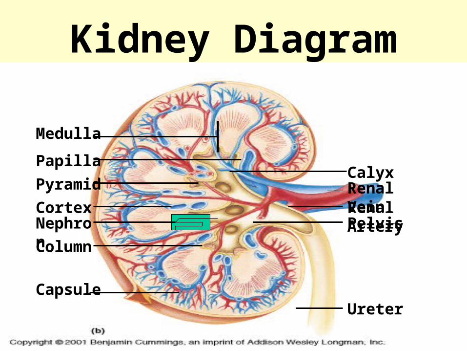
Kidney Diagram
Capsule
Renal VeinRenal ArteryCortex
Pyramid
PapillaCalyx
Pelvis
Ureter
Column
Medulla
Nephron

The renal artery• The Renal Artery
– Transports oxygenated blood from the aorta to the kidney for filtration

The renal vein• Renal Vein
– Transports filtered and deoxygenated blood from the kidney to the inferior vena cava and then the heart
Functions of Kidney Structures
• Renal Column– A passageway located between the renal
pyramids found in the medulla and used as a space for blood vessels

The nephron
• Nephron– The nephron is physiological unit
( functional unite )of the kidney used for filtration of blood and reabsorption and secretion of materials

The Kidney Structures
• The kidney is formed of cortex & medulla
• Cortex :– The outer layer of the kidney that contains
most of the nephron; main site for filtration, reabsorption and secretion

The Kidney Structures
• Medulla– inner core of the kidney that contains the
pyramids, columns, papillae, calyces, pelvis and parts of the nephron not located in the cortex; used for salt, water and urea absorption

The Kidney Structures
• Renal Pyramids– Triangular shaped units in the medulla that
house the loops of Henle and collecting ducts of the nephron; site for the counter-current system that concentrates salt and conserves water and urea

The Kidney Structures
• Renal Papilla– The tip of the renal pyramid that releases
urine into a calyx
Functions of Kidney Structures
• Calyx
• A collecting sac surrounding the renal papilla that transports urine from the papilla to the renal pelvis
• Renal Pelvis– Collects urine from all of the calyces in
the kidney
Functions of Kidney Structures
• Ureter– Transports urine from the renal pelvis to
the urinary bladder ( UB )

The Nephron
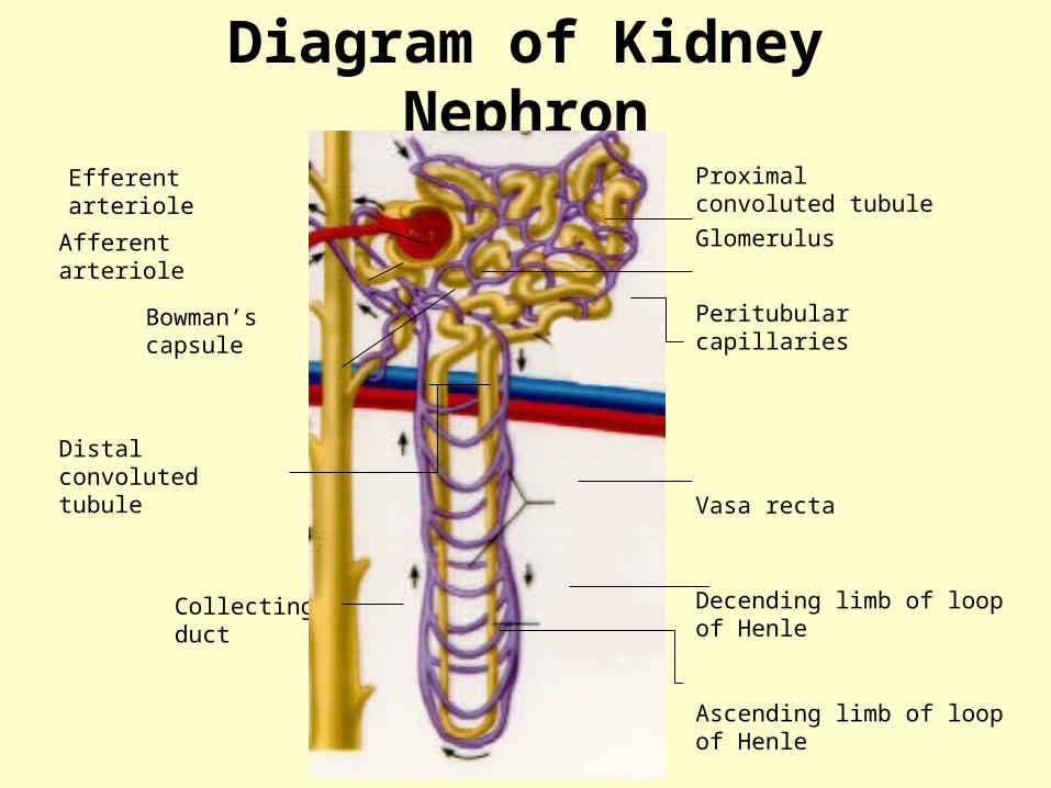
Diagram of Kidney Nephron
Efferent arteriole
Afferent arteriole
Bowman’s capsule
Collecting duct
Proximal convoluted tubule
Glomerulus
Peritubular capillaries
Vasa recta
Decending limb of loop of Henle
Ascending limb of loop of Henle
Distal convoluted tubule

Functions of Nephron Structures• AfferentArteriole• Transports arterial blood to the
glomerulus for filtration system• Efferent Arteriole
– Transports filtered blood from the glomerulus , through the peritubular capillaries and the vasa recta, and to the kidney venous
–

Functions of Nephron Structures
• Glomerulus– The site for blood filtration– operates as a nonspecific filter; in that, it
will remove both useful and non-useful material
– the product of the glomerulus is called filtrate

Functions of Nephron Structures
• Bowman’s Capsule– A sac that encloses Bowman’s Capsule and
transfers filtrate from the glomerulus to the Proximal Convoluted Tubule (PCT)

Functions of Nephron Structures
• Proximal Convoluted Tubule (PCT)– A thick, constantly actively segment of the
nephron that reabsorbs most of the useful substances of the filtrate: sodium (65%), water (65%), bicarbonate (90%), chloride (50%), glucose (nearly 100%!), etc.
– The primary site for secretion (elimination) of drugs, waste and hydrogen ions

Functions of Nephron Structures
• Decending Limb of the Loop of Henle– A part of the counter current multiplier
– freely permeable to water and relatively impermeable to solutes (salt particles)
– receives filtrate from the PCT, allows water to be absorbed and sends “salty”filtrate on the the next segment. “Saves water and passes the salt”

Functions of Nephron Structures
• Ascending Limb of the Loop of Henle– a part of the counter current multiplier
– impermeable to water and actively transports (reabsorbs) salt (NaCl) to the interstitial fluid of the pyramids in the medulla. “Saves salt and passes the water.”
– the passing filtrate becomes dilute and the interstitium becomes hyperosmotic
Functions of Nephron Structures
• Distal Convoluted Tubule (DCT)– receives dilute fluid from the ascending limb
of the Loop of Henle
– Variably active portion of the nephron
– When aldosterone hormone is present, sodium is reabsorbed and potassium is secreted. Water and chloride follow the sodium.
Functions of Nephron Structures
• Collecting Duct– receives fluid from the DCT– variably active portion of the Nephron– when antidiuretic hormone (ADH) is present, this
duct will become porous to water. Water from the collecting duct fluid then moves by osmosis into the “salty” (hyperosmotic) interstitium of the medulla.
– The last segment to save water for the body
Functions of Nephron Structures
• Peritubular Capillaries– transport reabsorbed materials from the
PCT and DCT into kidney veins and eventually back into the general circulation
– help complete the conservation process (reabsorption) that takes place in the kidney
Unit 1 - Objective 4

Site of Filtration
• Glomerulus– the Glomerulus is the site of filtration
– the filtration mechanism is sieve-like and consists of fenestrated glomerular capillaries, podocytes and a basement membrane that allows free passage of water and solutes smaller than plasma proteins

Location of the Glomerulus
Afferent Arteriole
Efferent Arteriole
Bowman’s Capsule
Proximal Convoluted Tubule
Glomerulus

Glomerular Filtration Mechanism
Bowman’s Capsule
Glomerulus
Fenestrated Capillary
Podocyte with Basement Membrane
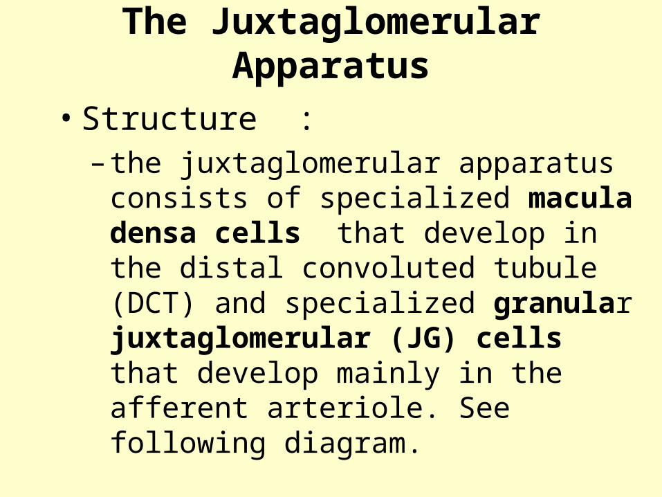
The Juxtaglomerular Apparatus
• Structure :– the juxtaglomerular apparatus consists of
specialized macula densa cells that develop in the distal convoluted tubule (DCT) and specialized granular juxtaglomerular (JG) cells that develop mainly in the afferent arteriole. See following diagram.
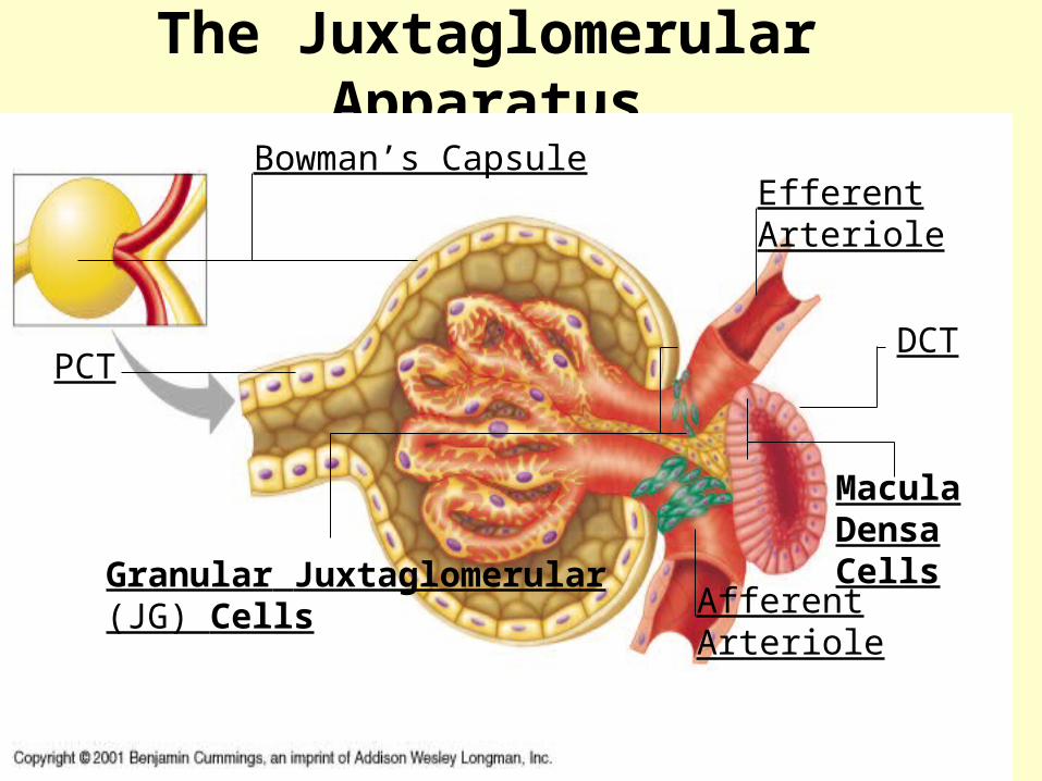
The Juxtaglomerular Apparatus
Afferent Arteriole
Efferent Arteriole
DCT
Macula Densa Cells
Granular Juxtaglomerular (JG) Cells
PCT
Bowman’s Capsule

The Juxtaglomerular Apparatus
• Used in maintaining blood pressure– if the blood pressure drops, the granular JG
cells release renin
– renin converts the blood protein angiotensinogen into angiotensin I which converts to angiotensin II
– angiotensin II acts as a vasoconstrictor to raise blood pressure. Continued on next slide.

The Juxtaglomerular Apparatus
• Used in maintaining blood pressure continued:– Angiotensin II also stimulates the release of
aldosterone hormone from the adrenal cortex
– aldosterone stimulates the DCT to reabsorb salt (NaCl). Continued on next slide.

The Juxtaglomerular Apparatus
• Used in maintaining blood pressure continued:– salt reabsorption attracts water to the blood
by osmosis and raises blood volume, as well as, contributing to the increase in blood pressure. Continued on next slide.
The Juxtaglomerular Apparatus
• Used in maintaining blood pressure continued:– the macula densa cells monitor the salt
content of the blood
– if the blood salt content gets too high, the macula densa cells begin to inhibit the granular cells and suppress renin release

The Juxtaglomerular Apparatus
• Used in maintaining blood pressure continued:– suppression of renin acts as a negative
feedback mechanism to prevent further increases in angiotensin II, Aldosterone and blood pressure
The Juxtaglomerular Apparatus
• Use in maintaining blood pressure continued:– eventually the blood pressure will come back
down
– the “push/pull” action of the granular cells and macula densa cells provide an effective mechanism for regulating blood pressure in the kidney

The Counter Current Mechanism
The counter current mechanism through which the kidneys excrete a concentrated urine by indicating the role of the following: sodium chloride, posterior pituitary, ADH, hypothalamus, collecting duct, active transport, osmosis, interstitial fluid, vasa recta, diffusion, loop of Henle and urea
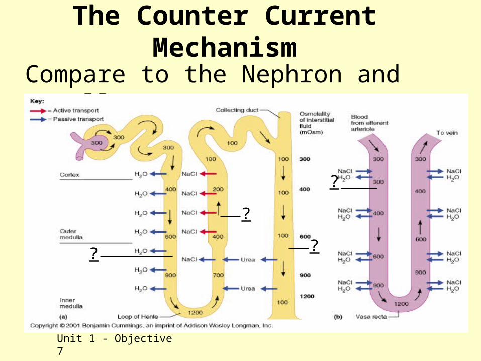
The Counter Current Mechanism
Compare to the Nephron and recall parts
Unit 1 - Objective 7
?
?
?
?
The Counter Current Mechanism
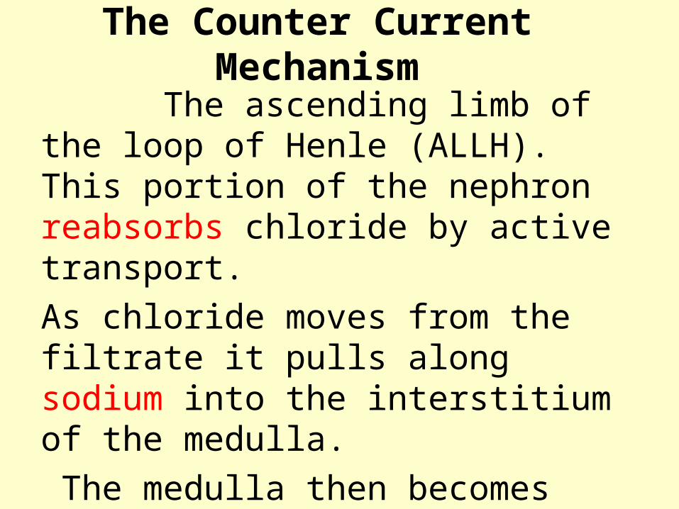
The ascending limb of the loop of Henle (ALLH). This portion of the nephron reabsorbs chloride by active transport.
As chloride moves from the filtrate it pulls along sodium into the interstitium of the medulla.
The medulla then becomes very hyperosmotic
The Counter Current Mechanism
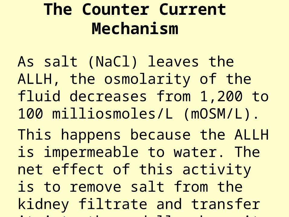
As salt (NaCl) leaves the ALLH, the osmolarity of the fluid decreases from 1,200 to 100 milliosmoles/L (mOSM/L).
This happens because the ALLH is impermeable to water. The net effect of this activity is to remove salt from the kidney filtrate and transfer it into the medulla where it can be saved for use by the body.

The Counter Current Mechanism
The accumulated salt in the interstitium of the medulla acts as an osmotic force which can be used to “draw” and conserve water from other parts of the nephron: the decending limb of the Loop of Henle (DLLH) and the collecting duct. The DLLH is a thin passive segment that is permeable to water, but, impermeable to salt.

The Counter Current Mechanism
As the DLLH gives up water to the medullary interstitium, the osmolarity of the fluid changes from 300 to 1,200 mOSM/L. The net effect of this process is to conserve water for the body. Thus, the loop of Henle actively transfers salt back into the kidney which can be used to save water osmotically. A remarkable process!

The Counter Current Mechanism
The hyperosmotic interstitium of the medulla will also “pull” and conserve water from the collecting duct, but, on a variable basis depending on the availibility of ADH. As water moves from the collecting duct, urea will follow. Thus, as water is conserved at this level, a certain amount of urea is also conserved. The urea contributes to the high osmolarity of the medulla
The Counter Current Mechanism
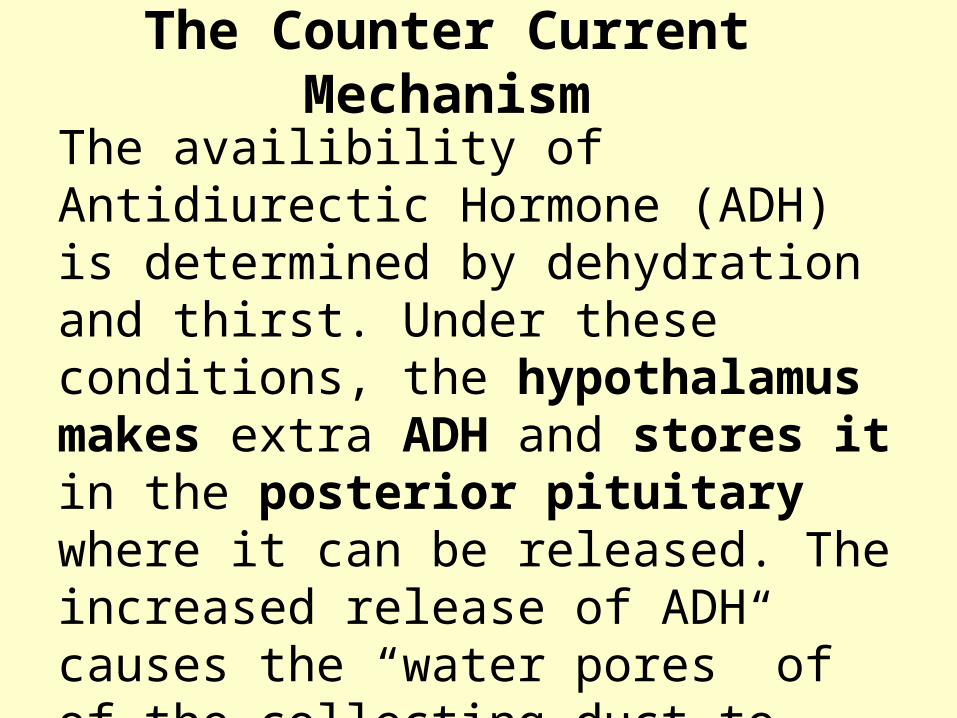
The availibility of Antidiurectic Hormone (ADH) is determined by dehydration and thirst. Under these conditions, the hypothalamus makes extra ADH and stores it in the posterior pituitary where it can be released. The increased release of ADH causes the “water pores” of of the collecting duct to open and allow water to move from the urine to the medulla.

The Counter Current Mechanism
As water leaves the collecting duct, the urine becomes progressively more concentrate. The osmolarity of the collecting duct fluid will increase from about 150-300 to 1,200 mOsm/l. under these conditions. If ADH is not present, water is not conserved and is lost as part of a dilute urine (100 mOsm/l).

The Counter Current Mechanism
The vasa recta is made up of a group of capillary like vessels and is freely permeable to salt and water. The vessels of the vasa recta roughly flow counter to the loop of Henle and acts as a counter current exchanger. As blood flows through the vasa recta it picks up water and leaves behind salt. Thus, the vasa recta returns conserved water back to the body and leaves the salt which maintains the hyperosmotic medulla.

Plasma Clearance
Plasma clearance is defined as the amount of plasma that is cleared or “cleansed” of a particular substance in one minute. The kidneys will carry out this clearance process through the use of filtration, reabsorption and secretion.

Plasma ClearanceFiltration will directly affect clearance. As filtration increases, more material will be removed from the blood plasma. Reabsorption will indirectly affect clearance. As reabsorption increases, less material will be removed from the blood plasma. Secretion will directly affect clearance. As secretion increases, more material will be removed from blood plasma.
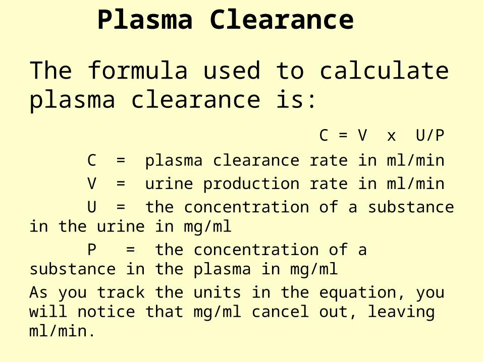
Plasma Clearance
The formula used to calculate plasma clearance is: C = V x U/P
C = plasma clearance rate in ml/min
V = urine production rate in ml/min
U = the concentration of a substance in the urine in mg/ml
P = the concentration of a substance in the plasma in mg/ml
As you track the units in the equation, you will notice that mg/ml cancel out, leaving ml/min.
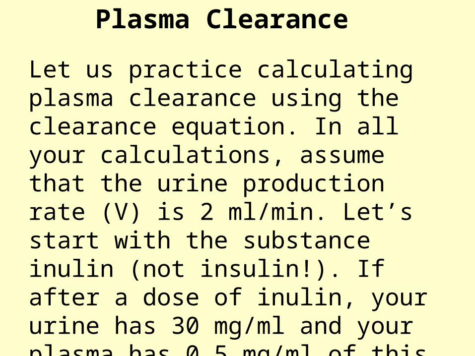
Plasma Clearance
Let us practice calculating plasma clearance using the clearance equation. In all your calculations, assume that the urine production rate (V) is 2 ml/min. Let’s start with the substance inulin (not insulin!). If after a dose of inulin, your urine has 30 mg/ml and your plasma has 0.5 mg/ml of this substance, what is the inulin clearance rate? If you got 120ml/min, you are correct!

Plasma Clearance
If you did not get 120ml/min, look at the following calculation and recheck your work.
120 ml/min = 2 ml/min x 30 mg/ml/ 0.5 mg/ml

Plasma ClearanceTest your ability to conduct further calculations by calculating the clearance rate for the following substances:Substance Urine concentration Plasma concentration
Urea 7.0 mg/ml 0.2 mg/ml
Glucose 0.0 mg/ml 1.0 mg/ml
Penicillin 298 mg/ml 0.7 mg/ml
Remember that the urine production rate (2ml/min) will be the same for all of the above calculations. The clearance rate for each of the above substances will be: Urea = 70 ml/min; Glucose = 0 ml/min; Penicillin = 851 ml/min. Were you able to get the right answers? If not, go back and restudy the clearance process.

Disorders of the Urinary System
• Renal Calculi (kidney stones)– caused by the crystallization of calcium,
magnesium or uric acid salts that precipitate in the renal pelvis.
– If the calculi become large and travel down the ureter, they can cause excruciating pain which radiate from the flank to the anterior abdominal wall on the same side.

Disorders of the Urinary System
• Cystitis– typically caused by bacteria from the anal
region, but, can also be caused by sexually transmitted diseases and various chemical agents
– can lead to inflammation, fever, increased urgency and frequency of urination and pain

Disorders of the Urinary System
• Glomerulonephritis ( Bright’s Disease)– caused by inflammation of the glomeruli
due to an abnormal immune response (autoimmune, streptococcal antibody complexes).
– Inflammation of the glomeruli leads to faulty filtration (passage of blood cells and proteins) and possible kidney failure.
Disorders of the Urinary System
• Incontinence– caused by loss of the ability to control
voluntary micturition (releasing urine from the bladder) due to age, emotional disorders pregnancy, damage to the nervous system, stress, excessive laughing and coughing
– leads to wetting of clothing, discomfort and embarassment

Dialysis Therapy
Dialysis is a process that artificially removes metabolic wastes from the blood in order to compensate for kidney (renal) failure. Kidney failure results in the rapid accumulation of nitrogen waste (urea, etc.) which leads to azotemia. Uremia and ion disturbances can also occur. This condition can cause acidosis, labored breathing, convulsions, coma and death.

Dialysis TherapyThe most common form of dialysis is hemodialysis which uses a machine to transfer patient’s blood through a semipermeable tube that is permeable only to selected substances. The dialysis machine contains an appropriate dialysis fluid that produces a diffusion gradient. This gradient allows abnormal substances to diffuse from the patient’s blood and produce a “cleaning” effect.
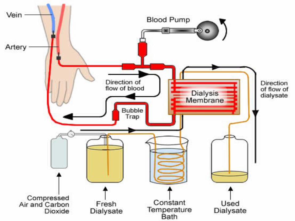
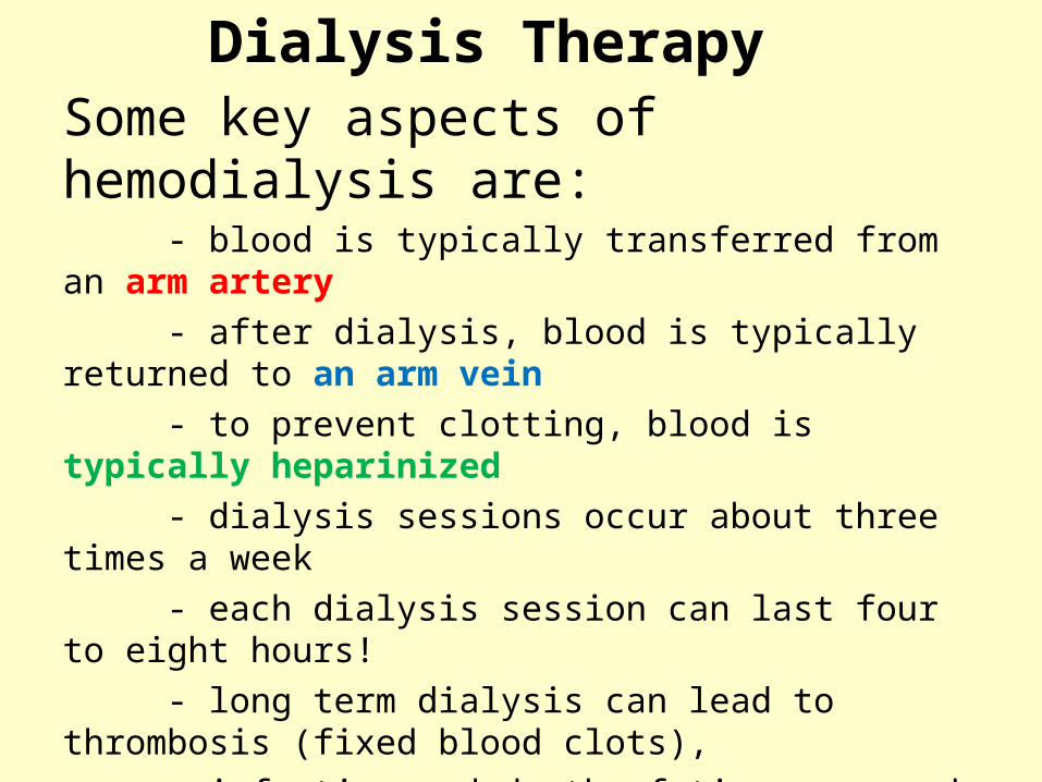
Dialysis TherapySome key aspects of hemodialysis are: - blood is typically transferred from an arm artery
- after dialysis, blood is typically returned to an arm vein
- to prevent clotting, blood is typically heparinized
- dialysis sessions occur about three times a week
- each dialysis session can last four to eight hours!
- long term dialysis can lead to thrombosis (fixed blood clots),
infection and death of tissue around a shunt (the blood access
site in the arm)


