

GE Healthcare VividClub
Application News
No 03-2011

GE Healthcare VividClub
- 2 -
AFI on TEE images
Automated Functional Imaging
Vivid E9 BT 11
Echo PAC BT 11

GE Healthcare VividClub
- 3 -
Table of content
AFI ON TEE IMAGES ...................................................................................................................................... 2
GETTING STARTED ........................................................................................................................................ 4
OPTIMISING IMAGES .................................................................................................................................... 4
MEASURE THE AVC ....................................................................................................................................... 4
From Doppler .............................................................................................................................................................................. 4
STARTING THE ANALYSIS ............................................................................................................................. 4
The measurement .................................................................................................................................................................... 4 Defining the ROI ........................................................................................................................................................................ 5 AV Closure ..................................................................................................................................................................................... 7 The quad screen ........................................................................................................................................................................ 8 Next analysis: .............................................................................................................................................................................. 8
THE SUMMARY .............................................................................................................................................. 9
Global Strain ................................................................................................................................................................................ 9
NOTE
This hand out is additional training material. For more information please refer to the user manual and/or reference manual.

GE Healthcare VividClub
- 4 -
Getting started 1. Create an exam 2. Connect the ECG
a. Press Physio and change ECG Lead to get the best signal b. Obtain a stable ECG trace
Optimising images 1. Sector width
o Not too small, the myocardium must be visible during the entire cardiac cycle. o Not too big, this lowers the frame rate
2. Frame rate
o Optimal between 40-90 fps o Optimise the frame rate with the rotary knob
3. Store loops from the following views
o Mid-oesophageal 4 CH o Mid-oesophageal 2 CH o Mid-oesophageal LAX o It is recommended acquiring all three views sequentially in order to get comparable
heart rates in all views.
Measure the AVC
From Doppler 1. Acquire a nice Doppler signal from the AV; most likely including the valve clicks. 2. Press Measure. 3. Open the folder for Event Timing. 4. Select AVC. 5. Set the marker for the closure.
Now the measurements are stored in the worksheet and will be used for the AFI analysis.
Starting the analysis
The measurement
1. Open the Mid-oesophageal LAX view 2. Press MEASURE 3. In the Measurement menu, select AFI.

GE Healthcare VividClub
- 5 -
4. Select APLAX o It is needed to start with the LAX view, because this allows checking the positioning
of the aortic valve closure (AVC).
Defining the ROI 1. Only three clicks! 2. Define the endocardial border on both basal points of the annulus and in the apex. 3. Follow the instructions on the pointer or in the status bar.

GE Healthcare VividClub
- 6 -
4. Correct ROI definition is crucial to get good tracking 5. After placing the three points the ROI is displayed.
6. The shape can be changed with the cursor (click on the points in the inner border and move them).
7. The processing of the whole loop starts automatically (when the cursor will not be moved any more).
8. The data is processed and the tracking validation screen is displayed.
GE Healthcare VividClub
- 7 -
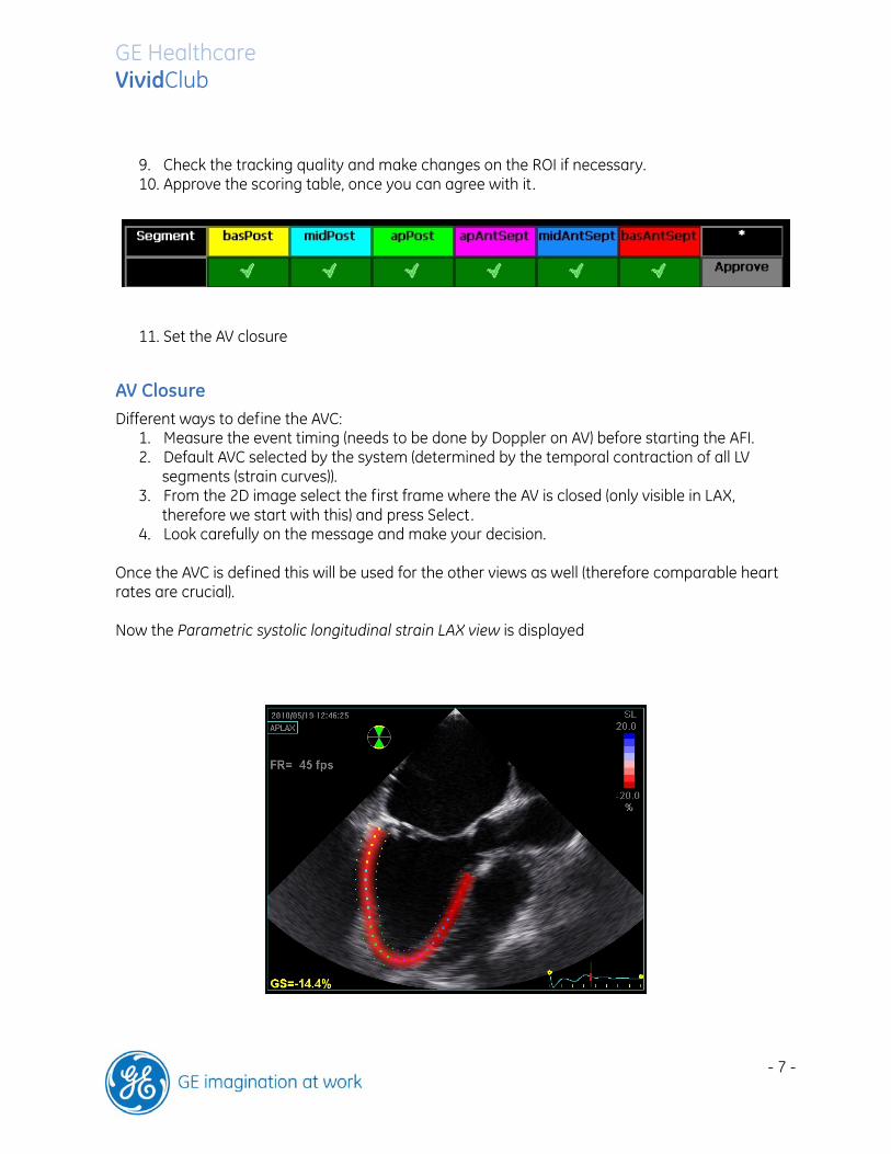
9. Check the tracking quality and make changes on the ROI if necessary. 10. Approve the scoring table, once you can agree with it .
11. Set the AV closure
AV Closure Different ways to define the AVC:
1. Measure the event timing (needs to be done by Doppler on AV) before starting the AFI. 2. Default AVC selected by the system (determined by the temporal contraction of all LV
segments (strain curves)). 3. From the 2D image select the first frame where the AV is closed (only visible in LAX,
therefore we start with this) and press Select. 4. Look carefully on the message and make your decision.
Once the AVC is defined this will be used for the other views as well (therefore comparable heart rates are crucial). Now the Parametric systolic longitudinal strain LAX view is displayed
GE Healthcare VividClub
- 8 -
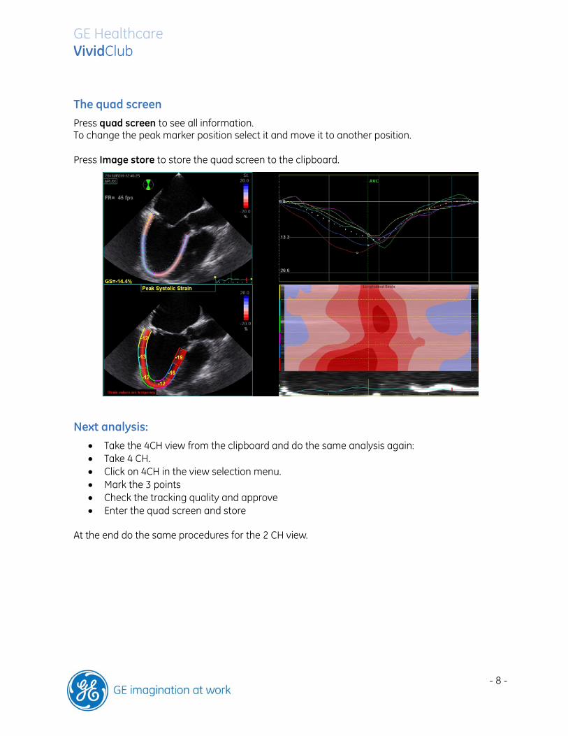
The quad screen
Press quad screen to see all information. To change the peak marker position select it and move it to another position. Press Image store to store the quad screen to the clipboard.
Next analysis:
Take the 4CH view from the clipboard and do the same analysis again: Take 4 CH. Click on 4CH in the view selection menu. Mark the 3 points Check the tracking quality and approve Enter the quad screen and store
At the end do the same procedures for the 2 CH view.

GE Healthcare VividClub
- 9 -
The Summary Once all views are analysed the system will come up with a bull’s eye and the traces from all three views.
Global Strain In order to get the global strain values for the different views, select the BE only. The bull’s eye gets enlarged over the screen and the global strain values are displayed.


