

BIOLOGY OF TOOTH WEAR: PREVENTIVE STRATEGIES
SARBIN RANJITKARBDS, BScDent (Hons)
Thesis submitted for the degree of Doctor of Philosophy
School of Dentistry
The University of Adelaide
August 2009

1
CHAPTER 1
INTRODUCTION

2
1.1 Definitions
Tooth wear is a complex process that includes attrition, abrasion and erosion, or a combination
of these factors. In dental terms, attrition is defined as tooth wear caused by tooth-to-tooth
contact without the presence of food. Abrasion is defined as the wearing of tooth substance
resulting from friction of exogenous material forced over the surface by incisive, masticatory
and grasping functions and tooth cleaning. Erosion refers to the superficial loss of dental hard
tissue due to chemical demineralization not involving bacteria.1,2 Recently, European
researchers have used the term ‘erosive tooth wear’ to refer to tooth wear caused by erosion
combined with mechanical factors, such as attrition and toothbrush abrasion.3 Some researchers
have also proposed abfraction as a separate wear process, leading to the formation of
characteristically wedge-shaped non-carious cervical lesions.4-6 However, there is a general lack
of firm clinical evidence to support the abfraction hypothesis that the lesions result from cuspal
flexure associated with heavy occlusal loading, and non-clinical models supporting this concept
has limitations.7
There is a lack of consistency in the use of terms referring to different types of tooth wear in the
literature.8,9 For example, some researchers use the term erosion to describe overall tooth wear.8
Anthropologists have also used all three terms, attrition, abrasion and erosion, inadvertently
when referring to abrasion.9 Furthermore, the context in which dental terms are used is different
to that in the discipline of tribology that investigates the relationship between lubrication,
friction and wear.10
In tribological terms, there are four fundamental frictional wear processes between opposing
surfaces, including adhesive wear, abrasive wear, fatigue wear and tribochemical wear.10,11
Adhesive wear occurs usually between sliding metals surfaces after asperities or microscopic
projections on one surface becomes cold-welded to the opposing surface as a result of high
attractive forces between them.10 Abrasive wear refers to wear between opposing surfaces

3
resulting from asperities of a harder surface ploughing into a softer surface.11 The surfaces are
rubbed away either by direct contact between the surfaces (two-body abrasion), or in the
presence of a slurry of abrasive particles (three-body wear).10 Fatigue wear occurs when plastic
deformation of the surface results in the propagation of subsurface micro-cracks, eventually
leading to the loss of unsupported material.10,11 Tribochemical wear occurs when a surface layer,
whose inter-molecular bonds are weakened by a chemical reaction (corrosion), is removed by
mechanical action of the opposing surface.12 The generic definition for ‘erosion’ in tribology is
the wear process of a surface caused by particles (solid or liquid) moving at a high velocity.1
Tribological terms for attrition and abrasion are two- and three-body wear respectively, and
chemical dissolution of a material (erosion) is referred to as corrosion in tribology.1 Although
tribological terms provide a more accurate representation of wear processes than conventional
dental terms, it is unlikely that they will be used routinely in dentistry. As the present study has
a strong emphasis on clinical dentistry, established dental terms will be used to describe tooth
wear processes throughout this thesis.
1.2 Clinical appearance of different mechanisms of tooth wear
1.2.1 Attrition
Teeth subjected to attritional wear present with well-defined, shiny wear facets that usually
match with wear facets from the opposing arch.1 Individuals with active tooth grinding can also
display enamel chipping on the incisal edges of teeth (Fig. 1.1) and symptoms related to
myofascial pain dysfunction.1 An uneven occlusal table is also a common observation in
individuals displaying heavy attrition (Fig. 1.2), but wear facets of teeth contributing to such
unevenness can usually be matched in extreme mandibular positions (Fig. 1.3). Extreme
mandibular position refers to the terminal phase of the tooth grinding (bruxing) cycle and does
not occur during mastication.13 In micrographs, wear facets display parallel striations following
the direction of tooth grinding (Fig. 1.4).

4
Figure 1.1. Evidence of enamel chipping on the incisal edges of maxillary central incisor teeth (white arrows).
Figure 1.2 Figure 1.3
Figure 1.2. Dentition displaying attritional wear with uneven occlusal table.
Figure 1. 3. Same dentition shown in Fig. 1.2 in an extreme right mandibular position. Wear facets of teeth 11 and 31, and of teeth 12 and 42, match with each other.

5
Figure 1.4. Micrograph of a wear facet showing parallel striations running (horizontally) along the direction of attrition (tooth grinding).
1.2.2 Abrasion
In pre-contemporary human populations, including Australian Aborigines, coarse food and
grasping functions relating to the use of teeth as tools were the most common causes of
abrasion, characterized by rounded off or blunt tooth cusps and pitting of tooth surfaces (Fig.
1.5).1 Abrasion can also be caused by pipe smoking and grasping functions relating to the use of
teeth as tools (for example, pencil chewing, nail biting, and seed crushing between teeth).1
Micrographs of the surface of such teeth display scratch marks arranged haphazardly together
with gouging and surface pitting (Fig. 1.6).2 Dentine may also be scooped in abraded teeth
because it is softer and wears faster than enamel (Fig. 1.5).1

6
Figure 1.5. Occlusal view of a mandibular right molar tooth displaying signs of abrasion, including pitting of the tooth surface.
Figure 1.6. Micrograph of the surface of a tooth displaying signs of abrasive wear which is characterized by scratch marks arranged haphazardly.

7
The most common form of abrasive tooth wear in modern human populations is believed to be
associated with the formation of non-carious cervical lesions (NCCLs) (Fig. 1.7).14 In a previous
study assessing the micro-morphology of NCCLs in a sample of extracted teeth, around half of
the specimens displayed evidence of ‘rippled’ horizontal furrows caused by toothbrush abrasion
and erosion (Fig. 1.8).15 Overall, abrasion and erosion were noted to be commonly associated
aetiological factors in the formation of wedge-shaped NCCLs.15 This finding contradicts the
abfraction hypothesis which states that NCCLs form as a result of a concentration of tensile
forces at the cervical region of teeth from cuspal flexure associated with heavy occlusal
loading.4-6
Figure 1.7. Variation in the appearance of non-carious cervical lesions that extend from the cemento-enamel junction to the subjacent root surface in a mandibular right lateral incisor (A) and in a mandibular left central incisor (B). (Adapted from Nguyen et al.15)
NOTE: This figure is included on page 7 of the print copy of the thesis held in the University of Adelaide Library.

8
Figure 1.8. A micrograph of an NCCL showing numerous horizontal furrows that are around 20–100µm wide (x100 magnification). These furrows appear smooth which is characteristic of erosion. (Adapted from Nguyen et al.15)
1.2.3 Erosion
The eroded enamel surface has a smooth and glazed appearance, with loss of
micromorphological details (Fig. 1.9).12 At an advanced stage, the tooth loses its original
morphology (Figs. 1.9, 1.10, 1.11).16 The convex profiles of the labial/ buccal and lingual
surfaces of teeth may flatten, or concavities may develop with their widths exceeding their
depths (Fig. 1.9).16 Active erosion also leads to the removal of stains and acquired pellicle from
the surface of the tooth, giving it a clean appearance.12 Loss of enamel leads to increased
translucency of the incisal and proximal surfaces of anterior teeth, and the tooth may appear
darker in shade as a result of shadowing of underlying dentine.12 On the occlusal surface,
dentine exposure results in occlusal scooping or cupping of the cusp tips (Fig. 1.10). In
micrographs, erosion lesions typically appear smooth and clean (Fig. 1.12) but may occasionally
show honeycomb structures on enamel and exposed tubules in dentine.
NOTE: This figure is included on page 8 of the print copy of the thesis held in the University of Adelaide Library.

9
Figure 1.9. Dental erosion is evident on the labial surfaces of maxillary left incisor and canine teeth, characterized by glazing, smoothing and loss of original tooth morphology.
Figure 1.10. Erosive tooth wear is evident on teeth 34, 35 and 36, characterized by scooping and cupping on the occlusal surface. The original occlusal morphology of these teeth has been lost.

10
Figure 1.11. Erosive tooth wear is evident on maxillary teeth characterized by cupping around cuspal regions of posterior teeth and notching of the palatal surfaces of incisor teeth.
Figure 1.12. Micrograph of an eroded lesion that appears smooth and clean compared with those of attrition (Fig. 1.4) and abrasion (Fig. 1.6).

11
1.3 The prevalence of tooth wear
1.3.1 The prevalence of tooth wear in pre-contemporary and modern populations
Tooth wear is a common occurrence in past and modern human populations.17 Heavy abrasive
tooth wear is characteristic of both ancient populations and present-day populations living under
non-industrialized conditions (for example, ancient Anglo-Saxons, Egyptians and Australian
Aboriginals), reflecting the nature of wear produced by abrasive diets (for example, fish, bones,
seeds, nuts and sand in the food).18 Furthermore, different patterns of tooth wear reflect cultural
differences between human populations in the use of teeth as tools for various functions,
including cracking and splitting bones, opening mollusc shells, cutting and tearing fresh meat,
and preparing tools.18 Barrett19 reported that heavy functional demands on teeth of Australian
Aboriginals before European settlement resulted in abrasive tooth wear characterized by
reduction in crown height, interproximal wear and reduction in the length of the dental arches.
This led to wider masticatory cycles and an edge-to-edge anterior bite relationship.19 In the
same population, the frequency of wear faceting resulting from tooth grinding (attrition) was
also very high in 18-year-old individuals (ranging from 65% of premolar teeth to 90% of molar
teeth), and the evidence of extreme mandibular movement was noted to occur in 93% of males
and 100% of females.13
Studies investigating the prevalence of tooth wear in medieval and modern human populations
indicate that erosive tooth wear is the primary wear process. Ganss et al.20 reported a high
prevalence of erosive tooth wear in medieval skulls from Germany compared with that of a
modern population. Wiegand et al.21 reported a high prevalence of erosion (32%) in primary
teeth of 463 German children aged between two- and seven-years, with its distribution being
highest in incisor teeth and lowest in molar teeth. The authors also found that 13.2% of children
had at least one tooth with erosive tooth wear extending to the dentine or pulp. Deery et al.22
reported that erosive tooth wear was common in adolescents in both the United States and the
United Kingdom. Some caution is needed in interpreting the findings on the prevalence of

12
erosive tooth wear because all three wear processes (attrition, abrasion and erosion) contribute
to general tooth wear in individuals.
Some researchers have described the general prevalence of tooth wear rather than focussing on
erosive tooth wear. The prevalence of tooth wear in a sample of 1,007 British dental patients
was about 97%, of which 7% of the tooth wear was classified as being pathological.23 Dugmore
and Rock24 reported loss of surface characteristics of enamel in 50% of 1,753 British children
aged 12-years. They also found that the proportion of individuals with extensive loss of enamel
increased from 4.9% to 13.1% over a period of two years, and those with exposed dentine
increased from 2.4% to 8.7%. In a recent British survey of 10,381 children aged between five-
and 15-years, tooth wear was scored using the tooth surface loss index (TSL) on the labial and
palatal surfaces of primary incisor and permanent incisor teeth and on the occlusal surface of
permanent first molar teeth.25 More than 50% of 5-year-old children displayed tooth wear, but
TSL was less common and less severe in the permanent dentition of 12- and 15-year-old
children. Bartlett26 assessed the degree and progression of tooth wear in adult British patients
who had been referred to a tooth wear clinic over a period of 18 years and found that it was a
slow, minimally progressive process.
In a longitudinal study of Swedish adults conducted over six years, the prevalence of occlusal
erosion increased from 3 to 8% in 26-30 year-old individuals and from 8 to 26% in 46-50 year-
old individuals.27 There is lack of information on the prevalence of tooth wear in adult
Australians, but clinical studies on patients who have been referred to tooth wear clinics indicate
that erosive tooth wear is common in this population.28 Springbett et al.29 also reported a high
prevalence of tooth wear in the primary dentition of two cohorts of South Australian children,
with 71% of all primary canines displaying exposed dentine in the younger age-group (with
mean age of 4.7 years) compared with 100% of primary maxillary canines in the older age-
group (with mean age of 10.6 years). The authors also noted that the frequency of faceting on
enamel surfaces ranged from 50% to 85% in the younger age group, and from 83% to 100% in

13
the older age group. Bartlett and Dugmore8 indicated that enamel wear is generally common in
children and adolescents in modern populations, but that dentine wear associated with the loss
of more than one-third of the crown is rare.
It is difficult to make direct comparisons about the prevalence of tooth wear between different
studies because of differences in the methods used to assess tooth wear, including tooth wear
indices. The two most commonly used tooth wear indices are those of Lussi et al.30 and Smith
and Knight,31 but these indices have undergone many modifications over the years.8,32 Inherent
problems with existing wear indices are that they do not relate to aetiology or management of
tooth wear.8 Recently, a group of international experts proposed an international standard to
record erosive tooth wear for both scientific and clinical purposes, termed the Basic Erosive
Wear Examination (BEWE) index.33 This index categorizes tooth wear into four grades
according to severity, and wear scores are calculated in six different sextants in the oral cavity.
Then, the highest scores from all sextants are added to obtain the final tooth wear score. This
score can then be used to assess the overall risk of tooth wear and to help in formulating
preventive and/or restorative management. Further research is needed to investigate its validity
and reliability as an international standard for tooth wear assessment. Given that most reports on
the prevalence of tooth wear have been based on around 1,000 subjects, it is also important to
ensure that these studies are conducted on a large number of representative subjects from
different populations.8
1.3.2 Multifactorial aetiology of tooth wear
Studies investigating the prevalence of tooth wear have provided some insight into its
multifactorial aetiology. Abrasion from harsh fibrous food in combination with heavy tooth
grinding has been noted to be the primary cause of heavy tooth wear in a pre-contemporary
Australian Aboriginal population.13 Abrasive wear is also a characteristic feature of dentitions
of pre-historic populations and hunter-gatherers.18 In comparison, erosive tooth wear from
dietary sources seems to be a modern-day condition.9 Based on clinical examinations and

14
questionnaires focussing on dietary habits, Deery et al.22 and Wiegand et al.21 did not find
associations between the occurrence of erosion and dietary factors. Other researchers have
reported significant associations between erosion and carbonated drinks,24 between erosion and
acidic beverages (herbal and lemon tea),34 and between erosion and citrus fruits, sports drinks,
vomiting and presence of gastric symptoms.35 There is a general consensus among clinicians
and researchers that acidic diets cause erosion, and some caution is needed when interpreting
data from questionnaires because of potential inaccuracy in subjects’ recollections about past
habits or events.35
It is generally believed that salivary factors, including acquired pellicle, are important in
protecting teeth against tooth wear,36,37 and that salivary dysfunction increases the risk of dental
erosion by both extrinsic (dietary) and intrinsic acids.38 Some in vitro, in situ and cohort
studies35 have also reported associations between dental erosion and compromised salivary
factors (for example, salivary calcium and phosphate levels), but other studies do not support
these findings.21,39 Overall, these findings highlight the difficulty in assessing salivary factors
with good reliability. In a review article, Dawes36 reported that concentrations of both salivary
electrolytes and proteins are influenced by glandular source, flow rate, nature and duration of
stimuli, biological rhythms and hormonal factors. Furthermore, salivary flow rate displays daily
and yearly variation and is associated with the degree of hydration of the body, body position,
gland size and drug use.36 Flink et al.40 reported a significant three- to six-fold increase in
unstimulated salivary flow rate (USFR) from 7.30am to 11.30am. The authors also found that a
USFR of less than 0.1mL/min, characteristic of hyposalivation and Sjogren’s syndrome,
increased to more than 0.1mL/min in 70.3% of subjects during this period, supporting the notion
that a lack of consideration of the variation in salivary properties can easily lead to misdiagnosis
of salivary hyposalivation. Thus, future epidemiological studies with robust study designs are
needed to further investigate the potential role of saliva in protecting against tooth wear.

15
1.3.3 Physiological versus pathological tooth wear
Some clinicians consider any signs of tooth wear to be pathological and believe that teeth
without wear are essential for a properly functioning “ideal” dentition.41 However, the general
consensus in clinical dentistry is that tooth wear is a slow, progressive process and is a
physiological process in most individuals.8,16 Kaidonis9 indicated that gradual tooth wear in a
pre-contemporary Aboriginal population was associated with physiological adaptation of the
masticatory system, including widening of the ‘teardrop’ nature of the masticatory cycle,
remodelling and flattening of the glenoid fossa, and continued tooth eruption. Based on the
notion that the human dentition is designed for wear in the context of evolutionary adaptation,42
tooth wear in this population can be considered to be physiological. However, the distinction
between physiological and pathological forms of tooth wear can be difficult. Excessive tooth
wear in modern populations is likely to be diagnosed as being pathological, even though the
dentition may be functional. It is the perception of modern societies that white, unworn teeth
represent health and youth, whereas a worn, yellowish dentition is usually attributed to old age
and loss of power.16 The diagnosis is more straight forward when the function is compromised
by severe dentinal hypersensitivity, acute pulpal pain or chronic pulpal necrosis with periapical
inflammation.16
Near or frank pulp exposure as a result of excessive tooth wear has been reported in around 11%
of patients referred to a tooth wear clinic in Queensland, particularly on the palatal surfaces of
maxillary incisor teeth and on the incisal surfaces of mandibular incisor teeth.43 In another
epidemiological survey of dental patients investigating the impact of various oral conditions in
South Australia, pulpal infection was the most common condition causing moderate to extreme
levels of discomfort followed by tooth wear and dentinal sensitivity.44 In this study, a greater
frequency of patients reported discomfort due to both tooth wear and dentinal hypersensitivity
compared with other oral conditions (such as caries, denture problems, tooth fracture and
periodontal disease). The impact of dentinal hypersensitivity on the quality of life, measured in
terms of disability-adjusted life years (DALYs), was comparable with other oral conditions,

16
except that it was lower than that of pulpal infection. These findings are supported by findings
from a different study, indicating that tooth wear had an impact on individuals’ appearance, oral
comfort and eating ability.45
There is a lack of standard guidelines to distinguish between physiological and pathological
forms of tooth wear. Existing data on the prevalence of pathological tooth wear using the Smith
and Knight index31 are not soundly scientific based, but rather on the subjective judgement of
the researchers.8 Donachie and Walls46 reported that the Smith and Knight index grossly
overestimated the level of pathological tooth wear in British individuals aged 45 years or more,
with the mean level of incisal wear scores in their study being classified as pathological
according to the Smith and Knight index. After the authors argued that the threshold for
detecting pathological tooth wear was inaccurate for the elderly population, Smith and Robb23
revised these threshold values in their subsequent epidemiological study of tooth wear in 1996.8
In this context, more defined and reproducible criteria need to be developed to differentiate
pathological tooth wear from physiological tooth wear.8
1.4 Methods for assessment of tooth wear
1.4.1 Significance of in vivo, in situ and in vitro studies
In vivo studies provide more realistic information about tooth wear but they need to be
conducted over a long period of time to detect significant changes, given that current methods
for assessment of minute amounts of tooth wear in living subjects or using serial dental models
are inadequate.47 In situ model “can be placed at some point on the continuum between a
clinical trial and an in vitro study”, yet provides clinically relevant information in a relatively
short period of time.48 This model involves human subjects wearing intra-oral appliances
containing slabs of enamel or dentine, but with demineralization and remineralization of tooth
substrate as well as tooth wear measurements are conducted in vitro.47 Given that in situ
methods are unsuitable to investigate tooth wear related to attrition and given also that in vivo
methods usually cannot separate the various components of tooth wear, in vitro studies have an

17
important role to play in our understanding of characteristics of tooth wear. Furthermore, in
vitro studies are useful because various components of tooth wear can be investigated
separately,49 and a large number of teeth can be examined over a relatively short period of time.
However, such results should only be used to provide information about general trends of tooth
wear and they should not be extrapolated directly to the in vivo situation.47
1.4.2 Different methods for assessment of tooth wear
Tooth wear can be assessed by using both quantitative and qualitative methods50 but they vary
in their precision from the macro- to nano-level. Attin51 indicated that quantitative data are
objective in nature and are desirable to investigate tooth wear. He also indicated that qualitative
assessment can be subjective, but it complements quantitative findings by enabling the wear
process to be visualized. The most popular method of tooth wear assessment in vivo involves
tooth wear indices which rely on visual assessment of changes on teeth intra-orally or from
study models or photographs.50 However, wear indices cannot detect minute amounts of tooth
wear. In comparison, most quantitative methods used in assessing tooth wear (for example,
profilometry, microhardness and nanohardness, microradiography, chemical analysis of
dissolved minerals and ultrasonic measurement of enamel thickness)51 have high precision but
are generally limited to in vitro and in situ situations. Most of these quantitative methods require
special sample preparation, including sectioning and polishing of specimens flat to a level of
around 1μm. Furthermore, they provide information on different aspects of tooth wear, and it is
desirable to use them in combination to provide a holistic approach to understand the complex
nature of tooth wear. Both quantitative and qualitative methods are used in the present study
enabling assessments of small amounts of tooth wear together with visualization of the wear
process.
Scanning electron microscopy is a popular method for qualitative assessment of tooth wear both
in vitro and in vivo. Other methods, such as atomic force microscopy and mass spectrometry
usually provide qualitative information but they can be used to provide partially quantitative

18
data with careful experimental design.51 Methods with high precision can detect minute amounts
of tooth wear but several areas of the same specimen need to be assessed to ensure
representation of the whole specimen. A discussion of methods for assessment of tooth wear in
this section will be limited to those methods which are commonly used, have high precision,
and are considered to be suitable to investigate erosive tooth wear in vitro.
Profilometry is the most commonly used method for the assessment of tooth wear in vitro. It can
involve a contacting profilometer (using a mechanical stylus) or a non-contacting profilometer
(using laser or lights of different colours) that read co-ordinates of an object in two or three
dimensions. Contacting profilometers provide accurate measurements but can damage the
external surface of the tooth, whereas the accuracy of non-contacting profilometers may depend
on the colour of the specimen tested. Furthermore, laser profilometers may produce ‘overshots’
at sharp edges, resulting in artefacts.51 Although profilometers can be used to detect
macroscopic changes to teeth,52 most studies have used them to detect microscopic changes.53-56
Some researchers have used profilometers to detect tooth wear below 1μm,53,57 even though it is
difficult to assess tooth wear accurately at that precision without reporting the validity and
reliability of methods.51 Profilometers require a stable reference plane to enable alignment of
scanned surfaces before and after wear. It is difficult to use profilometers in vivo because such
reference points need to be calculated using positions of best fit before and after wear.
Hardness measurements can provide reliable information about changes in the surface
characteristics of dental hard tissues following erosive demineralization and remineralization.
Two types of hardness measurements of teeth involve microhardness and nanohardness
indentation, with both methods using indentors of given geometrical shapes. The size of
indentation made on the tooth structure provides information about the hardness of the tooth
structure and its susceptibility to wear, but the measurements tend to be difficult in dentine
because of its elastic properties. Based on microhardness assessment, erosively demineralized
enamel has been shown to be softer than sound enamel.58 While microhardness indentation

19
provides general information about hardness of the bulk of the tooth structure, nanohardness
indentation can be used to obtain hardness measurements and elastic modulus measurements
more precisely. Previous studies have also shown that nanoindentation can be used to
characterize biomechanical properties of sound tooth structure,59,60 carious tooth structure61-63
and eroded tooth structure.64-66 Studies employing microhardness indentation typically use
longer periods of demineralization and remineralization to detect measurable changes in
mechanical properties of teeth, whereas nanoindentation is very sensitive to changes occurring
during early stages of enamel dissolution. For example, nanohardness measurements have
enabled researchers to detect enamel softening in vitro after erosion in citric acid (pH 3.3) for
2min67 and in sports drinks for 10min,64 whereas the experimental design for studies involving
microhardness measurements has involved eroding teeth for around 30min or longer during
each episode.68
Atomic force microscopy (AFM) and mass spectrometry are two relatively new techniques that
also have potential to elucidate changes in tooth structure during the early stages of erosion.
AFM scans the surface of a specimen at a very high resolution using a sharp tip and enables
both quantitative assessment (involving height loss, surface roughness and nanohardness
measurement) and qualitative assessment (involving high resolution surface topography). The
resolution of AFM is greater than that of a profilometer.69 Lippert et al.66 used AFM to show
that cola, lemonade and orange juice softened enamel. In a preliminary investigation, West and
Jandt47 showed that AFM could detect height loss of an abraded sealant in the order of around
5nm and they concluded that this technique might play a major role in future tooth wear
research. AFM is also capable of exploring areas that are not accessible by other microscopic
methods.47 Furthermore, specimens can be tested in an ambient environment and do not undergo
dehydration, unlike those used for scanning electron microscopy.
Secondary ion mass spectrometry (SIMS) is a very sensitive surface analytic technique, capable
of detecting concentrations of elements or molecules at subparts per million. Lewis and

20
colleagues70,71 have used this technique to identify corrosion products from CoCr orthopaedic
implants at the site of physical wear. Barbour and Rees69 described the application of SIMS in
creating surface mapping of calcium and magnesium of an eroded enamel surface. SIMS has the
potential to detect early mineral loss of eroded tooth surface, but further study is needed to
validate its suitability for erosion studies.51 SIMS has also been used to map chemical changes
across an incipient carious lesion63 and to elucidate the mechanism of fluoride uptake associated
with CO2 laser treatment on enamel.72 In this context, this technique has the potential to provide
insights into the mechanisms of tooth wear, including identification of third-body components
responsible for lubrication at the wear interface between opposing tooth/tooth and tooth/material
specimens.
Scanning electron microscopy (SEM) is commonly used to obtain high-resolution images of
tooth surfaces, and the predominant wear process can be identified based on micrographic
assessments. For example, a micrographic assessment of non-carious cervical lesions (NCCLs)
showed that toothbrush abrasion and erosion were noted to be commonly associated aetiological
factors.15 SEM has also been used to examine erosive tooth wear both in vitro73 and in vivo.74
The use of SEM assessment in combination with profilometry has also provided insights into
the complex relationship between wear rates, loads and pH values at the wear interface,
improving our understanding about the characteristics of opposing tooth surfaces and the role of
third body components in lubricating the wear interface.52,54 The main advantage of SEM over
other qualitative techniques is that tooth surfaces do not need to be prepared flat and polished,
thus natural tooth surfaces can be examined reliably. However, specimens may require removal
of a surface smear layer by treatment with chemicals (for example, acid etch and sodium
hypochlorite), which may alter the surface morphology. Caution is needed in interpreting cracks
in micrographs because dehydration of specimens under vacuum can cause crack propagation.

21
1.5 Interaction between common forms of tooth wear
It has been shown in many clinical studies that tooth wear usually occurs as a combination of
different wear processes (attrition, erosion and abrasion),7 and that these wear mechanisms in
combination can result in more destructive tooth wear than if these processes occurred
independently.3,75 However, it is often difficult during clinical examinations to determine the
major cause of tooth wear or to assess how different wear processes have interacted with each
other.7 An understanding of the characteristics of tooth wear caused by a combination of
different wear processes not only assists in clinical dentistry, but also provides insight into
tribological aspects of tooth wear that may be useful in developing new methods of wear
prevention.
1.5.1 Combination of attrition and erosion
Previous in vitro studies have focussed on dental erosion, partly due to the relative ease of
designing laboratory experiments to investigate the pathogenesis of erosion. Thus, the role of
attrition in tooth wear has been under-researched.49 Only a few in vitro models have been
described by researchers from Europe and Australia to investigate the characteristics of wear
between opposing tooth specimens.
Recently, Bartlett and colleagues from London (UK)76 described an attritional model to
investigate the effect of fluoride on dentine wear by treating flat, polished dentine specimens
obtained from mandibular third molar teeth with a concentrated fluoride varnish (containing
22,600ppm fluoride at pH 3.93) for 24 hours. The specimens were then slid against enamel
cusps of premolar teeth over a horizontal distance of 1mm for 5,000 cycles under a load of
150N using artificial saliva as a lubricant. The authors observed that fluoride application did not
reduce enamel wear. Eisenburger and Addy from Bristol (UK)54 described a model of
investigating enamel wear by sliding opposing enamel surfaces against each other in a tooth
wear machine for 2,280 cycles under a load of 6N (600gm) with citric acid (at pH 3.2, 5.5 and
7.0) and saline (pH 7.5) as lubricants. Attritional wear of enamel in a neutral environment was

22
observed to be greater than that in acidic conditions due to smoothing effect of erosion on
opposing enamel surfaces at the wear interface.
Kaidonis and his colleagues from Adelaide (Australia)77-79 have developed an electro-
mechanical tooth wear machine which has provided some insights into the complex nature of
wear in enamel, dentine and restorative materials under conditions simulating attrition and
erosion. A non-linear relationship between applied force and the rate of enamel reduction has
been established, with the combined effects of attrition and erosion resulting in very rapid rates
of tooth wear at low pH (simulating gastric regurgitation at pH 1.2).77 The rate of enamel
reduction was slow at all but the highest loads (>140N) in a moderately acidic environment (pH
3.0).77 It has also been shown that the rate of wear of enamel is lower than that of dentine at low
loads in a neutral environment but the rates are more comparable at higher loads, possibly due
to the harder but more brittle nature of enamel and the more elastic nature of dentine.78 A recent
study has established that the relationship between wear, loads and pH for wear occurring
between opposing enamel and dentine specimens is complex.52 In particular, when enamel
wears against dentine in an acidic environment, enamel will wear more rapidly at very low pH,
while under less acidic conditions dentine will wear faster than enamel.52 Wear facets of both
enamel and dentine produced at pH 3.0 (hydrochloric acid) were noted to be smoother than
those prepared at pH 6.1 (deionized water).52 These findings are supported by explanations
provided by Eisenburger and Addy,54 who described differences in wear mechanisms in
opposing enamel specimens at pH 3.0 and 7.0. Stress cracks and rough asperities break away
from enamel surfaces resulting in fatigue wear and ‘‘three-body’’ abrasion at around pH 7.0. In
contrast, at pH 3.0, enamel is softer and hydroxyapatite crystals at the surface become thin due
to erosive demineralization.7 Abrasion removes this softened layer, but not the sound, deeper
enamel structure, so that smoother asperities on opposing enamel surfaces contribute to well-
lubricated, ‘‘two-body’’ abrasion.54

23
Most models used to investigate erosive tooth wear involving attrition are based on the notion
that attrition and erosion occur concurrently. There are reports on the association between
bruxism and gastric reflux in individuals.80 There is some controversy regarding whether
bruxism occurs during rapid eye movement (REM) or non-REM stages of sleep. Lavigne et al.81
noted that rhythmic masticatory muscle activities (RMMA) occurred consistently in all stages of
sleep. Other studies have indicated that bruxism occurs during REM sleep stage, whereas some
authors have reported that bruxism occurs during both REM and non REM sleep stages.82
Lavigne et al.81 also found that bruxing episodes with grinding noises occurred during both
rapid eye movement (REM) and non-REM stages of sleep, and that the mean frequency of
bruxism episodes was 30.5 episodes per night (ranging from one to 84 episodes per night).
In comparison, gastric reflux episodes in individuals suffering from reflux oesophagitis have
been noted to occur more frequently during non-REM sleep.83 Gastro-oesophageal reflux is a
common physiological process that occurs for about an hour per day in healthy subjects, but
gastro-oesophageal reflux disease (GERD) is a pathological condition that produces signs and
symptoms of tissue injury within the oesophagus, oropharynx, larynx and respiratory track.84
Reflux oesophagitis is the most common form of GERD, recognized by the presence of
symptoms (e.g. heartburn) and the presence of mucosal inflammation in the oesophagus.84 It can
be caused by transient relaxation of lower esophageal sphincter, delayed gastric emptying and
hiatus hernia.85The frequency of reflux episodes has been reported to be greater than 100 during
an eight-hour sleep in individuals suffering from obstructive sleep apnoea syndrome (OSAS).86
Information about sleep bruxism and gastric regurgitation indicate that both bruxism and gastric
regurgitation can occur concurrently. Data on changes in intra-oral pH during sleep-related
reflux episodes are scarce because previous researchers have limited their measurements to the
distal oesophagus. Given that sleep facilitates proximal migration of acid and increases the
probability of pulmonary aspiration,87 regurgitation of gastric acid into the oral cavity may be a

24
common occurrence in individuals at high risk of GERD. It has been reported that OSAS and
obesity seem to predispose individuals to nocturnal GERD.83
The amount of force generated during bruxism has been estimated to be, on average, three times
that of functional chewing force of around 6.75 pounds (equivalent to 31N).82 Li et al.76 have
indicated that there is little consensus on selection of loads in previous in vitro studies of
attrition, with values ranging from 2 to 162N.54,77,88 Other researchers have reported higher
mean values for maximum voluntary bite (clenching) forces in humans, ranging from 189 to
1,181N,89-92 but reported maximum nocturnal bite forces, on average, are relatively lower and
range from 11 to 423N.91,93 Maximum clenching forces are static forces that occur rarely in
individuals,90,93 and dynamic forces are more relevant to tooth wear involving attrition.
Information on bite forces associated with protrusive and lateral movements during sleep is
scarce, but dynamic forces involved with chewing, on average, have been noted to be around 60
to 162N.90,94 Thus, a load of 100N selected in previous studies52,95 falls within the physiological
limits of dynamic bite forces in humans.
1.5.2 Combination of toothbrush abrasion and erosion
Normal tooth brushing habits with toothpaste are unlikely to produce excessive wear over a
lifetime but the super-imposed influence of acids is known to increase wear rates.7 However, an
eroded tooth surface is susceptible to mild toothbrushing action or friction from the tongue.7
Generally, toothbrush abrasion does not occur concurrently with erosion, but occurs after an
intake of erosive diet. Thus, most previous researchers have investigated erosive wear involving
toothbrush abrasion at around pH 3.0 simulating acidic diet rather than at lower pH values
simulating gastric regurgitation.
Eroded enamel has a shallow, softened subsurface layer, with crystals being thin96,97 and
vulnerable to mechanical wear (for example, toothbrush abrasion).55 Similarly, eroded dentine
has a demineralized, subsurface layer98 which is susceptible to wear by toothbrush abrasion.7

25
Dentine is more susceptible than enamel to toothbrush abrasion alone or in combination with
erosion.57 There is also some evidence showing that the abrasivity of toothpaste (often
quantified using Relative Dentine Abrasivity values) increases abrasive wear in dentine but not
in enamel.57
Our understanding of in vitro assessment of erosion on enamel and dentine, and erosive tooth
wear involving toothbrush abrasion, is primarily based on models developed by European
researchers (for example, Vieira et al.99). Their in vitro study design typically involves wear
regimes, each involving erosion of flat, polished tooth specimens at around pH 3.0 followed by
toothbrush abrasion in the presence of a slurry of toothpaste and artificial saliva. The specimens
are stored in artificial saliva between wear regimes to simulate remineralization by natural
saliva in vivo. It has been suggested that treatment of eroded enamel with artificial saliva for
two hours partially rehardens enamel in vitro,100 but Vanuspong et al.101 indicated that this is
less likely to occur in dentine. Nevertheless, Attin et al.102 have observed that delaying
toothbrushing by at least 30min after an erosive regime in situ provides some protection against
toothbrush abrasion. A total of 600 brushing strokes applied during three wear regimes on
bovine teeth was reported by Vieira et al.99 to correspond to around two years of wear.
Most toothbrush abrasion models have used reciprocal toothbrush action against stationary tooth
specimens, 99,103 but Parry et al.104 suggested that the movement of reservoirs containing tooth
specimens and toothpaste slurry against stationary toothbrushes would better maintain slurry
homogeneity at the wear interface. Parry et al.104 also reported that toothbrushing forces selected
in previous in vitro studies ranged from 0.1N to 8.8N. However, a force of around 2N has been
commonly used by most researchers to simulate normal toothbrushing force in vivo.56,99 To
enable detection of minute amounts of tooth wear using profilometry, most previous studies
have used flat and polished specimens rather than natural tooth surfaces. However, it should be
noted that natural tooth surfaces erode more slowly than polished surfaces.105

26
1.6 Current methods for tooth wear prevention and gaps in knowledge
As tooth wear can result occasionally in extensive loss of enamel and dentine, leading to
dentinal sensitivity and pulpal exposure,106 dental professionals have a responsibility to identify
individuals at risk and to preserve the long-term health of teeth.107 It is not always practical to
eliminate the causes of tooth wear, so effective preventive strategies need to be developed as
part of clinical management.108 Underlying medical conditions predisposing individuals to
dental erosion (for example, anorexia and bulimia nervosa, gastroesophageal reflux disease)
need to be recognized and managed as necessary, including referral to a medical practitioner or
a clinical psychologist.109 Previously suggested strategies for preventing erosive tooth wear
involving toothbrushing include the application of a protective resin coating on teeth108 or of
topical fluoride that increases the resistance of tooth structure against erosive wear.110 The
management of erosive tooth wear involving attrition includes dietary advice, stress
management, wearing a nightguard, use of fluoride and complex restorative work.37 Other
strategies for prevention of erosion include identification of the cause, enhancement of salivary
flow and buffering capacity, and maintenance of tooth surface integrity by using remineralizing
agents (such as fluoride and casein phosphopeptide - stabilized amorphous calcium phosphate,
CPP-ACP).111 Given that most recommendations for the management of erosion using
remineralizing agents have been based on in vitro studies and have been extrapolated from
evidence for the management of dental caries, well-designed clinical trials are needed to test
their effectiveness in managing dental erosion.111
Previous in vitro and in situ studies have provided a foundation for the development of
strategies for tooth wear prevention in vivo. Earlier reports indicated that fluoride provided
minimal protection to enamel against dental erosion112,113 and against toothbrush abrasion after
an erosive eopisode.114 However, more recent reports generally agree that fluoride protects
enamel and dentine against erosion at around pH 3.0.115-117 Hove et al.118 observed that intensive
application of fluoride provided significant protection against enamel erosion under conditions
simulating gastric reflux at pH 2.0. In contrast, Willumsen et al.119 found that sodium or

27
stannous fluoride did not protect enamel against erosion at pH 1.2, although stannous fluoride
provided greater protection against enamel erosion at pH 2.0 than sodium fluoride.118 Other
reports have indicated that topical fluoride can protect enamel and dentine against a
combination of erosion and toothbrush abrasion,102,103,120 but not against attritional wear between
opposing enamel and dentine specimens.76 Thus, there is still a need to improve preventive
strategies relating to erosive insults on teeth.107
1.6.1 Remineralizing agents, including CPP-ACP
In an in vitro study investigating the effect of frequent application of remineralizing agents on
enamel erosion, Lennon et al.14 found that highly fluoridated acidic amine gel (containing
12,500ppm F) provided significant protection to enamel from erosion, but Topacal
(casein/calcium-containing gel, CasCP), 250ppm F, or a combination of CasCP and 250ppm F
did not provide any protection. In a recent study on prevention of wine erosion, Tooth Mousse®
(TM, GC Corporation, Japan), which is a water-based cream containing a remineralizing agent
in the form of 10% casein phosphopeptide stabilized amorphous calcium phosphate (CPP-
ACP), reduced erosion by citric acid121 and white wine.122 These findings are supported by the
observation that four separate applications of TM within a period of 48hrs significantly
increased microhardness of enamel that had been eroded for 8min.68
Recent in vitro studies on mineral supplementation to acidic solutions have also provided
encouraging results in reducing tooth wear. Attin et al.123 reported that adding calcium,
phosphate and fluoride supplements to citric acid drink (pH 2.2) significantly reduced its
erosive potential. Jensdottir et al.124 also confirmed that addition of calcium and phosphate
reduced the erosive potential of orange juice. Ramalingam et al.125 showed that adding CPP-
ACP (casein phosphopeptide and amorphous calcium phosphate) ranging in concentration from
0.09 to 0.25% to the sports drink Powerade provided a protective effect against enamel
corrosion. Scanning electron micrographs of enamel treated with 0.63% to 0.25% CPP-ACP in
this study showed superficial granular structures adherent to enamel surface, which the authors

28
speculated to be remineralized enamel crystals, repair crystals, deposits formed as a result of
subsequent demineralization and remineralization, redeposited mineral phase from CPP-ACP,
or remineralization of the smear layer created during sample preparation. These findings suggest
three potential mechanisms by which CPP-ACP reduced erosion, including reduction in the
erosive potential of the sports drink, remineralization of the eroded lesion and physical
protection of the enamel surface by precipitates of CPP-ACP. Further research is needed to
clarify the mechanisms by which CPP-ACP reduces tooth wear.
The role of CPP-ACP as an anticariogenic agent has been well documented,126,127 and current
evidence from in vitro studies is promising in relation to its use in the management of erosion.
Its properties and potential for use in preventing tooth wear are discussed further in the
following chapters of this thesis. In a recent in vitro study, attritional wear of dentine was
almost eliminated with continuous application of a paste containing an anticariogenic
remineralizing agent CPP-ACP (casein phosphopeptide - amorphous calcium phosphate
nanocomplexes) compared with hydrochloric acid lubricant (pH 3.0) and deionized water
lubricant.95 Furthermore, intermittent application of CPP-ACP paste also reduced dentine wear
at both acidic and near neutral environments, highlighting its lubricating and remineralizing
properties in reducing erosive dentine wear.95 These findings are supported by observations that
CPP-ACP can reduce dental erosion by citric acid,121 white wine122 and sports drinks.125 These
findings warrant an investigation of the effect of CPP-ACP on reducing other forms of erosive
tooth wear involving attrition and toothbrush abrasion. These findings have the potential to lead
to the development of a new method for tooth wear prevention. Indeed, there is a need for better
strategies of tooth wear prevention because fluoride only provides partial protection against
erosive tooth wear. Furthermore, little is understood about the role of remineralizing agents in
reducing attritional wear, and nightguards, which are the only accepted method to prevent
attritional wear,49 may not be tolerated by some individuals.

29
1.6.2 Need for new, more effective methods of tooth wear prevention
Attempts to develop better strategies of tooth wear prevention conform with the paradigm of
minimal intervention dentistry, which indicates that clinical management of tooth wear should
focus on early detection and prevention before a restorative approach is considered.128 This is
becoming an issue of increasing importance in clinical dentistry, given that existing evidence
points to an increase in the prevalence of tooth wear as more elderly patients are retaining their
natural teeth to a stage when they present with extensive wear.128 In addition, more younger
patients are also presenting with erosive wear of both primary and permanent teeth.74,129 As
erosion in the primary teeth is an indicator of erosion in permanent teeth,130 early intervention is
desirable.
1.7 Preliminary findings, aims and hypotheses
1.7.1 Preliminary findings
A previous study investigating the role of wet and dry lubricants on enamel wear reduction has
shown that lubricants between opposing specimens reduced enamel wear by producing highly
polished wear facets.131 A recent study reported that attritional wear of dentine was almost
eliminated with continuous application of TM (pH 7.2) compared with hydrochloric acid
lubricant (pH 3.0) and deionized water lubricant (pH 6.1).95 Furthermore, intermittent
application of TM reduced dentine wear in both acidic and near neutral environments,
highlighting its lubricating and remineralizing properties in reducing attritional dentine wear.95
As TM was applied intermittently between erosive wear episodes and appeared to have been
washed off during visual assessment,95 it seems that traces of its ingredients (for example,
glycerol and CPP-ACP nanocomplexes) adhered to the enamel surface and then lubricated the
wear interface by acting as third-bodies. These findings point to the need to test the
effectiveness of TM in reducing erosive enamel wear and to better understand the types of third
bodies at the wear interface contributing to wear reduction.

30
Recent advances in sensitive surface analytic techniques have enabled the detection of traces of
materials on an implant or a tooth surface. Previous researchers have used ToF-SIMS to identify
corrosion products at sub-parts per million from CoCr orthopaedic implants at the site of
physical wear.70,71 SIMS has also been used to map chemical changes across an incipient carious
lesion63 and to elucidate the mechanism of fluoride uptake associated with CO2 laser treatment
on enamel.72 In this context, ToF-SIMS has the potential to identify the types of third-body
components at the wear interface, and to elucidate the mechanism by which TM reduces tooth
wear.
1.7.2 Aims and hypotheses
The aim of this research project was to determine the effect of TM on enamel wear under in
vitro conditions simulating three clinical situations: (i) heavy attrition at a load of 100N in
combination with severe acid erosion simulating gastric regurgitation at pH 1.2; (ii) toothbrush
abrasion after an exposure to dietary acid at pH 3.2; and (iii) erosion from gastric regurgitation
at pH 1.2. In addition, potential modes of action of TM in preventing enamel wear were
investigated by characterizing the enamel surface after treatment with TM using ToF-SIMS.
It was hypothesized that enamel wear would be reduced by intermittent application of TM under
all three conditions involving a combination of attrition and erosion, toothbrush abrasion and
erosion, and erosion alone. It was also hypothesized that ToF-SIMS analysis would be able to
detect traces of ingredients of TM (for example, calcium and glycerol) on the enamel surface
after treatment with TM. Although other ingredients of TM with a sticky consistency (for
example, D-sorbitol, propylene glycol, sodium carboxycellulose and guar gum) may contribute
to wear reduction, the present study was limited to the preliminary investigation of calcium,
phosphorous and glycerol contents.
The findings from this project have direct application in the clinical management of tooth wear.
They supplement other data on tooth wear by simulating tooth-to-tooth contact and three-body

31
wear, and will lead to the developmental of a tribological model of tooth wear from which
general conclusions should be able to be drawn about clinical situations, including bruxism and
acid regurgitation. They will supplement other ongoing studies of tooth wear, including a
longitudinal study of tooth wear in oenology students as well as other clinical and genetic
studies being undertaken by the Craniofacial Biology Research Group, School of Dentistry, The
University of Adelaide.

32
CHAPTER 2
GENERAL MATERIALS AND METHODS
This chapter provides a general description of the materials and methods used in the present
study. Specific experimental details on tooth wear prevention relating to a combination of
attrition and erosion, a combination of toothbrush abrasion and erosion, and erosion alone are
covered in Chapters 3, 4 and 5, respectively. Experimental details relating to the
characterization of enamel surfaces using ToF-SIMS are covered in Chapter 6.

33
2.1 Project outline
The effect of Tooth Mousse® (TM) in reducing enamel wear was tested using three established
models of tooth wear (Table 1). Experimental details relating to these models are provided further
in this chapter (section 2.3.2).A separate model was used for ToF-SIMS analysis. The effect of
TM in reducing enamel wear was also compared with that of Tooth Mousse Minus (TM- without
CPP-ACP) in these models. The attrition/erosion model to simulate wear of opposing enamel
specimens was based on a previous model that has been used extensively by researchers in the
School of Dentistry at the University of Adelaide in the investigation of enamel and dentine
wear.52,77
The toothbrush abrasion/erosion and erosion models were adopted from established models
developed by European researchers. The toothbrush erosion/abrasion model used in the present
study is similar to that applied by Vieira et al.99 to investigate the effect of fluoride treatment on
erosive enamel wear after 600 toothbrushing cycles. To simulate the intra-oral environment,
toothbrush abrasion was conducted in the presence of a toothpaste-artificial saliva slurry. A two-
hour remineralization period with artificial saliva was also included between wear regimes. The
erosion model was essentially similar to that of the toothbrush abrasion/erosion model, but without
the toothbrush abrasion component.
The ToF-SIMS model for characterization of enamel specimens treated with TM was adopted
from that of Lewis et al.70,71 This model has been used to identify corrosion products from CoCr
orthopaedic implants at the site of physical wear.

34
Table 1. Summary of the experiments conducted to investigate the effect of CPP-ACP paste on enamel wear under different conditions.
Experimental conditions Simultaneous occurrence of attrition (100N) &erosion (pH 1.2)
Erosion (pH 3.2) followed by toothbrush abrasion (2N)
Erosion alone (pH 1.2)
Type of electro-mechanical tooth wear machine
Tooth wear (attrition) machine designed in the University of Adelaide
Toothbrushing machine designed in King’s College London
-
Experimental groups (n = 12 per group)
Experimental group 1 (paste with CPP-ACP) Experimental group 2 (paste without CPP-ACP) Control group (no paste)
Experimental group 1 (paste with CPP-ACP) Experimental group 2 (paste without CPP-ACP) Control group (no paste)
Experimental group 1 (paste with CPP-ACP) Experimental group 2 (paste without CPP-ACP) Control group (no paste)
Contour of specimens (obtained from human third molar teeth)
Natural enamel surfaces with wear facets
Flat enamel surfaces from buccal and lingual surfaces of teeth polished to 1μm
Flat enamel surfaces from buccal and lingual surfaces of teeth polished to 1μm
Number of wear episodes (with each episode comprising wear cycles and demineralization and remineralization stages)
Around 63 episodes
10 episodes
10 episodes
Number of wear cycles in each wear episode
160 200 (No mechanical wear)
Steps in each wear episode Attrition under a load of 100N at 80 cycles per minute + erosion at pH 1.2 for 2min (160 cycles) Washing (30s) + air drying (15s) Application of paste with and without CPP-ACP (or no paste) for 5min Washing (2min) + air drying (15s)
Application of paste with and without CPP-ACP (or no paste) for 5min Washing (2min) + air drying (15s) Erosion in 0.3% citric acid (pH 3.2) for 10min Washing (1min) + blot-drying with paper towels Toothbrushing in a toothpaste and artificial saliva slurry (1:3 weight ratio) under a load of 2N at 100rpm Washing (1min) + blot-drying with paper towels Storage in artificial saliva (2hrs)
Application of paste with and without CPP-ACP (or no paste) for 5min Washing (2min) + air drying (15s) Erosion in 0.3% citric acid (pH 3.2) for 10min Washing (1min) + blot-drying with paper towels Storage in artificial saliva (2hrs)
Assessment of tooth wear Quantitative: Volume worn (mm3) using 3D
profilometry +
Qualitative: Scanning electron microscopy
(SEM)
Quantitative: Erosion depth (μm) using 3D
profilometry
Quantitative: Erosion depth (μm) using 2D
profilometry
Thesis chapter 3 4 5 Relevant publications Paper 2, Section 9.1.1.1 Paper 3, Section 9.1.1.1 -

35
2.2 Sample preparation
Fresh human third molar teeth were selected from a collection of extracted teeth for all four
models, including three models of tooth wear and a separate model of ToF-SIMS analysis. They
were collected as part of medical waste following routine dental care at the Adelaide Dental
Hospital and King’s College London. The teeth collected in the Adelaide Dental Hospital had
been stored in formalin (Chapters 3, 5 and 6) and those collected in the King’s College London
had been stored in saturated aqueous thymol solution (Chapter 4). The teeth were sectioned into
buccal and lingual halves and were then mounted so that mechanical testing could be
undertaken. In the attrition/erosion model, opposing enamel specimens were not polished flat
but the natural surfaces were worn against each other. To create a reference plane to enable the
assessment of tooth wear, three 2mm diameter spherical steel balls were mounted around each
specimen. For the other two models of tooth wear and the model for ToF-SIMS analysis, the
specimens were polished flat with silicon carbide sandpapers and diamond discs. Flat areas
adjacent to enamel specimens were used as reference planes to calculate wear depths in both the
toothbrush abrasion/erosion and erosion models.
2.3 Mechanical testing
2.3.1 Tooth wear machines and ToF-SIMS equipment
2.3.1.1 Tooth wear (attrition) machine
Two electro-mechanical tooth wear machines built to the same specifications were used to
investigate attritional wear in this study (Fig. 2.1). A detailed description of the electro-
mechanical tooth wear machine has been given by Kaidonis et al.77 This machine has two
components: an upper, mobile cam-driven unit and a lower fixed unit (Fig. 2.2). The machine is
driven by a 75-watt D.C. electric motor configured to operate the upper unit at variable speeds.
The motor powers a 10:1 reduction gearbox that moves a series of interchangeable cams in the
upper unit. Both units have holders that allow mounted specimens to be accurately repositioned
at all times. Depending on the selected cam, movement can be controlled in two dimensions

36
(horizontal and vertical). A simple adjustment screw on the cam follower accurately establishes
the degree of movement in the horizontal plane, allowing for control of the duration of contact
of opposing specimens. A magnetic counter, attached to the resultant drive of the gearbox,
records the number of wear cycles.
Figure 2.1. Electro-mechanical tooth wear machines used to investigate tooth wear at different loads and pH values. Each machine has a mobile upper unit and a fixed lower unit. A detailed description of these units is provided in Fig. 2.2.
The upper mobile compartment of the machine was designed to support weights for applying
loads to the specimens. Without the addition of load, the inherent weight of the upper
component is 32N. Additional weights can be added to the upper unit to investigate tooth wear

37
at higher loads. To reduce the load of the upper unit below 32N, loads can be added to the
upper unit to counterbalace its weight using an overhead pulley system.
Figure 2.2. A frontal view of the tooth wear machine showing the upper (mobile) and lower (fixed) units. Each unit is composed of a cylindrical holder (H) into which SEM stubs with enamel (E) anddentine (D) specimens can be mounted adjacent to three spherical steel reference balls using acrylic resin (AR). The upper, cam-driven unit rotates in a clockwise direction with an amplitude of 4mm. It weighs 32N and can support additional weights. A plastic drip (PD) supplies lubricants at the wear interface. (Adapted from Ranjitkar et al.52)
2.3.1.2 Toothbrushing machine
The design of the toothbrushing machine that simulates a horizontal scrub technique has been
described previously by Bartlett and colleagues (Fig. 2.3).114,132,133 This machine is driven by a
motor and has four reciprocating arms attached at each corner. The end of each arm has a
rectangular slot on which toothbrush heads can be mounted. A small cylindrical load is attached
to each brushing arm, and additional loads can be attached to each rectangular slot so that each

38
brush holder weighs 2N. Each reciprocating arm extends into a Perspex baths (120mm long x
60mm wide x 70mm deep) containing mounting bases to hold tooth specimens. During action,
each toothbrush head that is attached to a reciprocating arm can scrub against a tooth specimen
in a Perspex bath at a frequency of 100 revolutions per min (rpm). The bath can be filled with
various solutions, for example a slurry of tooth paste and artificial saliva.
Figure 2.3. Toothbrushing machine which is driven by a motor and has four reciprocating arms (with horizontal movements in the left-right direction), each extending to a Perspex bath containing a tooth specimen. The arrow for “holder for toothbrush head” points to the load attached on top of the holder. A toothbrush is attached to the lower end of this holder and a tooth specimen is positioned beneath it. (Note: A tooth specimen is not shown in this picture.)
2.3.1.3 ToF-SIMS equipment Secondary ion mass spectrometer (SIMS), one of the most sensitive of all the surface analytical
techniques, is capable of measuring concentrations at subparts per million for elements such as

39
calcium and phosphorous.70,71 Time of Flight-Secondary Ion Mass Spectrometry (ToF-SIMS)
involves desorption of ionised particles from the surface with an energetic primary ion beam,
followed by acceleration through a ToF-analyzer by applying a high voltage potential between
the sample surface and the mass analyser.134 Secondary ions travel through the ToF-analyzer
with different velocities, depending on their mass to charge ratio (Fig. 2.4). Finally, a full mass
spectrum is obtained for each primary ion pulse by converting arrival times of secondary ions to
their masses.134 ToF-SIMS is a highly surface sensitive technique, as only the secondary ions
generated from the outer 10-20�������������� ���������������������� �������������������
and analysis.134,135
ToF-SIMS measurements were performed using a PHI TRIFT 2100 ToF-SIMS equipped with a
gallium liquid metal ion gun (LMIG) operating with a beam current of 1nA and a net impact
energy of 20keV. In spectrographic mode the instrument allows chemical characterization for
selected areas and, with careful experiment design, it can be quantitative. In imaging mode, the
ToF-SIMS produces maps of chemical species on surfaces at concentrations of parts per
million, with the spatial resolution being 1-��������������������������������������������
����������������� !���"��������� � ������#�"�������������$�������� ������������rvals
from the central 100 x 90���������%� ��������������������������������������������������
material removal depths by calibrating the instrument with a 100nm Ta2O5 film on Ta foil and
using a Monte Carlo ion Trajectory computer program (TRIM) to estimate the sputter rates of
the relevant materials.

40
Figure 2.4. Schematic diagram of Time of Flight-Secondary Ion Mass Spectrometry (ToF-SIMS) equipment.134 When the surface is bombarded with a primary ion gun (for example, Ga+ gun), ionic particles are emitted from the surface and then accelerated into the time of flight chamber (including Sectors 1, 2 and 3). A full mass spectrum is obtained at the Detector by converting the arrival times of secondary ions to their masses (reproduced with permission from Physical Electronics).
2.3.2 General experimental protocols
In the attrition/erosion model, enamel surfaces were worn against each other under a load of
100N with hydrochloric acid lubricant (pH 1.2) in two electro-mechanical tooth wear machines.
A primary phase lasting 20,000 wear cycles under a load of 32N with deionized water lubricant
(pH 6.1) ensured that wear facets of similar surface areas were prepared well into enamel before
its wear characteristics were assessed. The characteristics of enamel were then assessed in the
secondary wear phase for around 10,000 cycles at pH 1.2. Enamel was worn nearly to the level
of the dentino-enamel junction by the end of this many cycles. As tooth wear cannot be tested
under all possible conditions that occur in the intraoral environment, a severe environment
simulating heavy attrition (under a load of 100N) and gastric regurgitation (at pH 1.2) was
chosen in this study. The rationale behind selection of a 100N load to simulate heavy attrition in

41
the tooth wear (attrition) machine has been described in a previous report52 and also discussed in
Chapter 1, Section 5.1 of this thesis.
In the toothbrush abrasion-erosion model, flat and polished enamel specimens were subjected
to 10 wear regimes in a toothbrushing machine as described by Vieira et al.99 Each wear regime
involved erosion in 0.3% citric acid (pH 3.2) for 10min followed by toothbrushing in a slurry of
fluoride-free toothpaste and artificial saliva (1:3 ratio by weight) under a load of 2N for 200
cycles. The specimens were immersed in artificial saliva for 2 hours between wear regimes.
The rationale behind selection of artificial saliva and the number of toothbrushing cycles has
been described by Vieira et al.99 and has also been discussed in Chapter 1, Section 5.2.
The erosion model was similar to the toothbrush abrasion/erosion model without the toothbrush
abrasion component, except that enamel specimens were subjected to erosion with hydrochloric
acid at pH 1.2 instead of citric acid at pH 3.2.
Experimental protocols for each of the three models of tooth wear (attrition/erosion, toothbrush
abrasion/erosion and erosion models) involved three experimental groups, comparing the
effectiveness of TM in reducing enamel wear with those of TM- (without CPP-ACP) and no
treatment. In experimental group 1, TM was applied for 5min and then the specimens were
washed and dried before the next wear episode continued. Similar protocols were used in
experimental group 2 and the other group that served as controls, but TM- was applied in
experimental group 2 and no TM was applied in control group.
The ToF-SIMS analysis was limited to only two flat, polished enamel specimens. The buccal
(experimental) specimen was subjected to treatment with TM for 5min followed by washing
with ultra-pure double deionized water for 2min before being dried with liquid nitrogen for
1min. No TM was applied to the lingual (control) specimen. As is indicated in Chapter 6, it is

42
intended to increase the sample size in future studies based on the preliminary findings reported
in this thesis.
2.4 Assessment of tooth wear and ToF-SIMS analysis
The investigations of the effect of TM in reducing enamel wear by attrition, toothbrush
abrasion and erosion were based primarily on quantitative assessments. Qualitative assessment
of wear facets was limited to an assessment of gross enamel loss in the attrition/erosion model.
Differences in the appearance of enamel surfaces in the different groups were obvious during
both visual and microscopic assessments. In contrast, the amounts of enamel wear in the
toothbrush abrasion/erosion and erosion models occurred at a microscopic level, and no
obvious differences in the appearances of enamel surfaces were noted between different
experiments (treatments).
2.4.1 Quantitative assessment
(i) Attrition/erosion model
The quantitative assessment of enamel wear was undertaken by measuring the reduction in
volume of enamel specimens before and after wear in the secondary wear phase. This involved
scanning the specimens by using a 3D profilometer (PIXA4, Roland DG, Tokyo, Japan)
provided with ‘Dr. PICZA' software. In this system, the heights (z) of surface mesh (x and y)
were recorded at the highest resolution (50 microns for x and y, and 25 microns for z). The data
in triplets (x,y,z) were then exported to purpose-written software developed using MATLAB,
which provided a series of options for defining a reference plane, graphing the data in 3D and
describing the volume of the scanned object.136
For data acquisition, a 3D scanner (PIX4A, Roland DG, Tokyo, Japan) interfaced with a
personal computer was used to record the heights (Z) of surface mesh points (X and Y). In this
system an active piezo sensor detects contact between its stylus and the scanned surface (Fig.
2.5). The X and Y mesh steps c������������"����!�������������!���������������&-axis

43
������������������� �������������'��(���)*+&,-�����"����./������0#�'�$��#�1���2�
provided with the scanner is a Windows or MAC OSX-based tool that allows the scan area to be
defined to accommodate the dimensions of the specimen and the scanning resolution to be set
according to the user’s needs. This decision involves balancing the need for high resolution
against the size of the resultant data set and the scanning duration, both of which are increased
with increasing resolution. In addition, a lower limit and the approximate X and Y coordinates
of the highest point of the specimen can be defined to further optimize the size of the data set
and shorten the scanner’s calibration and scanning times. The software allows basic
manipulation and visualization of the data and has the facility to export data in a range of
formats for subsequent analysis.
Figure 2.5. A 3D profilometer with a specimen mounted on scanner table. (Adapted from Liu et al.136)
For data analysis, a purpose-written software package was developed using MATLAB (version
6, The Mathworks Inc, Natick MA, U.S.A.). The package accepts data from “Dr PICZA” in the
form of (X, Y, Z) triples, where the X values are the west-east coordinates and the Y values the
north-south coordinates. To make optimal use of MATLAB and its graphic facilities, data sets
NOTE: This figure is included on page 43 of the print copy of the thesis held in the University of Adelaide Library.

44
were converted to a regular mesh grid and the Z-values were saved to a matrix (Z). The menu-
driven software package then provided a series of options for defining the reference plane,
graphing the data in 3D and deriving data describing the volume of the scanned object.
The mean changes in volumes before and after wear episodes were compared between different
experiments to obtain information about the effect of TM in reducing enamel wear under
conditions simulating heavy tooth grinding and gastric regurgitation.
(ii) Toothbrush abrasion/erosion model
Impressions of each specimen were obtained in polyvinyl siloxane (“Blue” Light Body-Fast
Set, 3M ESPE ExpressTM, St Paul, USA) before the first wear episode and after the final wear
episode, as described by Sundaram et al.133 Then, they were scanned on a Xyris 2000TL non-
contacting laser profilometer (Taicaan® Technologies, Southampton, UK), which consisted of
a transmitter emitting a 785nm red laser beam with a spot size diameter of 30μm. The accuracy
(maximum resolution) of the scanner was 0.1μm and its reliability under repeated
measurements was 0.3μm. Data acquisition and analysis was performed with Boddies v1.81
software (Taicaan® Technologies- Southampton UK). Tooth wear was calculated by measuring
the difference in step heights of tooth surfaces before and after the erosion-abrasion regime
using the surrounding stainless steel disc as a reference plane. A mean of three measurements
was recorded for each specimen. Negative replicas were used to quantify wear depths because
clear acrylic resin around enamel and dentine specimens could not be detected by the laser
beam. Furthermore, the accuracy of the scanning process was dependent on the homogeneity of
surface colour of polyvinyl-siloxane material, with blue colour providing more accurate surface
profiles than other colours.
The mean changes in heights of enamel specimens before and after wear regimes were
compared between different experiments to obtain information about the effect of TM on
reducing enamel wear under conditions simulating toothbrush abrasion after an erosive episode.

45
(iii) Erosion model
Quantitative measurements were undertaken for erosion depths by using a 2D contacting
profilometer ‘Zeiss Handysurf E-35A’ (Advanced Metrology Systems Ltd, Leicester, UK),
whose resolution in the z-axis is less than one micron (Fig. 2.6). In this system, an active piezo
sensor detects contact between the sensor and the scanned surface and data are then exported to
a personal computer to create a 2D surface topography. The maximum tracing distance for this
sensor is 4.0mm over a height of 150μm. Difference in the height of each specimen after
erosion was calculated using the flat surfaces on both sides as a reference plane. A total of three
erosion depths were calculated for each specimen, and the average value was used as the final
measurement for that specimen.
Figure 2.6. A 2D profilometer (towards the top left of the image) placed on a holder. Its piezo sensor with a stylus tip is positioned above a glass scanner table (towards the top right of the image). A control and display unit is shown at the lower part of the image.

46
The mean changes in heights of enamel specimens before and after erosion episodes were
compared between different experiments to obtain information about the ability of TM to
reduce enamel erosion wear under conditions simulating gastric regurgitation.
(iv) ToF-SIMS model
The SIMS library was searched for peak areas corresponding to the molecular weights of
different ions, including silicon, calcium, phosphate and glycerol. After the peak areas were
obtained in each spectrum, they were normalized to the total counts of peaks of interest (with
the total peak area for all the ions and molecules being unity). This allowed for any variation in
the peak intensity that could have been caused by variation in sample topography, differences
in sample positioning in the holder and instrument set-up.
Mean peak areas corresponding to calcium, phosphorous and glycerol were compared between
the experimental and control specimens to obtain information on the types of third-body
components on the enamel surface.
2.4.2 Qualitative assessment
For qualitative assessment of enamel wear in the attrition/erosion model, negative impressions
of enamel specimens were obtained with a light-body polyvinyl siloxane impression material
(Imprint II, 3M-ESPE Corporation, St Paul, USA). Epoxy resin replicas of enamel specimens
were prepared in clear epoxy resin and then splutter-coated with gold/carbon for further
assessment under a stereomicroscope (Leica StereoExplorer, Leica Microsystems Ltd,
Heerbrugg, Switzerland). This microscope captures two images from slightly different angles
using the associated optical data (objective working distance, magnification, pixel size) and
assembles the images to create a topographical surface model in three dimensions.

47
2.5 Statistical analyses
7���������"������� �������.��� ���������������8��9�!���;������*������������������#�<�9�!�!�;�
power, 1- =�9�!� !������������������������� ������"�������������������������es
displaying intermediate dispersion of individual values), 12 enamel specimens were initially
included in each experiment for separate investigations of enamel wear under conditions
simulating attrition/erosion, toothbrush abrasion/erosion and erosion alone. Although units of
measurements were different between these wear categories (for example, cubic mm for
attrition/erosion vs microns for toothbrush abrasion/erosion and erosion alone), their effect sizes
(referring to the ratios of the mean differences in enamel wear between two groups and their
standard deviations) were assumed to be similar. Linear mixed model analyses were designed
using SAS software (Proc Mixed, SAS 9.1, SAS Institute Inc, Cary, USA) to determine whether
there were significant differences in enamel wear between the three experiments described
under conditions simulating attrition/erosion and toothbrush abrasion/erosion. One-way
ANOVA was used to determine whether there were significant differences in mean erosion
depths between the experimental and control groups in the erosion. Statistical significance was
set at the 0.05 probability level.
As the findings from the present erosion alone study are preliminary, more experiments are
planned in near future comparing the erosion-inhibiting properties of CPP-ACP and fluoride.
Once these experiments are completed, a more sophisticated statistical analysis using linear
mixed model will be conducted.
The findings of ToF-SIMS analysis were treated as being preliminary because the analysis was
based on only one experimental and one control specimen. Thus, no statistical analysis was
conducted to compare mean peak areas for different ingredients of TM between these
specimens.

48
CHAPTER 3
THE EFFECT OF CPP-ACP ON ENAMEL WEAR UNDER SEVERE
EROSIVE CONDITIONS
This paper has been published in the Archives of Oral Biology and is attached in the Appendices (Paper 2 of Chapter 9, Section 1.1.1). Permission has been obtained
from the Elsevier to reproduce this paper in this thesis.

49
Statement of contribution by all authors
Title: The effect of CPP-ACP on enamel wear under severe erosive conditions
S Ranjitkar was primarily responsible for this project. S Ranjitkar designed and co-ordinated the
study, conducted the experiments, and collected and interpreted the data.
JA Kaidonis, LC Richards and GC Townsend provided critical comments and general
supervision.
All co-authors were involved with the preparation of the manuscript.
We give written permission for this paper to be included in S Ranjitkar’s thesis titled “Biology
of tooth wear: preventive strategies”.

50
The effect of CPP-ACP on enamel wear under severe erosive conditions
Sarbin Ranjitkar*
John A. Kaidonis
Lindsay C. Richards
Grant C. Townsend
School of Dentistry
The University of Adelaide
Adelaide SA 5005
Australia
Running title: Prevention of erosive enamel wear
Key words: abrasion, erosion, CPP-ACP, remineralization, lubrication, experimental studies
*Corresponding author
Dr Sarbin Ranjitkar
School of Dentistry
The University of Adelaide
Adelaide SA 5005, Australia
Phone: + 61 8 8303 3069
Fax: + 61 8 8303 3444
E-mail: [email protected]

51
3.1 Abstract
Objective: In addition to its role as a remineralizing agent in preventing dental caries, recent
evidence has shown that casein phosphopeptide- amorphous calcium phosphate (CPP-ACP) can
protect teeth against erosion. The aim of this study was to determine whether CPP-ACP could
reduce enamel under severe erosive conditions simulating heavy attrition and gastric
regurgitation.
Design: Enamel specimens were subjected to 10,000 cycles of wear at a load of 100N and pH
1.2 in tooth wear machines. The machines were stopped every 2min (160 cycles), and CPP-ACP
in the form of a paste (Tooth Mousse, GC Corporation, Japan) was applied for 5min in the
experimental group 1. A paste with the same formulation but without CPP-ACP was applied in
experimental group 2. No paste was applied in the control group.
Results: A linear mixed model analysis indicated that the mean wear rates in the experimental
group 1 (0.44 ± 0.05mm3 per 1,000 cycles) and in the experimental group 2 (0.63 ± 0.06mm3
per 1,000 cycles) were significantly lower than that in the control group (0.92 ± 0.11mm3 per
1,000 cycles) (p<0.05). The mean wear rate in experimental group 1 was also lower than that in
experimental group 2 (p<0.05). Wear facets in experimental groups 1 and 2 were noted to be
smoother and more polished than those in the control group.
Conclusions: Both remineralizing and lubricating properties of the paste containing CPP-ACP
appear to contribute to wear reduction in enamel. These findings may lead to new strategies for
the clinical management of tooth wear.

52
3.2 Introduction
There is an increasing awareness in clinical dentistry of the need to better understand the
aetiology and management of tooth wear17 as increasing numbers of elderly patients are
retaining their natural teeth to a stage when they present with extensive wear.128 In addition,
more younger patients are presenting with wear of both primary and permanent teeth.74,129 It has
been suggested that clinical management of tooth wear should focus on early detection and
prevention before a restorative approach is considered.128
Recent evidence indicates that fluoride can protect tooth structure from severe erosion at pH
2.3110 and pH 3.0.116,117 Hove et al.118 observed that intensive application of fluoride provided
significant protection against enamel erosion under conditions simulating gastric reflux at pH
2.0, but Willumsen et al.119 noted that fluoride did not protect enamel against erosion at pH 1.2.
Other reports have indicated that topical fluoride can protect enamel and dentine against a
combination of toothbrush abrasion and erosion at around pH 3.0,102,103,120 but not against
attritional wear between opposing enamel and dentine surfaces.76 Thus, there is still a need to
improve preventive strategies relating to erosive insults on teeth involving attritional wear of
opposing tooth surfaces.
Nightguards are the most commonly used devices to prevent attritional wear,49 but they may not
be tolerated by some individuals. An alternative preventive method could involve application of
a lubricating agent at the wear interface. For example, Kaidonis et al.131 showed that lubrication
at the wear interface, provided by calcium fluoride powder (CaF) and CaF/olive oil slurry,
reduced enamel wear compared with dry (no additional) lubrication. In a recent in vitro study,
attritional wear of dentine was almost eliminated with continuous application of a paste
containing an anticariogenic remineralizing agent CPP-ACP (casein phosphopeptide -
amorphous calcium phosphate nanocomplexes).95 Furthermore, intermittent application of CPP-
ACP paste also reduced dentine wear at both acidic and near neutral environments, highlighting
its lubricating and remineralizing properties in reducing erosive dentine wear.95 These findings

53
are supported by observations that CPP-ACP can reduce dental erosion by citric acid,121 white
wine122 and sports drinks,125 as well as erosive tooth wear involving toothbrush abrasion.137
Given these findings, an investigation of the effect of CPP-ACP on enamel wear by a
combination of attrition and erosion is warranted.
The aim of this study was to determine whether CPP-ACP could reduce enamel wear under in
vitro conditions simulating a combination of heavy attrition (at a load of 100N) and severe
erosion by gastric regurgitation (at pH 1.2). It was hypothesized that enamel wear would be
reduced by intermittent application of CPP-ACP.
3.3 Materials and methods
3.3.1 Sample preparation
Thirty-six freshly extracted, intact human third molar teeth were selected from a collection of
extracted teeth with unknown history. The teeth had been extracted as part of routine dental
treatment in dental practices in South Australia and subsequently stored in formalin. The
protocol for collection of extracted teeth was approved by the University of Adelaide Human
Research Ethics Committee (H/27/90). After discarding pulpal and periodontal tissues, each
tooth was sectioned longitudinally into a buccal and a palatal/lingual half and mounted on
scanning electron microscope (SEM) stubs, adjacent to three 2mm diameter spherical steel
reference balls, using autopolymerizing acrylic resin. The enamel specimens retained the
original contour of the buccal and palatal/lingual surfaces of the crown.
3.3.2 Mechanical testing
Opposing enamel specimens from the same tooth were worn along their longitudinal axes in one
of the two randomly selected electro-mechanical tooth wear machines to simulate heavy
attrition under a fixed load of 100N at a rate of 80 cycles per min. The design of the tooth wear
machines and assessment of tooth wear using a 3D profilometer have been described
previously.52 Enamel has been reported to wear in two phases, including a fast-wearing, short

54
primary phase and a slow-wearing, longer secondary phase. A primary phase lasting 20,000
cycles under a load of 32N with deionized water lubricant (pH 6.1) ensured that wear facets of
similar surface areas were prepared well into enamel before its wear characteristics were
assessed. Then, the specimens were coated with acid resistant nail varnish, except on wear
facets, and were worn under a load of 100N during the secondary wear phase. All wear
measurements for the purpose of this study were obtained during the secondary wear phase.
In experimental group 1 (n = 12), enamel specimens were worn for around 10,000 cycles in the
presence of hydrochloric acid at pH 1.2 (titratable acidity = 0.063M). The machine was stopped
every 2min (160 cycles) and specimens were washed for 30s and dried for 15s before a 5min
application of CPP-ACP in the form of a paste (Tooth Mousse®, GC Corporation, Japan).
Specimens were washed again for 2min and dried for 15s before the cycle was continued. The
experimental conditions are relevant to the sequence of acid exposure and CPP-ACP application
that might occur in vivo. The same protocol was followed for experimental group 2 (n = 12),
using a paste with the same formulation but without CPP-ACP (Tooth Mousse Minus, GC
Corporation, Japan). Specimens in the control group were worn at pH 1.2 but no paste was
applied. Nail varnish around the wear facets was removed prior to quantifying enamel wear
using 3D profilometry.
3.3.3 Assessment of enamel wear
The quantitative assessment of enamel wear was undertaken by measuring the reduction in
volume of enamel specimens using a 3D profilometer (PIXA-4®, Roland DG, Tokyo, Japan)
provided with ‘Dr. PICZA’ and MATLAB softwares.136 The maximum resolution of the
profilometer in X and Y axes is 50μm and that in the Z axis is 25μm. The validity of this system
for calculating volumes of objects with complex geometry is greater than 90%, and its intra- and
inter-examiner reliability is also high.136

55
For qualitative assessment of enamel wear, negative impressions of enamel specimens were
obtained with a light-body polyvinyl siloxane impression material (Imprint II®, 3M-ESPE
Corporation, St Paul, USA). Epoxy resin replicas of enamel specimens were prepared in clear
epoxy resin and then splutter-coated with gold/carbon for further assessment under a
stereomicroscope (Leica StereoExplorer, Leica Microsystems Ltd, Heerbrugg, Switzerland).
This microscope captures two images from slightly different angles using the associated optical
data (objective working distance, magnification, pixel size) and assembles the images to create a
topographical surface model in three dimensions.
3.3.4 Statistical analysis
Based on power calculations (assuming an effect size = 0.55; type I error �����������#�<�9�!�!�;�
power, 1- =�9�!� !�������������������������������"��������������������������������
values) and the time required for each experiment (approximately 30h), 12 specimens were
included in each group. Linear mixed models were designed using SAS software (Proc Mixed,
SAS 9.1, SAS Institute Inc, Cary, USA) to determine whether there were significant differences
in wear rates between the three experiments described. These models accounted for clustering
of data within the same teeth. As data were log-normally distributed, they were transformed
using natural logarithm for statistical analysis. Statistical significance was set at the 0.05
probability level.
3.4 Results
3.4.1 Quantitative analysis
Intervention involving treatment with pastes both with and without CPP-ACP was found to
significantly reduce enamel wear (p<0.01). Pairwise comparisons of wear rates between
different groups indicated significant differences between the experimental group 1 (mean ±
S.E., 0.44 ± 0.05mm3 per 1,000 cycles) and the control group (0.92 ± 0.11mm3 per 1,000 cycles)
(p<0.001), between the experimental group 2 (0.63 ± 0.06mm3 per 1,000 cycles) and the control
group (p<0.05), and between the experimental groups 1 and 2 (p<0.05). The mean wear rates of

56
enamel in experimental groups 1 and 2 were lower than that in the control group by 52.2% and
31.5% respectively.
0.00
0.20
0.40
0.60
0.80
1.00
1.20
Experimental group1(Paste + CPP-ACP)
Experimental group 2(Paste - CPP-ACP)
Control group (Nopaste)
Vol
ume
wor
n (c
ubic
mm
per
1,0
00 c
ycle
s)
Figure 3.1. Comparison of rates of mean enamel wear ± S.E. (mm3 per 1,000 cycles) between different groups at a load of 100N and pH 1.2 for 10,000 cycles.
a Pairwise comparison using a mixed model indicates a significant difference in wear rates
between experimental group 1 and control group (p<0.001) b Pairwise comparison using a mixed model indicates a significant difference in wear rates
between experimental group 2 and control group (p<0.05) c Pairwise comparison using a mixed model indicates a significant difference in wear rates
between experimental groups 1 and 2 (p<0.05)
3.4.2 Qualitative analysis
Wear facets of enamel specimens in experimental group 1 and experimental group 2 were noted
to be more polished, and appeared burnished in gold/carbon coated epoxy resin replicas (Figs.
3.2A and B), compared with those in the control group (Fig. 3.2C). Extensive surface
destruction was observed in wear facets in all groups, but this was more noticeable in control
specimens (Fig. 3.2). The shiny, burnished parts of the wear facets in experimental groups 1 and
2 corresponded to surfaces where sliding wear occurred between opposing specimens, but not
a, c
a, b
b, c
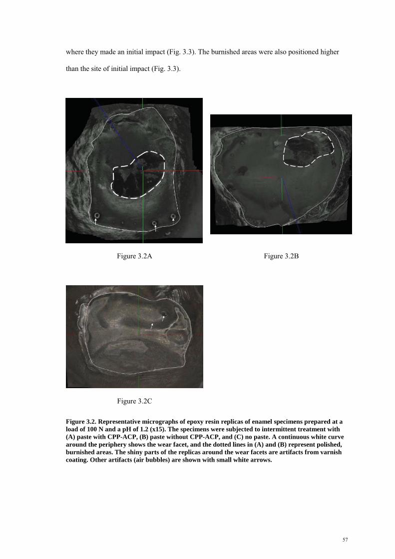
57
where they made an initial impact (Fig. 3.3). The burnished areas were also positioned higher
than the site of initial impact (Fig. 3.3).
Figure 3.2A
Figure 3.2B
Figure 3.2C
Figure 3.2. Representative micrographs of epoxy resin replicas of enamel specimens prepared at a load of 100 N and a pH of 1.2 (x15). The specimens were subjected to intermittent treatment with (A) paste with CPP-ACP, (B) paste without CPP-ACP, and (C) no paste. A continuous white curve around the periphery shows the wear facet, and the dotted lines in (A) and (B) represent polished, burnished areas. The shiny parts of the replicas around the wear facets are artifacts from varnish coating. Other artifacts (air bubbles) are shown with small white arrows.

58
Figure 3.3. The profile of an enamel specimen presented in Fig. 3.2A. The arrowheads in a circle above the specimen show the direction of movement of the opposing enamel specimen (anti-clockwise). A small white arrow on the left shows the area of the wear facet where the opposing specimen made an initial impact, and large straight white arrows show that the polished, burnished area was located higher than the site of initial impact.
3.5 Discussion
The attrition/erosion model used in this study is similar to that described by Kaidonis et al.,77
but it was modified to conform with intermittent application of CPP-ACP. The rationale behind
selection of load (100N) and a pH value of 1.2 has been described previously.52 A load of 100N
falls within a physiological range and represents a heavy attritional force.52 Bartlett et al.138
reported that the mean pH value of gastric juice in patients undergoing assessment for gastro-
esophageal reflux disease (GERD) was 2.9, ranging from 1.2 to 6.8. Previous researchers have
used a pH value of between 1.2 and 2.0 to simulate gastric regurgitation in vitro.52,118,139 Enamel
wear was limited to around 10,000 cycles because it was worn to its full thickness after this
many cycles. Some previous researchers have included stages of remineralization with artificial
saliva while investigating the characteristics of erosive tooth wear by dietary acids.53,118
However, a separate remineralization stage was not included in the present study, or in another
study simulating gastric regurgitation.139 Salivary factors are unlikely to provide protection

59
against strong erosive challenges (for example, gastric reflux).140 Furthermore, the rates of
salivary flow and acid clearance are lower during sleep-related gastric regurgitation than during
daytime regurgitation.83
We are unaware of previous reports confirming the effect of remineralizing agents on reducing
erosive tooth wear at a very low pH of 1.2. Furthermore, information on the effect of
remineralizing agents on the prevention of attritional tooth wear in combination with erosion is
limited. A previous study by Li et al.76 found that pre-treatment of dentine specimens with a
highly concentrated fluoride varnish (containing 22,600ppm fluoride at pH 3.9) did not reduce
wear between opposing enamel and dentine specimens. In this context, the findings of the
present study are encouraging in the management of tooth wear.
CPP-ACP was observed to reduce erosive enamel wear under severe erosive conditions
simulating a combination of heavy attrition and gastric regurgitation. However, the nature of
association between sleep bruxism and GERD is largely unexplored. Miyawaki et al.80 reported
that electromyographic (EMG) recording, and physiological gastroesophageal reflux (GER)
episodes corresponding to oesophageal pH values between 3.0 and 5.0, occurred more
frequently in bruxers than in healthy control subjects. However, their findings cannot be
extrapolated to individuals suffering from GERD. It is likely that sleep bruxism and GERD may
occur concurrently in heavy bruxers who also suffer from obstructive sleep apnoea syndrome
(OSAS) and obesity. It has been reported that OSAS and obesity seem to predispose individuals
to nocturnal GERD.83
Previous work has shown the potential of CPP-ACP as an anticariogenic agent both in vitro and
in situ,126,141,142 with CPP-ACP preventing demineralization and promoting remineralization of
sub-surface carious lesions in enamel and dentine.143,144 However, the mechanism by which
CPP-ACP reduces tooth wear is unclear.95,125 A recent study has shown that CPP-ACP can
increase microhardness of enamel and reduce erosion by cola drinks,68 implying that CPP-ACP

60
is capable of remineralizing eroded lesions. The process of remineralization of eroded lesions is
unclear, but it is likely to involve a repair process by deposition of mineral into the porous zone
rather than crystal regrowth.100
The differences in wear depths between experimental groups 1 and 2 are likely to reflect the
remineralizing potential of CPP-ACP in reducing erosive enamel wear. Interestingly, paste
without CPP-ACP also reduced erosive enamel wear, probably by lubricating the wear interface
(Fig. 3.2). This finding is consistent with the observation that parts of wear facets treated with
pastes with or without CPP-ACP were shiny and burnished, and corresponded to areas where
sliding wear occurred between opposing specimens (Figs. 3.2A and B). CPP-ACP paste seemed
to have provided greater protection against three-body abrasive wear (involving sliding wear of
opposing specimens) than impact stress (involving a greater vertical component of force at the
site of initial impact) (Fig. 3.3). The third bodies acting at the wear interface are likely to
include broken down enamel fragments, CPP-ACP and other ingredients of CPP-ACP paste
with a sticky consistency (for example, glycerol, D-sorbitol and propylene glycol). In addition,
the role of proteins in CPP-ACP paste (for example, casein) also needs to be investigated
because the protein phase can modify mechanical and tribological properties of enamel.
Recently, Swain and colleagues have noted that the protein phase between apatite crystals of
enamel helps to distribute shear strain,145 to deflect and arrest cracks146 and to sustain repetitive
cyclic contact loading.147
Further studies are needed to clarify the nature of lubricating and remineralizing potential of
CPP-ACP on tooth wear prevention, and to compare its effectiveness with nightguards, other
lubricating agents (for example, artificial saliva and food slurry) and remineralizing agents (for
example, fluoride products and a combination of CPP-ACP and fluoride). It is also unclear
whether there might be an additive effect of different preventive strategies on tooth wear.

61
On the basis of this investigation, it appears that CPP-ACP may help reduce enamel wear under
severe wear conditions, simulating heavy attrition and gastric regurgitation. Both remineralizing
and lubricating properties of CPP-ACP paste appear to contribute to wear reduction. The
lubricating effect of this paste seems to be more pronounced on areas of wear facets where
sliding wear occurred between opposing enamel surfaces than on surfaces subjected to impact
stress. Future in situ studies and clinical trials are needed to determine the true potential of CPP-
ACP in preventing erosive tooth wear.
3.6 Acknowledgements
This project was supported by grants from the Australian Dental Research Foundation Inc
(Grant no: 40/2005) and the National Health and Medical Council of Australia (Grant no:
349431). We would like to thank Ms Nancy Briggs for statistical analysis, G C Asia Pty Ltd for
providing Tooth Mousse® samples, and the 3M ESPE Corporation for donating impression
materials. Technical assistance provided by Dr My Anh Vu, Mr Victor Marino and Adelaide
Microscopy is also gratefully acknowledged.

62
CHAPTER 4
THE EFFECT OF CASEIN PHOSPHOPEPTIDE - AMORPHOUS
CALCIUM PHOSPHATE ON EROSIVE ENAMEL WEAR BY
TOOTHBRUSH ABRASION
This manuscript forms part of a paper titled “The effect of casein-phosphopeptide amorphous
calcium phosphate on erosive enamel and dentine wear by toothbrush abrasion”, which has been
published in the Journal of Dentistry (J Dent). This paper has been attached in the Appendices
(Paper 3 of Chapter 9, Section 1.1.1). As this thesis focuses on enamel wear prevention,
findings on dentine wear prevention are not included in this chapter. Experiments related to this
chapter (and the J Dent paper) were conducted during a sabbatical in King’s College London
between June 2007 till September 2007.
Permission has been obtained from the Elsevier to reproduce part of the J Dent paper in this
thesis.

63

64
The effect of casein phosphopeptide - amorphous calcium phosphate on erosive enamel wear by
toothbrush abrasion
S. Ranjitkara
J. M. Rodriguezb
J.A. Kaidonisa
L.C. Richardsa
G.C. Townsenda
D.W. Bartlettb,*
aSchool of Dentistry
The University of Adelaide
Adelaide SA 5005
Australia
bDepartment of Prosthodontics
King’s College London Dental Institute
Guy’s Hospital
London SE1 9RT
UK
Short title: Prevention of erosive tooth wear
Key words: abrasion, erosion, CPP-ACP, remineralization, lubrication
*Corresponding author
Prof David W. Bartlett
Professor/Hon Consultant in Prosthodontics
King’s College London Dental Institute
Floor 25, Guy’s Tower
London Bridge SE1 9RT, UK
Tel: +44 20 7188 5390; Fax: +44 20 7188 1792
E-mail: [email protected]

65
4.1 Abstract
Objective: In addition to its role as a remineralizing agent in preventing dental caries, calcium
product (CPP-ACP) delivered as a mousse (Tooth Mousse®, TM) can reduce erosion of
enamel. The aim of this study was to determine whether CPP-ACP could also reduce erosive
tooth wear involving toothbrush abrasion. Methods: Flat, polished enamel specimens (n = 36)
were subjected to 10 wear regimes, with each regime involving erosion in 0.3% citric acid (pH
3.2) for 10min followed by toothbrush abrasion in a slurry of fluoride-free toothpaste and
artificial saliva (1:3 ratio by weight) under a load of 2N for 200 cycles. The specimens were
immersed in artificial saliva for 2hrs between wear regimes. In the experimental group 1, TM
(containing CPP-ACP) was applied at the beginning of each wear episode for 5min whereas
TM- (without CPP-ACP) was applied in the experimental group 2. No mousse was applied in
the control group. Results: TM significantly reduced enamel wear (mean ± SE, 1.26 ± 0.33μm
in the experimental group 1 vs 3.48 ± 0.43μm in the control group). There was also a trend for
mean enamel wear in experimental group 2 (2.41 ± 0.50μm) to be less than that in control group
but this was not significantly different. Conclusion: The finding that TM reduced erosive
enamel wear involving toothbrush abrasion, probably by remineralizing and lubricating eroded
enamel surfaces, may have implications in the management of tooth wear.

66
4.2 Introduction
A combination of erosion with attrition or abrasion, referred to as erosive tooth wear, can result
in more destructive wear than if these processes occur independently.3,75 Eroded enamel has a
shallow, softened subsurface layer, with crystals being thin and vulnerable to mechanical
wear.96,97 Similarly, eroded dentine has a demineralized, subsurface layer98 which is susceptible
to wear by toothbrush abrasion.7
Normal tooth brushing habits with toothpaste are unlikely to produce excessive wear over a
lifetime but the super-imposed influence of acids is known to increase wear rates.7 Previously
suggested strategies for preventing erosive tooth wear by toothbrushing include the application
of a protective resin coating on teeth108 or of topical fluoride, which increases the resistance of
tooth structure against erosive wear.110 It is not always practical to eliminate the causes of tooth
wear in individuals, so it is desirable to develop other effective preventive strategies to manage
tooth wear.108
Recent in vitro studies have indicated that Tooth Mousse® (TM, GC Corporation, Japan),
containing a phosphopeptide that stabilizes amorphous calcium phosphate (CPP-ACP) may
reduce dental erosion caused by citric acid121,122 and an acidic sports drink.125 These findings
support the observation that TM reduces erosion from wine in both enamel and dentine.122
Recently, Ranjitkar et al.95 reported that attritional wear of dentine was almost eliminated in
vitro with continuous application of TM compared with hydrochloric acid lubricant (pH 3.0)
and deionized water lubricant (pH 6.1). Furthermore, intermittent application of TM also
reduced dentine wear in both acidic and near neutral environments, highlighting its lubricating
and remineralizing properties in reducing erosive dentine wear.95 However, the effectiveness of
TM in reducing erosive tooth wear involving toothbrush abrasion has not been assessed.

67
The aim of this study was to determine whether TM could reduce enamel wear under in vitro
conditions simulating toothbrush abrasion after an exposure to dietary acid. It was hypothesized
that CPP-ACP contained in a mousse would reduce enamel wear under these conditions.
4.3 Materials and methods
4.3.1 Sample preparation
Twelve intact, human third molars with unknown history were randomly selected from a pool of
extracted teeth that had been stored in saturated aqueous thymol solution at 4OC to reduce
deterioration in storage, following approval from the Guy’s Ethical Committee (reference no.
04/Q0704/57). Enamel specimens were obtained from the mesial, buccal, distal and lingual
surfaces of the mid-coronal portion of each tooth using a diamond blade (Diamond wafering
blade XL 12205, Benetec Ltd, London, UK). Each specimen was then mounted in a brass
specimen holder using chemically-cured acrylic resin, with a reference stainless steel ring
placed around the periphery to enable measurement of erosion depths. An outer layer of 400μm
of enamel was removed, based on protocols developed by Ganss et al.,110 by polishing the
specimens flat progressively with 800, 1200, 2400 and 4000 grit silicon carbide sandpapers
(SiC-Paper, Struers A/S, Copenhagen, Denmark) on a rotating polishing machine (Struers
Labopol-1, Denmark).
4.3.2 Study design
Enamel specimens (n = 36) were randomly allocated into two experimental groups and one
control group so that each group (n = 12) contained equal numbers of specimens from the
mesial, buccal, distal and lingual surfaces of teeth. CPP-ACP delivered in a mousse was applied
to enamel specimens in experimental group 1 (TM), whereas the mousse with the same
formulation but without CPP-ACP (TM-) was applied intermittently in experimental group 2.
No mousse was applied in the control group.

68
The effectiveness of TM and TM- in reducing enamel wear was tested in an abrasion-erosion
model using a toothbrushing machine to reproduce the oral environment.99 All specimens were
bathed in artificial saliva for two hours prior to experimentation. Artificial saliva was prepared
according to the protocol used by Eisenburger et al.100 and contained the following ingredients
in deionized water: CaCl2.2H2O 0.7mmol/l; MgCl2 0.2mmol/l; KH2PO4 4.0mmol/l; HEPES
buffer (acid form) 20.0mmol/l; KCl 30.0mmol/l, with the pH being adjusted to 7.0. Enamel
sections were subjected to 10 wear regimes, with each regime involving erosion in 0.3% citric
acid (pH adjusted to 3.2 using NaOH buffer at 23.80C) for 10min followed by toothbrush
abrasion in a slurry of fluoride-free toothpaste and artificial saliva (1:3 ratio by weight) under a
load of 2N for 200 cycles at 100rpm. The reference stainless steel rings were covered with an
adhesive tape to prevent abrasion by toothbrushing.
In experimental group 1, TM was applied for a total of 5min following manufacturer’s
recommendation. After an initial application for 3min, TM was diluted to a 30% solution with
artificial saliva during the remaining 2min to simulate the washing effect of saliva intra-orally.
The same protocol was used for experimental group 2 using TM-, and for control group but
without the mousse. At the end of each wear regime, TM and TM- were gently rinsed off the
surface with tap water for 2min and then air-dried. Specimens were subjected to erosion in 0.3%
citric acid for 10min. The titratable acidity of the acid solution, measured as the volume of
0.1mol NaOH required to raise the pH of the solution to pH 7, was 5.8ml. The specimens were
then washed with water for a further 1min and blot-dried gently using paper towels.
The specimens were subjected to toothbrush abrasion in a toothbrush machine using previously
published protocols.133 The tooth sections were placed on a mounting base and bathed in a
slurry of fluoride-free toothpaste (Kingfisher natural toothpaste, Kingfisher, Norwich, UK) and
artificial saliva (1:3 ratio by weight) before being subjected to toothbrushing with soft-bristled
toothbrush (Oral-B® Plus Size 40, Oral B, UK). The brush heads were replaced after a total of
4000 wear cycles. At the end of this stage, the specimens were washed for 1min and blot-dried

69
gently using paper towels and then stored in artificial saliva for 2hrs before the next wear
regime began.
4.3.3 Assessment of tooth wear
Impressions of each specimen were obtained in polyvinyl siloxane (“Blue” Light Body-Fast Set,
3M ESPE ExpressTM, St Paul, USA) before the first wear episode and after the final wear
episode as described by Sundaram et al.133 Then, they were scanned on a Xyris 2000TL non
contacting laser profilometer (Taicaan® Technologies, Southampton, UK), which consisted of
a transmitter emitting a 785nm red laser beam with a spot size diameter of 30μm. The accuracy
(maximum resolution) of the scanner was 0.1μm and its reliability under repeated measurements
was 0.3μm. Data acquisition and analysis was performed with Boddies v1.81 software
(Taicaan® Technologies- Southampton UK). Tooth wear was calculated by measuring the
difference in step heights of tooth surfaces before and after the erosion-abrasion regime using
the surrounding stainless steel disc as a reference plane, and a mean of three measurements was
recorded for each specimen. Negative replicas were used to quantify wear depths because clear
acrylic resin around enamel specimens could not be detected by the laser beam. Furthermore,
the accuracy of the scanning process was dependent on the homogeneity of surface colour of
polyvinyl-siloxane material, with blue colour providing more accurate surface profiles than
other colours.
4.3.4 Statistical analysis
Based on power calculations (assuming an effect ��8��9�!���;������*������������������#�<�9�!�!�;�
power, 1- =�9�!� !������������������������� ������"��������������������������
intermediate dispersion of individual values), 12 enamel specimens were included in each
group. A linear mixed model analysis was designed using SAS software (Proc Mixed, SAS 9.1,
SAS Institute Inc, Cary, USA) to determine whether there were significant differences in wear
depths between the three experiments described. This model accounted for clustering of data
from different surfaces of the same teeth. Data were log-normally distributed, so they were

70
transformed to natural logarithms for statistical analysis. Statistical significance for the final
model was set at the 0.05 probability level.
4.4 Results
Figure 1 shows the mean (SE) of enamel wear in different groups. The values for enamel wear
were 1.26μm (0.33μm) in experimental group 1 (with TM application), 2.41μm (0.50μm) in
experimental group 2 (with TM- application) and 3.48μm (0.43μm) in the control group (Fig.
4.1). Intervention involving TM and TM- was found to have a significant effect on both enamel
and dentine wear (p<0.01). Post-hoc multiple comparison tests for enamel wear showed that the
mean wear depth in experimental group 1 (TM) was significantly less than that in the control
group (p<0.001).
Figure 4.1. Comparison of mean wear depths ± S.E.(µm) in enamel specimens between three groups. a,b,c Pairwise comparisons using a mixed model indicate significant differences in mean
log(wear depths) between experimental group 1 (TM) and control group (p<0.001)a
a
a

71
4.5 Discussion
The toothbrush erosion/abrasion model used in this study is similar to that applied by Vieira et
al.99 to investigate the effect of fluoride treatment on erosive enamel wear after 600
toothbrushing cycles. To simulate the intra-oral environment, toothbrush abrasion occurred in
the presence of a toothpaste-artificial saliva slurry and a two-hour remineralization period with
artificial saliva was included between wear regimes. It has been suggested that treatment of
eroded enamel with artificial saliva for two hours partially rehardens enamel in vitro,100 but
Vanuspong et al.101 indicated that this is less likely to occur in dentine. Nevertheless, Attin et
al.102 have observed that delaying toothbrushing by at least 30min after an erosive regime in situ
provides some protection against toothbrush abrasion. A total of 600 brushing strokes applied
during three wear regimes on bovine teeth was reported by Vieira et al.99 to correspond to
around two years of wear. The present model, and those used by most previous researchers,99,103
have used reciprocal toothbrush action against stationary tooth specimens, but Parry et al.104
suggested that the movement of reservoirs containing tooth specimens and toothpaste slurry
against stationary toothbrushes would better maintain slurry homogeneity at the wear interface.
Parry et al.104 also reported that toothbrushing forces selected in previous in vitro studies ranged
from 0.1N to 8.8N. However, a force of around 2N has been used by most researchers to
simulate normal toothbrushing force in vivo.56,99 To detect minute amounts of tooth wear using
profilometry, flat and polished specimens were used in the present study. However, it should be
noted that natural tooth surfaces erode more slowly than polished surfaces and that coronal
dentine erodes faster than radicular dentine.105
TM (containing CPP-ACP) resulted in significantly less erosive wear of enamel sections than
TM- (without CPP-ACP) or no TM (control specimens) (Fig. 4.1). The effect of the TM-
probably reflects the lubricating potential of its ingredients (such as glycerol) in wear reduction,
and the differences in wear depths between the experimental group 1 (TM) and experimental
group 2 (TM-) are likely to reflect the remineralizing potential of CPP-ACP in reducing erosive

72
tooth wear (Fig. 4.1). However, these findings should be interpreted with some caution because
of relatively small differences in wear depths between different groups.
The potential of CPP-ACP as an anticariogenic agent has been reported both in vitro and in
situ,141,142 with CPP-ACP preventing demineralization and promoting remineralization of sub-
surface carious lesions in enamel and dentine.143,144 CPP-ACP maintains saturation levels of
calcium and phosphate at the tooth surface and provides a reservoir of neutral ion pairs
(CaHPO40), which inhibit demineralization and promote the formation of hydroxyapatite
crystals inside carious lesions.143 CPP-ACP is also detectable in the plaque matrix and the
surface of bacterial cells of subjects three hours after consuming CPP-ACP-containing
mouthrinse or chewing gum.141 The mechanisms by which CPP-ACP reduces erosive tooth wear
are unclear. However, the finding that TM increases hardness of enamel eroded by cola drink68
implies that its erosion-inhibiting potential probably involves remineralization action. Unlike
the process of remineralization of carious lesions, eroded tooth structure is likely to be repaired
by deposition of mineral into the porous zone rather than crystal regrowth.100 This hypothesis is
consistent with the observation that superficial granular structures were noted to form on the
enamel surface, probably representing remineralized enamel structure, after treatment with a
sports drink containing CPP-ACP.125
A non-fluoridated toothpaste was used instead of fluoridated toothpaste in the present study
because fluoride has been found to act synergistically with CPP-ACP in remineralizing carious
lesions,148 potentially making it a confounding variable. Previous studies have observed that
non-fluoridated toothpaste results in greater erosive tooth wear than fluoridated toothpaste,132
and that the level of protection also varies with fluoride formulation.107 In this context, the use
of non-fluoridated toothpaste in our experiments could have resulted in relatively higher
effectiveness of CPP-ACP in tooth wear prevention. Future studies are needed to compare the
effectiveness of TM and fluoride, when used individually or in combination, in preventing
erosive tooth wear.

73
On the basis of this investigation, it appears that TM application may help reduce erosive tooth
wear of coronal enamel by toothbrush abrasion. These findings provide a basis for future in situ
studies and clinical trials that will determine the true potential of TM in preventing erosive tooth
wear.
4.6 Acknowledgements
This project was completed during the tenure of the NHMRC (National Health and Medical
Council of Australia) Dental Postgraduate Research Award to the first author. It was also
supported by research grants from the Australian Dental Research Foundation Inc. and G C Asia
Pty Ltd, and by travelling awards to the first author from the University of Adelaide and the
NHMRC of Australia. The authors also wish to thank Prof Tim Watson for providing laboratory
facility to complete this study, Ms Nancy Briggs for conducting statistical analysis, and Mr
Peter Pilecki and Mr Richard Mallet for providing technical assistance.

74
CHAPTER 5
THE EFFECT OF CPP-ACP ON ENAMEL EROSION UNDER
CONDITIONS SIMULATING GASTRIC REGURGITATION
This manuscript has been written in the format for the Australian Dental Journal. The findings
of this study are preliminary, and it will be submitted for publication after further experiments
comparing the erosion-inhibiting potential of CPP-ACP and fluoride have been conducted. This
manuscript is listed as paper 1 under Chapter 9, Section 1.1.2.

75
Statement of contribution by all authors
Title: The effect of CPP-ACP on enamel erosion under conditions simulating gastric
regurgitation
S Ranjitkar was primarily responsible for this project. S Ranjitkar designed and co-ordinated the
study, and interpreted the data.
JA Kaidonis, LC Richards and GC Townsend provided critical comments and general
supervision.
All co-authors were involved with the preparation of the manuscript.
We give written permission for this paper to be included in S Ranjitkar’s thesis titled “Biology
of tooth wear: preventive strategies”.

76
The effect of CPP-ACP on enamel erosion under conditions simulating gastric regurgitation
Ranjitkar S*
Kaidonis J*
Richards LC*
Townsend GC*
*School of Dentistry
The University of Adelaide
Adelaide SA 5005
Key words: tooth wear, demineralization, remineralization, experimental studies
Number of figures: 1
Address for correspondence:
Dr Sarbin Ranjitkar
School of Dentistry
The University of Adelaide
Adelaide SA 5005
Telephone: +61 8 8303 3069
Fax: +61 8 8303 3444
E-mail: [email protected]

77
5.1 Abstract
Background: Tooth Mousse® (TM), containing an anticariogenic remineralizing agent in the
form of casein phosphopeptide – amorphous calcium phosphate (CPP-ACP), has been found to
reduce erosive enamel wear under conditions simulating heavy attrition and gastric
regurgitation. The aim of this in vitro study was to determine the effect of TM on enamel
erosion under conditions simulating gastric regurgitation alone.
Methods: Flat, polished enamel specimens were subjected to regimes of 10 erosion episodes,
with each episode involving application of a mousse containing CPP-ACP (TM), a mousse with
the same formulation but without CPP-ACP (TM-) or no mousse for 5min followed by erosion
in hydrochloric acid (HCl) at pH 1.2. The specimens were immersed in artificial saliva for 2 hrs
between erosion episodes. In the experimental group 1, TM was applied at the beginning of
each erosion episode for 5min whereas TM- was applied in the experimental group 2. No TM or
TM- was applied in the control group. One-way ANOVA was conducted to determine whether
there were significant differences in mean erosion depths between different groups.
Results: There were no significant differences in mean erosion depths between the three groups
(p>0.05).
Conclusions: TM did not protect enamel against erosion under conditions simulating gastric
regurgitation at pH 1.2. Comparison of this finding with those of a previous study involving
heavy attrition at pH 1.2 suggests that lubricating effect of TM, rather than its remineralizing
effect, was probably the primary factor responsible for reduction of wear in that study.
78
5.2 Introduction
There has been an increase in the prevalence of erosive tooth wear (referring to tooth wear by
erosion combined with mechanical factors, such as attrition and toothbrush abrasion) as
increasing numbers of elderly patients are retaining their natural teeth to a stage where they
present with extensive wear.128 In addition, more younger patients are also presenting with wear
of both primary and permanent teeth.74,129 In this context, clinical management should focus on
early detection and prevention before a restorative approach is considered.128
Larsen et al.112,113 reported that fluoride at high concentration provided minimal protection
against dental erosion. However, Hove et al.118 observed that intensive application of fluoride
provided significant protection against enamel erosion under conditions simulating gastric
reflux (at pH 2.0). Other researchers have indicated that fluoride can protect enamel and dentine
from both erosion53,110,116 and a combination of erosion and toothbrush abrasion,53,102 but not
from attritional wear between opposing enamel and dentine specimens.76 Thus, there is still a
need to improve preventive strategies to manage the risk of erosive insult on teeth.107
Tooth Mousse® (TM), manufactured by the GC Corporation, Japan, contains an anticariogenic
remineralizing agent CPP-ACP (amorphous calcium phosphate nanocomplex stabilized by
casein phosphopeptide) that prevents demineralization and promotes remineralization of sub-
surface carious lesions in both enamel and dentine.143,144 In addition to its anticariogenic
property, CPP-ACP has been found to reduce enamel erosion from citric acid121 and to reduce
the erosive potential of an acidic sports drink.125 A recent study reported that attritional wear of
dentine was almost eliminated with continuous application of TM compared with hydrochloric
acid lubricant (pH 3.0) and deionized water lubricant (pH 6.1).95 Furthermore, intermittent
application of TM reduced dentine wear in both acidic and near neutral environments,
highlighting its lubricating and remineralizing properties in reducing attritional dentine wear.95
These findings are supported by the observation that both remineralizing and lubricating

79
properties of TM appeared to contribute to reduction in enamel wear under severe conditions
simulating heavy attrition (100N) and gastric regurgitation (pH 1.2). However, the effect of TM
on enamel erosion under conditions simulating gastric reflux alone is unknown.
The aim of this in vitro study was to determine the effectiveness of TM in reducing enamel
erosion under conditions simulating gastric regurgitation at pH 1.2. It was hypothesized that TM
would prevent enamel erosion under this condition.
5.3 Materials and methods
5.3.1 Sample preparation
Twelve intact, human third molar teeth with unknown history were selected from a pool of
extracted teeth that had been stored in formalin after extraction as part of routine dental
treatment in South Australia. The protocol for collection of extracted teeth was approved by the
University of Adelaide Human Ethics Committee (H/27/90). Enamel specimens were obtained
from the mesial, buccal, distal and lingual surfaces of the mid-coronal portion of each tooth
using a diamond blade. Each specimen was then mounted in clear epoxy resin, and an outer
layer of 500μm of enamel was removed by polishing the specimens flat progressively with 500
grit silicon carbide sandpaper (SiC-Paper, Struers A/S, Copenhagen, Denmark) and diamond
discs with grain size of 15μm, 3μm and 1μm (MD-Dur cloths, Struers A/S, Copenhagen,
Denmark) on a rotating polishing machine (Struers Labopol-1, Copenhagen, Denmark).
5.3.2 Study design
Enamel specimens (n = 36) were randomly allocated into two experimental groups and one
control group so that each group (n = 12) contained equal number of specimens from the mesial,
distal, buccal and lingual surfaces of teeth. TM was applied intermittently to enamel specimens
in the experimental group 1, and Tooth Mousse with the same formulation but without CPP-
ACP (TM-) was applied intermittently in the experimental group 2. No TM or TM- was applied
in the control group.

80
The effectiveness of TM and TM- in reducing enamel erosion was tested in an erosion model
that is similar to the erosion-abrasion model99 used in a previous study [Chapter 4], but without
the abrasion component. All specimens were bathed in artificial saliva for two hours prior to
experimentation. Artificial saliva was prepared according to the protocol used by Eisenburger et
al.100 and contained the following ingredients in deionized water: CaCl2.2H2O 0.7mmol/l;
MgCl2 0.2mmol/l; KH2PO4 4.0mmol/l; HEPES buffer (acid form) 20.0mmol/l; KCl 30.0mmol/l,
with the pH being adjusted to 7.0.
Enamel specimens were subjected to regimes of 10 erosion episodes at room temperature
(around 23OC), with each regime involving application of TM or TM-, erosion in hydrochloric
acid (HCl) at pH 1.2 and then remineralization with artificial saliva. In experimental group 1,
TM was applied for a total of 5min following manufacturer’s recommendation. After an initial
application for 3min, TM was diluted with artificial saliva during the remaining 2min to
simulate the washing effect of saliva intra-orally. The same protocol was used for experimental
group 2 using TM-, and for the control group but without the application of TM or TM-. At the
end of each regime, TM and TM- were gently rinsed off the surface with tap water for 2min and
then air-dried. Specimens were then subjected to erosion in 0.063M hydrochloric acid (pH 1.2)
for 2min. The specimens were then washed with water for a further 1min and blot-dried gently
using paper towels. Then, they were stored in artificial saliva for 2hrs before the next erosion
episode began.
5.3.3 Assessment of tooth wear
Quantitative measurements were undertaken for erosion depths by using a 2D contacting
profilometer ‘Zeiss Handysurf E-35A’ (Advanced Metrology Systems Ltd, Leicester, UK),
whose accuracy (resolution in z-axis) is less than one micron. In this system, an active piezo
sensor detects contact between the sensor and the scanned surface and then data is exported to a
personal computer to create a 2D surface topography. The maximum tracing distance for this

81
sensor is 4.0mm over a height of 150μm. An average of three erosion depths was recorded for
each specimen. Nine specimens were discarded (two in the experimental group 1, four in the
experimental group 2 and three in the control group) because of the difficulty in creating surface
profiles of these specimens.
5.3.4 Statistical analysis
7���������"������� �������.��� ���������������8��9�!���;������*������������������#�<�9�!�!�;�
power, 1- =�9�!� !������������������������� ������"��������������������������
intermediate dispersion of individual values), 12 enamel specimens were initially included in
each group. One-way ANOVA was used to determine whether there were significant
differences in mean erosion depths between the experimental and control groups. Statistical
significance was set at the 0.05 probability level.
5.4 Results
Mean erosion depths and standard errors for enamel were 37.5 ± 2.4μm in the experimental
group 1, 30.1 ± 2.5μm in the experimental group 2 and 34.7 ± 2.5μm in the control group. There
were no significant differences in mean erosion depths between the three groups (p>0.05) (Fig.
5.1).

82
0
10
20
30
40
50
Tooth Mousse Tooth MousseMinus
Control
Treatment groups
Wea
r de
pths
(in
mic
rons
)
Figure 5.1. Comparison of the mean erosion depths ± S.E.(µm) between different groups treated with Tooth Mousse®, Tooth Mousse Minus (without CPP-ACP) or no mousse.
5.5 Discussion
The finding that TM did not protect enamel against erosion under conditions simulating gastric
regurgitation at pH 1.2 is consistent with that of Willumsen and colleagues,119 who concluded
that enamel was severely eroded regardless of pre-treatment with sodium or stannous fluoride
solutions at this pH value. Our finding was surprising, given that enamel wear was reduced by
52.2% with intermittent TM application and by 31.5% with intermittent TM- application in a
previous study conducted under similar conditions involving heavy attrition at pH 1.2 (Chapter
3). In that study, it was concluded that both remineralizing and lubricating properties of TM
probably contributed to the reduction in erosive wear. TM and TM- were delivered as a paste in
that study, and, in comparison, the experimental conditions in the present study were more
conducive for remineralization of eroded lesions. These findings indicate that the lubricating
effect of TM was probably the primary factor responsible for the reduction in erosive enamel
wear in the previous study (Chapter 3). An alternative explanation to the remineralizing
potential of TM in reducing erosive enamel wear in that study (Chapter 3) is that CPP-ACP
enhanced the lubricating potential of other ingredients of TM (for example, glycerol), resulting
in greater reduction in enamel wear with TM than with TM-.

83
The anticariogenic properties of TM, involving prevention of demineralization and
enhancement of remineralization, are well-documented.111,143 By maintaining saturation levels
of calcium and phosphate at the tooth surface, CPP-ACP provides a reservoir of neutral ion pair
(CaHPO40) that inhibits enamel demineralization and promotes formation of hydroxyapatite
crystals inside carious lesions.143 CPP-ACP is also detectable in the plaque matrix and the
surface of bacterial cells of subjects three hours after consuming CPP-ACP-containing
mouthrinse or chewing gum.141 TM has also been reported to increase hardness of enamel after
erosion in a cola drink (pH 2.7),68 implying that its erosion-inhibiting properties are likely to
involve prevention of demineralization and remineralization of eroded lesions.122 However, the
finding of the present study indicates that the erosion-inhibiting potential of TM is pH
dependent, with little protection being provided in a very strong erosive environment at pH 1.2.
The findings of the present study along with those of other studies involving CPP-ACP 121,122,125
have implications in the clinical management of tooth wear. Current recommendations for self-
application of TM for individuals at risk of dental erosion include application of TM before or
after the occurrence of erosive episodes.149 Liberali150 formulated a preventive management plan
involving Neutraflour toothpaste application (containing 5000ppm fluoride) twice daily and TM
application nightly in an individual suffering from gastro-oesophageal reflux disease. It has
been reported that neither fluoride119 nor CPP-ACP seem to protect enamel against erosion at
pH 1.2, although stannous fluoride provides greater protection against enamel erosion at pH 2.0
than sodium fluoride.118 These findings imply that a more effective erosion-inhibiting product is
needed for the clinical management of erosion in individuals suffering from gastric
regurgitation.
The present study did not compare the erosion-inhibiting effect of TM with that of fluoride as it
is difficult to make direct comparisons between our findings and those for fluoride118,119 due to

84
methodological differences. Future studies are needed to compare the relative benefits of CPP-
ACP and fluoride, when used individually or in combination, in preventing dental erosion.
5.6 Conclusion
Within the limitations of the present in vitro study, it is concluded that TM does not protect
enamel against erosion under conditions simulating gastric regurgitation at pH 1.2. This finding
is consistent with a previous finding that fluoride also does not protect enamel against erosion at
this pH value. Thus, a more effective erosion-inhibiting product is needed for the clinical
management of erosion in individuals suffering from gastric regurgitation. Comparison of our
finding with those of a previous study involving heavy attrition at pH 1.2 (Chapter 3) indicates
that lubricating effect of TM, rather than its remineralizing effect, was probably the primary
factor responsible for reduction of enamel wear in that study.
5.7 Acknowledgements
This project was supported by a research grant from the Australian Dental Research Fund Inc.
The second author was a recipient of the Dental Postgraduate Research Award from the
National Health and Medical Research Council of Australia (NHMRC). Technical assistance
provided by Dr Chin Nguyen, Adelaide Microscopy, Mr Victor Marino, Ms Archana Paneru
and Mr Nabaraj Dahal is gratefully acknowledged.

85
CHAPTER 6
SPECTRAL ANALYSIS OF THE ENAMEL SURFACE TREATED
WITH CPP-ACP
This manuscript has been written in the format for the Australian Dental Journal. The findings
of this study are preliminary, and it will be submitted for publication after further experiments
using a larger sample have been conducted. This manuscript has also been listed as paper 2
under Chapter 9, Section 1.1.2.

86
Statement of contribution by all authors
Title: Characterization of the enamel surface treated with CPP-ACP using mass spectrometry
S Ranjitkar was primarily responsible for this project. S Ranjitkar and A Lewis both designed
and co-ordinated the study, and collected and interpreted the data. A Lewis ran enamel
specimens under the ToF-SIMS equipment. V Marino participated in experimental design and
sample preparation.
JA Kaidonis, LC Richards and GC Townsend provided critical comments and general
supervision.
All co-authors were involved with the preparation of the manuscript.
We give written permission for this paper to be included in S Ranjitkar’s thesis titled “Biology
of tooth wear: preventive strategies”.

87
Spectral analysis of the enamel surface treated with CPP-ACP
S Ranjitkar*
A Lewis**
J Kaidonis*
V Marino*
LC Richards*
GC Townsend*
*School of Dentistry
The University of Adelaide
Adelaide SA 5005
**ACeSSS
The University of South Australia
Mawson Lakes SA 5095
Key words: tooth wear, abrasion, prevention, Tooth Mousse®, lubrication, ToF-SIMS
Number of figures: 2

88
6.1 Abstract
Background: Tooth Mousse® (TM) can reduce abrasive tooth wear by lubrication, but the
nature of third-body components at the wear interface is not properly understood. The aim of
this study was to characterize the enamel surface after treatment with TM and to clarify its
mode of action in reducing tooth wear.
Methods: Two flat, polished enamel specimens from the buccal and lingual halves of a human
third molar tooth were selected for this study. The buccal (experimental) specimen was
subjected to treatment with TM for 5min followed by washing with ultra-pure double deionized
water for 2min before being dried with liquid nitrogen for 1min. No TM was applied to the
lingual (control) specimen. Spectroscopy of the specimens was undertaken using ToF-SIMS
(Time of Flight - Secondary Ion Mass Spectrometry) to obtain mass spectra in six different
areas on each specimen.
Results: The peak areas for silicon and ethyl siloxane, which represented their relative
concentrations, were greater in the experimental specimen than in the control specimen. The
peak area for calcium was lower in the experimental specimen. Comparison of possible
substances representing glycerol (C3H8O3, C3H7O3, CH2O and CH3O) between the two
specimens yielded inconsistent results.
Conclusions: Traces of silicon and ethyl siloxane, found to be ingredients of TM, are potential
third-bodies leading to polishing at the wear interface and reduction in tooth wear. Lower
calcium content on the experimental specimen indicated that a thin layer of CPP-ACP was
probably present on its surface, with its calcium content being lower than that of the
hydroxyapatite of the control specimen. Further research is planned to obtain reference peaks
for glycerol and other organic ingredients of TM in order to improve the accuracy of the ToF-
SIMS analysis.

89
6.2 Introduction
To conform with the paradigm of minimal intervention dentistry, clinical management of tooth
wear should focus on early detection and prevention before a restorative approach is
considered.128 Recent evidence suggests that fluoride can protect enamel and dentine from both
erosion110,118 and a combination of erosion and toothbrush abrasion,53,102 but not against
attritional wear between opposing enamel and dentine specimens.76
An anticariogenic product (for example, Tooth Mousse®, TM, G C Asia Pty Ltd, Japan)
containing remineralizing agent in the form of CPP-ACP (amorphous calcium phosphate
nanocomplex stabilized by casein phosphopeptide) has been reported to reduce enamel erosion
from citric acid121 and white wine (pH 3.2),122 and to reduce erosive tooth wear involving
toothbrush abrasion (pH 3.2).137 However, it does not appear to be effective against erosion
simulating gastric regurgitation (pH 1.2) (Chapter 5). In another study investigating the effect of
intermittent application of TM on erosive enamel wear between opposing enamel specimens at
pH 1.2, TM with and without CPP-ACP was found to reduce erosive enamel wear (Chapter 3).
Ranjitkar et al.95 also reported that intermittent application of TM reduced dentine wear in both
acidic and near neutral environments, indicating that both its remineralizing and lubricating
properties were probably responsible for reduction in erosive dentine wear. Furthermore, the
observation that attritional wear of dentine was almost eliminated with continuous application
of TM compared with hydrochloric acid lubricant (pH 3.0) and deionized water lubricant (pH
6.1) suggests that the lubricating property of TM probably had a more profound effect on wear
reduction than its remineralizing property.95
As TM was applied intermittently between erosive wear episodes and appeared to have been
washed off during visual assessment in Chapter 3 and in our previous study,95 and it seems that
traces of its ingredients (for example, glycerol and CPP-ACP nanocomplexes) adhered to the
enamel surface and that they lubricated the wear interface by acting as third-bodies. Previous

90
researchers have used a very sensitive surface analytic technique, the secondary ion mass
spectrometry (SIMS) that is capable of detecting concentrations of elements or molecules at
subparts per million, to identify corrosion products from CoCr orthopaedic implants at the site
of physical wear.70,71 SIMS has also been used to map the chemical changes across an incipient
carious lesion63 and to elucidate the mechanism of fluoride uptake associated with CO2 laser
treatment on enamel.72 In this context, Time of Flight - Secondary Ion Mass Spectrometry (ToF-
SIMS) has the potential to identify the types of third-body components at the wear interface,
and to elucidate the mechanism by which TM reduces tooth wear.
By characterizing the enamel surface after treatment with TM, the aim of the present study was
to clarify the likely modes of action of TM in preventing enamel wear. It was hypothesized that
ingredients of TM would be detected on the enamel surface treated with TM. Although other
ingredients with a sticky consistency (for example, D-sorbitol, propylene glycol, sodium
carboxycellulose and guar gum) may contribute to wear reduction, the present study was limited
to preliminary investigation of calcium, phosphorous and glycerol contents in TM.
6.3 Materials and methods
6.3.1 Sample preparation
One intact, human third molar tooth was selected from a pool of extracted teeth that had been
stored in formalin. This tooth had been extracted as part of routine dental treatment in South
Australia. The protocol for collection of extracted teeth was approved by the University of
Adelaide Human Ethics Committee (H/27/90). After the tooth was sectioned into buccal and
lingual halves, each half was mounted in clear epoxy resin followed by the removal of an outer
layer of 500μm of enamel by polishing flat progressively with 500, 1000 and 2400 grit silicon
carbide sandpapers (SiC-Paper, Struers A/S, Copenhagen, Denmark) and diamond discs with
grain size of 15μm, 3μm and 1μm (MD-Dur cloths, Struers A/S, Copenhagen, Denmark) on a
rotating polishing machine (Struers Labopol-1, Copenhagen, Denmark). Ultra-pure, double

91
deionized water (pH 6.1) was used as a lubricant with silicon carbide sandpapers, and standard
diamond pastes and green lubricant were used with diamond discs.
6.3.2 Study design
After sample preparation, the specimens were washed thoroughly with ultra-pure, double
deionized water for 2min and dried gently with air. The experiments were conducted in a
laminar flow cabinet providing an aseptic and dust-free environment. The buccal (experimental)
specimen was treated with TM for 5min and then washed with ultra-pure deionized water for
2min before being dried with liquid nitrogen for 1min. The lingual (control) specimen was
washed and dried using similar protocols, but no TM was applied to it.
Spectroscopy of specimens was undertaken using ToF-SIMS to image mass spectra in six
different areas on each specimen. Spectral analysis was performed using a PHI TRIFT 2100
ToF-SIMS equipped with gallium liquid metal ion gun (LMIG) operating with a beam current
of 1nA and a net impact energy of 20kV. In spectrographic mode the instrument allows
chemical characterization for selected areas and, with careful experiment design, this
characterization can be quantified. In imaging mode, the ToF-SIMS produces maps of chemical
species on surfaces at concentrations of parts per million, with the spatial resolution being 1-
�����'��������� ������� �������������������������������������8������������������������
surface. Damage to the uppermost monolayers is minimized by applying low primary gallium
����� �������������������"���������������������������������������� !���"�����
continuous beam, with analysis being taken at regular time intervals from the central 100 x
�!���������%� ��������������������������������"������������������������������al depths by
calibrating the instrument with a 100-nm Ta2O5 film on Ta foil and using a Monte Carlo ion
Trajectory computer program (TRIM) to estimate the sputter rates of the relevant materials.
The SIMS library was searched for peak areas corresponding to the molecular weights of
different ions, including silicon, calcium, phosphate and glycerol. SIMS is very sensitive in

92
detecting traces of inorganic ions, but the identification of organic ions requires some caution
because of the possibility of desorption of atoms during the ionization process. To identify
glycerol (C3H8O3), the spectral analysis was conducted on two combinations of carbon,
hydrogen and oxygen elements (C3+H7+O3 and C3+H8+O3) as well as two molecules with
atomic masses of 91.05 (for C3H7O3) and 92.05 (for C3H8O3). As carbon chains of a molecule
can be disintegrated during the ionization process, SIMS analysis was also conducted for the
hydroxy methyl species of glycerol [-CH2-OH (-CH3O) and -CH-OH (-CH2O)]. Preliminary
investigations showed that there were no significant differences in the contents of C3+H7+O3
and C3+H8+O8 between the experimental and the control specimens, so final SIMS analysis for
glycerol was limited to glycerol ions (C3H7O3 and C3H8O3) and their methyl species (-CH3O and
-CH2O). Based on the assessment of molecular weights, a peak corresponding to amu 30.04 was
identified to represent -CH2O and peaks with amu 31.02 or 31.04 were identified to represent -
CH3O.
Given that electropositive and electronegative ions are represented in +ve spectrum (+SIMS)
and –ve spectrum (-SIMS) respectively, and that organic ions are represented in both +ve and –
ve spectra (+SIMS and –SIMS), the SIMS analyses for silicon (Si+4), calcium (Ca+2) and
phosphorous (P-3) were conducted on their respective spectra and the SIMS analysis for glycerol
was conducted on both +SIMS and –SIMS. These organic ions were identified on the spectra
after the SIMS library was calibrated for CH3, C2H5 and C3H7. The peak areas for these organic
ions (representing their relative concentration) were normalized to the total counts of peaks,
with the total peak area for all ions being 1. This allowed for any variation in the peak intensity
that could have been caused by the variation in sample topography, differences in sample
positioning in the holder and instrument set up. Total peak areas for the organic ions were then
compared between the experimental and the control specimens.
During preliminary analysis, peak areas for silicon and ethyl siloxane (C2H5SiO) were noted to
be greater in the experimental than the control specimens. As ethyl siloxane is a common

93
industrial contaminant, a separate investigation was conducted by characterizing the surface of a
clean silicon wafer after treatment with TM for 5min followed by washing and drying using
standard protocols. This analysis confirmed that ethyl siloxane was indeed present in TM. Thus,
spectral analysis for silicon and ethyl siloxane was also included in the overall spectral analysis.
As the spectral analysis was limited to sampling from six regions of one experimental and one
control specimen, no statistical analysis was conducted to compare the mean peak areas for
organic and inorganic ions because they were likely to be highly correlated. Thus, the
assessment of enamel surfaces treated with TM involved comparison of mean values for six
peak areas of inorganic and organic ions of interest between the specimens.
6.4 Results
The peak areas for silicon and ethyl siloxane were greater by 30.4 fold and 12.3 fold in the
experimental specimen than in the control specimen (Fig. 6.1). The peak area for calcium was
lower by 0.8 fold in the experimental specimen than in the control specimen (Fig. 6.1), but the
peak areas for phosphorous were similar between the specimens.
Figure 6.1. Comparison of the peak areas for silicon, calcium and ethyl siloxane between the experimental specimen (treated with Tooth Mousse®) and the control specimen.

94
Analysis of +SIMS library indicated that the peak area for a molecule of amu 92.04 (probably
representing C3H8O3) was greater by 6.0 fold in the experimental specimen compared with the
control specimen. However, the peak area for a molecule of amu 91.05 (probably representing
C3H7O3) was less by 0.5 fold in the experimental specimen than the control specimen. In the –
SIMS library, the peak areas for these organic ions were similar between the experimental and
the control specimens (Fig. 6.2).
Figure 6.2. Comparison of the relative concentration of molecules with atomic mass units of 91.054 and 92.0473, probably representing glycerol, between the experimental specimen (treated with Tooth Mousse®) and the control specimen.
Comparison of the peak areas for methyl species of glycerol (-CH2O and -CH3O) between the
two specimens also yielded inconsistent results. The peak areas for an organic ion with amu
30.04 (probably representing -CH2O) were similar between the experimental specimen (0.021)
and the control specimen (0.023) in the +SIMS library, but the peak area was greater in the
experimental specimen than the control specimen in the –SIMS library (0.204 vs 0.090
respectively). Two organic ions with amu 31.02 and 31.04 were identified as possible methyl
species -CH3O of glycerol. The peak areas for an organic ion with amu 31.02 were similar

95
between the experimental and control specimens in both +SIMS and –SIMS libraries. The peak
area for an organic ion with amu 31.04 was less in the experimental specimen than that in the
control specimen in the +SIMS library (0.003 vs 0.006 respectively), but these areas were
similar between the specimens in the –SIMS library.
6.5 Discussion
Traces of silicon and ethyl siloxane were found on the enamel surface after treatment with TM,
indicating that they were ingredients of TM. Silicon carbide discs were used to polish enamel
specimens in this study, and the possibility of silicon contamination from these discs cannot be
ruled out. However, this is unlikely to be a major problem because enamel specimens were
further polished with three diamond discs, and silicon particles were likely to have been
removed during this stage. Furthermore, the same polishing protocols were used for both the
experimental and control specimens, and silicon content was noted to be very low in the control
specimen. As the spectral analysis is sensitive to contamination in the laboratory, the
experiments were conducted in an ultra clean environment. Possible bias arising from the
variation in the enamel structure between teeth is also unlikely because both the experimental
and control specimens were obtained from the same tooth. Furthermore, anisotropic
microstructure of enamel is unlikely to affect ToF-SIMS analysis.63
The calcium content on the surface of the control specimen represented that of hydroxyapatite
in enamel. The observation that calcium content was lower in the experimental specimen
suggests that a thin layer of TM (including CPP-ACP) was present on its surface and that the
calcium content was lower in this film than that in hydroxyapatite. The calcium concentration in
the experimental specimen was probably lowered by presence of other ingredients in TM (for
example, casein phosphopeptide and other organic and non-organic ingredients) on the surface
of the experimental specimen.

96
The findings of the present study support the hypothesis that CPP-ACP nanocomplexes and
silicon in the form of silica and ethyl siloxane in TM lubricate the wear interface to reduce tooth
wear caused by both attrition95 and toothbrush abrasion.137 The mechanism by which silicon in
TM lubricates tooth surface may resemble that involved in polishing of tooth specimens using
silicon carbide discs in laboratories. The lubricating action of TM may also involve other
ingredients that have a sticky consistency (for example, D-sorbitol, propylene glycol, sodium
carboxycellulose and guar gum). Thus, further spectral analysis is needed for these ingredients
to better understand the nature of third-body components at the wear interface. In addition, the
role of proteins in TM (for example, casein) also needs to be investigated because they have a
potential to modify the mechanical and tribological properties of enamel. Recently, it has been
noted that the protein phase between apatite crystals of enamel helps to distribute shear strain,145
to deflect and arrest cracks146 and to sustain repetitive cyclic contact loading.147
Characterization of glycerol and its methyl species on the enamel surface yielded inconsistent
results, probably because of the lack of ‘reference peaks’ for glycerol. In this context, further
experiments need to be conducted to characterize enamel specimen after treatment with glycerol
alone. This will highlight which organic ions (with amu 92.04 or 91.05) and methyl species (-
CH3O or -CH2O) represent glycerol and will improve the reliability for the analysis of glycerol
content in TM. In this context, individual ‘reference peaks’ should be prepared for other
ingredients of TM that can lubricate the wear interface before conducting their spectral analyses.
This study has identified some third-bodies that are probably responsible for lubricating the
wear interface at a relatively neutral pH value. Further studies are needed to provide an insight
into the mechanisms involved in reducing erosive tooth wear. Barbour and Rees69 described the
application of SIMS in creating surface mapping of calcium and magnesium of an eroded
enamel surface. This method has the potential to enable early mineral loss of eroded tooth
surface, but further study is planned to validate its suitability for erosion studies.51

97
6.6 Conclusion
Improved understanding about the characteristics of opposing wear surfaces, and the role of
third-body components in lubricating the wear interface, should assist in the development of
new strategies for tooth wear prevention.52 In this context, this study has provided some insight
into potential third-body components from TM that can lubricate the wear interface. In
particular, silicon, ethyl siloxane and CPP-ACP nanocomplexes are likely to contribute to
polishing at the wear interface and therefore lead to reduction in tooth wear. Further research is
planned to better understand the role of other organic ingredients of TM in reducing tooth wear,
including proteins (for example, casein), that are likely to modify the mechanical properties of
enamel.
6.7 Acknowledgements
This project was supported by a research grant from the Australian Dental Research Foundation
Inc. We would like to thank Dr My Anh Vu for providing technical assistance.

98
CHAPTER 7
GENERAL DISCUSSION AND CONCLUSIONS

99
Understanding of the characteristics of tooth wear under different conditions (for example,
under different loads and pH values) has been based primarily on in vitro studies. Although
findings from in vitro studies should only be used to obtain general information about tooth
wear, and should not be extrapolated directly to in vivo situations, in vitro studies can separate
out different components of tooth wear. In vitro studies cannot replicate all possible conditions
in vivo, so three conditions involving enamel wear were chosen for the present study simulating
heavy attrition (under a load of 100N) in combination with gastric regurgitation (at pH 1.2),
toothbrush abrasion (under a load of 2N) after an erosive episode (at pH 3.2), and erosion
simulating gastric regurgitation alone (at pH 1.2).
TM was observed to reduce erosive enamel wear under conditions simulating heavy attrition in
combination with gastric regurgitation at pH 1.2 (Chapter 3). It also reduced enamel wear under
conditions simulating toothbrush abrasion after an erosive episode at pH 3.2 (Chapter 4). In
these two models, Tooth Mousse Minus (TM- without CPP-ACP) reduced erosive enamel wear,
probably by lubricating the wear interface. This hypothesis is consistent with the observation in
the attrition/erosion model that parts of wear facets of opposing enamel specimens treated with
Tooth Mousse® (TM) and TM- were shiny and burnished, and corresponded to areas where
sliding wear occurred between opposing specimens (Figs. 3.2A and 3.2B). The third bodies
acting at the wear interface are likely to include broken down enamel fragments and ingredients
of TM (such as CPP-ACP, glycerol and silica). It seems that the differences in the amounts of
tooth wear between experimental groups 1 and 2 reflect the remineralizing potential of CPP-
ACP in reducing erosive enamel wear. An alternative explanation to its remineralizing potential
is that CPP-ACP may have enhanced the lubricating potential of other ingredients of TM.
During the experiments, TM was indeed noted to be more viscous than TM- and was likely to
have adhered better to the enamel surface.

100
TM did not protect enamel against strong erosive demineralization at pH 1.2. Previous studies
have indicated that CPP-ACP can reduce enamel erosion125 and increase the microhardness of
enamel after erosion in a cola drink at a higher pH value of around 3.0,68 implying that its
erosion-inhibiting properties are likely to involve prevention of demineralization and
remineralization of eroded lesions.122 Thus, the erosion-inhibiting potential of CPP-ACP seems
to be pH dependent, with little protection being provided in a very strong erosive environment
at pH 1.2. These findings indicate that the mechanism by which TM reduces erosive tooth wear
is complex, with its lubricating effect probably being the primary factor in wear reduction in a
very strong acidic environment.
The findings of ToF-SIMS analysis support the hypothesis that CPP-ACP nanocomplexes and
silicon in TM can lubricate the wear interface and reduce tooth wear caused by attrition and
toothbrush abrasion. Traces of silicon and ethyl siloxane were found on the enamel surface after
treatment with TM, indicating that they were ingredients of TM. The mechanism by which
silicon in TM may have polished the wear interfaces in the attrition/erosion and toothbrush
abrasion/erosion models (including both enamel and dentine specimens) may resemble the way
that silicon carbide discs polish tooth specimens in laboratories. In the spectral analysis, the
calcium content on the surface of the control specimen represented that of hydroxyapatite in
enamel. The observation that calcium content was lower in the experimental specimen suggests
that a thin layer of TM (including CPP-ACP) was present on its surface and that the calcium
content was lower in this film than in hydroxyapatite. The calcium concentration in the
experimental specimen was probably lowered by the presence of other ingredients in TM (for
example, casein phosphopeptide and other organic and non-organic ingredients) on the surface
of the experimental specimen. The lubricating action of TM may also involve other ingredients
that have a sticky consistency (for example, D-sorbitol, propylene glycol, sodium
carboxycellulose and guar gum). Thus, further spectral analysis investigating a greater number
of ingredients of TM is needed to better understand the nature of third-body components at the
wear interface.

101
Our understanding of the potential role of TM in remineralizing eroded lesions is based mainly
on its anticariogenic properties. The potential of CPP-ACP as an anticariogenic agent has been
reported both in vitro and in situ,141,142 with CPP-ACP preventing demineralization and
promoting remineralization of sub-surface carious lesions in enamel and dentine.144 CPP-ACP
maintains saturation levels of calcium and phosphate at the tooth surface and provides a
reservoir of neutral ion pairs (CaHPO40), which inhibit demineralization and promote the
formation of hydroxyapatite crystals inside carious lesions.143 CPP-ACP is also detectable in the
plaque matrix and the surface of bacterial cells of subjects three hours after they have consumed
CPP-ACP-containing mouthrinse or chewed gum.141 The mechanism by which TM
remineralizes eroded enamel lesions may be fundamentally different to its anticariogenic
mechanisms. Given that erosion is predominantly a surface phenomenon and that it is likely to
result in only a shallow, softened subsurface layer compared with carious lesions,7,96,97,128
remineralization of eroded lesions is likely to involve repair by deposition of mineral into the
porous zone rather than growth of eroded crystals.100
The findings of the present study have direct clinical implications in the management of erosive
tooth wear involving a mechanical component. Current recommendations for self-application of
TM in the management of dental erosion include application of TM before or after the
occurrence of erosive episodes.149 Professional recommendations for the management of dental
erosion in individuals suffering from gastro-oesophageal reflux disease also include use of
concentrated fluoride products and TM.150 However, these recommendations may need to be
revised in light of findings of the present study. Although TM may provide protection against
erosive wear in heavy bruxers who also suffer from gastric regurgitation, it is unlikely to
provide protection against erosive demineralization alone at pH 1.2. A previous study has also
indicated that fluoride does not protect enamel against erosion at pH 1.2,119 although stannous
fluoride provides greater protection than sodium fluoride against enamel erosion at pH 2.0.118

102
These findings point to the need for the development of a more effective erosion-inhibiting
(remineralizing) agent to protect teeth from strong erosive challenges.
The present study did not compare the erosion-inhibiting effect of TM with that of fluoride, and
it is difficult to make direct comparisons between our findings and those for fluoride118,119 due to
methodological differences. Future studies are needed to compare the relative benefits of CPP-
ACP and fluoride in preventing dental erosion, when used individually or in combination.
Recent findings that calcium fluoride phosphate stabilized by CPP-ACP (CPP-ACFP) is more
effective in preventing dental caries and remineralizing incipient carious lesions than CPP-ACP
and fluoride products separately148 also point to the need to investigate the role of Tooth Mousse
Plus (containing ingredients of Tooth Mousse® along with 0.2% NaF) in tooth wear prevention.
The limitations of in vitro studies need to be considered when interpreting the findings of the
present study. The experimental design in the present study did not include fluoride, acquired
pellicle or natural saliva that would occur in vivo. The effectiveness of TM in reducing enamel
wear may have been slightly overestimated in this study compared with in vivo situations
because of additional protection provided by natural saliva37 and fluoride.102,110 However, the
incorporation of additional variables would have complicated our models, which may have then
been unable to investigate the erosion-inhibiting mechanisms of TM. Furthermore, spectral
analysis using ToF-SIMS had to be conducted with few variables because of the difficulty
associated with the identification of large organic ions. Addition of salivary factors in the model
would have resulted in desorption of salivary proteins, increasing the complexity of spectral
analysis.
The use of natural saliva in vitro can also be problematic because of individual and circadian
variation in its properties,36 and because of the need to implement strict infection control
protocols in laboratories. Furthermore, storage conditions may alter its properties. In this
context, artificial saliva was more desirable to use in the toothbrush abrasion/erosion model in

103
order to simulate the conditions in which erosive demineralization by dietary acid occurs
intraorally. Artificial saliva with the same ingredients as in the present study has been shown to
remineralize eroded enamel lesions,100 but it does not have protein components and is unlikely
to simulate all of the properties of natural saliva. Recent evidence suggests that proteins may
have the potential to modify the mechanical and tribological properties of enamel. Swain and
colleagues have suggested that shear strain on enamel is distributed along the protein phase
between apatite crystals,145 and that this protein phase helps to deflect and arrest cracks146 and to
sustain repetitive cyclic contact loading on enamel.147
On the basis of the findings of this study, it appears that the mechanism by which TM protects
enamel against erosive wear is complex. TM with and without CPP-ACP may help reduce
enamel wear under severe wear conditions simulating heavy attrition and gastric regurgitation,
and also reduce erosive enamel wear by toothbrush abrasion. Although previous reports have
indicated that TM protects enamel from erosion at around pH 3.0, probably by its
remineralizing action, this did not occur at pH 1.2 in the present study. These findings imply
that the lubricating effect of TM is more pronounced than its remineralizing effect in reducing
erosive enamel wear. Furthermore, qualitative analysis of wear facets in the attrition/erosion
indicates that anti-erosive potential of TM is more pronounced in areas subjected to sliding wear
than impact wear.
Further studies are needed to clarify the nature of lubricating and remineralizing potential of
TM on tooth wear prevention, and to compare its effectiveness with nightguards and other
lubricating agents (for example, artificial saliva and food slurry) and remineralizing agents (for
example, fluoride products and a combination of TM and fluoride) both individually and in
combination. A further study involving nanoindentation measurements is being conducted to
elucidate the effect of different remineralizing agents in reducing enamel erosion. At the
completion of this study, a similar study will be conducted on dentine but this will first require
establishment of baseline nanohardness data of sound and eroded dentine. The structure of

104
dentine is more complex than that of enamel, and, unlike enamel, hardness of dentine decreases
with proximity to the pulp because of an increase in the size and density of dentinal tubules
(containing softer intratubular dentine) and a decrease in microhardness of intertubular
dentine.151
Improved understanding about the characteristics of opposing wear surfaces, and the role of
third-body components in lubricating the wear interface, should assist in the development of
new strategies for tooth wear prevention.52 In this context, ToF-SIMS analysis has provided
some insight into potential third-body components from TM. In particular, silicon, ethyl
siloxane and CPP-ACP nanocomplexes are likely to contribute to polishing at the wear interface
and therefore lead to reduction in tooth wear. Further research is planned to better understand
the role of other organic ingredients of TM in reducing tooth wear, including proteins (for
example, casein) that are likely to modify the mechanical properties of enamel.
The findings reported in this thesis have clinical implications in the management of tooth wear
and may lead to the development of new strategies for preventing tooth wear. They provide a
basis for future in situ studies and clinical trials that may determine the true potential of TM in
preventing erosive tooth wear. Further studies are needed to compare its anti-erosive properties
with those of fluoride, both individually and in combination.


