

Blood

Objectives
• List the three primary functions of blood• List avg vol. of blood, viscosity, pH, % body weight• Distinguish between plasma and formed elements• Identify substances in plasma and distinguish among the 3
categories of proteins based on their purpose (albumins, globulins, fibrinogens)
• Explain the procedure for hematocrit separation • List the 3 types of formed elements• List primary purpose of erythrocyte• Describe the composition of hemogloblin• List the life span of an erythrocyte and the
destruction/recycling of the components when it is aged• Define hematopoiesis and list where in the body it occurs• List dietary requirements for erythropoiesis to occur
Functions of Blood
• Blood performs a number of functions dealing with:– Substance distribution
• Brings: nutrients from dig. organs, O2 from
lungs, hormones from endocrine glands
• Take: waste (CO2, urea, uric acid, etc)
– Regulation of blood levels of particular substances• Acid/Base balance of body fluids (buffer)• Vol. of blood flow to diff. areas of body
– Body protection• Phagocytic wbc and antibodies• Release clotting factors
Physical Characteristics of
Blood• Characteristics indicate individual health
state– Average volume of blood:
• 5–6 L for males; 4–5 L for females (Normovolemia)• Hypovolemia - low blood volume• Hypervolemia - high blood volume
– Viscosity (thickness) - 4 - 5 (where water = 1)• Due to formed elements (RBC)• Flows 5x slower than water
– The pH of blood is 7.35–7.45– Blood accounts for approximately 8% of body
weight

Composition of Blood• Blood is the body’s only fluid tissue (a
connective tissue)• 2 major components
– Liquid = plasma (55%)– Formed elements (45%)
• Erythrocytes, or red blood cells (RBCs)• Leukocytes, or white blood cells (WBCs)• Platelets - fragments of megakaryocytes in marrow

Components of Whole Blood
Withdraw blood and place in tube
1 2 Centrifuge
Plasma(55% of whole blood)
Formed elements
Buffy coat:leukocyctes and platelets(<1% of whole blood)
Erythrocytes(45% of whole blood)
• Hematocrit • Males: 47% ± 5%
• Females: 42% ± 5%

Blood Plasma
• Blood plasma components:• Clear, yellowish fluid suspends formed elements
– Water = 90-92%
– Proteins = 6-8%• Approx. 50 diff types found, classified into categories
• 3 Categories– Albumins
» Thicken blood– Globulins
» Serve as antibodies for immune response– Fibrinogen
» Serve as clotting proteins
Blood Plasma
– Additional dissolved substances (2%)• Organic nutrients – glucose, carbohydrates, amino acids
• Electrolytes – sodium, potassium, calcium, chloride, bicarbonate
• Nonprotein nitrogenous substances – lactic acid, urea, creatinine
• Respiratory gases – oxygen and carbon dioxide

Formed Elements
• Formed elements comprise 45% of blood• Erythrocytes, leukocytes, and platelets make up
the formed elements– Only WBCs are complete cells– RBCs have no nuclei or organelles, and platelets
are just cell fragments• Most formed elements survive in the bloodstream
for only a few days• Most blood cells do not divide but are renewed
by cells in bone marrow

Erythrocytes
RBCs

Erythrocytes Characteristics
• Biconcave disc– Folding increases surface area (30% more surface
area)• Anucleate, no centrioles, no organelles
– End result - no cell division– No mitochondria means they generate ATP anaerobically
• Prevents consumption of O2 being transported• Filled with hemoglobin (Hb) - 97% of cell contents
– Hb functions in gas transport• Most numerous of the formed elements
– Females: 4.3–5.2 million cells/cubic millimeter– Males: 5.2–5.8 million cells/cubic millimeter

Erythrocytes (RBCs)
Figure 17.3

Erythrocyte Function• Erythrocytes are dedicated to respiratory gas
transport• Hemoglobin reversibly binds with oxygen and
most oxygen in the blood is bound to hemoglobin• Composition of hemoglobin
– A protein called globin• made up of two alpha and two beta chains
– A heme molecule• Each heme group bears an atom of iron, which can bind to one oxygen molecule
• Each hemoglobin molecule thus can transport four molecules of oxygen

Structure of Hemoglobin
Figure 17.4

Hemoglobin
• Oxyhemoglobin – hemoglobin bound to oxygen– Oxygen loading takes place in the lungs
• Deoxyhemoglobin – hemoglobin after oxygen diffuses into tissues (reduced Hb)
• Carbaminohemoglobin – hemoglobin bound to carbon dioxide
– Carbon dioxide loading takes place in the tissues

Life Cycle of
Red Blood Cells

Fate and Destruction of Erythrocytes
• The life span of an erythrocyte is 100–120 days– Travels about 750 miles in that time
• Old erythrocytes become rigid and fragile, and their hemoglobin begins to degenerate
• Dying erythrocytes are engulfed by macrophages• Heme and globin are separated
– Iron is removed from the heme and salvaged for reuse• Stored as hemosiderin or ferritin in tissues• Transported in plasma by beta-globulins as transferrin

Fate and Destruction of Erythrocytes
• Heme is degraded to a yellow pigment called bilirubin– Liver secretes bilirubin into the intestines as
bile– Intestines metabolize bilirubin into urobilinogen – Urobilinogen leaves the body in feces, in a
pigment called stercobilin
• Globin is metabolized into amino acids which are then released into the circulation

Stages of Differentia
tion of Blood Cells
Figure 17.9

Production of Erythrocytes
• Hematopoiesis – blood cell formation– Occurs in the red bone marrow (myeloid tissue)
• Axial skeleton and girdles• Epiphyses of the humerus and femur• Marrow contains immature erythrocytes
• Hemocytoblasts give rise to ALL formed elements – Lymphoid stem cells - give rise to lymphocytes– Myeloid stem cells - give rise to all other blood
cells

Production of Erythrocytes: Erythropoiesis

• Circulating erythrocytes – the number remains constant and reflects a balance between RBC production and destruction– Too few red blood cells leads to tissue hypoxia– Too many red blood cells causes undesirable blood
viscosity
• Erythropoiesis is hormonally controlled and depends on adequate supplies of iron, amino acids, and B vitamins
Regulation and Requirements for Erythropoiesis
Hormonal Control of Erythropoiesis
• Erythropoietin (EPO) release by the kidneys is triggered by:– Hypoxia due to decreased RBCs– Decreased oxygen availability– Increased tissue demand for oxygen
• Enhanced erythropoiesis increases the: – RBC count in circulating blood– Oxygen carrying ability of the blood

Erythropoietin Mechanism
Figure 17.6
Imbalance
Reduces O2 levels in blood
Erythropoietin stimulates red bone marrow
Enhanced erythropoiesis increases RBC count
Normal blood oxygen levels Stimulus: Hypoxia due to decreased RBC count, decreased availability of O2 to blood, or increased tissue demands for O2
Imbalance
Start
Kidney (and liver to a smaller extent) releases erythropoietin
Increases O2-carrying ability of blood
• Erythropoiesis requires:– Proteins, lipids, and carbohydrates– Iron, vitamin B12, and folic acid
• The body stores iron in Hb (65%), the liver, spleen, and bone marrow
• Intracellular iron is stored in protein-iron complexes such as ferritin and hemosiderin
• Circulating iron is loosely bound to the transport protein transferrin
Dietary Requirements of Erythropoiesis

• Polycythemia– Abnormal excess of erythrocytes
• Increases viscosity, decreases flow rate of blood
• Leads to rise in BP• If untreated can cause blood clots (thrombosis) and hemorrhage
• Anemia – Anemia– Reduction in # of RBCs or in amt of Hg per unit of blood– Several types– It is a symptom rather than a disease itself– Blood oxygen levels cannot support normal
metabolism– Signs/symptoms include fatigue, paleness,
shortness of breath, and chills
Erythrocyte Disorders

Anemia: Insufficient Erythrocytes
• Hemorrhagic anemia – Result of acute or chronic loss of blood
• Hemolytic anemia – Prematurely ruptured erythrocytes
• Aplastic anemia– Destruction or inhibition of red bone marrow– aplastic anemia

• Iron-deficiency anemia results from:– A secondary result of hemorrhagic anemia– Inadequate intake of iron-containing foods– Impaired iron absorption
• Pernicious anemia results from:– Deficiency of vitamin B12
– Lack of intrinsic factor needed for absorption of B12
– Treatment is intramuscular injection of B12
Anemia: Decreased Hemoglobin Content

Anemia: Abnormal Hemoglobin
• Thalassemias – Absent or faulty globin chain in hemoglobin – Erythrocytes are thin, delicate, and deficient in
hemoglobin– documentary
• Sickle-cell anemia – Results from a defective gene– Codes for an abnormal hemoglobin called
hemoglobin S (HbS)– This defect causes RBCs to become sickle-shaped
in low oxygen situations

Leukocyte Objectives
• Differentiate between leukocytosis, leukopenia and leukemia
• Differentiate between two main categories of leukocytes• Name the 3 different types of lymphocytes listing each
purpose of the specific cell type• Be able to discuss differences between lymphocytes and
monocytes in terms of quantity, function and structure• List features of the granular cells that are different
that agranular cells ( ability to be stained, life span, nuclei)
• Name the 3 types of granular cells- distinguish between them in terms of %, function, life span
• Explain PMN/poly abbreviation for neutrophils• Define leukemia,origin of different types, treatment• Define platelets and explain function

Leukocytes (WBCs)• Leukocytes, the only blood components that
are complete cells:– 4,800 - 10,000/cubic millimeter
• Leukocytosis – WBC count over 11,000/mm3
– Normal response to bacterial or viral invasion
• Leukopenia – A decrease in WBC count below 4,800/mm3
• Leukemia - a cancer of WBC
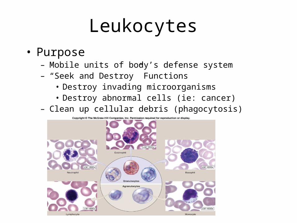
Leukocytes • Purpose
– Mobile units of body’s defense system– “Seek and Destroy” Functions
• Destroy invading microorganisms• Destroy abnormal cells (ie: cancer)
– Clean up cellular debris (phagocytosis)

Leukocyte Type
• Two main categories– Agranular
•Lymphocytes•Monocytes
– Granular•Neutrophils•Eosinophils•Basophils

5 - Types of WBC’s
Each WBC has a specific function
GranulocytesAgranulocytes

• Account for 25-30% or more of WBCs and:
• Absence of granules in cytoplasm when stained (however do contain lysosomes)
• Most important cells of the immune system
• There are three types of lymphocytes:– T cells – B cells– Natural killer cells
Agranulocytes: Lymphocytes

Agranulocytes: Lymphocytes
• T cells - attack foreign cells directly, helps restore immune system after infection; (cell- mediated immunity)
• B cells - produce antibodies that bind to pathogens to enable their destruction (humoral immunity)
• Natural killer cells – kills cells of the body that are displaying a signal to them (due to virus/cancer infection)

• Monocytes account for 3–7% of leukocytes – They are the largest leukocytes– U- or kidney-shaped nuclei– They leave the circulation, enter tissue, and differentiate into macrophages
– Clean up dead cell debris and attack microorganisms
Agranuloctes: Monocytes

Agranuloctes: Monocytes
Animation Microscope

Granulocytes
• Granulocytes – neutrophils, eosinophils, and basophils– Contain cytoplasmic granules that stain specifically (acidic, basic, or both) with Wright’s stain
– Are larger and usually shorter-lived than RBCs
– Have lobed nuclei– Are all phagocytic cells
• Account for 60-70% of total WBC’s• Life span short (~ 5 days)• Function
– Neutrophils are our body’s bacteria slayers
• AKA “polys” or PMN’s because multi-lobed nucleus can appear has multiple nuclei(polymorphonuclear)
• Present in large amounts of pus
Granulocytes: Neutrophils
(Polymorphonuclear leukocytes)

• Eosinophils account for 1–4% of WBCs
• Life span short (~10 days)• Function
– Lead the body’s counterattack against parasitic infections
– Lessen the severity of allergies by phagocytizing immune complexes (ending allergic reactions)
Granulocytes: Eosinophils

• Rare:Account for 0.5-1% of all WBCs
• Life span short (~few hours – few days)
• Function– Release histamine for inflammatory response•Histamine – inflammatory chemical that acts as a vasodilator and attracts other WBCs (antihistamines counter this effect)
Granulocytes: Basophils

• All leukocytes originate from hemocytoblasts– The mother of all blood stem cells
• Hemocytoblasts differentiate into myeloid stem cells and lymphoid stem cells– Myeloid stem cells become myeloblasts or monoblasts•Granulocytes form from myeloblasts•Monoblasts enlarge and form monocytes
– Lymphoid stem cells become lymphoblasts•Lymphoblasts develop into lymphocytes
Formation of Leukocytes

Formation of
Leukocytes
Figure 17.11

• Leukemia refers to cancerous conditions involving white blood cells
• Leukemias are named according to the abnormal white blood cells involved– Myelocytic leukemia – involves myeloblasts– Lymphocytic leukemia – involves lymphocytes
• Acute leukemia involves blast-type cells and primarily affects children
• Chronic leukemia is more prevalent in older people
Leukocytes Disorders: Leukemias

• Immature white blood cells are found in the bloodstream in all leukemias
• Bone marrow becomes totally occupied with cancerous leukocytes
• Severe anemia ensues due to excess production of WBC’s
• The white blood cells produced, though numerous, are not functional
• Death is caused by internal hemorrhage and overwhelming infections
• Treatments include irradiation, antileukemic drugs, and bone marrow transplants
Leukemia
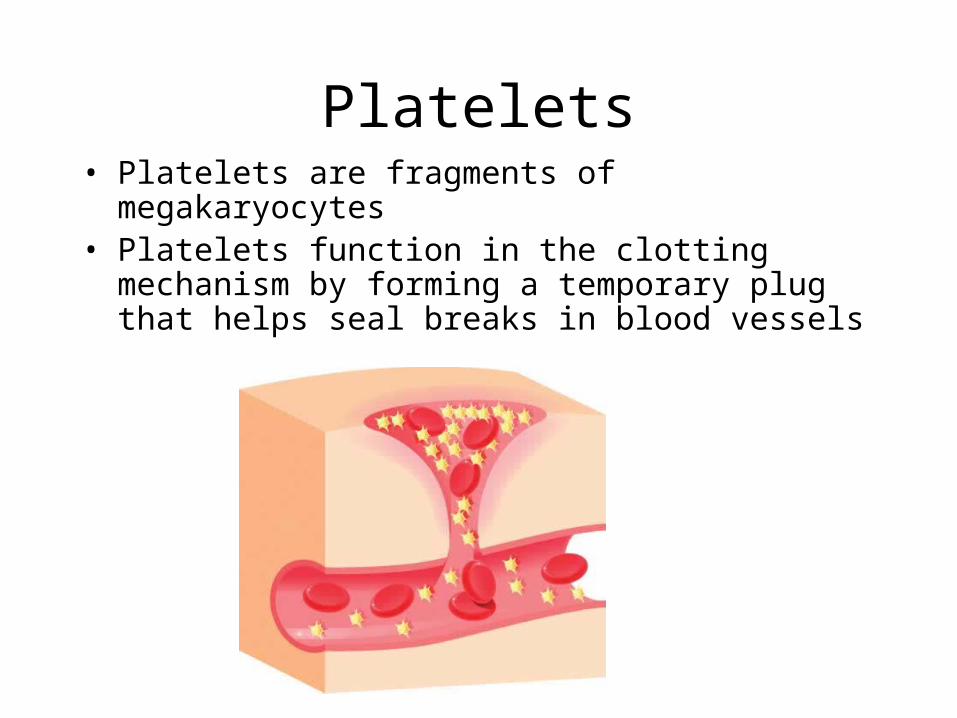
• Platelets are fragments of megakaryocytes
• Platelets function in the clotting mechanism by forming a temporary plug that helps seal breaks in blood vessels
Platelets


