
INFECTION AND IMMUNITY, Apr. 1989, p. 1151-11570019-9567/89/041151-07$02.00/0Copyright © 1989, American Society for Microbiology
Brucella abortus Regulates Bovine Macrophage-T-Cell Interactionby Major Histocompatibility Complex Class I1 and
Interleukin-1 ExpressionGARY A SPLITTER* AND KAREN M. EVERLITH
Department of Veterinary Science, University of Wisconsin-Madison, 1655 Linden Drive, Madison, Wisconsin 53706
Received 24 October 1988/Accepted 29 December 1988
T-cell activation is dependent on nominal antigen associated with major histocompatibility complex (MHC)class II molecules and interleukin-l (IL-1), both provided by antigen-presenting cells. We have studied theeffects of Brucella abortus and recombinant bovine gamma interferon (IFN-y) on bovine macrophageexpression of MHC class HI and IL-1 molecules and subsequent T-cell proliferation in response to B. abortus.When peripheral blood mononuclear cells were cocultured with B. abortus and IFN-y, increasing amounts ofIFN-y, from l to 100 U/ml, down regulated T-cell proliferation. Expression of MHC class II molecules on
macrophages was incr'eased independently by IFN-y or B. abortus treatment. Thus, class II moleculeexpression was not the cause of down regulation of T-cell proliferation as observed in other systems. However,B. abortus-IFN-y-treated 'macrophages obtained from overnight cultures had minimal membrane IL-1compared with macrophages treated 'with B. abortus alone. Loss of membrane IL-1 required IFN--y and theo-polysaccharide of the lipopolysaccharide. IFN-,y at 1 U/ml in combination with B. abortus produced a 32%decrease in T-cell response, while IFN-y at 100 U/ml added to B. abortus-treated cultures produced an 82%reduction in T-cell response. Membrane IL-1 levels were not altered when recombinant bovine IFN-a or therough strain 45/20 of B. abortus, which lacks the o-polysaccharide, was used. Secreted IL-1 levels were
unaffected by IFN-y and B. abortus treatment. The addition of recombinant bovine IL-1 (0.001 to 0.1 ng/ml)to B. abortus- and IFN--y-treated cultures failed to provide a signal necessary for T-cell proliferation. Thesedata suggest that membrane IL-1 has a key role in T-cell activation in response to B. abortus. When theo-polysaccharide of B. abortus lipopolysaccharide is combined with IFN--y at an inappropriate time during an
immune response, T-cell proliferation is prevented and cannot be restored by the addition of exogenous IL-1.
T-cell proliferation requires both nominal antigen associ-ated with major histocompatibility complex (MHC) class II
molecules and de novo synthesis of interleukin-1 (IL-1) bystimulated macrophages (42). The mechanisms that induceMHC class II and IL-1 molecules are not fully understood. Anumber of extrinsic as well as intrinsic factors that induce oraugment the expression ofMHC class II (40, 41) or IL-1 (10,23, 38) molecules have been identified. Facultative intracel-lular bacteria contribute to interferon (IFN) production bylymphocytes (2). IFN--y is known to augment MHC class II
molecule expression on macrophages (37). Lipopolysaccha-ride (LPS) (21) or a lymphokine produced by T cells (38) can
induce synthesis of IL-1 by macrophages. This lymphokineis distinct from IFN--y, tumor necrosis factor, and colonystimulating factor 1 and may be one mechanism by which Tcells signal macrophages to induce IL-1 (38). Two distinctbut structurally related forms of IL-1 (termed IL-la andIL-1i) have been identified (for a review, see reference 29);both forms appear to be associated with T-cell activation andcan be present in secreted or membrane-bound states (15,27). Both secreted and membrane-associated IL-1 are bio-logically active and participate in T-lymphocyte activation(18).Any process that alters macrophage expression of MHC
class II molecules or IL-1 may significantly influence im-mune responses. LPS can suppress expression of class II
molecules on macrophage surfaces (13), and it has beenreported that LPS inhibits both IFN--y induction and main-tenance of class II molecule expression on macrophages
* Corresponding author.
(36). In the absence of IFN--y, LPS had no significant effecton macrophage class II molecule expression. Furthermore,LPS or IL-1 activation of macrophages produces high levelsof prostaglandins which can modulate macrophage functions(5, 6).Bovine macrophages serve as important host cells for the
facultative intracellular bacterium Brucella abortus. Hostresistance to disease caused by facultative intracellularbacteria depends on appropriate interaction between specif-ically sensitized T cells and macrophages. Because macro-
phages are host cells for B. abortus, they might also becentral in initiating both positive and negative regulation ofimmune responses in infected cattle. We have shown previ-ously that bovine macrophages can present B. abortus in thecontext of MHC class II molecules to T cells (34). Toexamine the nature of the T-cell response to macrophage-associated B. abortus, we have studied MHC class II andIL-1 molecule expression by macrophages following treat-ment with the bacterium alone or in combination withIFN--y.
MATERIALS AND METHODS
Animals. Guernsey, Brown Swiss, and Holstein cattle(ages 6 months to 8 years) were maintained at a University ofWisconsin-Madison farm and vaccinated 2 to 4 times withthe viable attenuated smooth B. abortus 19 vaccine (Jensen-Salsbury Laboratory, Kansas City, Mo.). All animals were
serologically negative for antibodies to B. abortus at the timeof these studies.
T-lymphocyte enrichment. Peripheral blood mononuclear(PBM) cells were isolated by Ficoll-Hypaque as previously
1151
Vol. 57, No. 4
1152 SPLITTER AND EVERLITH
described (34) 6 months to 4 years following vaccination.Nonadherent cells were obtained from 60 x 106 PBM cellscultured overnight in 75-cm2 flasks containing RPMI 1640medium supplemented with 100 U of penicillin and 100 ,ug ofstreptomycin per ml, 2 mM L-glutamine, 25 mM N-2-hy-droxyethylpiperazine-N'-2-ethanesulfonic acid (HEPES), 5x 10⁻⁵ M 2-mercaptoethanol, and 10% fetal bovine serum.Nonadherent cells were washed, and contaminating macro-phages were removed by passage through a Sephadex G-10column (11). Further purification was accomplished by treat-ing the nonadherent cells with the anti-class II moleculemonoclonal antibody (MAb) H4 (One Lambda, Los Angeles,Calif.) (19) and anti-bovine light chain MAb DAS 9 (the gift,as was ascites fluid, from D. Goldsby) (35) followed bycomplement. MAb H4 was dialyzed to remove NaN3 andthen was used at a 1:10 dilution. DAS 9 ascites fluid was usedundiluted.APC. PBM cells were added to microdilution plates at 2 x
106 cells per well. Acetone-killed B. abortus smooth strain1119 or rough strain 45/20 (gifts of D. T. Berman) (24, 26)were added at the optimal concentration for T-cell prolifer-ation of 500 ,ug [dry weight] per ml (34). Recombinant bovineIFN-y or IFN-cL (Genentech, Inc., South San Francisco,Calif.) was added simultaneously with the bacteria to se-lected cultures at the concentrations indicated below. Afterovernight culture, nonadherent cells were removed, andadherent cells were washed three times with phosphate-buffered saline (PBS), irradiated (2,500 rad), and used asantigen-presenting cells (APC). By flow cytometry (EPICS;Coulter Electronics, Inc., Hialeah, Fla.), more than 80% ofthe adherent cells were present in a monocyte gate, andmore than 80% of the adherent cells stained for esterase.These adherent cells were termed macrophages and wereused as APC. Certain macrophage cultures were maintainedfor 7 days prior to use. Alternatively, macrophages wereobtained by culturing PBM cells overnight on glass, and afterremoval of nonadherent cells by treatment with EDTA, thecells were counted and added at selected concentrations tomicrodilution wells.APC fixation. After stimulation of APC by overnight
exposure to antigen or medium, they were washed and fixedwith 0.5 or 1% (wt/vol) paraformaldehyde for 15 min at roomtemperature. Following fixation, the cells were washed threetimes with PBS, and fresh medium was added. The fixedcells were incubated for 24 h at 370C to remove residualparaformaldehyde (16) and were washed prior to use ineither proliferative or membrane IL-1 determinations (17).
T-cell proliferation. The ability of APC to stimulate 4 x 10⁵autologous enriched T cells was assessed in a 6-day prolif-erative assay with [3H]thymidine (6.7 Ci/mmol; Dupont,NEN Research Products, Boston, Mass.) added during thefinal 16 h of incubation. In selected experiments, recombi-nant bovine IL-13 (Immunex, Seattle, Wash.) was added at0.1, 0.01, or 0.001 ng/ml to cultures containing enrichedbovine T cells or D10.G4.1 murine cells.
IL-1 assay. The IL-1-sensitive murine T-cell cloneD10.G4.1 (ATCC TIB 224) was maintained as describedelsewhere (12) by using C3H/HeJ (H-2k) spleen cells and 25U of recombinant human IL-2 (Biogen, Boston, Mass.) perml. To measure secreted IL-1, supernatants from unstimu-lated and stimulated APC were collected. Dilutions of thesesupernatant fluids were added to triplicate microdilutionwells in 100-,l1 portions, followed by 100 ,ul of 2 x 10⁴D10.G4.1 cells and 2.5 ,ug of concanavalin A (Sigma Chem-ical Co., St. Louis, Mo.) per ml. Supernatant from Esche-richia coli LPS-stimulated murine spleen cell cultures served
as a positive IL-1 control. Also, D10.G4.1 cells were addeddirectly to irradiated or paraformaldehyde-fixed APC as amethod of determining total IL-1 versus membrane-boundIL-1, respectively. [3H]thymidine was added during the final8 h of the 72-h incubation. Following culture, the cells wereharvested and isotope incorporation was determined in ascintillation counter.MHC class II molecule expression assays. PBM cells were
treated with recombinant bovine IFN--y, B. abortus, orIFN--y and B. abortus overnight in glass petri dishes. Fol-lowing removal of nonadherent cells, an enzyme-linkedimmunosorbent assay (ELISA) was used to detect MHCclass II molecule expression on adherent cells. Round-bottom microdilution plates were coated with 3% gelatin for2 h at 37°C and washed three times prior to use. Glass-adherent cells (2 x 10⁶) detached by 0.35% EDTA with 0.6%glucose in PBS were incubated in the microdilution plates for1 h with 10% heat-inactivated normal rabbit serum to blockFc receptors. After incubation, the cells were pelleted andsuspended in PBS containing 1% bovine serum albumin.MAb H4 was diluted 1:300 in PBS containing 1% bovineserum albumin and added in 100-pA portions to wells. Plateswere incubated for 1 h on ice and washed three times withPBS, and cells were assayed for class II molecules byELISA as previously described (33). MAb 262404, specificfor a bovine herpesvirus 1 glycoprotein, was used as anisotypic control (22).
Alternatively, class II molecule expression was detectedby flow cytometry by using MAb H4. Glass-adherent cells (2x 10⁶), obtained as described above, were incubated on icefor 1 h with a 1:300 dilution of MAb H4 and then washedwith PBS. Fluorescein-conjugated rabbit anti-mouse immu-noglobulin G (heavy and light chains) (Jackson Immunore-search Laboratories, Inc., Avondale, Pa.) was added to cellsuspensions, and they were incubated for an additional hour.After three washes, the cells were examined by flow cytom-etry. The rabbit anti-mouse IgG was used at an optimalsaturating concentration (data not shown).
RESULTS
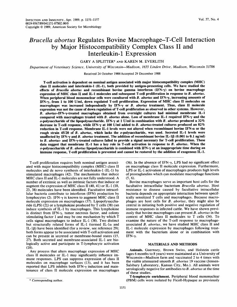
Activation of MHC class II molecules by IFN-y and B.abortus. On the basis of the ELISA, recombinant bovineIFN--y increased class II molecule expression on macro-phages. As little as 1 U of IFN--y per ml produced a fivefoldincrease in class II molecules detected (Fig. 1). Similarly, afourfold increase in class II molecule expression on macro-phages was observed when PBM cells were treated with B.abortus (Fig. 1). Simultaneous addition of IFN-y and B.abortus to PBM cells did not increase class II moleculeexpression on macrophages higher than either treatmentalone, suggesting that B. abortus and IFN--y at the levelstested do not have an additive effect. Therefore, class IImolecule expression can be increased independently by theaddition of recombinant bovine IFN-y or B. abortus to thecultures.
Detection of bovine IL-1 by using the murine D10.G4.1 cellline. Since IL-1 is not produced constitutively and extracel-lular stimuli induce its synthesis (14), is was possible that B.abortus could induce production of both membrane andsecreted forms of IL-1. An increasing concentration of B.abortus cultured with bovine macrophages for 24 h inducedmembrane IL-1 synthesis in a dose-dependent manner (Fig.2). To examine the effect of adherent cell numbers, selectednumbers of APC were cultured in microdilution wells with500 ,ug of B. abortus per ml to allow production of both
INFECT. IMMUN.
DOWN REGULATION OF MEMBRANE IL-1 1153
~~ ~ ~ ~ BU.r3(S g/ l
0.2
o02.
0
0.1
0.0
0 1 10 100 B. abortus .10 100
IFN (U/mi) (500 ug/mI) IFN (U/mi) &B. abortus (500 ug/mi)
FIG. 1. Detection by ELISA of MHC class II molecule expres-
sion. PBM cells were treated overnight with recombinant bovineIFN--y (U), B. abortus (500 ,ug/ml, 0), or recombinant bovine IFN--yand B. abortus (U). Nonadherent cells were removed, and a MAb toclass II molecules (H4) was added to adherent cells. The ELISA was
performed as described in Materials and Methods and is repre-
sentative of data obtained from seven animals. Isotype controlvalues were subtracted from treatment values. Error bars representstandard deviations. Data are representative of four experiments.
membrane and secreted IL-1. In parallel experiments, cellswere fixed with 1% paraformaldehyde to evaluate membraneIL-1 induction in the absence of secreted IL-1. The detectionlimit of our system required 10i or more APC for measure-ment of total or membrane IL-1 with the murine cell lineGlO.D4.1 (Fig. 3). B. abortus stimulated the production oftotal IL-1 from irradiated macrophages (Fig. 3A) and mem-
brane IL-1 from paraformaldehyde-treated macrophages(Fig. 3B). Less IL-1 was detected from the paraformalde-hyde-treated macrophages than from the nonfixed cells atconcentrations of 103 cells per well.
In contrast, adherent cells present in the PBM cell popu-
lation cultured with B. abortus and IFN--y showed a decreasein membrane IL-1 expression (Fig. 4). Cultures treatedovernight with B. abortus alone produced increased amounts
0.5
Ei
m10.0
50
20000 40000 60000 800003 H Thymidine Incorporation
CPUFIG. 2. Proliferation of D1O.G4.1 cells to paraformaldehyde-
fixed bovine macrophages treated with B. abortus. Adherent bovinecells were treated for 24 h with the indicated concentrations ofbacteria, washed, and fixed with 0.05% paraformaldehyde.D1O.G4.1 and 2.5 ,ug of concanavalin A per ml were added to thefixed cells, and proliferation of D1O.G4.1 was measured after 24 h.
of total IL-1 in comparison with control cultures. This issimilar to the findings shown in Fig. 3, which illustrates thatmacrophages treated with B. abortus alone produced bothsecreted and membrane IL-1. Cultures treated with B.abortus and IFN-y produced secreted IL-1, but the levels ofmembrane IL-1 were reduced more than fivefold comparedwith total IL-1 levels or membrane IL-1 levels in culturestreated with B. abortus only (Fig. 4).
T-cell proliferation in response to B. abortus- or B. abortus-IFN-y-treated macrophages. Because B. abortus inducedboth MHC class II molecule expression and IL-1 synthesis,it was important to correlate these observations with theability of autologous T-cells to proliferate in response tomacrophage-associated B. abortus. Macrophages within aPBM cell population were treated with B. abortus, recom-binant IFN--y, or both. The ability of the treated macro-phages expressing either total IL-1 (irradiated macrophages)or membrane IL-1 (paraformaldehyde-fixed macrophages) toinduce proliferation of antigen-specific T cells was deter-mined. Macrophages treated with B. abortus induced T-cellproliferation (Fig. 5). Likewise, macrophages treated with B.abortus and fixed with paraformaldehyde were still capableof inducing T-cell proliferation. However, cultures treatedwith B. abortus and IFN--y had a 62% reduction in PBMproliferation compared with cultures treated only with B.abortus. Similarly, cultures treated with B. abortus andIFN-y and then by paraformaldehyde fixation of the macro-phages had a 57% reduction in proliferation of PBM cells.MHC class II molecule expression on macrophages treatedin parallel culture and analyzed by flow cytometry with MAbH4 had 63 and 76% staining cells for irradiated and fixedmacrophages, respectively (data not shown). When IFN-ywas added to macrophages concurrently with B. abortus, nomarked reduction in class II expression was observed onunfixed (69% staining cells) or paraformaldehyde-fixed (52%staining cells) macrophages (data not shown). Down regula-tion of T-cell proliferation appeared dependent on the initialPBM cell population and macrophage state because downregulation was not observed when overnight-cultured mac-rophages only or 7-day-old macrophages were treated withB. abortus and IFN--y (data not shown).
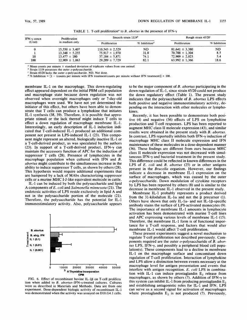
Determination of T-cell suppression due to IFN-y concen-tration and the strain of B. abortus. To determine if downregulation of T-cell proliferation by IFN--y and B. abortuswas associated with the LPS portion of the bacteria, as hasbeen suggested for E. coli (36), B. abortus smooth strain1119, which possesses the outer o-polysaccharide of LPS,was compared with rough strain 45/20, which lacks the outero-polysaccharide (24, 26). Increasing concentrations ofrecombinant bovine IFN--y were added to PBM cell culturescontaining B. abortus 1119. Following overnight incubation,nonadherent cells were removed, and enriched T cells wereadded. Table 1 shows a concordant decrease of proliferationas IFN concentrations increased. IFN--y at 1 U/ml caused a32% inhibition, and IFN--y at 100 U/ml caused an 82%inhibition of T-cell proliferation. In contrast, no markedinhibition of proliferation was observed with similar concen-trations of strain 45/20 and IFN-y. Also, cotreatment withrecombinant bovine IFN-a, ranging from 1 to 100 U/ml, andB. abortus 1119 failed to inhibit T-cell proliferation (data notshown).
Effect of adding IL-1 to cultures treated with B. abortus andIFN--y. Because membrane but not total IL-1 levels werereduced (Fig. 4) and a reduction of T-cell proliferation wasobserved with both unfixed and paraformaldehyde-fixedmacrophages (Fig. 5), the possibility that the bovine IL-1
VOL. 57, 1989
5H
1154 SPLITTER AND EVERLITH
A. Membrane and Secreted IL 1 B. Membrane IL 1
c
0
1-
0
ca0
r-atC.)C
E
I-
1o2 10 310 4
Number of Macrophages / well10 2 10 3 10 4Number of Macrophages / well
FIG. 3. Proliferation of D10.G4.1 cells to measure membrane and secreted IL-1. Glass-enriched macrophages of three animals were
cultured with B. abortus plus 710 (EL), 766 (*), or 549 (D), or medium only, i.e., 710 (O), 766 (D), or 549 (LI), overnight. Macrophages wereirradiated as a source of total (secreted and membrane) IL-1 (A) or paraformaldehyde fixed as a source of membrane IL-1 (B). D1O.G4.1 cellswere added to macrophages as described in Materials and Methods. Standard deviations were less than 5%.
produced was not biologically active on bovine cells wasconsidered. To determine if exogenous IL-1 could preventsuppression, recombinant bovine IL-ip was added to B.abortus-IFN-y-treated cultures at the time of T-cell addi-tion. The addition of IL-1 at selected doses failed to alter thesuppressed T-cell state (Fig. 6).
DISCUSSION
The present data demonstrate that when IFN-y and B.abortus were cocultured with PBM cells, there was a con-comitant decrease in macrophage membrane IL-1 expres-sion and T-cell proliferation. No alteration in total IL-1 wasobserved. MHC class II molecules on the APC were notresponsible for decreased T-cell proliferation, because bothrecombinant bovine IFN-y and B. abortus independentlyand in combination increased class II expression on macro-phages. T cells from immunized animals could proliferate
C
a
-x
0
a.m0
2
:I..
80000-
60000-
40000 -
20000-
0-
T* Secreted IL I
Membrane IL 1
wheh B. abortus was presented by macrophages without theaddition of recombinant IFN-y. Both total and membraneIL-1 were present when macrophages were treated with B.abortus.Demonstration of the decrease in membrane IL-1 and
T-cell proliferation was dependent on the simultaneousaddition of IFN--y and B. abortus to whole PBM cells prior toremoving nonadherent cells. A second factor, apparentlynecessary for the decrease in T-cell proliferation, was thepresence of the o-polysaccharide of B. abortus. Smooth B.abortus 1119 possesses the outer o-polysaccharide of theLPS and when combined with recombinant IFN--y produceda down regulation of the T-cell response. Rough strain 45/20does not possess the outer o-polysaccharide (24, 26) andwhen combined with IFN-y did not alter T-cell proliferation.The data are compatible with the hypothesis that IFN-y andthe outer o-polysaccharide of B. abortus can down regulateT-cell proliferation, perhaps by decreasing the expression of
0
a
S.
C)
I-
B. abortus B. abortus + IFN gamma
120000 -
100000 -
80000 -
60000 -
40000-
20000-
0-
Total IL IMembrane IL 1
B. abortus B. abortus + IFN gamma
Treatment
FIG. 4. Proliferation of D1O.G4.1 cells to measure membraneversus secreted IL-1 expression following macrophage treatment.Adherent cells present in 2 x 10⁶ PBM cells were treated overnightwith B. abortus or B. abortus and IFN--y. Following overnighttreatments, supernatants were removed as the source of secretedIL-1 and the adherent cells were fixed with 1% paraformaldehyde asthe source of membrane IL-1. D10.G4.1 cells were cultured withsupernatants (-) or fixed cells (U) to assay for IL-1. Data are
representative of eight experiments.
Treatment
FIG. 5. Proliferation of T cells added to unfixed (D) or paraform-aldehyde-fixed (X) macrophages previously treated with B. abortusor IFN--y and B. abortus. PBM cells were cultured overnight withIFN--y, B. abortus, B. abortus and IFN--y, or medium alone, andthen nonadherent cells were removed. Enriched T cells were added,and proliferation to the antigen-pulsed macrophages was determinedafter a 6-day culture. Response of cultures treated with medium orIFN--y alone was less than 10,000 cpm. Data are representative ofeight experiments.
00a
0US.0
eC)' :ECL0 0
E
cP
INFECT. IMMUN.
DOWN REGULATION OF MEMBRANE IL-1 1155
TABLE 1. T-cell proliferationa to B. abortius in the presence of IFN-y
IFN--y concn Proliferation Smooth strain lll9b Rough strain 45/20'
(U/mi) in medium only Proliferation % Inhibitiond Proliferation % Inhibition
0 15,550 ± 3,407 110,543 ± 2,529 ND 81,641 ± 3,380 ND1 13,348 ± 5,255 75,917 + 1,970 31.8 70,788 ± 1,304 8.5
10 13,477 + 180 37,184 ± 5,871 74.1 72,909 ± 2,023 5.4100 12,909 ± 1,063 29,289 ± 7,729 82.1 63,992 ± 1,366 18.6
a Mean counts per minute ± standard deviation of triplicate values from one animal.b Strain 1119 possesses the outer o-polysaccharide.c Strain 45/20 lacks the outer o-polysaccharide. ND, Not done.d % Inhibition = [1 - (counts per minute with IFN treatment/counts per minute without IFN treatment)] x 100.
membrane IL-1 on the macrophage. This down-regulatingeffect appeared dependent on the initial PBM cell populationand macrophage state because down regulation was notobserved when overnight macrophages only or 7-day-oldmacrophages were used. We have not yet determined theinitiator of this effect, but others have been able to demon-strate that T cells can produce a lymphokine that initiatesIL-1 synthesis (38, 39). Therefore, it is possible that appro-priate stimuli or the lack thereof might induce T cells toeffect a down regulation of macrophage membrane IL-1.Interestingly, an early description of IL-1 induction indi-cated that T-cell-induced IL-1 produced an additional com-ponent not present in LPS-induced IL-1 (23). This compo-nent might represent an altered, biologically inactive IL-1 ora T-cell-derived product, as was speculated by the authors(23). In support of a T-cell-derived product, IFN--y canmaintain the accessory function of APC for the induction ofsuppressor T cells (28). Presence of lymphocytes in themacrophage population when cultured with IFN and B.abortus might contribute to the simultaneous increase in theability to induce suppressor T cells, as shown by others (28).This hypothesis would require additional experiments thatare hampered by a lack of MAbs characterizing suppressorcells or a murine MHC I-J-like equivalent molecule in cattle.
IL-1 can be induced by both the polysaccharide and lipidA components of E. coli and Salmonella minnesota (21). Theendotoxic activities of LPS reside exclusively in lipid A andnot in the polysaccharide portion of the molecule (21).Therefore, the polysaccharide has the potential for IL-1immunostimulatory activity. Also, polysaccharide appears
B. abortus
B. ab/g. IFNE IL I (0.1)
ILl(0.01)IL1 (0.001)
0 10000 20000 30000 40000 500003 H Thymidine Incorporation
CPM
FIG. 6. Effect of recombinant bovine IL-lp on T-cell proliferation when added to B. abortus-IFN-y-treated cultures. Cultureswere as described in Materials and Methods. Data are from oneexperiment. Dose-dependent biologic activity of recombinant IL-1was demonstrated when the activity was assayed on D10.G4.1 cells.
to be the major component of B. abortus participating in thedown regulation of IL-1, since strain 45/20 could not producethe down regulatory effect (Table 1). The present studyindicates that the polysaccharide of B. abortus LPS effectsboth positive and negative immunostimulatory activity, de-pending on the interaction with other molecules or lympho-kines.
Recently, it has been possible to demonstrate both posi-tive (4) and negative (36) effects of LPS on lymphokineproduction and T-cell responses. LPS has been reported toaugment MHC class II molecule expression (43), and similarresults were obtained in the present study with B. abortus.In contrast, LPS reportedly inhibits both IFN--y induction ofmacrophage MHC class II molecule expression and IFNmaintenance of these molecules in a dose-dependent manner(36). These findings are different from ours because MHCclass II molecule expression was not decreased with simul-taneous IFN--y and bacterial treatment in the present study.This difference could be reflected in known differences in theLPS of E. coli and B. abortus (25) or in other antigenspresent in the Brucella organism. However, our findingsindicate a decrease in membrane IL-1 expression on thesurface of macrophages, which was caused by the outero-polysaccharide. Down modulation of cytokine receptorsby LPS has been reported by others (8) and is similar to thedecrease in membrane IL-1 observed in the present study.Membrane IL-1 probably represents a product derived
from the 31-kilodalton IL-la and not the ,B precursor (1).Others have shown that only IL-ia- and not IL-1l-specificantibody stains the surface of LPS-activated monocytes (9).The importance of membrane IL-1 amounts in lymphocyteactivation has been demonstrated with murine T-cell linesand APC expressing various levels of membrane IL-1 (15).Therefore, the membrane IL-1 form is of functional impor-tance for a T-cell response, and factors that would altermembrane IL-1 would affect T-cell proliferation.These present experiments suggest a novel mechanism to
regulate T-cell proliferation not described previously. Com-ponents required are the outer o-polysaccharide of B. abor-tus LPS, IFN-y, and possibly a peripheral blood cell popu-lation(s). These components lead to a decline in membraneIL-1 on the macrophage surface and concomitant downregulation of T-cell proliferation. Interaction of lymphokineand LPS allow a distinction between events necessary at themacrophage level for antigen presentation and events thatinterfere with antigen recognition. E. coli LPS in combina-tion with IL-1 can induce prostaglandin E2 release frommacrophages, as shown by others (7). Addition of IFN--y tothis system can inhibit IL-1 from producing prostaglandin E2and establishing antagonistic roles for IL-1 and IFN. LPScan serve as a second signal for activation of macrophageswhere prostaglandin E2 is not produced (7). Previously,
VOL. 57, 1989
1156 SPLITTER AND EVERLITH
others have not demonstrated whether IFN could alterexpression of IL-1 on macrophage surfaces. However, thefindings in the present study would extend the observation ofprevious workers (7) to suggest that IFN can down regulatemembrane IL-1 in the presence of LPS. Whether LPS fromother gram-negative bacteria can produce this effect orwhether it is restricted to B. abortus remains to be deter-mined. IL-1 inhibitors have been reported by others butappear to be different from either IFN or LPS (3, 20, 32), andviral infections have been noted to produce IL-1 inhibitoryfactors directly from macrophages (30, 31). These descrip-tions have indicated an effect on secreted IL-1, but littleevidence indicating an alteration in membrane but not se-creted IL-1 has been reported. The interaction of B. abortuscontaining the outer o-polysaccharide and IFN-y to downregulate membrane IL-1 appears unusual. Clearly, morework will be required to determine how host cytokinesinteract with foreign antigens and cells of the immune systemto down regulate molecules that participate in immuneresponses.
ACKNOWLEDGMENTS
This work was supported by U.S. Department of Agriculturegrants 86-CRCR-1-2210 and 58-6125-6-1 and by grants from Genen-tech, Inc., and the College of Agricultural and Life Sciences,University of Wisconsin.
LITERATURE CITED1. Bakouche, O., D. C. Brown, and L. B. Lachman. 1987. Subcel-
lular localization of human monocyte interleukin 1: evidence foran inactive precursor molecule and a possible mechanism for IL1 release. J. Immunol. 138:4249-4262.
2. Beller, D. I., and K. Ho. 1982. Regulation of macrophagepopulations. V. Evaluation of the control of macrophage Iaexpression in vitro. J. Immunol. 129:971-976.
3. Berman, M. A., C. I. Sandborg, B. S. Calabia, B. S. Andrews,and G. J. Friou. 1986. Studies of an interleukin-1 inhibitor:characterization and clinical significance. Clin. Exp. Immunol.64:136-145.
4. Blanchard, D. K., J. Y. Djeu, T. W. Klein, H. Friedman, andW. E. Stewart II. 1986. Interferon-y induction by lipopolysac-charide: dependence on interleukin 2 and macrophages. J.Immunol. 136:963-970.
5. Bonney, R. J., and J. L. Humes. 1984. Physiological andpharmacological regulation of prostaglandin and leukotrieneproduction by macrophages. J. Leukocyte Biol. 35:1-10.
6. Boraschi, D., and A. Tagliabue. 1984. Multiple modulation ofmacrophage functions by lymphokines: different effects of in-terferon and macrophage activating factor, p. 71. In E. Pick(ed.), Lymphokines, vol. 9. Academic Press, Inc., New York.
7. Browning, J. L., and A. Ribolini. 1987. Interferon blocks inter-leukin 1-induced prostaglandin release from human peripheralmonocytes. J. Immunol. 138:2857-2863.
8. Chen, B. D.-M., H.-S. Lin, and S. Hsu. 1983. Lipopolysaccha-ride inhibits the binding of colony-stimulating factor (CSF-1) tomurine peritoneal exudate macrophages. J. Immunol. 130:2256-2260.
9. Conlon, P. J., K. H. Grabstein, A. Alpert, K. S. Pricket, T. P.Hopp, and S. Gillis. 1987. Localization of human mononuclearcell interleukin 1. J. Immunol. 139:98-102.
10. Economou, J. S., and H. S. Shin. 1978. Lymphocyte-activatingfactor. I. Generation and physicochemical characterization. J.Immunol. 121:1446-1452.
11. Jerrells, T. R., J. H. Dean, G. L. Richardson, and R. B.Huberman. 1980. Depletion of monocytes from human periph-eral blood mononuclear leukocytes: comparison of the Sepha-dex G10 column method with other commonly used techniques.J. Immunol. Methods 32:11-29.
12. Kaye, J., S. Gillis, S. B. Mizel, E. M. Shevach, T. R. Malek,C. A. Dinarello, L. B. Lachman, and C. A. Janeway. 1984.
Growth of a cloned helper T cell line induced by a monoclonalantibody specific for the antigen receptor: interleukin 1 isrequired for the expression of receptors for interleukin 2. J.Immunol. 133:1339-1345.
13. Koerner, T. J., T. A. Hamilton, and D. 0. Adams. 1987.Suppressed expression of surface Ia on macrophages by lipo-polysaccharide: evidence for regulation at the level of accumu-lation of mRNA. J. Immunol. 139:239-243.
14. Kurt-Jones, E., H. W. Virgin IV, and E. R. Unanue. 1986. Invivo and in vitro expression of macrophage membrane interleu-kin 1 in response to soluble and particulate stimuli. J. Immunol.137:10-14.
15. Kurt-Jones, E. A., D. I. Beller, S. B. Mizel, and E. R. Unanue.1985. Identification of a membrane-associated interleukin 1 inmacrophages. Proc. Natl. Acad. Sci. USA 82:1204-1208.
16. Kurt-Jones, E. A., J.-M. Kiely, and E. R. Unanue. 1985.Conditions required for expression of membrane IL 1 on B cells.J. Immunol. 135:1548-1550.
17. Kurt-Jones, E. A., H. W. Virgin IV, and E. R. Unanue. 1985.Relationship of macrophage Ia and membrane IL 1 expressionto antigen presentation. J. Immunol. 135:3652-3654.
18. Le, J., D. Weinstein, U. Gubler, and J. Vilcek. 1987. Induction ofmembrane-associated interleukin 1 by tumor necrosis factor inhuman fibroblasts. J. Immunol. 138:2137-2142.
19. Lewin, H. A., C. C. Calvert, and D. Bernoco. 1985. Cross-reactivity of a monoclonal antibody with bovine, equine, ovine,and porcine peripheral blood B lymphocytes. Am. J. Vet. Res.46:785-788.
20. Liao, Z., A. Haimovitz, Y. Chen, J. Chan, and D. L. Rosen-streich. 1985. Characterization of a human interleukin 1 inhibi-tor. J. Immunol. 134:3882-3886.
21. Loppnow, H., L. Brade, H. Brade, E. T. Rietschel, S. Kusumoto,T. Shiba, and H.-D. Flad. 1986. Induction of human interleukin1 by bacterial and synthetic lipid A. Eur. J. Immunol. 16:1263-1267.
22. Marshall, R. L., L. L. Rodriguez, and G. J. Letchworth III.1986. Characterization of envelope proteins of infectious bovinerhinotracheitis virus (bovine herpesvirus 1) by biochemical andimmunological methods. J. Virol. 57:745-753.
23. Mizel, S. B., J. J. Oppenheim, and D. L. Rosenstreich. 1978.Characterization of lymphocyte-activating factor (LAF) pro-duced by a macrophage cell line, P388D1. II. Biochemicalcharacterization of LAF induced by activated T cells and LPS.J. Immunol. 120:1504-1508.
24. Moreno, E., L. M. Jones, and D. T. Berman. 1984. Immuno-chemical characterization of rough Brucella lipopolysaccha-rides. Infect. Immun. 43:779-782.
25. Moreno, E., R. S. Kurtz, and D. T. Berman. 1984. Induction ofimmune and adjuvant immunoglobulin G responses in mice byBrucella lipopolysaccharide. Infect. Immun. 46:74-80.
26. Moreno, E., M. W. Pitt, L. M. Jones, G. G. Schurig, and D. T.Berman. 1979. Purification and characterization of smooth andrough lipopolysaccharides from Brucella abortus. J. Bacteriol.138:361-369.
27. Nagelkerken, L. M., and P. J. C. van Breda Vriesman. 1986.Membrane-associated IL 1-like activity on rat dendritic cells. J.Immunol. 136:2164-2170.
28. Noma, T., and M. E. Dorf. 1985. Modulation of suppressor Tcell induction with y-interferon. J. Immunol. 135:3655-3660.
29. Oppenheim, J. J., E. J. Kovacs, K. Matsushima, and S. K.Durum. 1986. There is more thant one interleukin 1. Immunol.Today 7:45-56.
30. Roberts, N. J., A. H. Prill, and T. N. Mann. 1986. Interleukin 1and interleukin 1 inhibitor production by human macrophagesexposed to influenza virus or respiratory syncytial virus. J. Exp.Med. 163:511-519.
31. Rodgers, B. C., D. M. Scott, J. Mundin, and J. G. Sissons. 1985.Monocyte-derived inhibitor of interleukin 1 induced by humancytomegalovirus. J. Virol. 55:527-532.
32. Scala, G., Y. D. Kuang, R. E. Hall, A. V. Muchmore, and J. J.Oppenheim. 1984. Accessory cell function of human B cells. I.Production of both interleukin 1-like activity and an interleukin1 inhibitory protein by an EBV-transformed human cell line. J.
INFECT. IMMUN.
DOWN REGULATION OF MEMBRANE IL-1
Exp. Med. 159:1637-1652.33. Splitter, G., J. Burkholder, K. O'Reilly, and A. Janzer. 1987.
Anti-BoLA-w8 monoclonal antibody: production of a tissuetyping reagent after blocking monomorphic sites on bovinemononuclear cells. Tissue Antigens 30:122-127.
34. Splitter, G. A., and K. M. Everlith. 1986. Collaboration ofbovine T lymphocytes and macrophages in T-lymphocyte re-sponse to Brucella abortus. Infect. Immun. 51:776-783.
35. Srikumaran, S., A. J. Guidry, and R. A. Goldsby. 1983. Produc-tion and characterization of monoclonal bovine immunoglob-ulins G1, G2, and M from bovine x murine hybridomas. Vet.Immunol. Immunopathol. 5:323-342.
36. Steeg, P. S., H. M. Johnson, and J. J. Oppenheim. 1982.Regulation of murine macrophage Ia antigen expression by animmune interferon-like lymphokine: inhibitory effect of endo-toxin. J. Immunol. 129:2402-2406.
37. Steeg, P. S., R. N. Moore, H. M. Johnson, and J. J. Oppenheim.1982. Regulation of murine macrophage Ia antigen expressionby a lymphokine with immune interferon activity. J. Exp. Med.156:1780-1793.
38. Takacs, L., J. A. Berzofsky, J. York-Jolley, T. Akahoshi, E.Blasi, and S. K. Durum. 1987. IL 1 induction by murine T cellclones: detection of an IL 1-inducing lymphokine. J. Immunol.138:2124-2131.
39. Unanue, E. R., and P. M. Allen. 1987. The basis for theimmunoregulatory role of macrophages and other accessorycells. Science 236:551-557.
40. Unanue, E. R., D. I. Beller, C. Y. Lu, and P. M. Allen. 1984.Antigen presentation: comments on its regulation and mecha-nism. J. Immunol. 132:1-5.
41. Wentworth, P. A., and H. K. Ziegler. 1987. Induction ofmacrophage Ia expression by Listeria monocytogenes in con-
genitally athymic nude mice. J. Immunol. 138:3167-3173.42. Windle, J. J., H. S. Shin, and J. F. Morrow. 1984. Induction of
interleukin 1 messenger RNA and translation in oocytes. J.Immunol. 132:1317-1322.
43. Ziegler, H. K., L. K. Staffileno, and P. Wentworth. 1984.Modulation of macrophage Ia-expression by lipopolysaccha-ride. I. Induction of Ia expression in vivo. J. Immunol. 133:1825-1835.
VOL. 57, 1989 1157


