
Burns and Other Skin Lesions
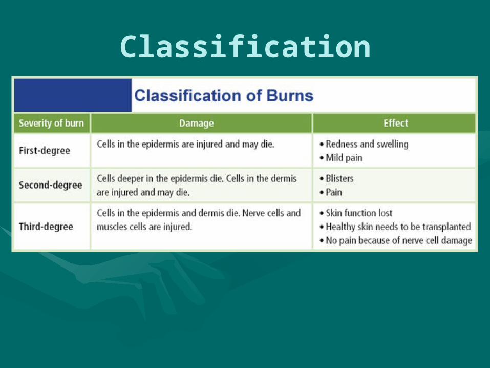
Classification

First Degree Burn: Epidermis only

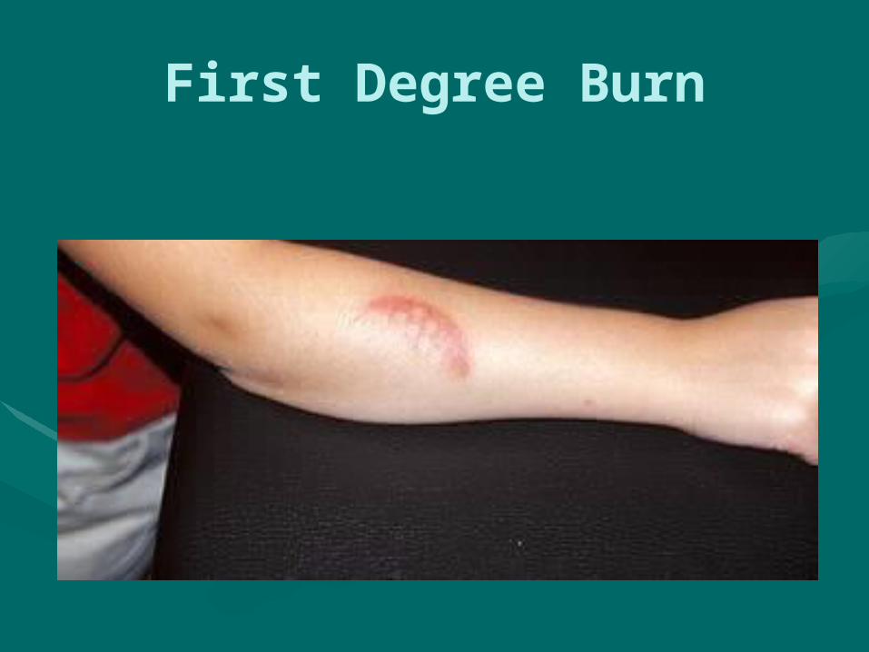
First Degree Burn

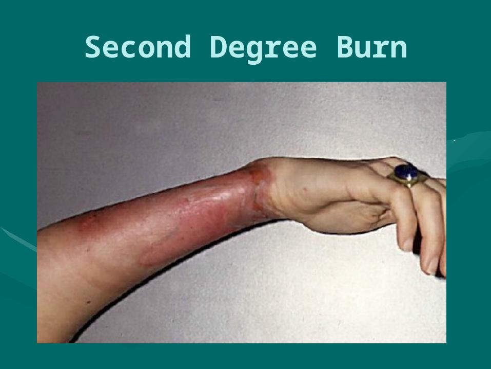
Second Degree Burn

Second Degree Burn: Epidermis and upper layers of the dermis

Second Degree Burn
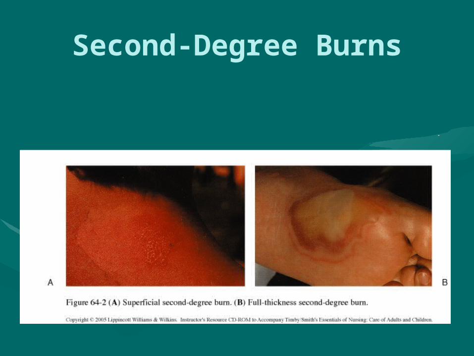
Second-Degree Burns

Third Degree Burn: Complete destruction of the epidermis & dermis


Estimating percentages

Regeneration

Regeneration/Scarring

Scar
• Occurs whenever wound/ulcer has occurred; reflects healing
• Keloid: Abnl formation of CT; form w/ dermal tissue damage; initially are thick but may w/ time become white and atrophic


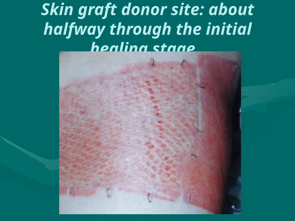
Skin graft donor site: about halfway through the initial healing stage

Papule
• Firm, raised lesion (less than 1cm in diameter)

Papule

Plaque
• Large, raised lesion (greater than 1 cm in diameter)

Plaque - Psoriasis
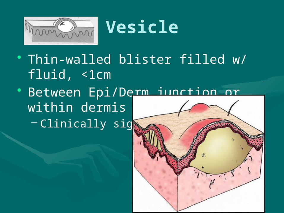
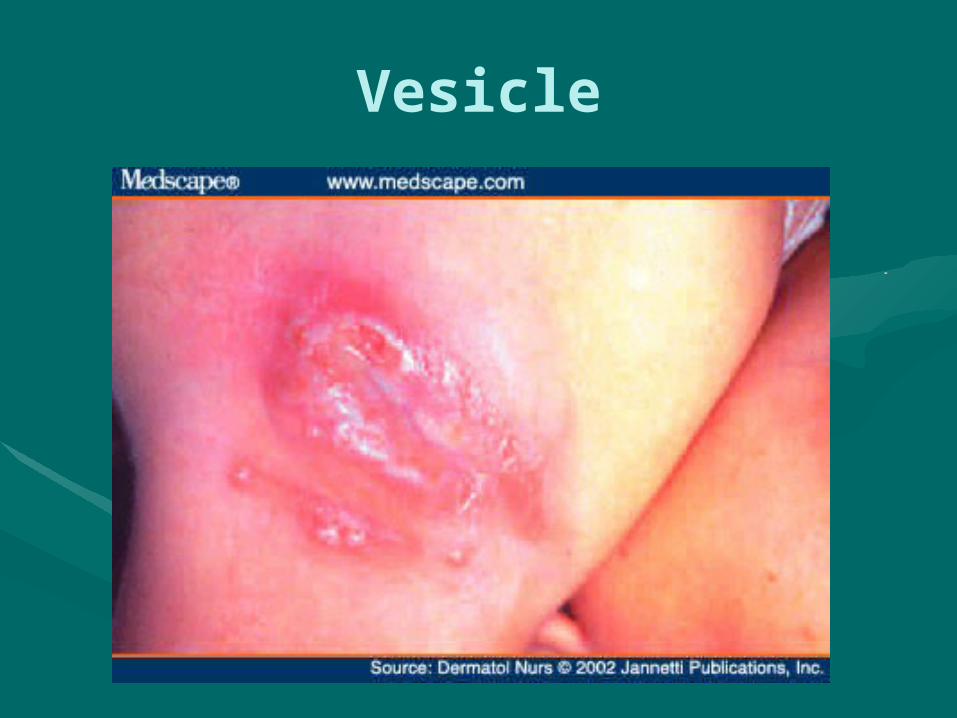
Vesicle
• Thin-walled blister filled w/ fluid, <1cm• Between Epi/Derm junction or within dermis
– Clinically significant
Vesicle

Pustule
• Elevated lesion filled w/ pus

Pustule

Crust
• Hardened deposits of serum and cellular debris
• result when serum, blood, or purulent (pus) exudates dries on skin surface
• “scab”

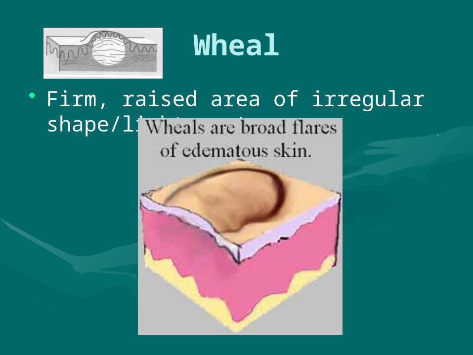
Wheal
• Firm, raised area of irregular shape/light center

Wheal

Cyst
• Nodule that contains fluid or semisolid material

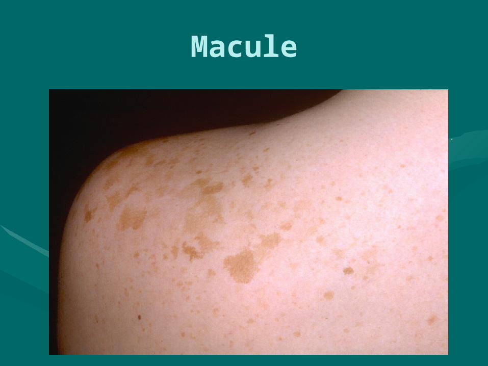
Macule
• Distinguished from surrounding skin by color
Macule

Abrasion
• Epidermis removed, revealing dermis
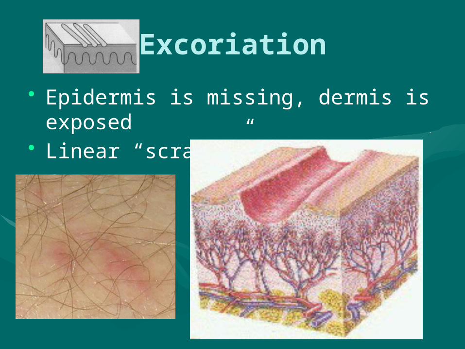
Excoriation
• Epidermis is missing, dermis is exposed• Linear “scratch”

Ulcer
• Craterlike lesion caused by disintegration of the skin (epidermis and dermis)
• Heals w/ scarring

Bed Sore• Compression of skin between bony
prominence and other surface• Cuts off circulation
– leads to necrosis of tissue• - Lack of blood supply prevents proper healing• promotes infection

Fissure
• Linear crack or break from epidermis to dermis


