

University of Naples “Federico II”
School of Medicine and Surgery
Dipartimento di Biologia e Patologia Cellulare e Molecolare “L. Califano”
Research Doctorate Program in Molecular Oncology and Endocrinology –
XVIII Cycle
Coordinator: Professor G. Vecchio
Tutor: Professor V.E. Avvedimento
cAMP links PI3K to multiple signaling pathways
Candidate Dr. C. Cosentino
Year 2005-2006

Administrative Location
Dipartimento di Biologia e Patologia Cellulare e Molecolare “L. Califano”
Università degli Studi di Napoli Federico II Partner Institutions Italian Institutions Università di Napoli Federico II, Naples, Italy Istituto di Endocrinologia ed Oncologia Sperimentale “G. Salvatore”, CNR, Naples, Italy Seconda Università di Napoli, Naples, Italy Università del Sannio, Benevento, Italy Università di Genova, Genoa, Italy Università di Padova, Padova, Italy Foreign Institutions Johns Hopkins University, Baltimore, MD, USA National Institutes of Health, Bethesda, MD, USA Ohio State University, Columbus, OH, USA Université Paris Sud XI, Paris, Francia Supporting Institutions Università di Napoli “Federico II”, Naples, Italy Ministero dell’Istruzione, dell’Università e della Ricerca Istituto Superiore di Oncologia (ISO) Polo delle Scienze e delle Tecnologie per la Vita, Università di Napoli Federico II Polo delle Scienze e delle Tecnologie, Università di Napoli Federico II Terry Fox Foundation Istituto di Endocrinologia ed Oncologia Sperimentale “G. Salvatore”, CNR, Naples, Italy Centro Regionale di Competenza in Genomica (GEAR)

Faculty
Italian Faculty
Giancarlo Vecchio, MD, Co-ordinator
Francesco Beguinot, MD
Angelo Raffaele Bianco, MD
Francesca Carlomagno, MD
Gabriella Castoria, MD
Angela Celetti, MD
Fortunato Ciardiello, MD
Sabino De Placido MD
Pietro Formisano, MD
Massimo Imbriaco, MD
Paolo Laccetti, MD
Antonio Leonardi, MD
Barbara Majello, PhD
Rosa Marina Melillo, MD
Claudia Miele, PhD
Pacelli Roberto, MD
Giuseppe Palumbo, PhD
Silvio Parodi, MD
Renata Piccoli, PhD
Giuseppe Portella, MD
Antonio Rosato, MD
Massimo Santoro, MD
Giampaolo Tortora, MD
Donatella Tramontano, PhD
Giancarlo Troncone, MD
Bianca Maria Veneziani, MD
Foreign Faculty
National Institutes of Health (USA)
Michael M. Gottesman, MD
Silvio Gutkind, PhD
Derek LeRoith, MD
Stephen Marx, MD
Ira Pastan, MD
Johns Hopkins University (USA)
Vincenzo Casolaro, MD
Pierre Coulombe, PhD
James G. Herman MD
Robert Schleimer, PhD
Ohio State University, Columbus (USA)
Carlo M. Croce, MD
Université Paris Sud XI, Paris, Francia
Martin Schlumberger, MD

“cAMP links PI3K to multiple signaling pathways”

TABLE OF CONTENTS Page
List of Publications 5
List of Figures and Tables 6
Acknowledgements 8
Abstract 10
Background 12
1. cAMP and Protein Kinase A (PKA) 13
2. Ras 15
2a. Pathways downstream of Ras 15
2b. Ras function and its role in cancer 16
3. PI3Ks 17
3a. Structure and function of p85PI3K 18
3b. Pathways downstream of PI3K 20
3c. PI3K function and its role in cancer 22
4. Crosstalk between different signaling pathways:
the paradox of cAMP 24
Aims of the study 27
Materials and Methods 28
1. Plasmid construction 28
2. Materials and Reagents 28
3. Cell culture and transfections 29
4. Cell lysis and immunoprecipitation 30
5. GST pull-down assay 30
6. Western blot 30
7. In vitro phosphorylation 31
8. In vitro protein synthesis 31
9. Apoptosis assays 32
10. Cell growth analysis 33
11. 5’-bromo-2’-deoxyuridine (BrdU) labelling 33
12. Lipid kinase assay 34
2

Results 35
1. PKA phosphorylates serine 83 of p85αPI3K 35
1a. In vitro phosphorylation 35
1b. In vivo phosphorylation 36
2. Biological effects of the phosphorylation of
p85αPI3K in NIH 3T3 38
2a. cAMP induced survival depends on phosphorylation
of p85αPI3K 38
2b. cAMP mediated G1-S arrest requires phosphorylation
of p85αPI3K 39
3. Molecular mechanisms affected by phosphorylation
of serine 83 of p85αPI3K 41
3a. The phosphorylation of p85αPI3K increases the
formation of the complex Ras-PI3K 41
3b. The disruption of the phosphorylation site on p85αPI3K
did not abolish the binding p85αPI3K- p110αPI3K 42
3c. cAMP-PKA activates PI3K in vitro 44
3d. Phosphorylation of serine 83 on p85αPI3Kalters cAMP
induced PI3K activity both in vitro and in vivo 45
4. Biological effects of the phosphorylation of p85αPI3K
TSH-cAMP dependent cells: FRTL5 and NTCRII 47
4a. p85A is lethal in TSH-cAMP dependent cells 48
4b. Phosphorylation of p85αPI3K is necessary for
S-G2/M transition 49
5. Molecular mechanisms affected by phosphorylation
of serine 83 of p85αPI3K in FRTL-5 and NTCRII cells 51
5a. Phosphorylated p85αPI3K interacts with RIIβ 51
5b. Phosphorylation of serine 83 on p85αPI3Kalters
cAMP induced PI3K activity both in vitro and in vivo 53
3

6. cAMP-PKA amplifies estrogen binding and
signaling to PI3K 55
Discussion 58
1. cAMP-PKA selectively influences Ras signaling 58
2. cAMP-PKA regulates cell cycle progression
through p85αPI3K phosphorylation 59
3. cAMP cytoprotective action requires serine 83
phopshorylation 61
4. PKA-RIIβ expression switches cells from cAMP-dependent
proliferation to cAMP dependent arrest 61
5. cAMP-PKA phosphrylates p85αPI3K and amplifies
estrogen signaling 62
Conclusions 63
References 64
4

LIST OF PUBLICATIONS This dissertation is based upon the following publications:
Claudia Cosentino, Marina Di Domenico, Antonio Porcellini, Concetta Cuozzo, Giorgia De Gregorio, M.Rosaria Santillo, Savina Agnese, Rosina Di Stasio, Antonio Feliciello, Antimo Migliaccio and Enrico V. Avvedimento. p85 regulatory subunit of PI3K mediates cAMP-PKA and estrogens biological effects on growth and survival. Oncogene. 2006 Oct 2; [Epub ahead of print] Giorgia De Gregorio, Anna Coppa, Claudia Cosentino, Severine Ucci, Samantha Messina, Arianna Nicolussi, Sonia D’Inzeo, Alba Di Pardo, Enrico V. Avvedimento, Antonio Porcellini. The p85 regulatory subunit of PI3K mediates TSH-cAMP-PKA growth and survival signals. Oncogene. 2006 Oct 9; [Epub ahead of print]
5

List of Figures and Tables
Page
Table 1. Properties of Mammalian G Proteins Linked to GCPRs 14
Figure 1: MAPK cascade 16
Figure 2: Classes of PI3Ks 18
Figure 3: Structure of p85αPI3K 19
Figure 4: Activation of PI3K 20
Figure 5: PI3K-AKT pathway 21
Figure 6: cAMP through Rap1 regulates ERK activity 25
Figure 7: AKT-Raf crosstalk in MCF-7 cells 26
Figure 8: Alignment of p85αPI3K sequences from different species 35
Figure 9: In vitro phosphorylation of serine 83 in p85αPI3K by
cAMP-PKA 36
Figure 10: In vivo phosphorylation of serine 83 in p85αPI3K by
cAMP-PKA 37
Figure 11: Analysis of cell survival 39
Figure 12: Analysis of cell proliferation 40
Figure 13: Effects of the substitution of serine 83 on the association
Ras-PI3K 42
Figure 14: Effect of the substituion of serine 83 of p85αPI3Kon the
association with p110αPI3K 43
Figure 15: PI3K in vitro activity assay 45
Figure 16 Activation of PI3K-AKT pathway in response to cAMP 46
Figure 17: Effect of TSH on cell proliferation on engineered NIH 3T3 47
Figure 18: Plating efficiency of p85WT, A or D expressing clones 48
Figure 19: Effects of serine 83 phosphorylation on TSH-cAMP
deprivation induced apoptosis in NTCRII cells 49
Figure 20: Effects of serine 83 phosphorylation on cell cycle
progression 50
6
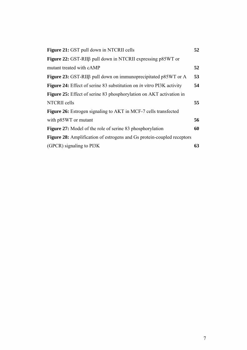
Figure 21: GST pull down in NTCRII cells 52
Figure 22: GST-RIIβ pull down in NTCRII expressing p85WT or
mutant treated with cAMP 52
Figure 23: GST-RIIβ pull down on immunoprecipitated p85WT or A 53
Figure 24: Effect of serine 83 substitution on in vitro PI3K activity 54
Figure 25: Effect of serine 83 phosphorylation on AKT activation in
NTCRII cells 55
Figure 26: Estrogen signaling to AKT in MCF-7 cells transfected
with p85WT or mutant 56
Figure 27: Model of the role of serine 83 phosphorylation 60
Figure 28: Amplification of estrogens and Gs protein-coupled receptors
(GPCR) signaling to PI3K 63
7

Acknowledgements
There are many people I wish to thank for reaching this goal; first and
foremost, my supervisor, Professor Vittorio Enrico Avvedimento. I joined his
lab 8 years ago for the laurea diploma and after the first year he gave me a
project to continue on my own. Obviously, I could have never managed to do it
without his guide and supervision. He taught me to find the "good side" of any
experiment; he taught me that even if the assay didn´t work, then it was at least
"technically correct" and informative... even a negative result is a result...at
least he tried! And I thank him because he is so keen on science that working
with him is impossible not to be more enthusiastic of this job everyday.
I would like to thank Prof. Antonio Feliciello, for being a second tutor during
all these years. Every time I needed advice or explanation about what I was
going to do or what I did he has always been a good point of reference, a really
great one!
I sincerely thank all the people that worked on this project, above all Prof.
Antimo Migliaccio, Marina di Domenico, Antonio Porcellini, and Giorgia De
Gregorio. Without their efforts we would have never finished it.
I thank Mariarosaria Santillo for the helpful advice and training for the FACS
experiments.
I´m really grateful to Savina, obviously for collaborating on this project, but
above all for the many coffee-breaks during these years; essential to get
through the bad days...
8

Thanks to Annalisa (A.lisa), for the scientific discussion we had and the
primary antibodies she started on many Sundays...and above all, because every
time I got to do something of importance, she was always by my side.
I would like to thank Prof. Angela Acquaviva, Prof. Domenico Grieco, Prof.
Annamaria Musti, Silvana Cassano, Rita Cerillo, Adriana Gallo, Alessandra
(AleB), Annalisa, Ilaria, Imma, Mary, Vincenzo and all the students in our lab.
All of them helped me to finish this thesis with good advice, "technical" help
and a smile.
I can´t forget that I spent the last year of the Doctorate Programme at Clare
Hall. So I would like to thank Vincenzo Costanzo and his group Ali, Eloise,
Kristina and Sarah for the warm welcome and I’m grateful to Eloise also for
being my last minute proofreading. Thanks to Alessia, Jean-Yves and Tiz...The
Super-Thursday Committee...the greatest help ever during the stressful days of
studying! Well, thanks to the late lifts...when the last bus was gone Alberto and
Bale represented the only solution to go home! Let me say one more word to
Bale...thanks for all the advice regarding difficult labelling (what about 14C?)
but above all for the hearty laughs in those sad evenings!
And at least, but not last, I wish to thank all my friends, especially Gigi and Isa,
and of course my grandparents, my uncles Adriano and Rocco, my aunt Nadia,
Rosaria and my family. In many occasions it has been difficult for my parents
and my brother to handle my stress or my often "lab based life-table" but in
many moments they supported and encouraged me.
9
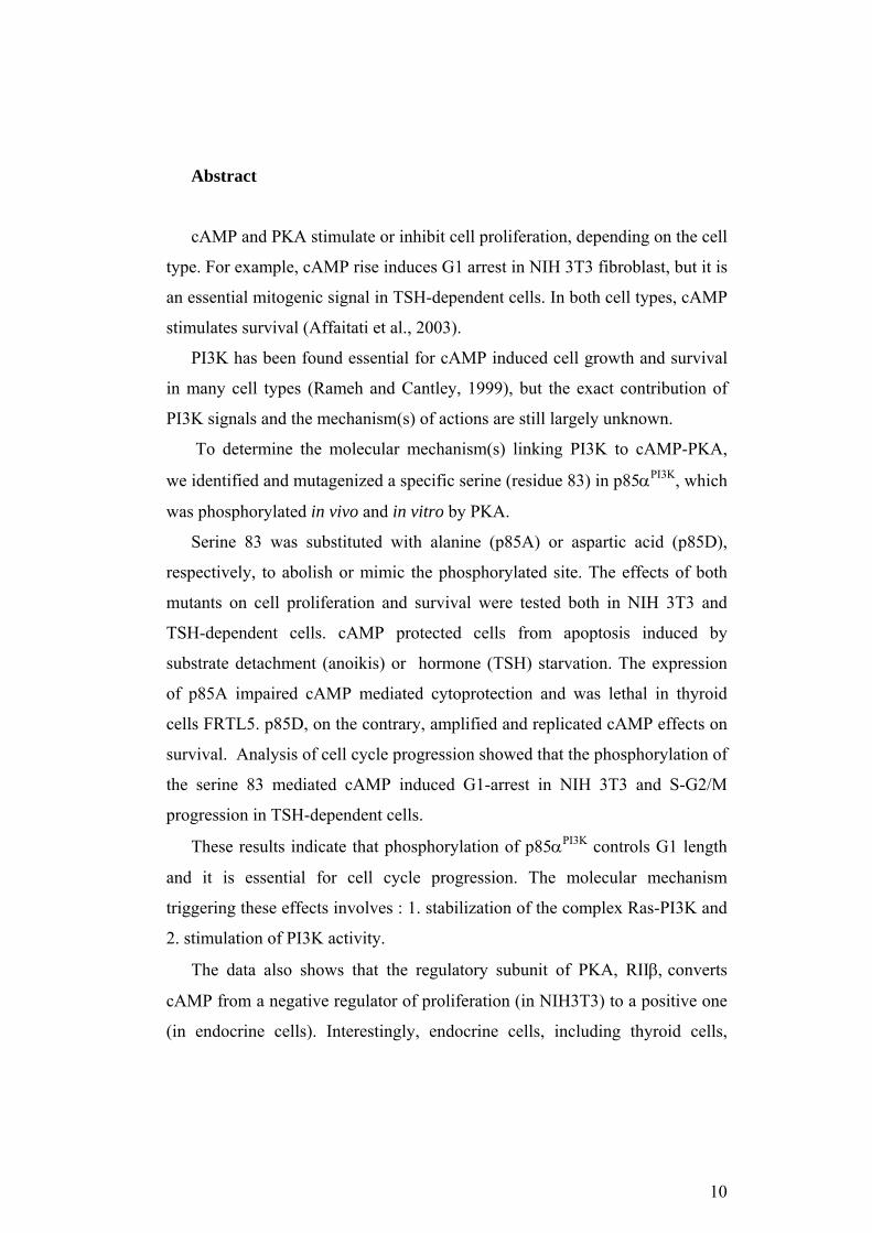
Abstract
cAMP and PKA stimulate or inhibit cell proliferation, depending on the cell
type. For example, cAMP rise induces G1 arrest in NIH 3T3 fibroblast, but it is
an essential mitogenic signal in TSH-dependent cells. In both cell types, cAMP
stimulates survival (Affaitati et al., 2003).
PI3K has been found essential for cAMP induced cell growth and survival
in many cell types (Rameh and Cantley, 1999), but the exact contribution of
PI3K signals and the mechanism(s) of actions are still largely unknown.
To determine the molecular mechanism(s) linking PI3K to cAMP-PKA,
we identified and mutagenized a specific serine (residue 83) in p85αPI3K, which
was phosphorylated in vivo and in vitro by PKA.
Serine 83 was substituted with alanine (p85A) or aspartic acid (p85D),
respectively, to abolish or mimic the phosphorylated site. The effects of both
mutants on cell proliferation and survival were tested both in NIH 3T3 and
TSH-dependent cells. cAMP protected cells from apoptosis induced by
substrate detachment (anoikis) or hormone (TSH) starvation. The expression
of p85A impaired cAMP mediated cytoprotection and was lethal in thyroid
cells FRTL5. p85D, on the contrary, amplified and replicated cAMP effects on
survival. Analysis of cell cycle progression showed that the phosphorylation of
the serine 83 mediated cAMP induced G1-arrest in NIH 3T3 and S-G2/M
progression in TSH-dependent cells.
These results indicate that phosphorylation of p85αPI3K controls G1 length
and it is essential for cell cycle progression. The molecular mechanism
triggering these effects involves : 1. stabilization of the complex Ras-PI3K and
2. stimulation of PI3K activity.
The data also shows that the regulatory subunit of PKA, RIIβ, converts
cAMP from a negative regulator of proliferation (in NIH3T3) to a positive one
(in endocrine cells). Interestingly, endocrine cells, including thyroid cells,
10

express significant amount of RIIβ. In this context RIIβ binds and targets PKA
to PI3K and the membrane, stimulating growth and proliferation.
Moreover, phosphorylation of serine 83 of p85αPI3K is essential also for
estrogen signaling. p85A expression in MCF-7 impairs the ERα binding to
p85αPI3K and, as a consequence, abolishes estrogen induced AKT activity.
Taken together, the data suggests a general mechanism of PI3K regulation
by cAMP, operating in various cell types under different conditions.
11

Background
The control of cell growth and survival is a very complex mechanism
subject to different signals. Each “signaling molecule” (hormones and growth
factors) interacts with a specific receptor, and this is the first step of the signal
transduction (Alberts, c2002).
1. The cellular receptor may be of two types:
1) Intracellular: typically steroid hormones and small peptides are
able to diffuse through the plasma membrane and interact with their
own receptor inside the cells. Usually the complex ligand-receptor
enters the nucleus and modulates the expression of specific gene
through the binding to specific responsive elements on the DNA
(Alberts, c2002);
2) Extracellular: Most of growth factors, proteic hormones and
neurotransmitter interact with receptors present on the plasma
membrane, which can be “tyrosine kinase” or “G protein coupled”
receptor (Alberts, c2002).
Briefly, the tyrosine kinase receptors (RTKs) are constituted of two
subunits, their extracellular domain binds the ligand, while the catalytic
domain is in the intracellular region. Upon the binding of the ligand this
receptors dimerize and the tyrosine residues in the catalytic domain
undergo autophosphorylation. The G protein coupled receptor
(GPCRs), instead, are characterized by an extracellular domain, seven
transmembrane segments and an intracellular domain. When the ligand
binds the receptor it undergoes a conformational change that enables the
activation of a specific trimeric G protein. The G proteins are
constituted of three subunits: α, β, γ; in their inactive state the α subunit
binds GDP. After the binding of the ligand to the receptor the GDP is
substituted by GTP, the α subunit detaches from the β/γ complex and
this is the starting point of the transduction pathway in the cell. The
12

GTP is soon hydrolyzed to GDP, and the G protein returns in its
inactive state (Lodish, c1999 ; Alberts, c2002).
The signal transduced by the receptor is then amplified through a kinase
cascade inside the cells.
Another important aspect of the signal transduction is that the different
molecules regulating the cell behavior are not fully independent from the
others. The activated pathway are overlapping, redundant sometimes and
linked at many points, they establish a complicate network known as crosstalk.
An example of crosstalk between receptors is the one between GCPRs leading
to cAMP increase and RTKs that activate Ras and PI3K.
1. cAMP and Protein Kinase A (PKA)
GCPRs may bind different kind of G proteins (Table 1), which can act
through various mechanisms and second messengers (cAMP, Ca2+, inositol
triphosphate, diacylglycerol or cGMP) (Lodish, c1999 ).
Specific receptors associated to Gs protein, such as TSH receptor (TSHR),
activate adenylyl cyclase. This enzyme converts AMP in cAMP that, as a main
effect, leads to the activation of PKA. PKA is a holoenzyme composed by two
regulatory (R) and two catalytic subunits (cPKA) (Feliciello et al., 2001,
2005). cAMP binds to the regulatory subunits leading to the release of the
catalytic ones, which in turn phosphorylate many nuclear and cytoplasmic
substrates controlling multiple cell functions, including motility, metabolism,
differentiation, synaptic transmission, ion channel activities, growth and gene
transcription (Edelman et al., 1987; Haynes et al., 1992; Meinkoth et al., 1993;
Feliciello et al., 2001). There are two different types of PKA:
1) PKA I: it binds the regulatory subunit RIα that has an high
affinity for cAMP, this entails that it is activated by low level of cAMP;
13
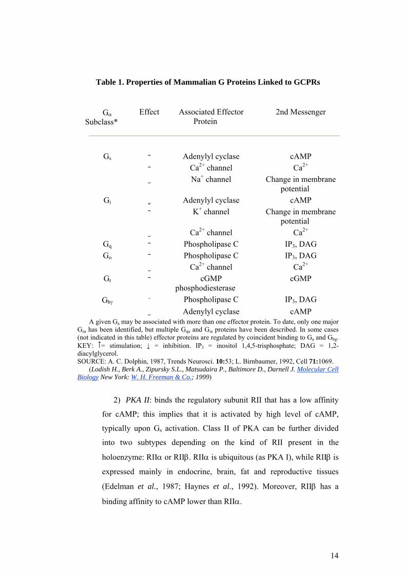
Table 1. Properties of Mammalian G Proteins Linked to GCPRs
Gα Subclass*
Effect Associated Effector Protein
2nd Messenger
Gs Adenylyl cyclase cAMP Ca2+ channel Ca2+
Na+ channel Change in membrane potential
Gi Adenylyl cyclase cAMP K+ channel Change in membrane
potential Ca2+ channel Ca2+
Gq Phospholipase C IP3, DAG Go Phospholipase C IP3, DAG
Ca2+ channel Ca2+
Gt cGMP phosphodiesterase
cGMP
Gbγ Phospholipase C IP3, DAG Adenylyl cyclase cAMP
A given G may be associated with more than one effector protein. To date, only one major G has been identified, but multiple G and G proteins have been described. In some cases (not indicated in this table) effector proteins are regulated by coincident binding to G and G .
a
sa qa ia
a bg KEY: = stimulation; ↓ = inhibition. IP = inositol 1,4,5-trisphosphate; DAG = 1,2-diacylglycerol.
3
SOURCE: A. C. Dolphin, 1987, Trends Neurosci. 10:53; L. Birnbaumer, 1992, Cell 71:1069. (Lodish H., Berk A., Zipursky S.L., Matsudaira P., Baltimore D., Darnell J. Molecular Cell
Biology New York: W. H. Freeman & Co.; 1999)
2) PKA II: binds the regulatory subunit RII that has a low affinity
for cAMP; this implies that it is activated by high level of cAMP,
typically upon Gs activation. Class II of PKA can be further divided
into two subtypes depending on the kind of RII present in the
holoenzyme: RIIα or RIIβ. RIIα is ubiquitous (as PKA I), while RIIβ is
expressed mainly in endocrine, brain, fat and reproductive tissues
(Edelman et al., 1987; Haynes et al., 1992). Moreover, RIIβ has a
binding affinity to cAMP lower than RIIα.
14
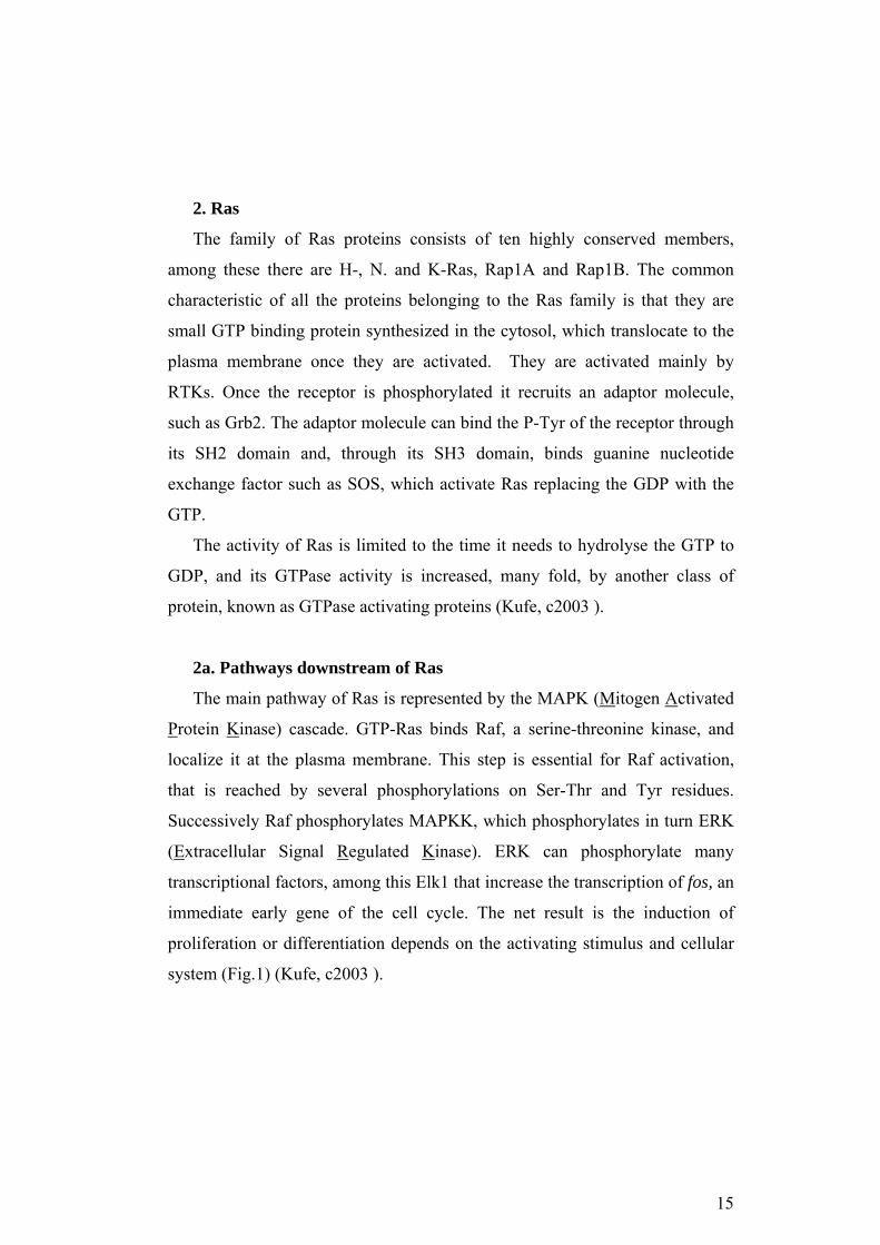
2. Ras
The family of Ras proteins consists of ten highly conserved members,
among these there are H-, N. and K-Ras, Rap1A and Rap1B. The common
characteristic of all the proteins belonging to the Ras family is that they are
small GTP binding protein synthesized in the cytosol, which translocate to the
plasma membrane once they are activated. They are activated mainly by
RTKs. Once the receptor is phosphorylated it recruits an adaptor molecule,
such as Grb2. The adaptor molecule can bind the P-Tyr of the receptor through
its SH2 domain and, through its SH3 domain, binds guanine nucleotide
exchange factor such as SOS, which activate Ras replacing the GDP with the
GTP.
The activity of Ras is limited to the time it needs to hydrolyse the GTP to
GDP, and its GTPase activity is increased, many fold, by another class of
protein, known as GTPase activating proteins (Kufe, c2003 ).
2a. Pathways downstream of Ras
The main pathway of Ras is represented by the MAPK (Mitogen Activated
Protein Kinase) cascade. GTP-Ras binds Raf, a serine-threonine kinase, and
localize it at the plasma membrane. This step is essential for Raf activation,
that is reached by several phosphorylations on Ser-Thr and Tyr residues.
Successively Raf phosphorylates MAPKK, which phosphorylates in turn ERK
(Extracellular Signal Regulated Kinase). ERK can phosphorylate many
transcriptional factors, among this Elk1 that increase the transcription of fos, an
immediate early gene of the cell cycle. The net result is the induction of
proliferation or differentiation depends on the activating stimulus and cellular
system (Fig.1) (Kufe, c2003 ).
15
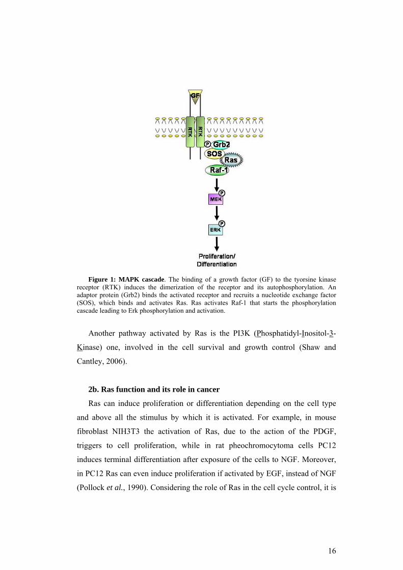
Figure 1: MAPK cascade. The binding of a growth factor (GF) to the tyorsine kinase
receptor (RTK) induces the dimerization of the receptor and its autophosphorylation. An adaptor protein (Grb2) binds the activated receptor and recruits a nucleotide exchange factor (SOS), which binds and activates Ras. Ras activates Raf-1 that starts the phosphorylation cascade leading to Erk phosphorylation and activation.
Another pathway activated by Ras is the PI3K (Phosphatidyl-Inositol-3-
Kinase) one, involved in the cell survival and growth control (Shaw and
Cantley, 2006).
2b. Ras function and its role in cancer
Ras can induce proliferation or differentiation depending on the cell type
and above all the stimulus by which it is activated. For example, in mouse
fibroblast NIH3T3 the activation of Ras, due to the action of the PDGF,
triggers to cell proliferation, while in rat pheochromocytoma cells PC12
induces terminal differentiation after exposure of the cells to NGF. Moreover,
in PC12 Ras can even induce proliferation if activated by EGF, instead of NGF
(Pollock et al., 1990). Considering the role of Ras in the cell cycle control, it is
16

not surprising that different mutations of these genes are found in human
cancers. The main hot-spots for activating Ras mutations are in the GTP
binding domain, these alterations lead to the constitutive activation of Ras
disabling it from hydrolyzing GTP. The main aminoacid for this function is the
Gly in position 12, which is frequently substituted with Val or Asp. The nature
of the mutation correlates even with the aggressivity of the cancer. In fact, Ras-
Val12 is more frequently associated to advanced and metastatic colon-
carcinoma, while Ras-Asp12 is more often present in benign human colorectal
cancer. On a molecular point of view the difference between these two
mutations is that Ras-Val12 activates the Erk pathway, on the other end Ras-
Asp12 stimulates the PI3K and FAK pathway (Cespedes et al., 2006).
3. PI3Ks
PI3Ks are some of the main players in the pathways regulating cell
proliferation, survival and motility. They phosphorylate the inositol on the
position 3 and can generate inositol 3 monophosphate, 1,3 diphosphate and
3,4,5 triphosphate. Depending on the structure and the substrate specificity
PI3Ks are divided into 3 classes (Fig.2) (Walker et al., 1999).
Class I PI3Ks preferentially phosphorylates phosphatidilinositol 4,5
diphosphate (PtdIns(4,5)P2) in vivo, this class can be divided into two
subclasses: IA and IB. The PI3Ks IA are p110α, β and δ, all of which bind an
adaptor molecule of 85 KDa (p85) that is required for the binding to the
tyrosine kinase receptor, by which the enzyme is activated. The class IB is
activated by heterotrimeric G-proteins subunit and they require the binding to a
p101 adaptor molecule for their full activation. All the enzyme belonging to
class I are characterized by an N-terminal Ras Binding Domain (RBD), this
implies that Ras can activate them.
Class II enzymes are large proteins (170-210 KDa) characterized by the
PIK domain, 50% similar to the one of PI3Ks of class I, and a C2 C-term
domain. Another characteristic domain is the PX, common to molecule such as
17

NADPH-oxidase, phox-40 and phox-47. In vitro class II enzymes
preferentially phosphorylate PtdIns and PtdIns-4-P.
The prototype of class III enzymes, VPS34, was first identified in yeast in a
screening for mutants defective in protein sorting. This protein is associated
with a serine-threonine kinase, VPS15, which is essential for the intracellular
trafficking. VPS15 recruits VPS34 under the cellular membrane and enhances
VPS34 lipid kinase activity. The preferential substrate of VPS34 is PtdIns. The
analog in mammalian cell has been identified; it is a heterodimeric protein and
is associated with a phosphatidylinositol transfer protein, which stimulates its
activity. This class of PI3Ks lacks the RBD (Fruman et al., 1998;
Vanhaesebroeck and Waterfield, 1999; Walker et al., 1999).
Figure 2: Classes of PI3Ks. The table illustrates the classes of PI3Ks and the defining
characteristic of each one. (Vanhaesebroeck B.,and Waterfield M. D., Experimental Cell Research,1999; 253, 239–254)
3a. Structure and function of p85PI3K
p85PI3K is the regulatory subunit of PI3K, there are three different isoforms,
α, β and γ. They share a high homology and their characteristic domains are: a)
an N-terminal SH3 binding domain; b) a Proline Rich Domain (PRD); c) a
BCR domain, which is homolog to the GTPase of the Rho family; d) a second
PRD; e) two SH2 domains separated by a region called iSH2 (inter-SH2
domain) (Fig.3).
18

Figure 3: Structure of p85αPI3K. P1 and P2 are respectively the Proline Rich Domain 1 and 2.
It has been shown that the SH3 and the BCR domain are involved in the
dimerization of p85PI3K, that, in turn, may be involved in the stabilization of
p110PI3K (Harpur et al., 1999). The interaction between p85PI3K and p110PI3K is
mediated by iSH2 domain, which binds the N-terminal of p110PI3K. Previous
studies have shown that p85PI3K can both stabilize p110PI3K increasing its
activity and inhibit it. The actual model to explain the opposite effects of
p85PI3K on PI3K activity is that the binding of p110PI3K to p85PI3K is necessary
to stabilize the protein. The binding to p85PI3K itself is not sufficient to activate
the lipid kinase activity of p110PI3K, to reach the effect it is necessary a
conformational change, which is induced by the phosphopeptide binding to the
SH2 domain. Experiment with deletion mutant showed that the nSH2 is
necessary and sufficient for the activation of p110PI3K. In contrast, if the
phosphopetides bind cSH2, to activate p110PI3K, the first 322 aminoacids (SH3
domain, the first PRD and the BH domain) and the nSH2 are required
(Carpenter et al., 1993; Klippel et al., 1993; Dhand et al., 1994a; Holt et al.,
1994; Hu and Schlessinger, 1994; Yu et al., 1998). Moreover, both the binding
of phosphopeptide to the nSH2 and the one to the cSH2 induce a
conformational change at the nSH2 (Fig. 4).
19
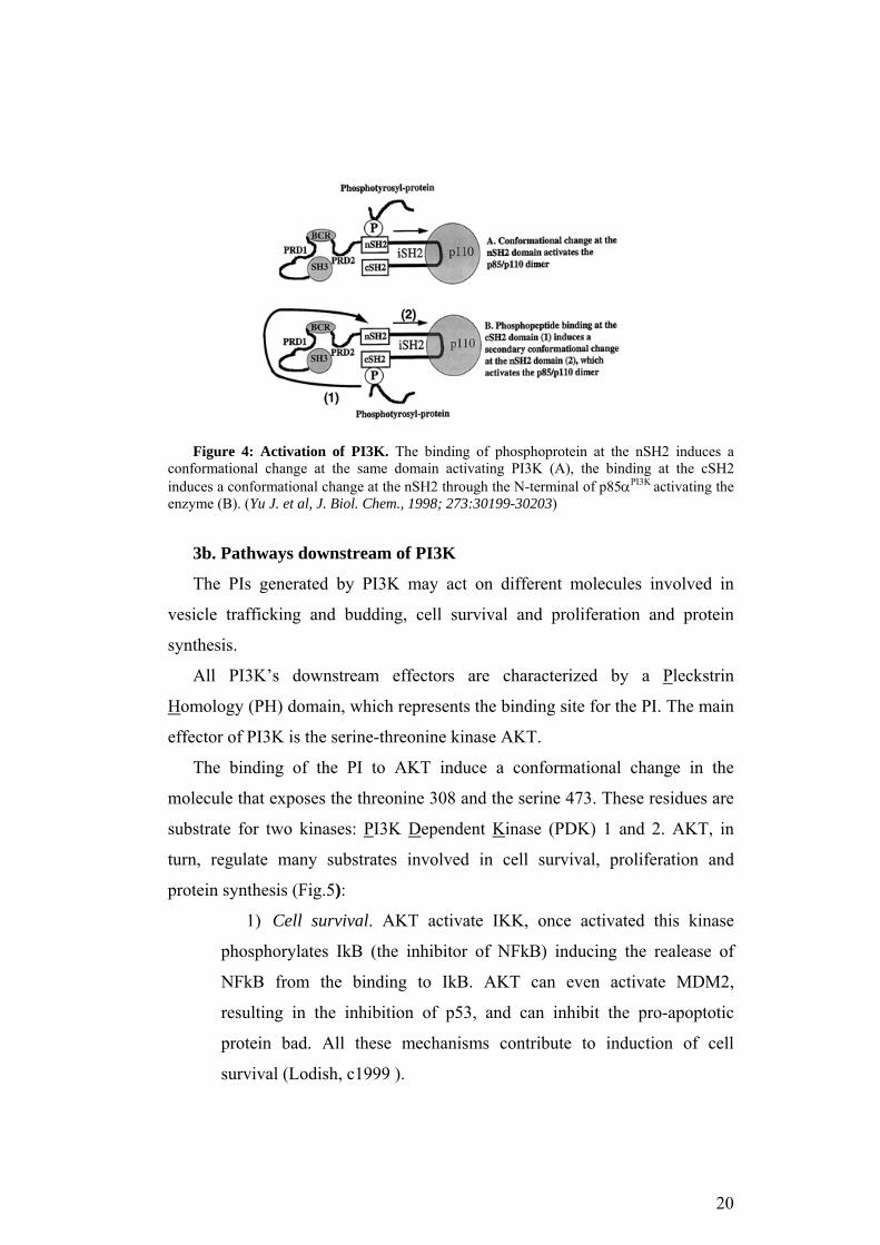
Figure 4: Activation of PI3K. The binding of phosphoprotein at the nSH2 induces a
conformational change at the same domain activating PI3K (A), the binding at the cSH2 induces a conformational change at the nSH2 through the N-terminal of p85αPI3K activating the enzyme (B). (Yu J. et al, J. Biol. Chem., 1998; 273:30199-30203)
3b. Pathways downstream of PI3K
The PIs generated by PI3K may act on different molecules involved in
vesicle trafficking and budding, cell survival and proliferation and protein
synthesis.
All PI3K’s downstream effectors are characterized by a Pleckstrin
Homology (PH) domain, which represents the binding site for the PI. The main
effector of PI3K is the serine-threonine kinase AKT.
The binding of the PI to AKT induce a conformational change in the
molecule that exposes the threonine 308 and the serine 473. These residues are
substrate for two kinases: PI3K Dependent Kinase (PDK) 1 and 2. AKT, in
turn, regulate many substrates involved in cell survival, proliferation and
protein synthesis (Fig.5):
1) Cell survival. AKT activate IKK, once activated this kinase
phosphorylates IkB (the inhibitor of NFkB) inducing the realease of
NFkB from the binding to IkB. AKT can even activate MDM2,
resulting in the inhibition of p53, and can inhibit the pro-apoptotic
protein bad. All these mechanisms contribute to induction of cell
survival (Lodish, c1999 ).
20

2) Cell proliferation. PI3K activity is required at different steps of
the cell cycle, first at the transition G0-G1, a second peak is at mid G1 and
it is necessary for the entry in S-phase and induction of DNA synthesis and
for the G2-M transition. AKT is involved at all the steps. Its main role in
cell cycle control is the inhibition of Glycogen Synthase Kinase 3β
(GSK3β) and the transcriptional factors FOXO. GSK is a negative
regulator of cell cycle since it targets cyclin E, cyclin D and Myc for
degradation. FOXO is the O subgroup of Forkhead transcriptional factors
(TFs) family. These TFs induce the expression of molecule essential for
quiescence maintenance, such as p27KIP , p130 and cyclin G2 (Martinez-
Gac et al., 2004; Garcia et al., 2006).
3) Protein Synthesis. AKT activates mTOR and p70S6K inducing
protein synthesis, and, in turn, increase of cell mass (cell growth) that is
essential for cell division (Garcia et al., 2006).
Figure 5: PI3K-AKT pathway. The activation of PI3K leads to the formation of PtdIns(3,4,5)P3 (PIP3) that activates directly or indirectly AKT. AKT phosphorylates different substrates controlling cell proliferation, survival and growth (protein synthesis).
21

3c. PI3K function and its role in cancer
Class IA PI3Ks play a main role in control cell replication, migration,
survival and glucose homeostasis (Kufe, c2003 ). The oncogenic potential of
PI3K and its target AKT was first revealed by two retroviruses (Bader et al.,
2005; Kang et al., 2005):
1) the avian sarcoma virus ASV16, which encodes a constitutive
active homolog of p110αPI3K, P3k, that is fused to the Gag sequence of
the virus;
2) the murine lymphoma virus AKT8, that encodes for
constitutively active AKT.
The oncogenicity of the two viral proteins is due to the constitutive
membrane addresses, because of the myristoylation, and their constitutive
kinase activity. In fact, if the wild type p110αPI3K is overexpressed in normal
chicken embryo fibroblast no alteration of cell growth is observed. In contrast,
mutant p110αPI3K can induce strong oncogenic transformation in the same
cellular system.
In the few past years, different mutations of PIK3CA, the gene encoding
p110αPI3K, have been identified, outlining the role of PI3K in human cancer.
These mutations are somatic missense ones, they are tumor-specific and they
map to a few hot-spots. The three most common mutations are E542K, E545K
and H1014R. The H1014R mutation is in the substrate binding pocket and this
suggest an increased binding affinity of mutant PI3K for PtdIns(4,5)P2. The
other two mutation, instead, are in the helical domain. It is not likely that they
increase the catalytic activity of the enzyme since they are too far from the
catalytic domain. Considering that both p85PI3K and Ras bind to the N-terminal
of p110PI3K (even if not to the helical domain), it is possible that the E542K and
the E545K substitution alter the binding to these proteins, or to other unknown
regulatory proteins.
Other mutations that constitutively activate PI3K are the p85PI3K mutations.
p65αPI3K is the first oncogenic variant of p85PI3K that has been identified.
22
Jiemenez et al.in 1998 (Jimenez et al., 1998), in fact, cloned p65αPI3K from a
murine lymphoma generated through X-Ray irradiation. This variant lacks part
of the iSH2 and the cSH2, can still bind p110αPI3K and can localize the PI3K
complex at the plasmamembrane. This results in the constitutive activity of the
enzyme and contributes to cellular transformation. Another oncogenic variant
of p85αPI3K is p76αPI3K, this lacks the cSH2 and it has been found in a
Hodgkin’s lymphoma cell line (Jucker et al., 2002). In human ovarian and
colon cancer Philip et al. found a few mutation in splicing site leading to exon
13 skipping, so to the deletion of the region of the iSH2 proximal to serine608
(Philp et al., 2001). The serine608 is an important auotregulatory site. Its
phosphorylation by p110αPI3K results in a three-sevenfold decrease of the lipid
kinase activity (Carpenter et al., 1993; Dhand et al., 1994b). Even the
oncogenic variants previously described impair the ser608 phosphorylation.
Other mutations frequently found in human cancer are:
1) mutation of the upstream receptor, such as PDGF, EGF, ErbB2.
The increased expression and activation of the receptors result in
increased activity of the downstream effectors (Osaki et al., 2004);
2) mutation of phosphatase PTEN (phosphatase and tensin
homologue deleted on chromosome 10). PTEN dephosphorylates PIP3
to generate PIP2 downregulating the PI3K pathway. It is a tumor
suppressor gene frequently inactivated in primary cancers in thyroid,
breast, prostate, uterus, central nervous system, soft tissue and above all
colorectal cancer (Osaki et al., 2004).
3) mutation of AKT, amplification of AKT2 are present in ≈15% of
human ovarian cancer and ≈10% of human pancreatic cancer. Its
amplification is not due to polysomy of the chomosome 19, where AKT
gene is located (Cheng et al., 1996). Moreover, AKT alteration in
cancer correlates with a poor prognosis because of the increased cell
motility,that results in an higher tumor invasiveness (Balsara et al.,
2004). In recent studies it has been shown a strong prognostic
23

significance for AKT constitutive activation in acute myeloid leukemia,
it correlates, in fact, with a shorter overall survival (Min et al., 2003).
4. Crosstalk between different signaling pathways: the paradox of
cAMP
The different signaling pathways in the cell form complicate networks,
where the many transduction systems communicate with each other in feed-
forward and feed-back regulatory loops. Considering that it exists a wide range
of specialized cell types, it is essential for the ubiqitously expressed signal
transduction systems to be adapted to meet the specific requirements of the
cell. It is paradigmatic, in this context, the role of cAMP. In fact, in cell such as
rat thyroid cell FRTL-5 and Swiss 3T3 fibroblast cAMP induces proliferation
(Lee et al., 1998; Ariga et al., 2000), while in most cell types (Magnaldo et al.,
1989) it inhibits proliferation. How is it possible is not yet fully understood. It
has been proposed that cAMP inhibits proliferation through the inhibition of
ERK. This is due to mechanisms PKA-dependent and others cAMP, but not
PKA, dependent. First, PKA can phosphorylate Raf-1 on serine 43, 259 and
621 blocking Raf-1 activation (Cook et al., 1993; Mischak et al., 1996; Dhillon
et al., 2002). Second, cAMP activates some cAMP dependent nucleotide
exchange factor (EPAC) leading to the activation of Rap1. Rap1 is a small G-
protein highly homologue to Ras and binds Raf-1 inhibiting it (Stork and
Schmitt, 2002). This leads to the cAMP mediated inhibition of proliferation in
cell as NIH3T3. In cell where proliferation is activated by cAMP, it has been
proposed that ERK is activated by cAMP in a PKA- independent manner. In
fact, it has been shown that Rap1 can stimulate the ERK pathway through B-
Raf (Zwartkruis et al., 1998). These observations suggest that the effect of
cAMP on cell proliferation may depend on the differential expression of Raf
isoforms in the distinct cell types (Fig.6).
24

Figure 6: cAMP through Rap1 regulates ERK activity. cAMP activates Rap1, in B-Raf
negative cells this leads to Raf-1 inhibition and suppresses ERK activity (A), instead, in B-Raf positive cells (B), Rap1 activates ERK.(Stork and Schmitt, TRENDS in Cell Biology, 2002; 258-266)
Another example of crosstalk between two pathways is the Raf-AKT one.
RTKs activate both the PI3K and Ras pathways. In MCF-7 cells, a human
breast cancer cell line, Insulin-like Growth Factor I (IGF-I) induces cell
proliferation and survival through PI3K-AKT and growth arrest and
differentiation through Ras-Raf. According to Moelling et al (2002), the effect
of IGF-I on MCF-7 cells depends on the concentration of the growth factor. In
particular, high doses of IGF-I activate AKT quickly and strongly enough to
suppress Raf-1 activity. AKT, in fact, can phosphorylate Raf-1 on serine 259
inhibiting it. In this context proliferation is stimulated. Low doses of IGF,
instead, are not enough to suppress Raf-1, so both pathways are active and the
differentiation signals are favored over the mitogenic ones (Fig.7, (Moelling et
al., 2002).
25
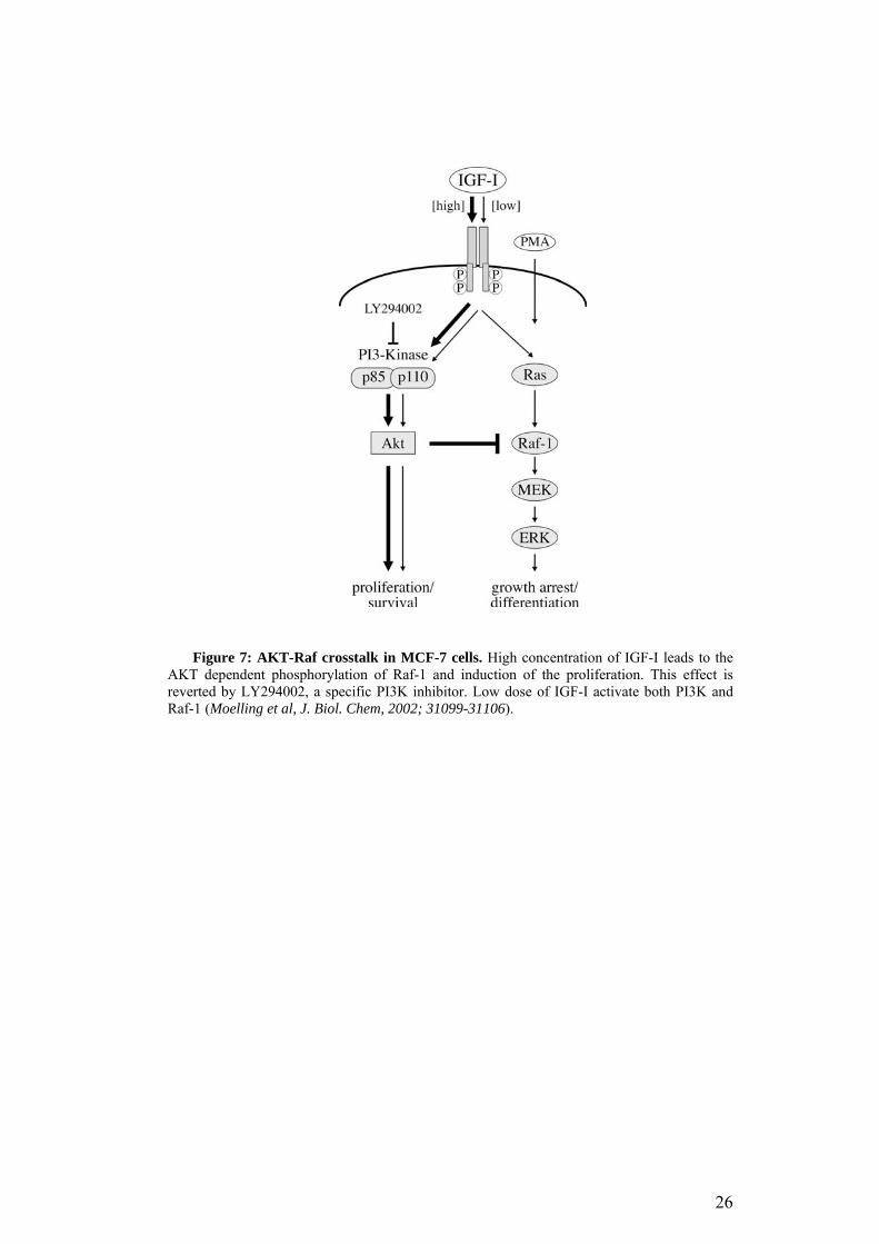
Figure 7: AKT-Raf crosstalk in MCF-7 cells. High concentration of IGF-I leads to the AKT dependent phosphorylation of Raf-1 and induction of the proliferation. This effect is reverted by LY294002, a specific PI3K inhibitor. Low dose of IGF-I activate both PI3K and Raf-1 (Moelling et al, J. Biol. Chem, 2002; 31099-31106).
26

Aims of the study
It has been previously shown in our laboratory that PI3K interacts with
Ras upon cAMP stimulation, and this complex is essential for G1-S transition
in these cells.
Moreover, the formation of the complex was H89 sensitive, indicating
that PKA was involed in the regulation of the interaction. In vitro kinase assay
revealed that p85αPI3K was efficiently phosphorylated. These data suggested
that cAMP-PKA selectively influences Ras effector pathway through p85αPI3K
phosphorylation.
1) The first part of the work presented here, focuses on the
identification and validation of the PKA phosphorylation site on p85αPI3K.
2) The second part, instead, investigates the biological role of this
phosphorylation in different cell types. It is worth noting that cAMP leads to
opposite effects on proliferation depending on the cellular system (Pastan et
al., 1975). To investigate the role of phosphorylation of p85αPI3K and its
association with Ras in the regulation of cell proliferation, the experiments
presented were performed both in cells where proliferation was inhibited
(fibroblasts NIH 3T3) or stimulated (TSH-dependent cells) by cAMP.
27

Materials and Methods
1. Plasmid construction
The cDNA encoding for bovine p85αPI3K wild type (acc. n.:163476) was
cloned in the pSG5 vector (gift of Dr. J. Downward). The region of p85αPI3K
from the Xho I restriction site in position 1014 (acc. n.:163476) was amplified
by PCR with a 3’end primer containing the Flag sequence (MDYKDDDDK)
and a BamHI restriction site.
The PCR product was sub-cloned in the II-TOPO vector (Invitrogen),
digested with Xho I and BamHI, extracted and ligated to a pSG5-p85αPI3K
vector previously digested with the same restriction enzymes. The vector
encoding p85αPI3K-flag was then used as template for site-directed mutagenesis
(QuickChange Site-Directed Mutagenesis Kit, Stratagene) to convert serine 83
in alanine or aspartic acid. All the constructs were verified by DNA sequence
analysis.
2. Materials and Reagents
Unless otherwise specified, drugs and chemicals were obtained from Sigma
Aldrich and cell culture supplies were purchased from standard suppliers, e.g.
Falcon, Life Technologies inc., Hyclone.
The antibodies used were: anti-pan-Ras (clone 10, mouse monoclonal,
UBI), anti-Raf1 (rabbit polyclonal, Santa Cruz), anti-p85PI3K (rabbit polyclonal,
UBI), anti-P-Serine (rabbit polyclonal, Zymed), anti-Erk 1/2 (rabbit polyclonal,
Santa Cruz), anti-P-Erk 1/2 (mouse monoclonal, Santa Cruz), anti-P-Akt ser
473 (rabbit polyclonal, Cell Signaling), anti-Akt (rabbit polyclonal, Cell
Signaling), anti P-Gsk ser 21/9(rabbit polyclonal, Cell signaling), ant GSKα/β
(mouse monoclonal, UBI). The anti-flag antibody was the mouse monoclonal
Sigma M2 antibody.
28
3. Cell culture and transfections
Cell lines used in the experiments were grown as follows:
1. murine fibroblasts NIH 3T3 in DMEM 10% bovine serum;
2. rat thyroid cells FRTL-5 in medium with 5% calf serum and six
hormones (1mU/ml TSH, 1µg/ml Insulin, 3.6 µg/ml
Hydrocortisone, 5 µg/ml Transferrin, 10 ng/ml Somatostatin, 20
µg/ml Glycil-histidil-lysine);
3. NTCRII cells in DMEM 10% foetal bovine serum. Considering
that these cells were conditional stable clones of RIIβ and TSHR,
the medium was supplemented with puromycin 2.5 µg/ml and
geneticin (G418) 200 µg/ml to maintain the selection and
tetracycline 1.0 µg/ml to keep the genes silenced. The selection and
tetracycline were removed 48 hr before starting the experimental
procedures described in the results (Porcellini et al., 2003);
4. MCF-7 breast cancer cells in DMEM with 5% foetal bovine
serum, supplemented with 6 ng/ml insulin and 3,75 ng/ml
hydrocortisone;
5. HeLa cells in RPMI 10% foetal bovine serum.
All the media were supplemented with penicillin/streptomycin
100mU/mL and 2 mM glutamine.
Cells were transfected with lipofectamine according to the
manufacturer’s recommendations (Gibco Invitrogen). Briefly cells were
transfected at 80% confluence with 4µg of DNA for each 100mm dish. The
lipofectamine was used 1µL for each µg of DNA. The mix DNA-
lipofectamine was incubated 45 minutes at room temperature to allow the
formation of the precipitates. Before adding the mixture cells were washed
with PBS and the growing medium was replaced with medium without
serum and antibiotics. 5 hours after the adding of the mixture the normal
concentration of serum was restored.
29

4. Cell lysis and immunoprecipitation
Cells were collected in ice-cold PBS and spun at 1500 rpm for 3 minutes.
Pellets were re-suspended in lysis buffer (50 mM Tris-HCl pH 7.4, 1% Nonidet
NP-40, 100 mM NaCl, 2 mM EDTA 50 mM NaF, 0.1 mM NaVO3 1 mM β-
glycerophosphate, 2.5 mM sodium pyrophosphate and a protease inhibitor
cocktail). After 15 minutes incubation on ice the samples were spun at 13000
rpm for 10 minutes. Cell lysates were transferred to other tubes and quantified
at the spectrophotometer using the Bradford assay (BioRad protein assay).
Protein lysates were diluted to 2 mg/ml and were incubated with 4 µg of
antibody/0.5-1 mg of protein at 4°C in gentle rock agitation overnight. At the
end of incubation, 20 µl of A/G plus were added to samples and the
immunoprecipitates were washed three times with lysis buffer and then
collected by centrifugation. The bound proteins were eluted with one volume
of 2× Laemmli buffer or, when indicated, using 0.1 M glycine HCl pH 3.5. In
the latter case, 1M Tris-HCl pH 8 was added to the eluted proteins to neutralize
the pH.
5. GST pull-down
GST pull-down were performed as described by (Grieco et al., 1996).
Briefly, cells were lysed in 200mM NaCl, 50mM Tris-Hcl pH 7.5, 2mM
MgCl2, 10% glycerol, 1% NP 40, 10µg/ml Trypsin inhibitor, 1µg/ml aprotinin,
1µg/ml leupeptin, 10mM NaF, 10mM Na3VO4 (Pull Down buffer). 1 mg of
protein extract was incubated with 1 microgram of GST-RIIβ fusion protein or
the control protein (GST) for 4 hours at 4°C in gentle rock agitation. The
pellets were washed 5 times in pull down buffer and re-suspended in one
volume of 2× Laemmli buffer.
6. Western blot
Total cell extracts and immunoprecipitates were separated on 10% SDS-
PAGE and transferred onto nitrocellulose filter. The filters were blocked in
30

TBS 0,1% TWEEN (TBS-T) 5% not-fat-dry-milk (NFM) for 1 hour at room
temperature. The filters were washed three times with TBS-T and incubated
with the indicated primary antibody. Primary antibodies were diluted according
to the manufacturer’s recommendations. The filters were successively washed
three times with TBS-T and incubated with the peroxidase conjugated antibody
diluted 1:3000 in TBS-T 3% NFM. The signal was detected with
chemiluminescence system (Feliciello et al., 2000).
7. In vitro phosphorylation
HeLa cells were transiently transfected with p85αPI3K-flag and p85A. 48
hours after transfection, cell lysates were immunoprecipitated with non-
immune IgG or anti-flag antibody 15 h at 4°C. Protein A/G bound
immunoprecipitates were washed twice with lysis buffer and finally with
Kinase buffer (Hepes 20 mM, MgCl2 10 mM, pH 7.4). The washed
immunoprecipitates were treated with 0.4 µg of Protein Kinase A. Each aliquot
was incubated in a final volume of 30 µl of Kinase buffer containing 10-5 M
cAMP, 100 µM ATP and 10 µCi[γ32P-ATP] for 30 minutes at 30°C. The
reaction was stopped by adding one volume of 2× Laemmli buffer (Ciullo et
al., 2001).
8. In vitro protein synthesis
p110αPI3K and p85αPI3K wild type or p85A or p85D were co-transcribed
and co-translated in vitro in [35S]methionine-containing reticulocyte lysate
according to the manufacturer’s recommendations. The conditions of the
reaction were optimized to reach the same efficiency of synthesis of the co-
transcripted and co-translated proteins. The amount of template used was 1µg,
and the optimal ratios of the two template were: p85αPI3K wt / p110αPI3K 1:2;
p85A/ p110αPI3K and p85D/ p110αPI3K 2:1. The reactions were incubated for
1h 30’ at 30°C and diluted 1:100 with PBS conatining 0.5% Triton X-100 and
31

protein inhibitors. The control reaction was performed using only p110αPI3K as
template.
The diluted lysates were immunoprecipitated with the anti-Flag antibody,
as described previously. The immunocomplexes were washed three times with
PBS-0.5% Triton X-100, solubilized in 2× Laemmli buffer and boiled. The
samples were separated on SDS-PAGE. The gel was fixed 30 minutes in a
solution containing 10% acetic acid and 20% methanol, washed three times in
deionized water and treated with 100 mM salycilate to enhance the radioactive
signal. The gel was then dried and expose on a autoradiography film. On the
gel the input (1 µl of the reaction mix using 1 µg of plasmid as template) was
loaded as control of the reaction.
9. Apoptosis assays
NIH 3T3 were co-transfected with GFP and the indicated vector. 48 hours
later cells were plated in DMEM 0,1% bovine serum -/+ 200µM CPT-cAMP
on plates covered with 2% agarose (anoikis) . After a 5 hours incubation cells
were collected and washed three times with PBS before the 5 minutes
incubation with propidium iodide. Successively cells were analyzed by
Fluorescent Activated Cell Sorter (FACS) using CELLQuest software (Becton
Dickinson). The percentage of death (PI positive cells, i.e. red population) was
calculated on the population positive for GFP (green population). The
experiments were performed in triplicate.
5*105 NTCRII cells were transfected as described. 24 hours later cells the
normal medium was replaced by 0,5% serum medium with or without
10mU/mL TSH or 100µM cAMP. 18 hr after treatment, cells were fixed in 2%
paraformaldehyde/1X PBS, 10 min, RT and washed one time in PBS + 50 mM
glycine for 10 min at RT and 3 times for 5 min in PBS. Cells were
permeabilized with 0.5% triton X-100/ 1X PBS for 10 min, washed 3 x 5 min
in PBS and incubated with 100 µl of 1X TdT reaction mix. TUNEL reaction
was carried out at 37°C for 60 min using 15 Units of TdT (ROCHE) and 2 µl
32

of 2mM BrdUTP. BrdUTP incorporation was revealed by anti-BrdU-FITC and
the samples were then stained in Propidium Iodide. The data were acquired and
analyzed by CELLQuest software for bivariate-analysis of DNA content versus
BrdU. Experiments were performed in triplicate.
10. Cell growth analysis
5*105 cells were transfected with the indicated vectors. 48 hours later cells
were plated in 60 mm dishes and growth in 0,5% serum containing medium.
After 18 hr cells were induced into the cycle with 10 mU/ml TSH or EGF 100
ng/ml. Cells were collected and washed twice with PBS. Successively cells
were fixed in 70% ethanol and stained for 30 min at room temperature in 0.1%
triton-X100, 0.2 mg/ml DNase-free RnaseA, 20 µg/ml Propidium Iodide.
Cells were acquired using the FACScan Flow Cytometer (Becton
Dickinson) and analyzed by Cell Fit Cell-Cycle Analysis Version 2 to define
the percentage of cells in the different phases of cell cycle.
11. 5’-bromo-2’-deoxyuridine (BrdU) labelling
BrdU incorporation was assayed in a pulse-chase experiment. Cells were
labelled for 30 min with BrdU to a final concentration of 20 µg/ml and
harvested at 0, 90 and 270 min. After treatment, cells were fixed in ice-cold
70% ethanol for 4 hr at +4 °C and washed 3 times for in PBS. Cell pellet was
re-suspended in 0.25 ml of 1N HCL and let stand 20 min at room temperature.
After acidic denaturation of DNA, cells were washed 2 times in
phosphate/citric buffer (0.2 M Na2HPO4; pH 7.4). BrdU incorporation was
revealed by anti-BrdU-FITC and then stained for 30 min at room temperature
in 0.1% triton-X100, 0.2 mg/ml DNase-free RnaseA, 20 µg/ml Propidium
Iodide. Fluorescence was determined by using the FACScan Flow Cytometer.
Experiments were performed in triplicate. The data were acquired and analyzed
by CELLQuest software for bivariate-analysis of DNA content versus BrdU
and by Cell Fit Cell-Cycle Analysis Version 2 for DNA content analysis.
33

12. Lipid kinase assay
Lipid kinase activity was determined as described by (Maier et al., 1999).
Briefly, the assays were carried out in a final volume of 50 µl containing 0.1%
bovine serum albumin, 1 mM EGTA, 120 mM NaCl, 40 mM HEPES, pH 7.4,
1 mM dithiothreitol, 1 mM -glycerophosphate, 7mM MgCl2 (buffer E). Lipid
vesicles (30 µl containing 320 µM phosphatidylethanolamine, 300 µM
phosphatidylserine, 140 µM phosphatidylcholine, 30 µM sphingomyelin,
supplemented with 40 µM PI-4,5-P2 in buffer E) were sonicated 1 hour and
incubated on ice 10 min. The immunoprecipitates were added to the lipid
mixture and incubated for 10 min at 4°C in a final volume of 40 µl. The
reaction was started by adding 40 µM ATP (1 µCi of [-32P]ATP in 10 µl of the
assay buffer. The reaction was incubated 15 minutes at 30°C and then stopped
with 150 µl of 1 N ice-cold HCl. The lipids were extracted by vortexing
samples with 500 µl of chloroform/methanol (1:1). After centrifugation the
organic phase was washed twice with 200 µl of 1 N HCl. Phosphorylated lipids
were separated by TLC developed in CHCl3/CH3OH/H2O/NH4OH
(60:47:11.3:2), dried, and visualized by autoradiography and quantified with
Phosphor-Imager.
34

RESULTS
1. PKA phosphorylates serine 83 of p85αPI3K
It has been previously shown in our laboratory that PKA efficiently
phosphorylates p85αPI3K in vitro (Ciullo et al., 2001). The sequence analysis
revealed a PKA consensus in the sequence of bovine p85αPI3K (KKIS). This
consensus is highly conserved in evolution (Fig.8): KKIS in bovine/human and
KRIS in mouse/rat. Moreover, no PKA consensus was found in p85βPI3K.
Figure 8: Alignment of p85αPI3K sequences from different species. p85αPI3K presents an
highly conserved PKA consensus at the residues 80-83.
To determine if this is a bona fide PKA phosphorylation site and its
biologiacal role, the serine in the consensus (serine 83) was substituted with
alanine to prevent the phosphorylation or aspartate to mimic it. To distinguish
the exogenous from the endogenous protein, wild-type p85αPI3K encoding
cDNA was fused to a C-terminal Flag sequence (MDYKDDDDK ) and
subcloned in a pSG5 vector. The tagged wild-type cDNA was used as a
template for the mutagenesis reaction (see Materials and Methods).
1a. In vitro phosphorylation
The wild-type and the alanine tagged proteins were transiently expressed in
Hela cells. The cell lysates were immunoprecipitated with anti-Flag antibody
or non-immune IgG (SNI) as described in Materials and Methods. The
35

immunoprecipitates were incubated in vitro in kinase buffer containing 10-5 M
cAMP, 100 µM ATP and 10 µCi[γ32P-ATP] for 30 minutes at 30°C, with or
without recombinant PKA (cPKA). The immunoprecipitates were separated on
SDS-PAGE and analysed by western blot with anti-p85 antibody (Fig. 9, upper
panel) and by autoradiography (Fig.9, bottom panel).
Figure 9: In vitro phosphorylation of serine 83 in p85αPI3K by cAMP-PKA. HeLa cells
were transiently transfected with p85αPI3K-flag wild type or p85A; 48 hours after transfection, cell lysates were immunoprecipitated and in vitro phosphorylated with cPKA as described in Material and Methods. Sample aliquots were run on SDS-PAGE and subjected to immunoblot (upper panel) or autoradiography (bottom panel).
p85αPI3K wild-type, but not p85A, was efficiently phosphorylated in vitro
by PKA. This reaction was dependent on cPKA, because omission of cPKA
from the mixture did not results in p85αPI3K phosphorylation. The western blot
in the upper panel shows that the immunoprecipitation efficiency was
comparable in all the samples.
This result indicates that serine 83 is the PKA phosphorylation site on
p85αPI3K.
1b.In vivo phosphorylation
To verify that PKA phosphorylates p85αPI3K not only in vitro but also in
vivo quiescent breast carcinoma cells (MCF7), were transiently transfected
with p85WT or p85A encoding vector. 24 hours after transfection cells were
stimulated with 100µM CPT-cAMP or left untreated. Protein lysates were
immunoprecipitated with anti-Flag antibody and the immunoprecipitates were
36
separated on SDS-PAGE. The proteins were transferred onto nitrocellulose
filter and analyzed by western blot with anti-p85 and anti-phospho-serine (anti-
P-ser) antibodies. Figure 10A shows that p85WT, but not p85A, was efficiently
phosphorylated following cAMP treatment (bottom panel). The amount of
protein in all samples was the same (upper panel).
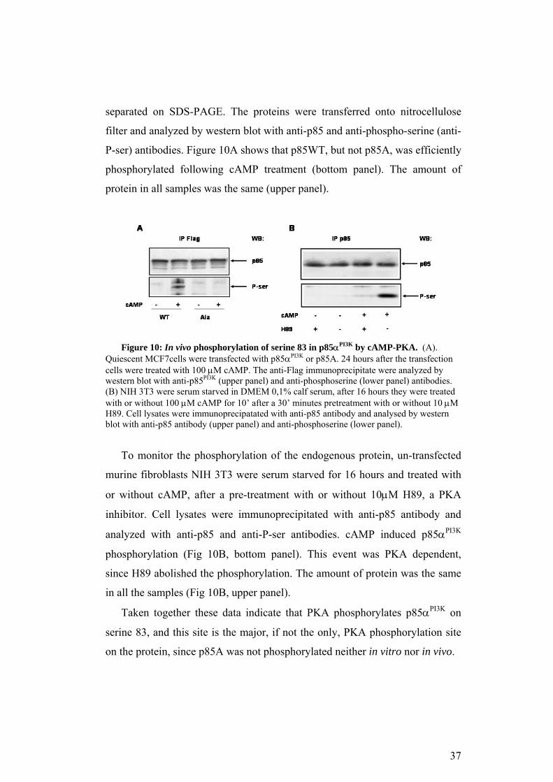
igure 10: In vivo phosphorylation of serine 83 in p85α by cAMP-PKA. (A). Quiescen αPI3K ion cell
d
o monitor the phosphorylation of the endogenous protein, un-transfected
mu
t PKA phosphorylates p85αPI3K on
ser
F PI3K
t MCF7cells were transfected with p85 or p85A. 24 hours after the transfects were treated with 100 µM cAMP. The anti-Flag immunoprecipitate were analyzed by
western blot with anti-p85PI3K (upper panel) and anti-phosphoserine (lower panel) antibodies. (B) NIH 3T3 were serum starved in DMEM 0,1% calf serum, after 16 hours they were treatewith or without 100 µM cAMP for 10’ after a 30’ minutes pretreatment with or without 10 µMH89. Cell lysates were immunoprecipatated with anti-p85 antibody and analysed by western blot with anti-p85 antibody (upper panel) and anti-phosphoserine (lower panel).
T
rine fibroblasts NIH 3T3 were serum starved for 16 hours and treated with
or without cAMP, after a pre-treatment with or without 10µM H89, a PKA
inhibitor. Cell lysates were immunoprecipitated with anti-p85 antibody and
analyzed with anti-p85 and anti-P-ser antibodies. cAMP induced p85αPI3K
phosphorylation (Fig 10B, bottom panel). This event was PKA dependent,
since H89 abolished the phosphorylation. The amount of protein was the same
in all the samples (Fig 10B, upper panel).
Taken together these data indicate tha
ine 83, and this site is the major, if not the only, PKA phosphorylation site
on the protein, since p85A was not phosphorylated neither in vitro nor in vivo.
37

2. Biological effects following the phosphorylation of p85αPI3K in NIH
3T
oth cAMP and PI3K regulate cell survival and growth (see Background).
In
-AKT is
one
a. cAMP induced-survival depends on phosphorylation of p85αPI3K
is
an
83 on the anoikis, NIH 3T3 cells
we
3
B
particular cAMP protects cells from serum deprivation induced apoptosis
(Affaitati et al., 2003) and regulates cell proliferation in different manner
depending on cell type (Pastan et al., 1975).In fact, cAMP induces proliferation
in such cells as thyroid cell FRTL5 (Lee et al., 1998; Ariga et al., 2000), while
it inhibits proliferation in most cell types, such as NIH 3T3 (Magnaldo et al.,
1989). The mechanisms underlying these effects are not yet defined.
On the other side, as discussed in the Background Section, PI3K
of the most important pathway promoting survival and cell growth. These
observations suggest that cAMP-PKA dependent phosphorylation of serine 83
may affect these important biological functions.
2
To validate our hypothesis, inhibition of anoikis has been tested. Anoikis
apoptotic pathway triggered by loss of cell adhesion to the extracellular
matrix and strictly dependent on PI3K (Frisch and Francis, 1994; Khwaja et
al., 1997). In cell culture it can be easily assayed by culturing the cells on
plates covered with a thin 2% agarose layer.
To test the effects of mutagenesis of serine
re co-transfected with a GFP encoding vector and the wild type or mutant
vesions of p85αPI3K. 48h later, the cells were plated in 0,1% CS -/+ 200 µM
CPT-cAMP medium on 2% agarose. After 5h cells were stained with
propidium iodide and the percentage of apoptosis determined by FACS
analysis. The histogram in figure 11B shows that cAMP protected cells from
apoptosis, and the over-expression of p85WT amplified the effect of cAMP.
38

Figure 11: Analysis of cell survival. NIH 3T3 were co-transfected with a GFP encoding
vector and the wild type and mutant of p85αPI3K. (A) The expression was analyzed by western blot. the exogenous molecule is the upper band of the doublet. (B) fraction of apoptotic cells after anoikis was determined by FACS analysis . The data are the mean of three independent experiments; * indicates p ≤ 0.01 comparing cell death in the presence of cAMP in all samples.
The expression of p85A abolished the cAMP mediated protection (*). On
the other side, the p85D expressing cells are more resistant to anoikis in
absence of cAMP (**). This indicates that, at least in part, the S83D
substitution can mimic cAMP effects on survival. It is worth noting that p85D
expressing cells showed a reduced response to cAMP, probably caused by low
expression of p85D (fig 11A).
These data indicate that p85αPI3K mediates the cytoprotective effect of
cAMP during anoikis and its phosphorylation is necessary for the cAMP
induced survival.
2b. cAMP mediated G1-S arrest requires phosphorylation of p85αPI3K
To test if the phosphorylation of p85αPI3K is involved in cAMP growth
arrest, the wild type and the mutant p85αPI3K were expressed in NIH 3T3.
Cells were serum-starved 15 hours and induced into the cycle with 2 % serum
in the presence or absence of cAMP 200 µM. 12 hours later cells were
collected in ice cold PBS, fixed with 70% ice cold ethanol and stained with
propidium iodide and then FACS analyzed. Fig.12A and 12B show that cAMP
39
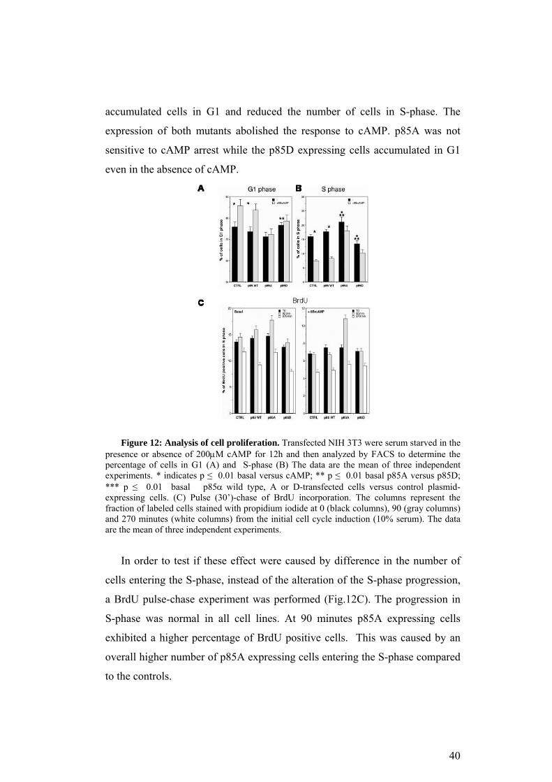
accumulated cells in G1 and reduced the number of cells in S-phase. The
expression of both mutants abolished the response to cAMP. p85A was not
sensitive to cAMP arrest while the p85D expressing cells accumulated in G1
even in the absence of cAMP.
Figure 12: Analysis of cell proliferation. Transfected NIH 3T3 were serum starved in the
presence or absence of 200µM cAMP for 12h and then analyzed by FACS to determine the percentage of cells in G1 (A) and S-phase (B) The data are the mean of three independent experiments. * indicates p ≤ 0.01 basal versus cAMP; ** p ≤ 0.01 basal p85A versus p85D; *** p ≤ 0.01 basal p85α wild type, A or D-transfected cells versus control plasmid-expressing cells. (C) Pulse (30’)-chase of BrdU incorporation. The columns represent the fraction of labeled cells stained with propidium iodide at 0 (black columns), 90 (gray columns) and 270 minutes (white columns) from the initial cell cycle induction (10% serum). The data are the mean of three independent experiments.
In order to test if these effect were caused by difference in the number of
cells entering the S-phase, instead of the alteration of the S-phase progression,
a BrdU pulse-chase experiment was performed (Fig.12C). The progression in
S-phase was normal in all cell lines. At 90 minutes p85A expressing cells
exhibited a higher percentage of BrdU positive cells. This was caused by an
overall higher number of p85A expressing cells entering the S-phase compared
to the controls.
40

These data indicate that PKA-dependent phosphorylation of p85αPI3K
mediates selectively G1 arrest induced by cAMP, since S phase progression
was not influenced in cells expressing the mutant versions.
3. Molecular mechanisms regulated by phosphorylation of serine 83 of
p85αPI3K
It has been previously shown that cAMP-PKA stimulated the interaction
between PI3K and Ras and this event was necessary for cell cycle progression
in cells FRTL-5 (Ciullo et al., 2001). Moreover, Ras and PI3K are among the
major effectors in transduction pathway modulating cell cycle and survival. It
is possible that cAMP promotes cell survival and induces G1 arrest in NIH
3T3 stabilizing PI3K/Ras complex through serine 83 phosphorylation and, in
turn, activating PI3K-AKT pathway.
3a. The phosphorylation of p85αPI3K increases the formation of the
complex Ras-PI3K
In order to test our hypothesis, NIH 3T3 were transfected with p85WT or
mutant encoding vectors. 36 hours later the cells were serum starved and,
after 16 hours, were treated 10 minutes with or without 200µM cAMP. Cell
lysates were immunoprecipitated with anti-Flag antibody. The immuno-
precipitates were separated on SDS-PAGE and analyzed by western blot with
anti-Flag and anti-Ras antibodies. Figure 13A, bottom panel, shows that Ras
co-immunoprecipitated with p85WT after cAMP treatment, the substitution of
serine 83 with aspartic acid amplified this effect. On the contrary, Ras was not
present in p85A immunoprecipitates. Ras was barely detected in all the
untreated samples, while the amount of p85αPI3K was the same in all samples
(upper panel).
The histogram in Figure 13B is the p85/Ras ratio determined as an
average of 3 independent experiments.
41

Figure 13: Effects of the substitution of serine 83 on the association Ras-PI3K. NIH
3T3 were transfected with p85 WT, A or D and straved for 16h. Later on , cells were treated 10 min with 200 µM CPT-cAMP. The total lysates were immunoprecipitated with anti-Flag antibody and analyzed by western blot with anti-p85PI3K and anti-Ras antibody (A). The histogram (B) represents the mean of the densitometric analysis of 3 independent experiments.
These data indicate that serine 83 phosphorylation was necessary to
induce PI3K/Ras interaction, since p85A lost is ability to bind Ras, although it
was not sufficient, because p85D/Ras association was still dependent on
cAMP.
3b. The disruption of the phosphorylation site on p85αPI3K did not
abolish the binding p85αPI3K- p110αPI3K
Considering that p110αPI3K, and not p85αPI3K, mediates PI3K/Ras
interaction (Rodriguez-Viciana et al., 1996) it is possible that serine 83
substitution with alanine may alter the folding of p85αPI3K disrupting the
p85αPI3K- p110αPI3K binding.
Since the commercial antibodies versus p110αPI3K are not satisfactory in
terms of specificity in immunoblot analysis, we decided to study the
association between p85αPI3K and p110αPI3K using the recombinant proteins.
To this end we transcribed and translated the vector encoding p85αPI3K and
42
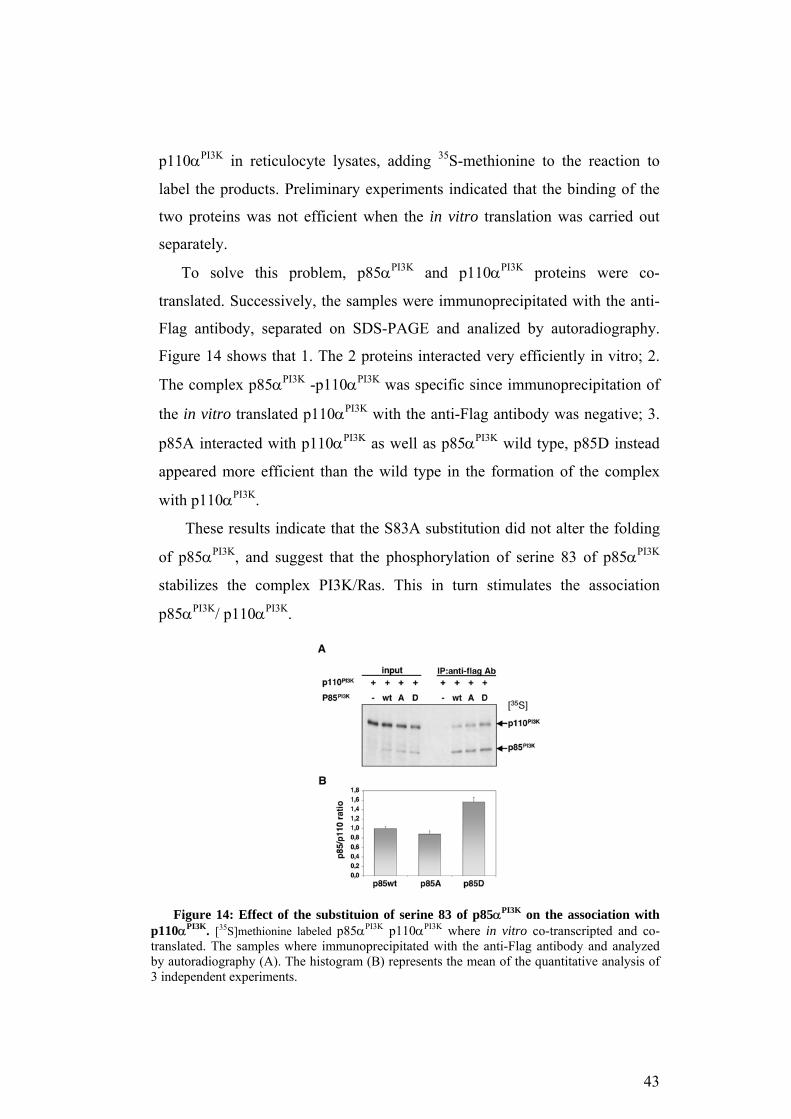
p110αPI3K in reticulocyte lysates, adding 35S-methionine to the reaction to
label the products. Preliminary experiments indicated that the binding of the
two proteins was not efficient when the in vitro translation was carried out
separately.
To solve this problem, p85αPI3K and p110αPI3K proteins were co-
translated. Successively, the samples were immunoprecipitated with the anti-
Flag antibody, separated on SDS-PAGE and analized by autoradiography.
Figure 14 shows that 1. The 2 proteins interacted very efficiently in vitro; 2.
The complex p85αPI3K -p110αPI3K was specific since immunoprecipitation of
the in vitro translated p110αPI3K with the anti-Flag antibody was negative; 3.
p85A interacted with p110αPI3K as well as p85αPI3K wild type, p85D instead
appeared more efficient than the wild type in the formation of the complex
with p110αPI3K.
These results indicate that the S83A substitution did not alter the folding
of p85αPI3K, and suggest that the phosphorylation of serine 83 of p85αPI3K
stabilizes the complex PI3K/Ras. This in turn stimulates the association
p85αPI3K/ p110αPI3K.
Figure 14: Effect of the substituion of serine 83 of p85αPI3K on the association with
p110αPI3K. [35S]methionine labeled p85αPI3K p110αPI3K where in vitro co-transcripted and co-translated. The samples where immunoprecipitated with the anti-Flag antibody and analyzed by autoradiography (A). The histogram (B) represents the mean of the quantitative analysis of 3 independent experiments.
43

3c. cAMP-PKA activates PI3K in vitro
To determine if the phosphorylation of serine 83 influenced PI3K lipid
kinase activity, NIH 3T3 were transfected with the p85WT encoding vector.
Cells were serum starved for 16 hours before a 10 minutes treatment with
200µM cAMP in the presence or absence of H89. Cell lysates were
immunoprecipitated with anti-Flag antibody, and the immunoprecipitates
divided into two aliquots. One aliquot was used to test the in vitro lipid kinase
assay (see Material and Methods). Following a 15 minutes incubation the
reaction was stopped with 1N HCl and separated by TLC. Considering that
the lipid substrate added to the mixture was the PI 4,5 diphostate the only
phosphorylated product detected was the PI 3,4,5 triphosphate (PIP3). The
other aliquots were used to normalize for total p85αPI3K present in the
immunoprecipitates, by western blot with anti-p85 antibody.
Figure 15A shows that cAMP activated PI3K in a PKA dependent
manner, since this effect was reversed by H89 (upper panel and histogram).
The amount of p85WT immunoprecipitated was comparable in all the
samples (bottom panel).
To verify if cAMP activated PI3K through serine 83 phopshorylation, NIH
3T3 were transfected with the mutant versions of p85αPI3K. Figure 15B
shows the result of the lipid kinase assay performed on the mutant enzyme. In
p85A expressing cells cAMP did not activate PI3K, on the contrary in p85D
expressing cells the basal activity was slightly higher compared to p85A.
Moreover, the serine 83 substitution did not significantly affect PI3K activity
induced by PDGF.
Taken together these data indicate that cAMP induces PI3K activity
through serine 83 phosphorylation.
44

Figure 15: PI3K in vitro activity assay. NIH were transfected with p85αPI3K-Flag
encoding vectors. 24h after transfection cells were starved for 16h.(A) p85WT expressing cells were treated with or without 200µM CPT-cAMP for 10 min after a 30 min pretreatment with or without 10 µΜ Η89. The cell lysates were immunoprecipitate with the anti-Flag antibody. The immunoprecipitates were divided into 2 aliquots. One was analyzed by western blot with anti-p85PI3K antibody (bottom panel) and the other one was used for the activity assay (upper panel). The histogram shows the quantitative analysis from 3 independent experiments. (B) p85A or D expressing cells were treated 10 min with 200µM CPT-cAMP or 15 min with 100ng/mL PDGF. Cellular extracts were immunoprecipitated with anti-Flag antibody and divided into 2 aliquots. One of these was analyzed by western blot (bottom panel) and the other was used for the activity assay (upper panel). The histogram is the mean of the quantitative analysis from 3 independent experiments.
3d. Phosphorylation of serine 83 on p85αPI3Kalters cAMP induced
PI3K signalling in vivo
To verify that cAMP activate in vivo PI3K, NIH 3T3 were serum starved
for 16 hours and treated 10 minutes with 200µM cAMP in the presence or
absence of H89. In vivo activity of PI3K can be measured determining the
phosphorylation state of the downstream targets, such as AKT and GSK
(Marte and Downward, 1997; Rameh and Cantley, 1999). Figure 16A shows
the results of anti-P-AKT and anti-P-GSK antibodies. cAMP-induced
phosphorylation of both proteins in a PKA dependent manner, since it was
reversed by H89. The total amount of protein was comparable in all the
samples, as shown by anti-AKT western blot (bottom panel).
45
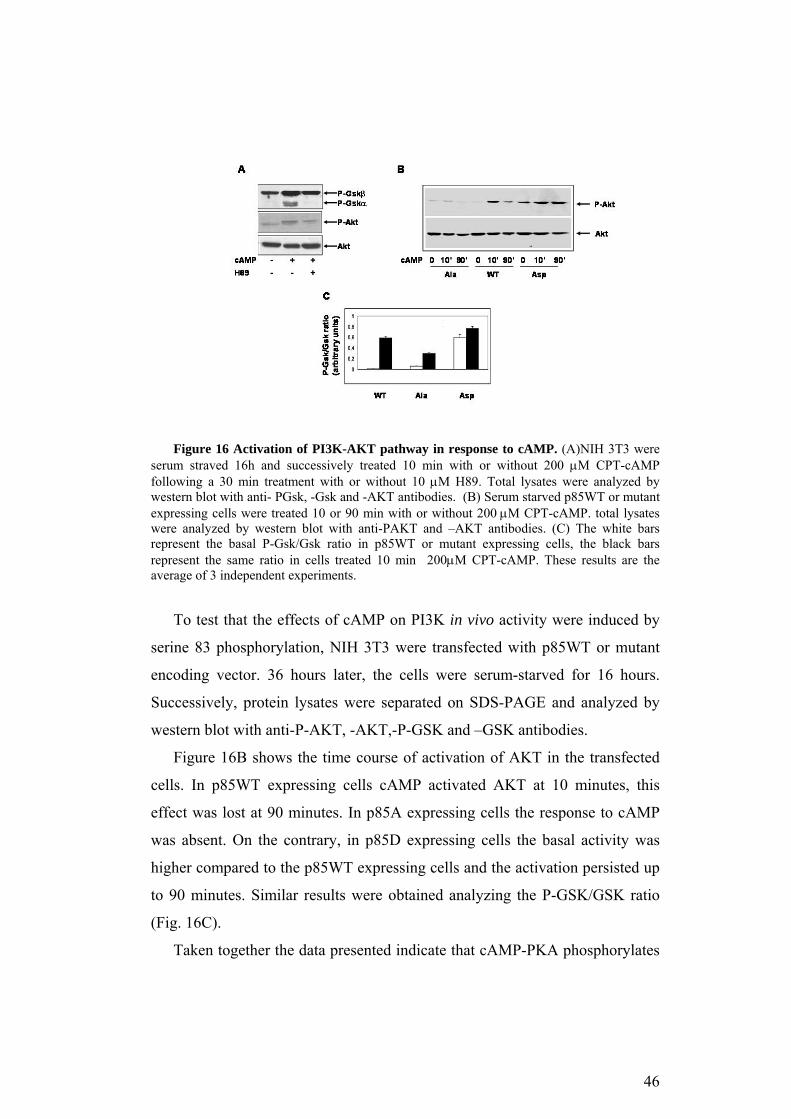
Figure 16 Activation of PI3K-AKT pathway in response to cAMP. (A)NIH 3T3 were serum straved 16h and successively treated 10 min with or without 200 µM CPT-cAMP following a 30 min treatment with or without 10 µM H89. Total lysates were analyzed by western blot with anti- PGsk, -Gsk and -AKT antibodies. (B) Serum starved p85WT or mutant expressing cells were treated 10 or 90 min with or without 200 µM CPT-cAMP. total lysates were analyzed by western blot with anti-PAKT and –AKT antibodies. (C) The white bars represent the basal P-Gsk/Gsk ratio in p85WT or mutant expressing cells, the black bars represent the same ratio in cells treated 10 min 200µM CPT-cAMP. These results are the average of 3 independent experiments.
To test that the effects of cAMP on PI3K in vivo activity were induced by
serine 83 phosphorylation, NIH 3T3 were transfected with p85WT or mutant
encoding vector. 36 hours later, the cells were serum-starved for 16 hours.
Successively, protein lysates were separated on SDS-PAGE and analyzed by
western blot with anti-P-AKT, -AKT,-P-GSK and –GSK antibodies.
Figure 16B shows the time course of activation of AKT in the transfected
cells. In p85WT expressing cells cAMP activated AKT at 10 minutes, this
effect was lost at 90 minutes. In p85A expressing cells the response to cAMP
was absent. On the contrary, in p85D expressing cells the basal activity was
higher compared to the p85WT expressing cells and the activation persisted up
to 90 minutes. Similar results were obtained analyzing the P-GSK/GSK ratio
(Fig. 16C).
Taken together the data presented indicate that cAMP-PKA phosphorylates
46

the serine 83 of p85αPI3K, stimulating the formation of the Ras/PI3K complex.
The association between these molecules triggers the activation of the PI3K-
AKT pathway which results in inhibition of apoptosis and G1 arrest.
4. Biological effects of the phosphorylation of p85αPI3K TSH-cAMP
dependent cells: FRTL5 and NTCRII
Ciullo et al. in 2001 demonstrated that the association between PI3K and
Ras was essential for the G1-S transition in thyroid cells FRTL5. Considering
the data presented above concerning the role of serine 83 phosphorylation in
the formation of Ras/PI3K complex and the regulation of cell survival and
proliferation in NIH 3T3, we investigated the possibility that serine 83 was a
key regulator of cAMP effects also in cAMP-dependent cells.
The following experiments were performed in thyroid cells FRTL5 and
NTCRII cells. FRTL5 cells proliferate in response to TSH, an hormone who
activates a Gs coupled receptor (see Background). NTCRII cells are modified
NIH 3T3 fibroblasts that became TSH-cAMP dependent. Porcellini et al. in
2003 showed that NIH 3T3 expressing both TSH receptor and the PKA
regulatory subunit RIIβ proliferated in dependence of TSH-cAMP,
recapitulating the characteristics of thyroid cells (Fig. 17).
Figure 17: Effect of TSH on cell proliferation on engineered NIH 3T3. The
expression of TSHR (T6321) or RIIβ induces cell death in response to TSH added to the culture media, while TSH induces proliferation in NIH 3T3 expressing both TSHR and RIIβ. (Porcellini et al., JBC, 2003; 278, 40621-40630)
47

4a. p85A is lethal in TSH-cAMP dependent cells
FRTL5 cells were co-transfected with a G-418 resistance encoding vector
and p85WT or mutant encoding vector. 96 hours after the transfection 400
µg/ml of G-418 was added to the media but very few clones expressing p85A
survived selection. To test if p85A expression in FRTL5 impaired cell
survival, we tested the plating efficiency of the various transfected lines.
5x10⁵ cells were transfected with the G-418 resistance encoding vector and
the indicated vector. 96 hours after the transfection 400 µg/ml of G-418 was
added to the media. After 15 days, the G-418 resistant clones were counted.
Figure 18 shows that the numbers of p85A expressing clones was
significantly lower than the control and p85WT expressing clones. Moreover,
the number of p85D expressing clones was slightly higher than the number of
p85WT expressing clones.
Figure 18: Plating efficiency of p85WT, A or D expressing clones. The ability to form
G-418 resistant clones was determined by transfecting 5*105 FRTL-5 cells with p85WT, A or D expressing vector and selecting the clones in the presence of 400 µg/ml of G-418 for 15 days. The histogram represents the number of clones obtained, and it is the mean of three experiments in triplicate.
The few surviving clones expressing p85A were expressed low levels of
p85A protein (data not shown). These data indicate that p85A expression
inhibited growth or/and survival in FRTL5, suggesting that phosphorylation
of serine 83 of p85αPI3K is a key step in the transmission of survival signals in
FRTL5. To test this hypothesis, NTCRII cells were transiently transfected
with p85WT, A or D or control vectors. Cells were grown for 18 hours in the
48
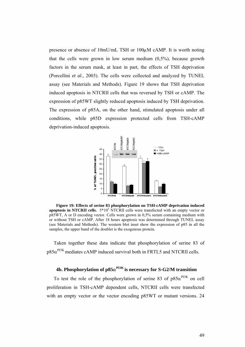
presence or absence of 10mU/mL TSH or 100µM cAMP. It is worth noting
that the cells were grown in low serum medium (0,5%), because growth
factors in the serum mask, at least in part, the effects of TSH deprivation
(Porcellini et al., 2003). The cells were collected and analyzed by TUNEL
assay (see Materials and Methods). Figure 19 shows that TSH deprivation
induced apoptosis in NTCRII cells that was reversed by TSH or cAMP. The
expression of p85WT slightly reduced apoptosis induced by TSH deprivation.
The expression of p85A, on the other hand, stimulated apoptosis under all
conditions, while p85D expression protected cells from TSH-cAMP
deprivation-induced apoptosis.
Figure 19: Effects of serine 83 phosphorylation on TSH-cAMP deprivation induced
apoptosis in NTCRII cells. 5*105 NTCRII cells were transfected with an empty vector or p85WT, A or D encoding vector. Cells were grown in 0,5% serum containing medium with or without TSH or cAMP. After 18 hours apoptosis was determined through TUNEL assay (see Materials and Methods). The western blot inset show the expression of p85 in all the samples, the upper band of the doublet is the exogenous protein.
Taken together these data indicate that phosphorylation of serine 83 of
p85αPI3K mediates cAMP induced survival both in FRTL5 and NTCRII cells.
4b. Phosphorylation of p85αPI3K is necessary for S-G2/M transition
To test the role of the phosphorylation of serine 83 of p85αPI3K on cell
proliferation in TSH-cAMP dependent cells, NTCRII cells were transfected
with an empty vector or the vector encoding p85WT or mutant versions. 24
49

hours after the transfection, the colture media was replaced with 0,5% serum
containing medium. 18 hours later, 10mU/mL of TSH were added to the media
to induce cell cycle entry. Cells were analyzed by BrdU labelling, as described
in Materials and Methods. Figure 20 shows that only the expression of p85A
affected cell cycle progression. In particular, p85A expressing cells
accumulated in S-phase at 270 minutes (left panel). The S-phase arrest of p8A
expressing cells correlates with the reduction of the number of cells in G2/M
phase at 270 minutes (right panel).
Figure 20: Effects of serine 83 phosphorylation on cell cycle progression. NTCRII cells
were transiently transfected with the p85WT, p85A, p85D mutants or with the empty vector (control). 24 hours after transfection, the cells were starved in 0,5% serum containing medium. 18 hours later cells were induced with 10 mU/ml TSH. BrdU incorporation was assayed by a pulse-chase experiment. Cells were labeled for 30 min with BrdU and harvested at 0, 90 and 270 min. DNA content was determined by propidium iodide stainig. Cells were subjected to FACS analysis for bivariate-analysis of DNA content versus BrdU.
We also monitored EGF-induced proliferation, since NCTRII cells in the
presence of serum respond to EGF. p85A affected selectively cAMP-TSH
induced proliferation but not the EGF induced proliferation (data not shown,
(De Gregorio et al., 2006). These data indicate that phosphorylation of serine
83 is necessary for cell cycle progression.
50

5. Molecular mechanisms affected by phosphorylation of serine 83 of
p85αPI3K in FRTL-5 and NTCRII cells
As in NIH 3T3, cAMP induced the binding of Ras to PI3K, containing
p85WT but not p85A (De Gregorio et al., 2006). The formation of this
complex was tested both in FRTL5 or NTCRII cells, respectively stably or
transiently expressing p85WT or mutant versions. This indicates that the same
molecular mechanism, i.e., stimulation of the complex PI3K/Ras, exerts
opposing effects: cell proliferation in TSH-cAMP dependent cells and G1
arrest in NIH 3T3. One explanation of this paradox may involve the differential
expression of RII isoforms in the cell lines analyzed. Both NTCRII and
FRTL5, in fact, express RIIβ, while this protein is present at low level, if not
absent, in NIH 3T3 fibroblasts (Porcellini et al., 2003).
5a. Phosphorylated p85αPI3K interacts with RIIβ
To verify if RIIβ influences the biological effects of serine 83
phosphorylation, the binding between RIIβ-PKA and PI3K has been tested.
p85WT, A or D have been transiently expressed in NTCRII cells. 24 hours
after the transfection, cells were serum starved for 6 hours before the treatment
with 10mU/mL TSH (20 minutes). Cell lysates were incubated in vitro with
GST or GST-RIIβ (Grieco et al., 1996). Figure 21 shows the western blot with
anti-Flag, -GST and -phosphoserine (P-Ser) antibodies on the bound proteins
(pull down assay). p85WT formed a complex with RIIβ following incubation
with TSH. The substitution S83A abolished the binding, while the substitution
S83D determined constitutive (in the absence of cAMP) binding
RIIβ/p85αPI3K.
51
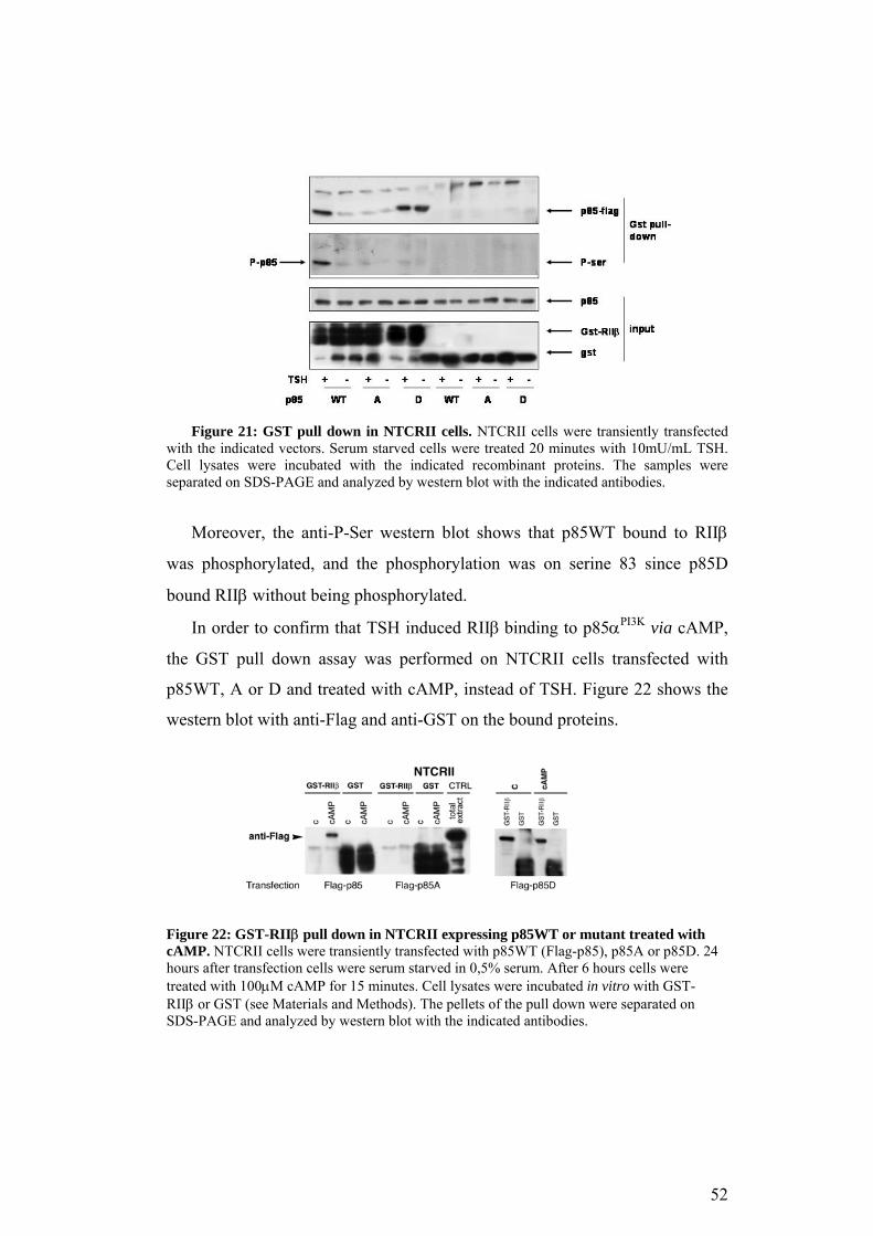
Figure 21: GST pull down in NTCRII cells. NTCRII cells were transiently transfected
with the indicated vectors. Serum starved cells were treated 20 minutes with 10mU/mL TSH. Cell lysates were incubated with the indicated recombinant proteins. The samples were separated on SDS-PAGE and analyzed by western blot with the indicated antibodies.
Moreover, the anti-P-Ser western blot shows that p85WT bound to RIIβ
was phosphorylated, and the phosphorylation was on serine 83 since p85D
bound RIIβ without being phosphorylated.
In order to confirm that TSH induced RIIβ binding to p85αPI3K via cAMP,
the GST pull down assay was performed on NTCRII cells transfected with
p85WT, A or D and treated with cAMP, instead of TSH. Figure 22 shows the
western blot with anti-Flag and anti-GST on the bound proteins.
Figure 22: GST-RIIβ pull down in NTCRII expressing p85WT or mutant treated with cAMP. NTCRII cells were transiently transfected with p85WT (Flag-p85), p85A or p85D. 24 hours after transfection cells were serum starved in 0,5% serum. After 6 hours cells were treated with 100µM cAMP for 15 minutes. Cell lysates were incubated in vitro with GST-RIIβ or GST (see Materials and Methods). The pellets of the pull down were separated on SDS-PAGE and analyzed by western blot with the indicated antibodies.
52

Also cAMP stimulates the binding of p85WT to GST-RIIβ but not of p85A,
the binding of p85D instead was constitutive.
To test if the binding between the two proteins was direct, p85WT or A
was transiently expressed in NTCRII. Protein lysates from cAMP induced
cells were immunoprecipitated with anti-Flag antibody and p85αPI3K was
eluted with 0,1M glycine pH 3,5. The eluted proteins were used for the GST-
RIIβ pull down assay. Figure 23 shows that cAMP induced the binding to
RIIβ οf p85 WT, not of the mutated version, p85A.
Figure 23: GST-RIIβ pull down on immunoprecipitated p85WT or A. Exogenous
p85PI3K was immunoprecipitated with anti-FLAG antibody from NTCRII cells transfected with p85WT or p85A and exposed to 100 µM 8BrcAMP for 15 min. The immunoprecipitates were extensively washed in RIPA buffer containing 1% Triton X-100 -0.1% SDS and purified by elution with 0.1M glycine pH 3.5. Input indicates the eluted fraction; +glycine represents the pellet after elution. The purified p85PI3K was incubated in vitro with GST-RIIβ in a pull-down experiment as described in Materials and Methods.
Together these data indicate that phosphorylation of serine 83 in p85αPI3K is
important for the binding to RIIβ-PKA. This results, possibly, in the anchoring
of PKA under the plasma membrane and the phosphorylation of a particular
subset of substrates.
5b. Phosphorylation of serine 83 on p85αPI3Kalters cAMP induced
PI3K activity both in vitro and in vivo
To determine if phosphorylation of p85αPI3K correlates with an increase of
activity of PI3K, as in NIH 3T3, NTCRII cells were transiently transfected
53
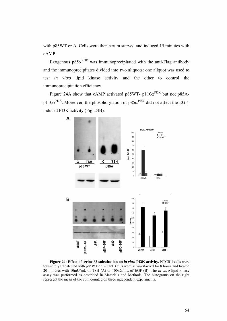
with p85WT or A. Cells were then serum starved and induced 15 minutes with
cAMP.
Exogenous p85αPI3K was immunoprecipitated with the anti-Flag antibody
and the immunoprecipitates divided into two aliquots: one aliquot was used to
test in vitro lipid kinase activity and the other to control the
immunoprecipitation efficiency.
Figure 24A show that cAMP activated p85WT- p110αPI3K but not p85A-
p110αPI3K. Moreover, the phosphorylation of p85αPI3K did not affect the EGF-
induced PI3K activity (Fig. 24B).
Figure 24: Effect of serine 83 substitution on in vitro PI3K activity. NTCRII cells were
transiently transfected with p85WT or mutant. Cells were serum starved for 8 hours and treated 20 minutes with 10mU/mL of TSH (A) or 100nG/mL of EGF (B). The in vitro lipid kinase assay was performed as described in Materials and Methods. The histograms on the right represent the mean of the cpm counted on three independent experiments.
54

These data were confirmed in vivo by determining the level of
phosphorylation of AKT in NTCRII cells transiently transfected with p85WT
or mutant versions and treated with TSH.
Figure 25 shows that p85A expressing cells fail to activate AKT at all time
points considered. On the other end, expression of p85D slightly amplified
TSH signaling. P-AKT levels were, in fact, higher in p85D expressing cells
than in the controls both at 0 and 5 minutes. These results confirm that
phosphorlation of p85αPI3K was necessary to activate PI3K through cAMP-
PKA pathway.
Figure 25: Effect of serine 83 phosphorylation on AKT activation in NTCRII cells.
NTCRII cells were transfected with the indicated vector, 30 hours later the normal medium was replaced with a low serum medium (0,5% serum). After 18 hours cells were treated with 10mU/mL TSH for the indicated time. Protein lysate were analyzed by western blot with anti-P-AKT and –AKT antibodies. The histogram in B is the mean of three independent experiments.
6. cAMP-PKA amplifies estrogen binding and signaling to PI3K
Considering that PI3K is essential also in estrogen mediated AKT
activation (Simoncini et al., 2000; Castoria et al., 2001) and that p85αPI3K was
efficiently phosphorylated in MCF-7 cells (Fig. 10A) it was possible that serine
83 phosphorylation was required for estrogen signaling. To verify this
55
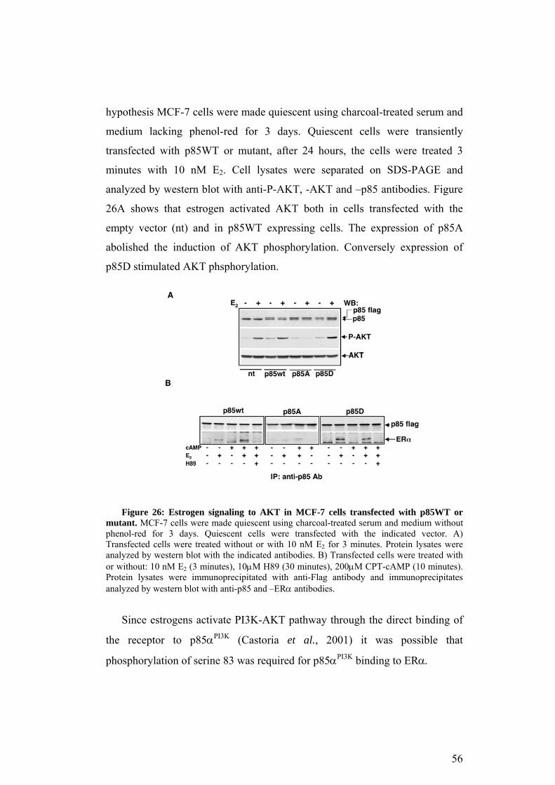
hypothesis MCF-7 cells were made quiescent using charcoal-treated serum and
medium lacking phenol-red for 3 days. Quiescent cells were transiently
transfected with p85WT or mutant, after 24 hours, the cells were treated 3
minutes with 10 nM E2. Cell lysates were separated on SDS-PAGE and
analyzed by western blot with anti-P-AKT, -AKT and –p85 antibodies. Figure
26A shows that estrogen activated AKT both in cells transfected with the
empty vector (nt) and in p85WT expressing cells. The expression of p85A
abolished the induction of AKT phosphorylation. Conversely expression of
p85D stimulated AKT phsphorylation.
Figure 26: Estrogen signaling to AKT in MCF-7 cells transfected with p85WT or
mutant. MCF-7 cells were made quiescent using charcoal-treated serum and medium without phenol-red for 3 days. Quiescent cells were transfected with the indicated vector. A) Transfected cells were treated without or with 10 nM E2 for 3 minutes. Protein lysates were analyzed by western blot with the indicated antibodies. B) Transfected cells were treated with or without: 10 nM E2 (3 minutes), 10µM H89 (30 minutes), 200µM CPT-cAMP (10 minutes). Protein lysates were immunoprecipitated with anti-Flag antibody and immunoprecipitates analyzed by western blot with anti-p85 and –ERα antibodies.
Since estrogens activate PI3K-AKT pathway through the direct binding of
the receptor to p85αPI3K (Castoria et al., 2001) it was possible that
phosphorylation of serine 83 was required for p85αPI3K binding to ERα.
56
In order to test this hypothesis, quiescent MCF-7 cells were transiently
transfected with p85WT or mutant. 24 hours later, cells were treated with E2,
H89 and/or cAMP. Protein lysates were immunoprecipitated with anti-Flag
antibody and analyzed by western blot with anti-p85 and -ERα antibodies.
Figure 26B shows that estrogen induced p85WT binding to the receptor, cAMP
on its own did not induce the binding but amplified estrogen-stimulated
binding. In fact, the amount of receptor bound to p85αPI3K was significantly
higher in cAMP and estrogen-treated cells than in cells exposed to estrogen
alone. Moreover, the binding was PKA-dependent, since H89 inhibited the
effects of estrogens.
cAMP-PKA modulates ERα- p85αPI3K binding through the
phosphorylation of p85αPI3K. In fact p85A failed to bind the receptor in all the
conditions considered. Once more, it is worth noting that the phosphorylation
of serine 83 of p85αPI3K is absolutely necessary to mediate cAMP-PKA
effects, but it is not sufficient. In fact, p85D binding to ERα in E2 treated cells
was inhibited by H89. These data indicate that phosphorylation of serine 83 is
required for the binding of p85αPI3K to ERα and the consequential activation of
PI3K-AKT, but it still requires PKA, because these events are H89 sensitive. It
is worth noting that ΕRα itself may be a PKA substrate (Cui et al., 2004;
Michalides et al., 2004). These observations suggest the possibility that the
receptor has to be phosphorylated by PKA to efficiently bind p85α.
57
Discussion
The data presented here indicate that phopshorylation of p85αPI3K on serine
83 is a critical step in the cAMP-PKA signaling. Biological effects of cAMP
and PKA on cell cycle and growth have been extensively studied (Pastan et al.,
1975; Lee et al., 1998; Ariga et al., 2000; Stork and Schmitt, 2002) but the
molecular mechanism(s) underling their action are not yet defined. Our data
may shed light on cAMP mediated phenotypes and may provide some
mechanistic insights into transduction signalling in a variety of cell types.
1. cAMP-PKA selectively influences Ras signaling
Previous data indicate that cAMP inhibits Raf1 association with Ras and
down-regulates the MAPK pathway through two different mechanisms.
The first is cAMP, but not PKA, dependent. In fact cAMP stimulates
directly EPAC, a guanine nucleotide exchange factor for the Ras-like small
GTPases (Rap1 and Rap2) (Bos, 2003). Rap1 binds Raf1 and inhibits its kinase
function (Hu et al., 1999). The importance of Rap1 mediated regulation of
Raf-1 is confirmed by the complementary action of insulin. In fact, insulin, in
the absence of cAMP-PKA, decreases Rap1 bound to Raf1 and increases the
association Ras-Raf1, with a concomitant stimulation of Raf1-MEK-ERK
cascade (Okada et al., 1998). The second mechanism is PKA dependent, PKA
in fact phosphorylates serines 43 and 259 in Raf1. Phophorylation of serine 43
decreases the binding to Ras, while serine 259 appears to be the sole target for
PKA inhibition of ERK1/2 (Dhillon et al., 2002).
The inhibition of Raf-1 signalling does not necessarily correspond to
MAPK inhibition by cAMP. In fact cAMP may stimulate rather than inhibit
MAPK (Stork and Schmitt, 2002; Norum et al., 2003). Our data indicate that
the major target of cAMP-PKA action is PI3K. Ciullo et. al in 2001 showed
that Ras binding to PI3K increases when the cells were exposed to cAMP.
58
Moreover, the formation of this complex was dependent on PKA activity, since
H89 reversed cAMP-induced association PI3K/Ras.
The results reported here indicate that PKA phosphorylates serine 83 of
p85αPI3K and this event is necessary for the binding to Ras, although not
sufficient. This has been shown in NIH 3T3 and confirmed in TSH-dependent
cells (De Gregorio et al., 2006), suggesting that it is a general, not a tissue
specific mechanism. Our data indicate that phosphorylation of serine 83 in
p85αPI3K stabilized the binding with p110αPI3K, resulting in stimulation of the
enzymatic activity both in vivo and in vitro (Figs 14, 15 and 24). Stabilization
of p110αPI3K-p85αPI3K complex is important for two reasons: 1. It may
facilitate translocation to the cellular membrane (see Background); 2. It
increases local specific activity. We suggest that phosphorylated p85αPI3K
associates more efficiently to p110αPI3K, stabilizes the enzyme and localizes to
the membrane. This results in an increased binding of PI3K to Ras and to the
activation of the PI3K-AKT pathway.
2. cAMP-PKA regulates cell cycle progression through p85αPI3K
phosphorylation
cAMP inhibits proliferation in NIH 3T3. In fact the phosphorylation of
p85αPI3K leads to G1 arrest in fibroblasts, as shown by transfecting the mutant
p85D. On the contrary, preventing the phosphorylation (p85A), NIH 3T3 cells
become resistant to cAMP-induced G1 arrest (Fig. 12) . A different effect was
observed in TSH-dependent cells. The phosphorylation of p85αPI3K is in fact
essential for a correct progression of the cells through S and G2/M phases (Fig.
20). How can the same phosphorylation lead to opposing phenotypes?
It is worth noting that NIH 3T3 fibroblasts display a very short cell cycle,
approximately 24 hours, to complete 1 cycle. This implies a short G1. On the
other hand, TSH-dependent cells display a 48 hours long cell cycle with an
extended (ca. 36 h) G1. We suggest that cAMP-PKA regulates the length of
G1. This effect is not detectable in TSH-dependent cells since they do not
59
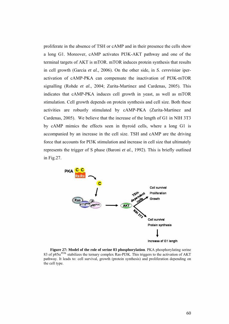
proliferate in the absence of TSH or cAMP and in their presence the cells show
a long G1. Moreover, cAMP activates PI3K-AKT pathway and one of the
terminal targets of AKT is mTOR. mTOR induces protein synthesis that results
in cell growth (Garcia et al., 2006). On the other side, in S. cerevisiae iper-
activation of cAMP-PKA can compensate the inactivation of PI3K-mTOR
signalling (Rohde et al., 2004; Zurita-Martinez and Cardenas, 2005). This
indicates that cAMP-PKA induces cell growth in yeast, as well as mTOR
stimulation. Cell growth depends on protein synthesis and cell size. Both these
activities are robustly stimulated by cAMP-PKA (Zurita-Martinez and
Cardenas, 2005). We believe that the increase of the length of G1 in NIH 3T3
by cAMP mimics the effects seen in thyroid cells, where a long G1 is
accompanied by an increase in the cell size. TSH and cAMP are the driving
force that accounts for PI3K stimulation and increase in cell size that ultimately
represents the trigger of S phase (Baroni et al., 1992). This is briefly outlined
in Fig.27.
Figure 27: Model of the role of serine 83 phosphorylation. PKA phosphorylating serine
83 of p85αPI3K stabilizes the ternary complex Ras-PI3K. This triggers to the activation of AKT pathway. It leads to: cell survival, growth (protein synthesis) and proliferation depending on the cell type.
60

3. cAMP cytoprotective action requires serine 83 phopshorylation
cAMP protects cells from apoptosis induced by different signals in different
cell types (Affaitati et al., 2003). In this study the resistance to anoikis has been
evaluated. This apoptotic pathway is triggered by the loss of cell-matrix signals
and it is highly dependent on PI3K. This system allowed us to define the role
of serine 83 phopshorylation without the interferences of other survival
pathway. The result was that phosphorylation of serine 83 was critical for
cAMP mediated protection from anoikis. Moreover p85D expression replicated
the effects of cAMP, at least in part (Fig. 11). This suggests that the
phosphorylation of serine 83 of p85αPI3K is the main player of cAMP
cytoprotective signals.
The function of phosphorylated serine 83 was essential in TSH-dependent
cells. In fact, the absence of TSH or cAMP induced apoptosis in p85WT but
not p85D-expressing cells. Moreover thyroid and thyroid-like cells undergo
apoptosis when expressed p85A, also in the presence of TSH (Figs 18 and 19)
This further indicates that p85αPI3K is a key player in TSH-cAMP induced
survival.
4. PKA-RIIβ expression switches cells from cAMP-dependent
proliferation to cAMP-dependent arrest
Different isoforms of PKA exist, depending on the characteristics of the
regulatory subunit (Feliciello et al., 2001). We can distinguish RI and RII
subunits, the first has a higher sensitivity to cAMP. This indicates that RI-PKA
responds to low level of cAMP while RII-PKA to high level of intracellular
cAMP. Thyroid cells express high level of PKA-RII, and in particular PKA-
RIIβ. Moreover, NIH 3T3 cells expressing TSH receptor but not RIIβ, do not
proliferate in response to TSH. Only cells expressing in the meantime TSHR
and RIIβ become TSH dependent cell for growth (Porcellini et al., 2003). This
indicates that RIIβ levels distinguish cAMP-dependent cells from independent
cells.
61
Here we show that RIIβ binds phosphorylated p85αPI3K and
phosphorylation on serine 83 is the only requirement to this event, since p85D
constitutively bind RIIβ (Figs 21 and 22). This association anchors PKA at the
cellular membrane, determining a highly localized kinase activity (Feliciello et
al., 2001).
5. cAMP-PKA phosphorylates p85αPI3K and amplifies estrogens
signaling
The data presented in this study indicate that cAMP cooperates with
estrogens in the stimulation of AKT. Moreover, this effect is dependent on
p85αPI3K phosphorylation. In fact, p85A was unable to bind the estrogen
receptor and p85A expressing cells did not activate AKT in response to
estrogens. On the other hand, the substitution S83D amplified the association
ERα-p85αPI3K induced by estrogens. Moreover, p85D expression enhanced
estrogens mediated activation of AKT. These observations indicate that
phosphorylation of serine 83 is essential for estrogens signaling. Our data also
indicate that this step is necessary but not sufficient, because p85D expressing
cells still respond to cAMP to amplify AKT signaling (Fig. 26). PKA can
phosphorylate the receptor itself (Cui et al., 2004; Michalides et al., 2004), and
this phosphorylation may be required for fully activation of the ERα-p85αPI3K
circuit. Considering that estrogens activate the adenylyl cyclase (Aronica et al.,
1994) and H89 prevents the association ERα-p85αPI3K, it is likely that PKA-
dependent phosphorylation occurs even in cells treated only with estrogens,
and that the bound p85αPI3K is only the phosphorylated form. This may explain
why p85A acts as a negative dominant.
62
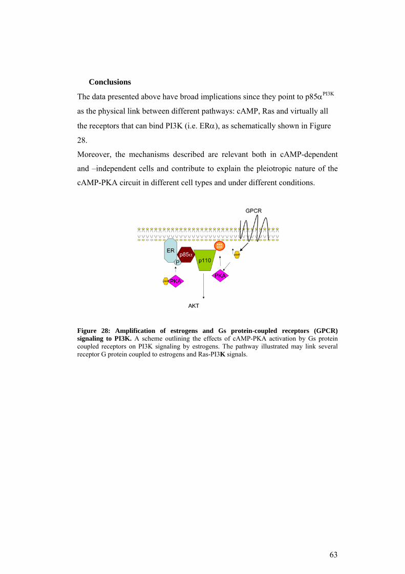
Conclusions
The data presented above have broad implications since they point to p85αPI3K
as the physical link between different pathways: cAMP, Ras and virtually all
the receptors that can bind PI3K (i.e. ERα), as schematically shown in Figure
28.
Moreover, the mechanisms described are relevant both in cAMP-dependent
and –independent cells and contribute to explain the pleiotropic nature of the
cAMP-PKA circuit in different cell types and under different conditions.
Figure 28: Amplification of estrogens and Gs protein-coupled receptors (GPCR) signaling to PI3K. A scheme outlining the effects of cAMP-PKA activation by Gs protein coupled receptors on PI3K signaling by estrogens. The pathway illustrated may link several receptor G protein coupled to estrogens and Ras-PI3K signals.
63
References
Affaitati, A., Cardone, L., de Cristofaro, T., Carlucci, A., Ginsberg, M.D., Varrone, S., Gottesman, M.E., Avvedimento, E.V., and Feliciello, A. (2003). Essential role of A-kinase anchor protein 121 for cAMP signaling to mitochondria. The Journal of biological chemistry 278, 4286-4294. Alberts, B.J., Alexander; Lewis, Julian; Raff, Martin; Roberts, Keith; Walter, Peter (c2002). Molecular Biology of the Cell. Garland Science: New York and London. Ariga, M., Nedachi, T., Akahori, M., Sakamoto, H., Ito, Y., Hakuno, F., and Takahashi, S. (2000). Signalling pathways of insulin-like growth factor-I that are augmented by cAMP in FRTL-5 cells. Biochem J 348 Pt 2, 409-416. Aronica, S.M., Kraus, W.L., and Katzenellenbogen, B.S. (1994). Estrogen action via the cAMP signaling pathway: stimulation of adenylate cyclase and cAMP-regulated gene transcription. Proc Natl Acad Sci U S A 91, 8517-8521. Bader, A.G., Kang, S., Zhao, L., and Vogt, P.K. (2005). Oncogenic PI3K deregulates transcription and translation. Nature reviews 5, 921-929. Balsara, B.R., Pei, J., Mitsuuchi, Y., Page, R., Klein-Szanto, A., Wang, H., Unger, M., and Testa, J.R. (2004). Frequent activation of AKT in non-small cell lung carcinomas and preneoplastic bronchial lesions. Carcinogenesis 25, 2053-2059. Baroni, M.D., Monti, P., Marconi, G., and Alberghina, L. (1992). cAMP-mediated increase in the critical cell size required for the G1 to S transition in Saccharomyces cerevisiae. Experimental cell research 201, 299-306. Bos, J.L. (2003). Epac: a new cAMP target and new avenues in cAMP research. Nat Rev Mol Cell Biol 4, 733-738. Carpenter, C.L., Auger, K.R., Duckworth, B.C., Hou, W.M., Schaffhausen, B., and Cantley, L.C. (1993). A tightly associated serine/threonine protein kinase regulates phosphoinositide 3-kinase activity. Molecular and cellular biology 13, 1657-1665. Castoria, G., Migliaccio, A., Bilancio, A., Di Domenico, M., de Falco, A., Lombardi, M., Fiorentino, R., Varricchio, L., Barone, M.V., and Auricchio, F. (2001). PI3-kinase in concert with Src promotes the S-phase entry of oestradiol-stimulated MCF-7 cells. The EMBO journal 20, 6050-6059.
64

Cespedes, M.V., Sancho, F.J., Guerrero, S., Parreno, M., Casanova, I., Pavon, M.A., Marcuello, E., Trias, M., Cascante, M., Capella, G., and Mangues, R. (2006). K-ras Asp12 mutant neither interacts with Raf, nor signals through Erk and is less tumorigenic than K-ras Val12. Carcinogenesis 27, 2190-2200. Cheng, J.Q., Ruggeri, B., Klein, W.M., Sonoda, G., Altomare, D.A., Watson, D.K., and Testa, J.R. (1996). Amplification of AKT2 in human pancreatic cells and inhibition of AKT2 expression and tumorigenicity by antisense RNA. Proceedings of the National Academy of Sciences of the United States of America 93, 3636-3641. Ciullo, I., Diez-Roux, G., Di Domenico, M., Migliaccio, A., and Avvedimento, E.V. (2001). cAMP signaling selectively influences Ras effectors pathways. Oncogene 20, 1186-1192. Cook, S.J., Rubinfeld, B., Albert, I., and McCormick, F. (1993). RapV12 antagonizes Ras-dependent activation of ERK1 and ERK2 by LPA and EGF in Rat-1 fibroblasts. The EMBO journal 12, 3475-3485. Cui, Y., Zhang, M., Pestell, R., Curran, E.M., Welshons, W.V., and Fuqua, S.A. (2004). Phosphorylation of estrogen receptor alpha blocks its acetylation and regulates estrogen sensitivity. Cancer research 64, 9199-9208. De Gregorio, G., Coppa, A., Cosentino, C., Ucci, S., Messina, S., Nicolussi, A., D'Inzeo, S., Di Pardo, A., Avvedimento, E.V., and Porcellini, A. (2006). The p85 regulatory subunit of PI3K mediates TSH-cAMP-PKA growth and survival signals. Oncogene. Dhand, R., Hara, K., Hiles, I., Bax, B., Gout, I., Panayotou, G., Fry, M.J., Yonezawa, K., Kasuga, M., and Waterfield, M.D. (1994a). PI 3-kinase: structural and functional analysis of intersubunit interactions. The EMBO journal 13, 511-521. Dhand, R., Hiles, I., Panayotou, G., Roche, S., Fry, M.J., Gout, I., Totty, N.F., Truong, O., Vicendo, P., Yonezawa, K., and et al. (1994b). PI 3-kinase is a dual specificity enzyme: autoregulation by an intrinsic protein-serine kinase activity. The EMBO journal 13, 522-533. Dhillon, A.S., Pollock, C., Steen, H., Shaw, P.E., Mischak, H., and Kolch, W. (2002). Cyclic AMP-dependent kinase regulates Raf-1 kinase mainly by phosphorylation of serine 259. Mol Cell Biol 22, 3237-3246. Edelman, A.M., Blumenthal, D.K., and Krebs, E.G. (1987). Protein serine/threonine kinases. Annual review of biochemistry 56, 567-613.
65
Feliciello, A., Gallo, A., Mele, E., Porcellini, A., Troncone, G., Garbi, C., Gottesman, M.E., and Avvedimento, E.V. (2000). The localization and activity of cAMP-dependent protein kinase affect cell cycle progression in thyroid cells. J Biol Chem 275, 303-311. Feliciello, A., Gottesman, M.E., and Avvedimento, E.V. (2001). The biological functions of A-kinase anchor proteins. Journal of molecular biology 308, 99-114. Feliciello, A., Gottesman, M.E., and Avvedimento, E.V. (2005). cAMP-PKA signaling to the mitochondria: protein scaffolds, mRNA and phosphatases. Cellular signalling 17, 279-287. Frisch, S.M., and Francis, H. (1994). Disruption of epithelial cell-matrix interactions induces apoptosis. J Cell Biol 124, 619-626. Fruman, D.A., Meyers, R.E., and Cantley, L.C. (1998). Phosphoinositide kinases. Annual review of biochemistry 67, 481-507. Garcia, Z., Kumar, A., Marques, M., Cortes, I., and Carrera, A.C. (2006). Phosphoinositide 3-kinase controls early and late events in mammalian cell division. The EMBO journal 25, 655-661. Grieco, D., Porcellini, A., Avvedimento, E.V., and Gottesman, M.E. (1996). Requirement for cAMP-PKA pathway activation by M phase-promoting factor in the transition from mitosis to interphase. Science 271, 1718-1723. Harpur, A.G., Layton, M.J., Das, P., Bottomley, M.J., Panayotou, G., Driscoll, P.C., and Waterfield, M.D. (1999). Intermolecular interactions of the p85alpha regulatory subunit of phosphatidylinositol 3-kinase. The Journal of biological chemistry 274, 12323-12332. Haynes, J., Jr., Robinson, J., Saunders, L., Taylor, A.E., and Strada, S.J. (1992). Role of cAMP-dependent protein kinase in cAMP-mediated vasodilation. The American journal of physiology 262, H511-516. Holt, K.H., Olson, L., Moye-Rowley, W.S., and Pessin, J.E. (1994). Phosphatidylinositol 3-kinase activation is mediated by high-affinity interactions between distinct domains within the p110 and p85 subunits. Molecular and cellular biology 14, 42-49. Hu, C.D., Kariya, K., Okada, T., Qi, X., Song, C., and Kataoka, T. (1999). Effect of phosphorylation on activities of Rap1A to interact with Raf-1 and to suppress Ras-dependent Raf-1 activation. The Journal of biological chemistry 274, 48-51.
66

Hu, P., and Schlessinger, J. (1994). Direct association of p110 beta phosphatidylinositol 3-kinase with p85 is mediated by an N-terminal fragment of p110 beta. Molecular and cellular biology 14, 2577-2583. Jimenez, C., Jones, D.R., Rodriguez-Viciana, P., Gonzalez-Garcia, A., Leonardo, E., Wennstrom, S., von Kobbe, C., Toran, J.L., L, R.B., Calvo, V., Copin, S.G., Albar, J.P., Gaspar, M.L., Diez, E., Marcos, M.A., Downward, J., Martinez, A.C., Merida, I., and Carrera, A.C. (1998). Identification and characterization of a new oncogene derived from the regulatory subunit of phosphoinositide 3-kinase. The EMBO journal 17, 743-753. Jucker, M., Sudel, K., Horn, S., Sickel, M., Wegner, W., Fiedler, W., and Feldman, R.A. (2002). Expression of a mutated form of the p85alpha regulatory subunit of phosphatidylinositol 3-kinase in a Hodgkin's lymphoma-derived cell line (CO). Leukemia 16, 894-901. Kang, S., Bader, A.G., Zhao, L., and Vogt, P.K. (2005). Mutated PI 3-kinases: cancer targets on a silver platter. Cell cycle (Georgetown, Tex 4, 578-581. Khwaja, A., Rodriguez-Viciana, P., Wennstrom, S., Warne, P.H., and Downward, J. (1997). Matrix adhesion and Ras transformation both activate a phosphoinositide 3-OH kinase and protein kinase B/Akt cellular survival pathway. Embo J 16, 2783-2793. Klippel, A., Escobedo, J.A., Hu, Q., and Williams, L.T. (1993). A region of the 85-kilodalton (kDa) subunit of phosphatidylinositol 3-kinase binds the 110-kDa catalytic subunit in vivo. Molecular and cellular biology 13, 5560-5566. Kufe, D.W.P., Raphael E.; Weichselbaum, Ralph R.; Bast, Robert C., Jr.; Gansler, Ted S.; Holland, James F. . (c2003 ). Cancer Medicine Frei III, Emil,editors.: Hamilton (Canada): BC Decker Inc. Lee, Y.H., Park, J.S., Park, C.H., and Lee, S.K. (1998). Synergistic effect of cyclic AMP and insulin on the expression of cyclin A gene in Swiss 3T3 cells. Biochem Biophys Res Commun 244, 843-848. Lodish, H.B., Arnold; Zipursky, S. Lawrence; Matsudaira, Paul; Baltimore, David; Darnell, James E. (c1999 ). Molecular Cell Biology Freeman & Co.: New York: W. H. . Magnaldo, I., Pouyssegur, and Paris, S. (1989). Cyclic AMP inhibits mitogen-induced DNA synthesis in hamster fibroblasts, regardless of the signalling pathway involved. FEBS Lett 245, 65-69.
67
Maier, U., Babich, A., and Nurnberg, B. (1999). Roles of non-catalytic subunits in gbetagamma-induced activation of class I phosphoinositide 3-kinase isoforms beta and gamma. The Journal of biological chemistry 274, 29311-29317. Marte, B.M., and Downward, J. (1997). PKB/Akt: connecting phosphoinositide 3-kinase to cell survival and beyond. Trends in biochemical sciences 22, 355-358. Martinez-Gac, L., Alvarez, B., Garcia, Z., Marques, M., Arrizabalaga, M., and Carrera, A.C. (2004). Phosphoinositide 3-kinase and Forkhead, a switch for cell division. Biochemical Society transactions 32, 360-361. Meinkoth, J.L., Alberts, A.S., Went, W., Fantozzi, D., Taylor, S.S., Hagiwara, M., Montminy, M., and Feramisco, J.R. (1993). Signal transduction through the cAMP-dependent protein kinase. Molecular and cellular biochemistry 127-128, 179-186. Michalides, R., Griekspoor, A., Balkenende, A., Verwoerd, D., Janssen, L., Jalink, K., Floore, A., Velds, A., van't Veer, L., and Neefjes, J. (2004). Tamoxifen resistance by a conformational arrest of the estrogen receptor alpha after PKA activation in breast cancer. Cancer cell 5, 597-605. Min, Y.H., Eom, J.I., Cheong, J.W., Maeng, H.O., Kim, J.Y., Jeung, H.K., Lee, S.T., Lee, M.H., Hahn, J.S., and Ko, Y.W. (2003). Constitutive phosphorylation of Akt/PKB protein in acute myeloid leukemia: its significance as a prognostic variable. Leukemia 17, 995-997. Mischak, H., Seitz, T., Janosch, P., Eulitz, M., Steen, H., Schellerer, M., Philipp, A., and Kolch, W. (1996). Negative regulation of Raf-1 by phosphorylation of serine 621. Molecular and cellular biology 16, 5409-5418. Moelling, K., Schad, K., Bosse, M., Zimmermann, S., and Schweneker, M. (2002). Regulation of Raf-Akt Cross-talk. The Journal of biological chemistry 277, 31099-31106. Norum, J.H., Hart, K., and Levy, F.O. (2003). Ras-dependent ERK activation by the human G(s)-coupled serotonin receptors 5-HT4(b) and 5-HT7(a). The Journal of biological chemistry 278, 3098-3104. Okada, S., Matsuda, M., Anafi, M., Pawson, T., and Pessin, J.E. (1998). Insulin regulates the dynamic balance between Ras and Rap1 signaling by coordinating the assembly states of the Grb2-SOS and CrkII-C3G complexes. Embo J 17, 2554-2565.
68

Osaki, M., Oshimura, M., and Ito, H. (2004). PI3K-Akt pathway: its functions and alterations in human cancer. Apoptosis 9, 667-676. Pastan, I.H., Johnson, G.S., and Anderson, W.B. (1975). Role of cyclic nucleotides in growth control. Annual review of biochemistry 44, 491-522. Philp, A.J., Campbell, I.G., Leet, C., Vincan, E., Rockman, S.P., Whitehead, R.H., Thomas, R.J., and Phillips, W.A. (2001). The phosphatidylinositol 3'-kinase p85alpha gene is an oncogene in human ovarian and colon tumors. Cancer research 61, 7426-7429. Pollock, J.D., Krempin, M., and Rudy, B. (1990). Differential effects of NGF, FGF, EGF, cAMP, and dexamethasone on neurite outgrowth and sodium channel expression in PC12 cells. J Neurosci 10, 2626-2637. Porcellini, A., Messina, S., De Gregorio, G., Feliciello, A., Carlucci, A., Barone, M., Picascia, A., De Blasi, A., and Avvedimento, E.V. (2003). The expression of the thyroid-stimulating hormone (TSH) receptor and the cAMP-dependent protein kinase RII beta regulatory subunit confers TSH-cAMP-dependent growth to mouse fibroblasts. The Journal of biological chemistry 278, 40621-40630. Rameh, L.E., and Cantley, L.C. (1999). The role of phosphoinositide 3-kinase lipid products in cell function. The Journal of biological chemistry 274, 8347-8350. Rodriguez-Viciana, P., Marte, B.M., Warne, P.H., and Downward, J. (1996). Phosphatidylinositol 3' kinase: one of the effectors of Ras. Philosophical transactions of the Royal Society of London 351, 225-231; discussion 231-222. Rohde, J.R., Campbell, S., Zurita-Martinez, S.A., Cutler, N.S., Ashe, M., and Cardenas, M.E. (2004). TOR controls transcriptional and translational programs via Sap-Sit4 protein phosphatase signaling effectors. Mol Cell Biol 24, 8332-8341. Shaw, R.J., and Cantley, L.C. (2006). Ras, PI(3)K and mTOR signalling controls tumour cell growth. Nature 441, 424-430. Simoncini, T., Hafezi-Moghadam, A., Brazil, D.P., Ley, K., Chin, W.W., and Liao, J.K. (2000). Interaction of oestrogen receptor with the regulatory subunit of phosphatidylinositol-3-OH kinase. Nature 407, 538-541. Stork, P.J., and Schmitt, J.M. (2002). Crosstalk between cAMP and MAP kinase signaling in the regulation of cell proliferation. Trends in cell biology 12, 258-266.
69
Vanhaesebroeck, B., and Waterfield, M.D. (1999). Signaling by distinct classes of phosphoinositide 3-kinases. Experimental cell research 253, 239-254. Walker, E.H., Perisic, O., Ried, C., Stephens, L., and Williams, R.L. (1999). Structural insights into phosphoinositide 3-kinase catalysis and signalling. Nature 402, 313-320. Yu, J., Wjasow, C., and Backer, J.M. (1998). Regulation of the p85/p110alpha phosphatidylinositol 3'-kinase. Distinct roles for the n-terminal and c-terminal SH2 domains. The Journal of biological chemistry 273, 30199-30203. Zurita-Martinez, S.A., and Cardenas, M.E. (2005). Tor and cyclic AMP-protein kinase A: two parallel pathways regulating expression of genes required for cell growth. Eukaryot Cell 4, 63-71. Zwartkruis, F.J., Wolthuis, R.M., Nabben, N.M., Franke, B., and Bos, J.L. (1998). Extracellular signal-regulated activation of Rap1 fails to interfere in Ras effector signalling. The EMBO journal 17, 5905-5912.
70

ORIGINAL ARTICLE
p85 regulatory subunit of PI3K mediates cAMP–PKA and estrogensbiological effects on growth and survival
C Cosentino1, M Di Domenico2, A Porcellini3,4, C Cuozzo1, G De Gregorio4, MR Santillo5,S Agnese1, R Di Stasio2, A Feliciello1, A Migliaccio2 and EV Avvedimento1
1Dipartimento di Biologia e Patologia Molecolare e Cellulare, Istituto di Endocrinologia ed Oncologia Sperimentale del CNR,Universita Federico II, Napoli, Italy; 2Dipartimento di Patologia Generale, II Universita di Napoli, Napoli, Italy; 3Dipartimento diMedicina Sperimentale e Patologia, Universita di Roma ‘La Sapienza’, Roma, Italy; 4INM Neuromed, Pozzilli, Italy and5Dipartimento di Neuroscienze-Sezione di Fisiologia, Universita Federico II, Napoli, Italy
Cyclic adenosine 3050 monophosphate (cAMP) and proteinkinase A (PKA) cooperate with phosphatidylinositol 30
kinase (PI3K) signals in the control of growth andsurvival. To determine the molecular mechanism(s)involved, we identified and mutagenized a specific serine(residue 83) in p85aPI3K, which is phosphorylated in vivoand in vitro by PKA. Expression of p85aPI3K mutants(alanine or aspartic substitutions) significantly altered thebiological responses of the cells to cAMP. cAMPprotection from anoikis was reduced in cells expressingthe alanine version p85aPI3K. These cells did not arrest inG1 in the presence of cAMP, whereas cells expressing theaspartic mutant p85D accumulated in G1 even in theabsence of cAMP. S phase was still efficiently inhibited bycAMP in cells expressing both mutants. The binding ofPI3K to Ras p21 was greatly reduced in cells expressingp85A in the presence or absence of cAMP. Conversely,expression of the aspartic mutant stimulated robustly thebinding of PI3K to p21 Ras in the presence of cAMP.Mutation in the Ser 83 inhibited cAMP, but not PDGFstimulation of PI3K. Conversely, the p85D asparticmutant amplified cAMP stimulation of PI3K activity.Phosphorylation of Ser 83 by cAMP–PKA in p85aPI3K
was also necessary for estrogen signaling as expression ofp85A or p85D mutants inhibited or amplified, respec-tively, the binding of estrogen receptor to p85a and AKTphosphorylation induced by estrogens. The data presentedindicate that: (1) phosphorylation of Ser 83 in p85aPI3K iscritical for cAMP–PKA induced G1 arrest and survival inmouse 3T3 fibroblasts; (2) this site is necessary foramplification of estrogen signals by cAMP–PKA andrelated receptors. Finally, these data suggest a generalmechanism of PI3K regulation by cAMP, operating invarious cell types and under different conditions.Oncogene advance online publication, 2 October 2006;doi:10.1038/sj.onc.1210027
Keywords: cAMP; PI3K; p21Ras; growth
Introduction
Cyclic adenosine 3050 monophosphate (cAMP) regulatesthe growth of many cells types (Pastan et al., 1975).Although cAMP can promote the growth of some cells,such as Swiss 3T3 fibroblasts and thyrocytes (Lee et al.,1998; Ariga et al., 2000), it inhibits proliferation inmost cells (Magnaldo et al., 1989). However, the precisemechanism by which cAMP inhibits cell cycle entryand progression remains undefined (Houslay andKolch, 2000). cAMP prevents cells from entering Sphase and arrests the cells in G1 (Kato et al., 1994).Furthermore, cAMP inhibits proliferation stimulated byeither G protein-coupled receptors or receptor tyrosinekinases (Magnaldo et al., 1989), suggesting that ittargets a signaling pathway that is central to cell cycleprogression.
Stimulation of growth by cAMP–protein kinase A(PKA) in selected cell types, as thyroid cells, is tightlydependent on Ras and phosphatidylinositol 30 kinase(PI3K) (Ciullo et al., 2001). Also, cAMP and PKA arepowerful survival signals in several cell types (Affaitatiet al., 2003). To date the mechanism(s) and the relevantplayers mediating cAMP effects on growth and survivalare not completely known.
We have previously shown that cAMP and PKAregulate Ras signaling, by selectively stimulating Ras–PI3K complex. Also, we have found that p85aPI3K wasan efficient PKA substrate in vitro (Ciullo et al., 2001).
Here we report that Ser 83 in the p85aPI3K isphosphorylated in vivo by PKA and that the corre-sponding mutants (p85A or p85D) inhibit or amplifycAMP biological effects on growth and survival. Thissite is also relevant for the interaction of p85aPI3K withestrogen receptor a, as the mutants inhibit or amplifyestrogen signaling to PI3K. Finally, we demonstrate thatcAMP in the absence of serum stimulates PI3K and thatp85aPI3K mutants selectively interfere with cAMP stimu-lation of PI3K activity.
These data provide a molecular framework thatexplains the synergism between cAMP and differenttypes of receptors, and add another level of com-plexity to the regulation of PI3K and Ras p21 bycAMP–PKA.
Received 9 February 2006; revised 24 August 2006; accepted 28 August2006
Correspondence: Professor EV Avvedimento, Dipartimento di Biolo-gia e Patologia Molecolare, Istituto di Endocrinologia, ed OncologiaSperimentale del CNR, Via S. Pansini, 5, Napoli, Italy.E-mail: [email protected]
Oncogene (2006), 1–9& 2006 Nature Publishing Group All rights reserved 0950-9232/06 $30.00
www.nature.com/onc
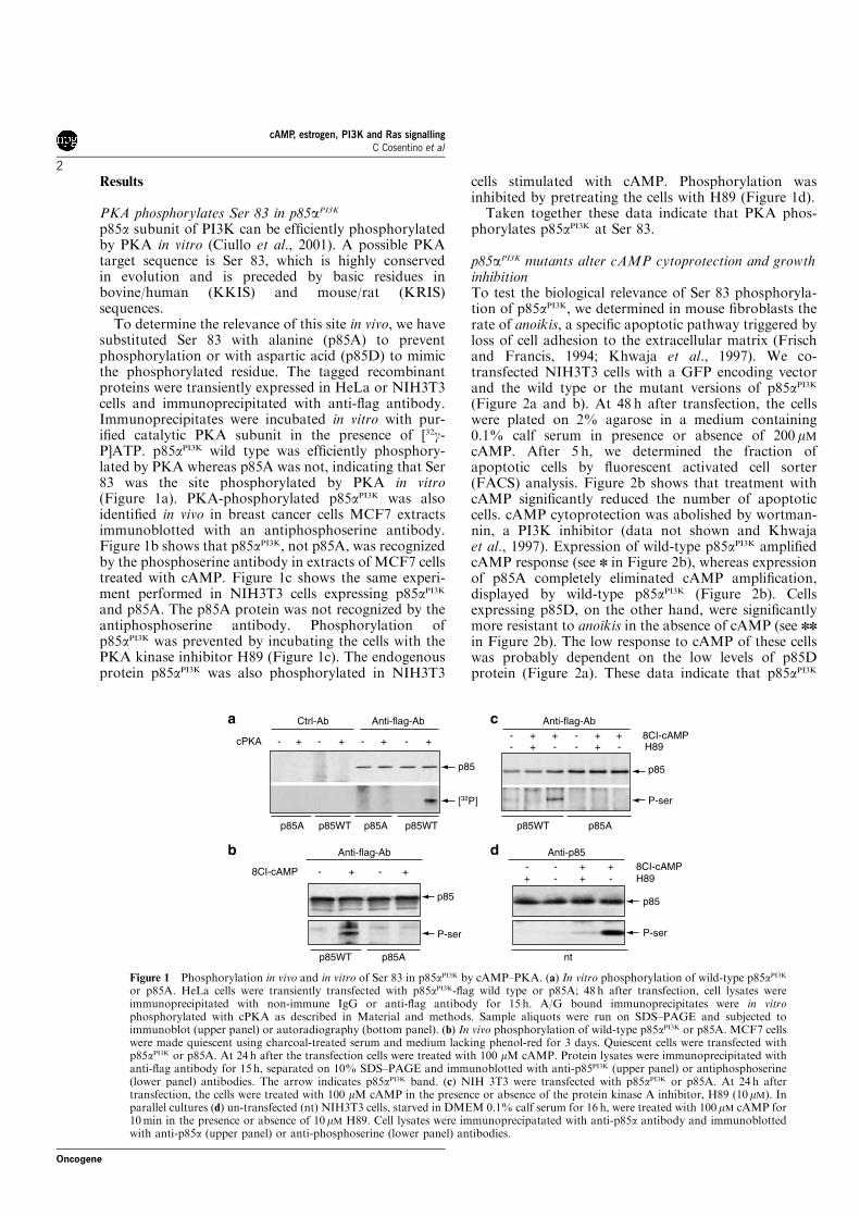
Results
PKA phosphorylates Ser 83 in p85aPI3K
p85a subunit of PI3K can be efficiently phosphorylatedby PKA in vitro (Ciullo et al., 2001). A possible PKAtarget sequence is Ser 83, which is highly conservedin evolution and is preceded by basic residues inbovine/human (KKIS) and mouse/rat (KRIS)sequences.
To determine the relevance of this site in vivo, we havesubstituted Ser 83 with alanine (p85A) to preventphosphorylation or with aspartic acid (p85D) to mimicthe phosphorylated residue. The tagged recombinantproteins were transiently expressed in HeLa or NIH3T3cells and immunoprecipitated with anti-flag antibody.Immunoprecipitates were incubated in vitro with pur-ified catalytic PKA subunit in the presence of [32g-P]ATP. p85aPI3K wild type was efficiently phosphory-lated by PKA whereas p85A was not, indicating that Ser83 was the site phosphorylated by PKA in vitro(Figure 1a). PKA-phosphorylated p85aPI3K was alsoidentified in vivo in breast cancer cells MCF7 extractsimmunoblotted with an antiphosphoserine antibody.Figure 1b shows that p85aPI3K, not p85A, was recognizedby the phosphoserine antibody in extracts of MCF7 cellstreated with cAMP. Figure 1c shows the same experi-ment performed in NIH3T3 cells expressing p85aPI3K
and p85A. The p85A protein was not recognized by theantiphosphoserine antibody. Phosphorylation ofp85aPI3K was prevented by incubating the cells with thePKA kinase inhibitor H89 (Figure 1c). The endogenousprotein p85aPI3K was also phosphorylated in NIH3T3
cells stimulated with cAMP. Phosphorylation wasinhibited by pretreating the cells with H89 (Figure 1d).
Taken together these data indicate that PKA phos-phorylates p85aPI3K at Ser 83.
p85aPI3K mutants alter cAMP cytoprotection and growthinhibitionTo test the biological relevance of Ser 83 phosphoryla-tion of p85aPI3K, we determined in mouse fibroblasts therate of anoikis, a specific apoptotic pathway triggered byloss of cell adhesion to the extracellular matrix (Frischand Francis, 1994; Khwaja et al., 1997). We co-transfected NIH3T3 cells with a GFP encoding vectorand the wild type or the mutant versions of p85aPI3K
(Figure 2a and b). At 48 h after transfection, the cellswere plated on 2% agarose in a medium containing0.1% calf serum in presence or absence of 200 mMcAMP. After 5 h, we determined the fraction ofapoptotic cells by fluorescent activated cell sorter(FACS) analysis. Figure 2b shows that treatment withcAMP significantly reduced the number of apoptoticcells. cAMP cytoprotection was abolished by wortman-nin, a PI3K inhibitor (data not shown and Khwajaet al., 1997). Expression of wild-type p85aPI3K amplifiedcAMP response (see n in Figure 2b), whereas expressionof p85A completely eliminated cAMP amplification,displayed by wild-type p85aPI3K (Figure 2b). Cellsexpressing p85D, on the other hand, were significantlymore resistant to anoikis in the absence of cAMP (see nn
in Figure 2b). The low response to cAMP of these cellswas probably dependent on the low levels of p85Dprotein (Figure 2a). These data indicate that p85aPI3K
Ctrl-Ab
p85A p85WT
p85WT p85A nt
p85A p85Ap85WT p85WT
p85
p85
P-ser
p85
8CI-cAMPH89
P-ser
p85
8CI-cAMPH89
P-ser[32P]
cPKA
8CI-cAMP - + - + - - + ++ - + -
- + - + - + - + - -+ +- -
+--+
++
Anti-flag-Ab Anti-flag-Ab
Anti-flag-Ab Anti-p85
a c
b d
Figure 1 Phosphorylation in vivo and in vitro of Ser 83 in p85aPI3K by cAMP–PKA. (a) In vitro phosphorylation of wild-type p85aPI3Kor p85A. HeLa cells were transiently transfected with p85aPI3K-flag wild type or p85A; 48 h after transfection, cell lysates wereimmunoprecipitated with non-immune IgG or anti-flag antibody for 15 h. A/G bound immunoprecipitates were in vitrophosphorylated with cPKA as described in Material and methods. Sample aliquots were run on SDS–PAGE and subjected toimmunoblot (upper panel) or autoradiography (bottom panel). (b) In vivo phosphorylation of wild-type p85aPI3K or p85A. MCF7 cellswere made quiescent using charcoal-treated serum and medium lacking phenol-red for 3 days. Quiescent cells were transfected withp85aPI3K or p85A. At 24 h after the transfection cells were treated with 100 mM cAMP. Protein lysates were immunoprecipitated withanti-flag antibody for 15 h, separated on 10% SDS–PAGE and immunoblotted with anti-p85PI3K (upper panel) or antiphosphoserine(lower panel) antibodies. The arrow indicates p85aPI3K band. (c) NIH 3T3 were transfected with p85aPI3K or p85A. At 24 h aftertransfection, the cells were treated with 100 mM cAMP in the presence or absence of the protein kinase A inhibitor, H89 (10mM). Inparallel cultures (d) un-transfected (nt) NIH3T3 cells, starved in DMEM 0.1% calf serum for 16 h, were treated with 100 mM cAMP for10min in the presence or absence of 10mM H89. Cell lysates were immunoprecipatated with anti-p85a antibody and immunoblottedwith anti-p85a (upper panel) or anti-phosphoserine (lower panel) antibodies.
cAMP, estrogen, PI3K and Ras signallingC Cosentino et al
2
Oncogene

mediates cAMP inhibition of anoikis and that phos-phorylation of Ser 83 is an important signal for cAMP-induced-survival.
To test if p85aPI3K mediated also cAMP inhibition ofgrowth in mouse NIH3T3 fibroblasts, we analysed thebiological effects of p85aPI3K mutants on growth arrestinduced by cAMP. NIH3T3 fibroblasts were cultured inlow serum in the presence or absence of cAMP. Underthese conditions, cAMP induced a robust and reversibleinhibition of cell growth by accumulating the cells in G1phase and reducing the fraction of cells entering S phase(see n in Figure 2c and d). Cells expressing p85A did notefficiently arrest in G1 in the presence of cAMP(Figure 2c). Conversely, in cell lines expressing theaspartic mutant, cAMP response was lost and thefraction of cells accumulating in G1 was significantlyhigher than in cells expressing p85A (see nn inFigure 2c). The fraction of cells arrested in S phase in
the presence of cAMP was robustly stimulated in thecontrol and cells expressing the wild-type p85aPI3K (see n
in Figure 2d). Cells expressing the mutant versions ofp85aPI3K did not respond to cAMP and the fraction ofcells transiting S increased (see Figure 2d, p85Annn) ordecreased (see Figure 2d, p85Dnnn) relative to controlor wild-type p85aPI3K cells. These data indicate a higher(p85D) or lower rate of G1 arrest (p85A) of these celllines, independently on cAMP presence in the medium.The high statistical significance of S phase values,relative to G1, is due to the lower value of S phase cellfraction. To discriminate more precisely the effects ofcAMP in G1 or S phases in cells expressing p85aPI3K
mutants, we performed a kinetic analysis of the cell cycleby labeling the cells with BrdU. Under these conditions,the entry and the progression in S phase of transfectedcells can be precisely determined. Figure 2e shows thatp85A or p85D did not alter S phase progression,
a
c d e
b
CTRL
G1 phase90
-+8BrcAMP
-+8BrcAMP
Basal +8BrcAMPT090 min270 min
T090 min270 min
80
70
60
50 0
5
10
15
20
0 0
2
4
6
8
10
12
5
10
15
20
25
30
% o
f cel
ls in
G1
phas
e
% o
f cel
ls in
S p
hase
% o
f Brd
U p
ositi
ve c
ells
in S
pha
se
CTRL p85 WT p85A p85D CTRL p85 WT p85A p85D CTRL p85 WT p85A p85D CTRL p85 WT p85A p85D
S phase BrdU
WT ALA ASP
CTRL
% o
f cel
l dea
th
p85 WT
***
* *
** ***
****
*
ALA ASP
p85cAMP+-+-+-+-
50
40
30
20
10
0
Figure 2 Phosphorylation of p85aPI3K Ser 83 mediates cAMP biological effects on growth and survival. NIH3T3 cells were transientlytransfected with p85aPI3K-flag wild type or mutant versions. A GFP expression vector encoding green fluorescence protein was used asreporter. At 48 h after transfection, cells were plated on 2% agarose in DMEM 0.1% CS without or with cAMP 200 mM for 5 h and thefraction of apoptotic cells was determined by FACS analysis. (a) Extracts of transfected cells immunoblotted with anti-p85aPI3Kantibody. Exogenous p85aPI3K is represented by the upper band in the doublet. (b) Quantitative analysis of FACS data. White andblack columns represent, respectively, untreated or cAMP-treated samples. The data are the mean of four independent experiments; nindicates Pp0.01 comparing cell death in the presence of cAMP in all samples. The p85A (ALA) sample is significantly different fromp85aPI3K (WT), but not from CTRL or p85D (ASP). To analyse the growth, NIH3T3 cells were transiently transfected with theconstructs indicated above. At 24 h after the transfection, the cells were serum-starved 15 h and induced into the cycle with 2% serum inthe presence or absence of cAMP (200 mM for 6 h). After 12 h the cells were subjected to FACS analysis. The quantitative analysis ofFACS data relative to G1 phase (c) or S phase (d) was derived from three independent experiments and analysed by Student paired T-test. Black and gray columns represent respectively untreated or cAMP-treated samples. n indicates Pp0.01 basal versus cAMP;nnPp0.01 basal p85A versus p85D; nnnPp0.01 basal p85a wild type, A or D-transfected cells versus control plasmid-expressing cells.(e) Pulse (300)-chase of BrdU incorporation in transfected cell lines in the presence or absence of cAMP. The columns represent thefraction of labeled cells stained with propidium iodide at 0 (black columns), 90 (gray columns) and 270min (white columns) from theinitial cell cycle induction (10% serum). The data are the mean of three independent experiments.
cAMP, estrogen, PI3K and Ras signallingC Cosentino et al
3
Oncogene

although p85A-expressing cells transited S phase veryrapidly, compared to control or p85D expressing lines inthe presence of cAMP. Cells expressing p85A wereunrestrained in G1 in the presence of cAMP and enteredS phase more efficiently than control or p85D-expres-sing cells. Taken together, these data indicate that Ser 83in the p85aPI3K is critical for cAMP-induced G1 arrest oncell cycle progression. Alanine substitution of Ser 83abolished G1 arrest by cAMP. p85D, on the other hand,increased the number of cells arrested in G1 in theabsence of cAMP (see nn in Figure 2d).
p85aPI3K (ser83) phosphorylation increases association ofto p21 RasWe have previously shown that cAMP PKA selectivelyincreased the association between PI3K and p21 Ras(Ciullo et al., 2001). As this complex can mediatecAMP–PI3K effects on growth and survival, we set outto determine the formation of p21 Ras–PI3K complex incells expressing wild-type p85aPI3K or p85A or p85D.NIH3T3 were transfected with p85aPI3K wild type orp85A or p85D and 36 h after transfection, the cells wereserum starved for 16 h before 10min treatment withcAMP. Cell lysates were immunoprecipitated with anti-flag antibody and then analysed by Western blot withanti-Ras antibody. In the presence of cAMP, p85aPI3K
wild type efficiently was found associated with p21 Ras.This association was significantly inhibited in cellsexpressing p85A and stimulated in p85D expressingcells. In the absence of cAMP, Ras–PI3K complex wasbarely detectable in all the samples. In p85D-expressingcells, however, cAMP stimulated robustly association ofPI3K to p21 Ras, suggesting that Ser 83 in p85aPI3K
cooperates with another cAMP–PKA signal to regulatePI3K association with p21 Ras (Figure 3a and b). Wealso tested the effects of H89 on the formation of the
Ras–PI3K complex. H89 treatment reduced the complexRas–PI3K to the levels found in p85A-expressing cells(Figure 3c). These data indicate that under conditions ofcAMP stimulation, the interaction Ras–PI3K is sig-nificantly favored.
p85aPI3K (Ser 83) phosphorylation selectively amplifiesPI3K signaling by cAMPTo directly test the hypothesis that cAMP stimulationamplified Ras–PI3K signalling, we stimulated the cellswith cAMP or PDGF and determined AKT and GSKphosphorylation, downstream targets of PI3K.Figure 4a shows that cAMP and PKA stimulated thephosphorylation of AKT and GSKa and that H89reversed cAMP effects. To test if p85aPI3K mutantsinterfere also with other pathways that signal to PI3K,we determined PDGF stimulation of AKT and ERK1/2,the two major kinases downstream to PDGFR.Figure 4b shows that expression of p85A and p85Ddid not significantly influence PDGF stimulation ofERK1/2 or P-AKT accumulation. p85A-expressingcells, on the other hand, poorly phosphorylated AKTor GSKa in response to cAMP. Conversely, expressionof p85D increased the basal P-AKT and P-GSKa androbustly amplified cAMP response (Figure 4c and d).
So far we have analysed the effects of p85aPI3K
variants on indirect effectors of PI3K signaling. Todirectly test the relevance of cAMP induced phosphor-ylation of p85aPI3K on PI3K activity, we have measuredthe activity of the enzyme in cells expressing wild type orthe mutant versions of p85aPI3K. We have previouslyreported that cAMP in the presence of serum did notstimulate PI3K activity (Ciullo et al., 2001). As thepresence of serum can mask some important biologicaleffects of cAMP–PKA (Porcellini et al., 2003) and (DeGregorio et al., 2006, in press), we carried out the
a
b
c
Anti-p85-flag
Anti-ras
IP:
cAMP
CMV p85WT p85D
p85WT p85A
p85Ap85Dp85WT
ras/
p85
ratio
(A
.U.)
p85A
- +
cAMP -1
0.8
0.6
0.4
0.2
0
+ - + - +
cAMP - -+ + +H89 - -- + -
- + - + - + WB:
WB:
p85
p21ras
p85
p21ras
Figure 3 cAMP stimulates the formation of PI3K/Ras complex. NIH-3T3 cells were transiently transfected with wild type or mutantversions of p85aPI3K-flag. At 36 h after transfection, the cells were starved for 16 h in DMEM, 0.1% calf serum and treated with cAMP200mM for 10min. (a) Cells lysates were immunoprecipitated with anti-flag antibody and blotted with anti-Ras or anti-p85aPI3Kantibodies. (b) The histograms represent the ratio of Ras-p85aPI3K bands in the p85aPI3K immunoprecipitates, derived from threeindependent experiments. (c) NIH-3T3 were transiently transfected with wild-type p85aPI3K-flag or p85A. At 36 h after transfection, thecells were starved for 16 h in DMEM, 0.1% calf serum and treated with cAMP 200mM for 10min in the presence or absence of H89.Cells lysates were immunoprecipitated with anti-Ras antibody and blotted with anti-Ras or anti-flag antibodies.
cAMP, estrogen, PI3K and Ras signallingC Cosentino et al
4
Oncogene
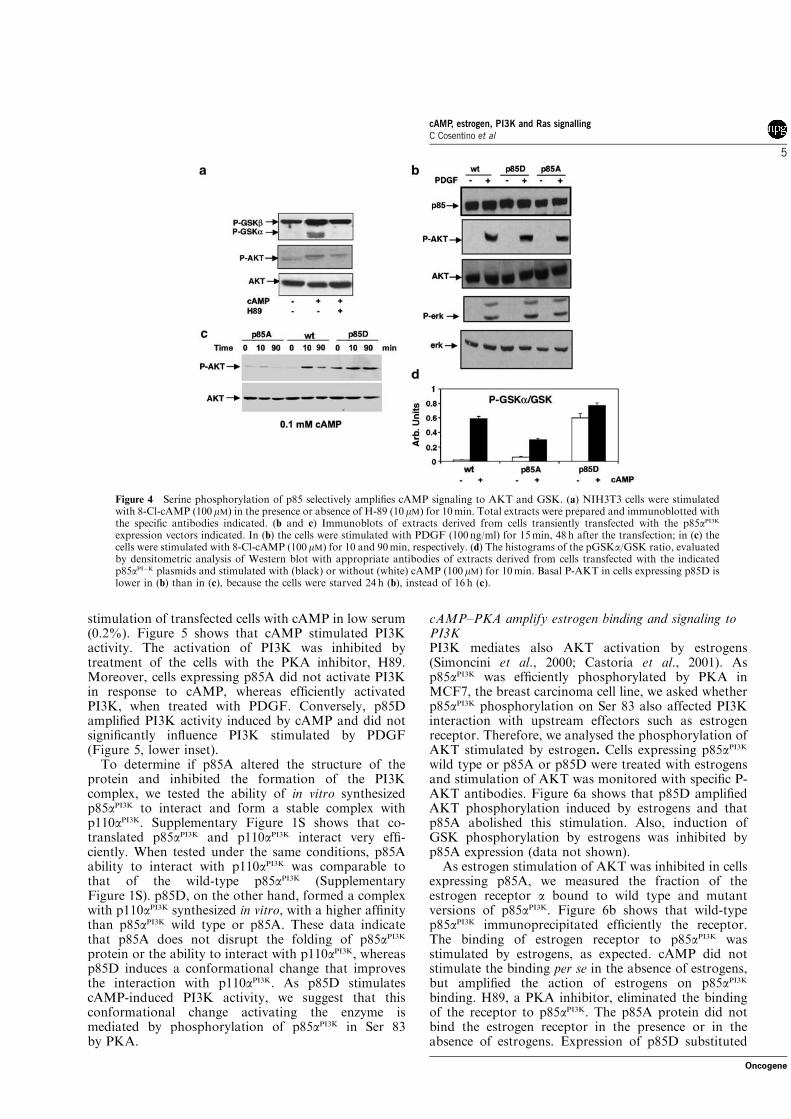
stimulation of transfected cells with cAMP in low serum(0.2%). Figure 5 shows that cAMP stimulated PI3Kactivity. The activation of PI3K was inhibited bytreatment of the cells with the PKA inhibitor, H89.Moreover, cells expressing p85A did not activate PI3Kin response to cAMP, whereas efficiently activatedPI3K, when treated with PDGF. Conversely, p85Damplified PI3K activity induced by cAMP and did notsignificantly influence PI3K stimulated by PDGF(Figure 5, lower inset).
To determine if p85A altered the structure of theprotein and inhibited the formation of the PI3Kcomplex, we tested the ability of in vitro synthesizedp85aPI3K to interact and form a stable complex withp110aPI3K. Supplementary Figure 1S shows that co-translated p85aPI3K and p110aPI3K interact very effi-ciently. When tested under the same conditions, p85Aability to interact with p110aPI3K was comparable tothat of the wild-type p85aPI3K (SupplementaryFigure 1S). p85D, on the other hand, formed a complexwith p110aPI3K synthesized in vitro, with a higher affinitythan p85aPI3K wild type or p85A. These data indicatethat p85A does not disrupt the folding of p85aPI3K
protein or the ability to interact with p110aPI3K, whereasp85D induces a conformational change that improvesthe interaction with p110aPI3K. As p85D stimulatescAMP-induced PI3K activity, we suggest that thisconformational change activating the enzyme ismediated by phosphorylation of p85aPI3K in Ser 83by PKA.
cAMP–PKA amplify estrogen binding and signaling toPI3KPI3K mediates also AKT activation by estrogens(Simoncini et al., 2000; Castoria et al., 2001). Asp85aPI3K was efficiently phosphorylated by PKA inMCF7, the breast carcinoma cell line, we asked whetherp85aPI3K phosphorylation on Ser 83 also affected PI3Kinteraction with upstream effectors such as estrogenreceptor. Therefore, we analysed the phosphorylation ofAKT stimulated by estrogen. Cells expressing p85aPI3K
wild type or p85A or p85D were treated with estrogensand stimulation of AKT was monitored with specific P-AKT antibodies. Figure 6a shows that p85D amplifiedAKT phosphorylation induced by estrogens and thatp85A abolished this stimulation. Also, induction ofGSK phosphorylation by estrogens was inhibited byp85A expression (data not shown).
As estrogen stimulation of AKT was inhibited in cellsexpressing p85A, we measured the fraction of theestrogen receptor a bound to wild type and mutantversions of p85aPI3K. Figure 6b shows that wild-typep85aPI3K immunoprecipitated efficiently the receptor.The binding of estrogen receptor to p85aPI3K wasstimulated by estrogens, as expected. cAMP did notstimulate the binding per se in the absence of estrogens,but amplified the action of estrogens on p85aPI3K
binding. H89, a PKA inhibitor, eliminated the bindingof the receptor to p85aPI3K. The p85A protein did notbind the estrogen receptor in the presence or in theabsence of estrogens. Expression of p85D substituted
Figure 4 Serine phosphorylation of p85 selectively amplifies cAMP signaling to AKT and GSK. (a) NIH3T3 cells were stimulatedwith 8-Cl-cAMP (100mM) in the presence or absence of H-89 (10mM) for 10min. Total extracts were prepared and immunoblotted withthe specific antibodies indicated. (b and c) Immunoblots of extracts derived from cells transiently transfected with the p85aPI3Kexpression vectors indicated. In (b) the cells were stimulated with PDGF (100 ng/ml) for 15min, 48 h after the transfection; in (c) thecells were stimulated with 8-Cl-cAMP (100 mM) for 10 and 90min, respectively. (d) The histograms of the pGSKa/GSK ratio, evaluatedby densitometric analysis of Western blot with appropriate antibodies of extracts derived from cells transfected with the indicatedp85aPI!K plasmids and stimulated with (black) or without (white) cAMP (100 mM) for 10min. Basal P-AKT in cells expressing p85D islower in (b) than in (c), because the cells were starved 24 h (b), instead of 16 h (c).
cAMP, estrogen, PI3K and Ras signallingC Cosentino et al
5
Oncogene

cAMP by amplifying estrogen stimulated binding of thereceptor to p85aPI3K. H89 inhibited the binding of p85Dto estrogen receptor, indicating that another phosphor-ylation PKA-dependent was necessary for the formationof the complex estrogen-p85aPI3K.
These data indicate that phosphorylation by cAMP–PKA of Ser 83 in p85aPI3K is required for estrogenbinding and signalling to PI3K.
Discussion
The data presented here indicate that the phosphoryla-tion of p85aPI3K in a critical serine residue mediatescAMP action on growth and survival. More specifically,phosphorylation of Ser 83 in p85aPI3K shifts the signalingflow towards PI3K-driven pathways. Such a conclusionis supported by several observations. The substitution ofSer 83 with alanine inhibited cAMP effects on cellsurvival and G1 arrest in NIH 3T3 fibroblasts.Conversely, the aspartic mutant of Ser 83 in p85aPI3K
stimulated cell survival in the absence of cAMP andslowed down cell cycle cycle progression, replicating theeffects of cAMP. Ras binding to PI3K and AKTphosphorylation were inhibited by expressing p85Aand were stimulated by p85D.
cAMP effects on growth and cell cycle progression arevery selective. cAMP–PKA act on at least three phases
E2 - + - + - + - + WB:p85 flagp85
P-AKT
AKT
nt p85wt
p85wt p85A
IP: anti-p85 Ab
p85D
p85 flag
ER!cAMP - - + + +E2 - + - + +H89 - - - - +
- - + + +- + - + +- - - - +
- + + +- - + +- - - +
p85A p85D
a
b
Figure 6 cAMP phosphorylation of p85aPI3K amplifies estrogenbinding and signaling to PI3K. MCF-7 cells were made quiescentby charcoal-treated serum and medium lacking phenol-red for 3days. (a) Quiescent cells were transfected with p85aPI3K wild type orp85A or p85D. After 24 h, the cells were treated with 10 nM E2 for3min. Cell lysates were analysed by Western blot with anti-p85aPI3K(upper panel), P-AKT (middle panel) or AKT (lower panel). (b)Quiescent cells were transfected with the wild type or the mutantversions of p85aPI3K. After 24 h, the cells were treated with 10 nM E2
for 3min or for 10min with 200mM cAMP, or with 10 nM E2 for3min following a 10-min stimulation with 200mM cAMP. The celllysates were immunoprecipitated with anti-flag antibody 15 h, asdescribed above. Sample aliquots were immunoblotted with anti-ERa and anti-p85aPI3K antibodies to detect the PI3K/ER complex.Lanes 5 and 14 show the complex estrogen receptor-p85aPI3K incells pretreated with 10 mM H89 for 30min.
a
b
PIP3
PIP3
p85A p85D
p85!Flag
p85!Flag
cAMP - + +H89
cAMP - - - -+ +- + - +- -PDGF
- - +Basal
p85A p85DBasal cAMP PDGF Basal cAMP PDGF
cpm
cpm
("1
00)
0
1000
0
50
100
150
200
250
300
2000
3000
4000
5000
cAMP cAMP+H89
Figure 5 cAMP stimulates PI3K activity in the absence of serum. Effects of p85aPI3K mutants on PI3K catalytic activity. NIH3T3 cellswere transfected with the p85aPI3K expression vectors indicated and stimulated with cAMP (100 mM) 20min in the presence or absenceof H89 (10mM) or PDGF (100 ng/ml) 15min, 48 h after transfection. Total extracts were prepared and subjected toimmunoprecipitation with anti-flag antibody. Immunoprecipitates were quantified by immunoblot with anti-p85aPI3K antibodies(p85aPI3K-flag) and assayed for PI3K activity by using a mixture of lipids containing PI4-5 (see Materials and methods). PI-4-5 labeledat position 3 is indicated by the arrow. The upper inset (a) shows PI3K activity associated with p85aPI3K-flag wild type and theimmunoblot with anti-p85aPI3K antibody of the p85aPI3K-flag immunoprecipitate. The lower inset (b) shows PI3K activity associatedwith p85A and p85D, immunoprecipitated with the anti-flag antibody. The immunoblot with the p85aPI3K antibody is shown below.The histograms on the right show the statistical analysis of (a) and (b) derived from three experiments.
cAMP, estrogen, PI3K and Ras signallingC Cosentino et al
6
Oncogene

of cell cycle. cAMP slows down G1, blocks S phase andG2–M transition (Kato et al., 1994; Kurokawa andKato, 1998; Houslay and Kolch, 2000). Our dataindicate that p85aPI3K phosphorylation of Ser 83 controlsselectively G1–S transition (Figure 2).
PI3K is an important regulator of cell growth as itlinks cell size, growth and cycle progression. Forexample, constitutive expression of p110a increases cellmass and size by stimulating the biosynthetic capacity ofthe cells. However, if not restrained, p110 activityimpairs exit from the cell cycle (Alvarez et al., 2003).We suggest that Ras–PI3K signals regulate the length ofG1 phase. cAMP by amplifying and stabilizing PI3K,lengthens G1 and slows down G1–S transition. The netresult is an apparent G1–S block, which results inincreased biosynthetic activity and cell mass prior todivision. This is better shown in thyroid cells, FRTL5,which are exquisitely dependent on TSH and cAMP forgrowth and differentiation. In these cells, Ras and PI3Kare selectively required for G1 phase progression (Ciulloet al., 2001). In thyroid cells, TSH induces PI3K activityand stabilizes the complex Ras–PI3K. Also, expressionof p85 mutant (p85A) inhibits selectively the stimulationof growth and PI3K activity induced by TSH (DeGregorio et al., in press). In thyroid cells and in cellsexpressing high levels of the regulatory subunit of PKA,RIIb, p85aPI3K binds and anchors PKA. This binding isamplified in cells expressing p85D and it is H89 resistant(De Gregorio et al., in press). This suggests thatanchoring PKA to the membrane is a prerequisite forcAMP–PKA biological effects.
Also, cAMP–PKA stimulate in the absence (Figure 5),not in the presence of serum (Ciullo et al., 2001), PI3Kactivity. This stimulation requires phosphorylation ofSer 83 in in p85aPI3K, probably because phosphorylatedp85aPI3K induces a conformational change in thep110aPI3K molecule (Supplementary Figure 1S) orincreases its affinity to PKA (De Gregorio et al., inpress). The stimulation of PI3K activity by cAMPappears to be very selective, as p85A or p85D mutantsdid not interfere with PDGF (Figure 4) or EGF (DeGregorio et al., in press) induced PI3K activity.
Although the cell lines we have used are not robustlydependent on cAMP signaling for growth or differentia-tion, our data indicate that PI3K-Ras signals areamplified by cAMP both in cAMP-independent(NIH3T3 fibroblasts or breast MCF7 cells) or depen-dent (thyroid) cells. In these latter cells, amplification ofPI3K by cAMP is mainly triggered by TSH and PKA,which binds p85aPI3K. The amplification of the pathwayRas–PI3K induced by cAMP is redundant in 3T3, butnot in thyroid cells, because in these latter cellsexpression of p85A triggers apoptosis (De Gregorioet al., in press).
We suggest that Ser 83 in p85aPI3K is an importantpoint of convergence of two parallel pathways: cAMP–PKA and PI3K signals. In this framework, it is worthnoting that in Saccharomyces cerevisiae inactivationof PI3K–TOR signalling is partly compensated byiper-activation of cAMP–PKA (Rohde et al., 2004;Zurita-Martinez and Cardenas, 2005).
Our data indicate that estrogen receptor signallingwas profoundly altered when p85A was expressed.Accordingly, p85aPI3K binding to estrogen receptor wasseverely impaired. Although PKA phosphorylates di-rectly AKT and estrogen receptor (Cui et al., 2004), theeffects we described were mainly dependent on p85aPI3K,as they were inhibited or amplified by expressing thep85aPI3K alanine or aspartic mutants, respectively.Moreover, we have data indicating that also inductionof estrogen responsive genes is inhibited by H89 and it issignificantly altered in p85A-expressing cells (data notshown). Estrogens stimulate adenylyl cyclase (Aronicaet al., 1994) and cAMP–PKA stimulate differentiationof granulosa cells (Knecht et al., 1984). Our dataindicate that phosphorylation of Ser 83 in p85aPI3K bycAMP PKA stabilizes the binding of activated receptorto PI3K and stimulates PI3K activity.
General implicationsThe data presented above have broad implicationsbecause they point to p85aPI3K as the physical linkbetween Ras, estrogen receptor and cAMP–PKA(Figure 7). Also, we find in many and unrelated celltypes that cAMP amplification of PI3K is essential fornegative or positive cAMP effects on growth andsurvival. Apparently, all receptors binding p85aPI3K cancooperate with cAMP–PKA signals via phosphorylationof Ser 83 in p85aPI3K. This may explain the pleiotropicnature of the effects exerted by cAMP–PKA on several,apparently unrelated, signaling cascades and illustrateshow cAMP–PKA can influence Ras p21 effectors atmultiple levels.
Materials and methods
Plasmid constructionThe cDNA encoding for p85aPI3K-flag wild type was generatedby fusing the flag sequence (MDYKDDDDK) to the C-
GPCR
ER
P p110p85!
PKA
AKT
PKAcAMP
cAMP
RASGTP
Figure 7 Amplification of estrogen and Gs protein-coupledreceptors (GPCR) signaling to PI3K. A scheme outlining theeffects of cAMP–PKA activation by Gs protein coupled receptorson PI3K signaling by estrogens. PKA and cAMP arrows on theright side indicate other cAMP–PKA targets that amplify Ras–PI3K interaction. p85aPI3K links receptors coupled to G proteins toestrogens and Ras–PI3K signals.
cAMP, estrogen, PI3K and Ras signallingC Cosentino et al
7
Oncogene

Terminus of bovine p85aPI3K cDNA in the pSG5 vector (gift ofDr J Downward). The region of p85aPI3K from the XhoIrestriction site in position 1014 (Acc. No: 163476) wasamplified by PCR with a 30 end primer containing the flagsequence and a BamHI restriction site. The PCR product wassubcloned in the II-TOPO vector (Invitrogen), digested withXhoI and BamHI, extracted and ligated to a pSG5-p85aPI3Kvector previously digested with the same restriction enzymes.The vector encoding p85aPI3K-flag was then used as templatefor site-directed mutagenesis (QuickChange Site-DirectedMutagenesis Kit, Stratagene) to convert Ser 83 in alanine oraspartic acid. All the plasmid constructs were verified by DNAsequence analysis.
Cell culture and transfectionsHeLa and NIH3T3 cells were grown in RPMI with 10% fetalcalf serum or Dulbecco’s modified Eagle’s medium (DMEM)with 10% calf serum, respectively. MCF-7 cells were grown inDMEM with 5% fetal bovine serum, supplemented with:penicillin/streptomycin 100mU/ml, 2mM glutamine, 6 ng/mlinsulin and 3.75 ng/ml hydrocortisone. Cells were transfectedwith lipofectamine (Gibco-Invitrogen, Carlsbad, CA, USA). Inall transfections, RSV-LacZ was included to determine andnormalize transfection efficiency. Experiments varying in thetransfection efficiency above 20% were discarded.
Antibodies, immunoprecipitation and immunoblotThe antibodies used were: anti-Ras (mouse monoclonal pan-Ras clone 10, UBI (an affiliate of Millipore, Billerica, MA,USA)), anti-p85PI3K (rabbit polyclonal, UBI), anti-P-Serine(rabbit polyclonal, Zymed, San Francisco, CA, USA), anti-Erk 1/2 (rabbit polyclonal, Santa Cruz, Santa Cruz, CA,USA), anti-P-Erk 1/2 (mouse monoclonal, Santa Cruz), anti-P-Akt Ser 473 (rabbit polyclonal, Cell Signaling, Beverly, MA,USA), anti-Akt (rabbit polyclonal, Cell Signaling), anti-P-GskSer 21/9(rabbit polyclonal, Cell signaling), anti-GSKa/b(mouse monoclonal, UBI). The anti-flag antibody was themouse monoclonal Sigma M2 antibody.
ImmunoprecipitationTotal extracts were prepared by lysing the cells on ice 5minwith 50mM Tris-HCl pH 7.4, 1% Nonidet NP-40, 100mM
NaCl, 2mM EDTA 50mM NaF, 0.1mM NaVO3 1mM b-glycerophosphate, 2.5mM sodium pyrophosphate and aprotease inhibitor cocktail. Cell lysates (2mg/ml) wereincubated with 4mg of antibody/0.5–1mg of protein at 41Cin gentle rock agitation 15 h. At the end of incubation, 20ml ofA/G plus were added to samples and the immunoprecipitateswere collected by centrifugation. SDS–PAGE and immuno-blots were performed as previously described (Feliciello et al.,2000).
AnoikisAt 48 h after transfection, the cells were collected andmaintained in suspension on dishes covered with a thin layerof 2% agarose. Cells were plated in medium containing 0.1%serum with or without 200mM CPT-cAMP (Sigma) for 5 h. Atthe end of incubation, the cells were collected, washed twicewith phosphate-buffered saline (PBS) and incubated 5minwith propidium iodide and analysed by FACS.
Analysis of DNA content and 50-bromo-20-deoxyuridine (BrdU)incorporation2" 106 transfected cells (48 h after transfection) were plated in100mm dishes and grown in low serum (0.5% fetal bovine
serum) for 18 h in the presence or in absence of 50mM 8-ClcAMP. Cells were labelled for 30min with BrdU to a finalconcentration of 20mg/ml and harvested at 0, 90 and 270min.After treatment, the cells were fixed in ice-cold 70% ethanolfor 4 h at # 41C and washed three times in PBS. Cells were re-suspended in 0.25ml of 1N HCl and maintained 20min atroom temperature. After acidic denaturation of DNA, the cellswere washed two times in phosphate/citric buffer (0.2M
Na2HPO4; pH 7.4). BrdU incorporation was revealed byanti-BrdU-FITC (Becton Dickinson, Franklin Lakes, NJUSA) and then stained for 30min at room temperature in0.1% Triton X-100, 0.2mg/ml DNase-free RnaseA, 20 mg/mlpropidium iodide. Fluorescence was determined by using theFACScan Flow Cytometer (Becton Dickinson). Experimentswere performed in triplicate. The data were acquired andanalysed by CELLQuest software (Becton Dickinson) and byCell Fit Cell-Cycle Analysis Version 2 for bivariate analysis ofDNA content versus BrdU incorporation.
In vitro phosphorylationHeLa cells were transiently transfected with p85aPI3K-flag andp85A. At 48 h after transfection, cell lysates were immunopre-cipitated with non-immune IgG or anti-flag antibody 15 h at41C. Protein A/G bound immunoprecipitates were washedtwice with lysis buffer and finally with kinase buffer (N-2-hydroxyethylpiperazine-N0-2-ethane sulfonic acid (HEPES)20mM, MgCl2 10mM, pH 7.4). The washed immunoprecipi-tates were treated with 0.4 mg of partly purified catalyticsubunit of protein kinase A. Each aliquot was incubated in afinal volume of 30 ml of kinase buffer containing 10!5 M cAMP,100 mM ATP and 10 mCi[g32P-ATP] for 30min at 301C. Thereaction was terminated by adding 2" Laemmli buffer.
Lipid kinase assayLipid kinase activity was determined as described by Maieret al. (1999). Briefly, the assays were carried out in a finalvolume of 50ml containing 0.1% bovine serum albumin, 1mM
EGTA (ethylene glycol-bis(b-aminoethyl ether)-N,N,N’,N’,-tetraacetic acid), 120mM NaCl, 40mM HEPES, pH 7.4, 1mM
dithiothreitol, 1mM glycerophosphate, 7mM MgCl2 (buffer E).Lipid vesicles (30 ml containing 320mM phosphatidylethanola-mine, 300 mM phosphatidylserine, 140 mM phosphatidylcholine,30 mM sphingomyelin, supplemented with 40mM PI-4,5-P2 inbuffer E) were sonicated 1 h and incubated on ice 10min. Somealiquots were preincubated with LY294002, 0.5 mM (Calbio-chem (EMD Biosciences, Inc, an Affiliate of Merck KGaA,Darmstadt, Germany)) at 371C. The immunoprecipitates wereadded to the lipid mixture and incubated for 10min at 41C in afinal volume of 40ml. The reaction was started by adding 40mMATP (1 mCi of [-32P]ATP in 10 ml of the assay buffer at 301C.After 15min, the reaction was stopped with ice-cold 150mlHCl (1 N) on ice. The lipids were extracted by vortexing thesamples with 500ml of chloroform/methanol (1:1). Aftercentrifugation the organic phase was washed twice with200 ml of 1N HCl. Phosphorylated lipids were separated byTLC, detected by autoradiography and quantified withPhosphor-Imager.
Acknowledgements
This work was partly supported by AIRC (AssociazioneItaliana Ricerca Cancro), NOGEC (Naples OncogenomicCenter), MIUR (Italian Ministry of Education, Universityand Research). Concetta Cuozzo is a recipient of an FIRCFellowship. This work is dedicated to the loving memory ofStelio Varrone.
cAMP, estrogen, PI3K and Ras signallingC Cosentino et al
8
Oncogene

References
Affaitati A, Cardone L, de Cristofaro T, Carlucci A, GinsbergMD, Varrone S et al. (2003). Essential role of A-kinaseanchor protein 121 for cAMP signaling to mitochondria. JBiol Chem 278: 4286–4294.
Alvarez B, Garrido E, Garcia-Sanz JA, Carrera AC. (2003).Phosphoinositide 3kinase activation regulates cell divisiontime by coordinated control of cell mass and cell cycleprogression rate. J Biol Chem 278: 26466–26473.
Ariga M, Nedachi T, Akahori M, Sakamoto H, Ito Y,Hakuno F et al. (2000). Signalling pathways of insulin-likegrowth factor-I that are augmented by cAMP in FRTL-5cells. Biochem J 348(Part 2): 409–416.
Aronica SM, Kraus WL, Katzenellenbogen BS. (1994).Estrogen action via the cAMP signaling pathway: stimula-tion of adenylate cyclase and cAMP-regulated gene tran-scription. Proc Natl Acad Sci USA 91: 8517–8521.
Castoria G, Migliaccio A, Bilancio A, Di Domenico M, deFalco A, Lombardi M et al. (2001). PI3Kinase in concertwith Src promotes the S-phase entry of oestradiol-stimulatedMCF-7 cells. EMBO J 20: 6050–6059.
Ciullo I, Diez-Roux G, Di Domenico M, Migliaccio A,Avvedimento EV. (2001). cAMP signaling selectivelyinfluences Ras effectors pathways. Oncogene 20:1186–1192.
Cui Y, Zhang M, Pestell R, Curran EM, Welshons WV,Fuqua SA. (2004). Phosphorylation of estrogen receptoralpha blocks its acetylation and regulates estrogen sensitiv-ity. Cancer Res 64: 9199–9208.
De Gregoria G, Coppa A, Cosentino C, Ucci S, Messina S,Nicolussi A et al. (2006). The p85 regulatory subunit ofPI3K mediates TSH-cAMP-PKA growth and survivalsignals. Oncogene, in press.
Feliciello A, Gallo A, Mele E, Porcellini A, Troncone G, GarbiC et al. (2000). The localization and activity of cAMP-dependent protein kinase affect cell cycle progression inthyroid cells. J Biol Chem 275: 303–311.
Frisch SM, Francis H. (1994). Disruption of epithelialcell–matrix interactions induces apoptosis. J Cell Biol 124:619–626.
Houslay MD, Kolch W. (2000). Cell-type specificintegration of cross-talk between extracellular signal-regulated kinase and cAMP signaling. Mol Pharmacol 58:659–668.
Kato JY, Matsuoka M, Polyak K, Massague J, Sherr CJ.(1994). Cyclic AMP-induced G1 phase arrest mediated by aninhibitor (p27Kip1) of cyclin-dependent kinase 4 activation.Cell 79: 487–496.
Khwaja A, Rodriguez-Viciana P, Wennstrom S, Warne PH,Downward J. (1997). Matrix adhesion and Ras transforma-tion both activate a phosphoinositide 3-OH kinase andprotein kinase B/Akt cellular survival pathway. EMBO J 16:2783–2793.
Knecht M, Darbon JM, Ranta T, Baukal AJ, Catt KJ. (1984).Estrogens enhance the adenosine 30,50-monophosphate-mediated induction of follicle-stimulating hormone andluteinizing hormone receptors in rat granulosa cells.Endocrinology 115: 41–49.
Kurokawa K, Kato J. (1998). Cyclic AMP delays G2progression and prevents efficient accumulation of cyclinB1 proteins in mouse macrophage cells. Cell Struct Funct 23:357–365.
Lee YH, Park JS, Park CH, Lee SK. (1998). Synergisticeffect of cyclic AMP and insulin on the expression of cyclinA gene in Swiss 3T3 cells. Biochem Biophys Res Commun244: 843–848.
Magnaldo I, Pouyssegur J, Paris S. (1989). Cyclic AMPinhibits mitogen-induced DNA synthesis in hamster fibro-blasts, regardless of the signalling pathway involved. FEBSLett 245: 65–69.
Maier U, Babich A, Nurnberg B. (1999). Roles of non-catalytic subunits in gbetagamma-induced activation of classI phosphoinositide 3Kinase isoforms beta and gamma. JBiol Chem 274: 29311–29317.
Pastan IH, Johnson GS, Anderson WB. (1975). Role ofcyclic nucleotides in growth control. Annu Rev Biochem 44:491–522.
Porcellini A, Messina S, De Gregorio G, Feliciello A, CarlucciA, Barone M et al. (2003). The expression of the thyroid-stimulating hormone (TSH) receptor and the cAMP-dependent protein kinase RII beta regulatory subunitconfers TSH-cAMP-dependent growth to mouse fibroblasts.J Biol Chem 278: 40621–40630.
Rohde JR, Campbell S, Zurita-Martinez SA, Cutler NS, AsheM, Cardenas ME. (2004). TOR controls transcriptional andtranslational programs via Sap-Sit4 protein phosphatasesignaling effectors. Mol Cell Biol 24: 8332–8341.
Simoncini T, Hafezi-Moghadam A, Brazil DP, Ley K, ChinWW, Liao JK. (2000). Interaction of oestrogen receptor withthe regulatory subunit of phosphatidylinositol-3-OH kinase.Nature 407: 538–541.
Zurita-Martinez SA, Cardenas ME. (2005). Tor and cyclicAMP-protein kinase A: two parallel pathways regulatingexpression of genes required for cell growth. Eukaryot Cell 4:63–71.
Supplementary Information accompanies the paper on the Oncogene website (http://www.nature.com/onc).
cAMP, estrogen, PI3K and Ras signallingC Cosentino et al
9
Oncogene

ORIGINAL ARTICLE
The p85 regulatory subunit of PI3K mediates TSH–cAMP–PKA growthand survival signals
G De Gregorio1, A Coppa2, C Cosentino3, S Ucci1, S Messina1, A Nicolussi2, S D’Inzeo2,A Di Pardo1, EV Avvedimento3 and A Porcellini1,2
1Dipartimento di Patologia Molecolare IRCCS Neuromed, Localita Camerelle, Pozzilli, Italy; 2Dipartimento di MedicinaSperimentale, Universita ‘La Sapienza’, Viale Regina Elena, Roma, Italy and 31 Dipartimento di Biologia e Patologia Molecolare eCellulare, Istituto di Endocrinologia ed Oncologia Sperimentale del CNR, Universita Federico II, Napoli, Italy
Phosphatidylinositol 3-kinase (PI3K) is necessary forthyroid stimulating hormone (TSH)-induced cell cycleprogression. To determine the molecular mechanismlinking PI3K to TSH, we have identified a serine residuein p85aPI3K phosphorylated by protein kinase A (PKA) invitro and in vivo. Expression of an alanine mutant (p85A)abolished cyclic AMP/TSH-induced cell cycle progres-sion and was lethal in thyroid cells (FRTL-5). Theaspartic version of the p85aPI3K (p85D) inhibited apoptosisfollowing TSH withdrawal. The p85aPI3K wild type not thep85A bound PKA regulatory subunit RIIb in cellsstimulated with cAMP or TSH. The binding of theaspartic version of p85aPI3K to RIIb was independent ofcAMP or TSH stimulation. Similarly, binding of PI3K top21Ras and activation of AKT, a downstream PI3Ktarget, were severely impaired in cells expressing the p85Amutant. Finally, we found that the catalytic activity ofPI3K was stimulated by TSH in cells expressing the wild-type p85aPI3K but not in cells expressing p85A. This lattermutant did not affect the epidermal growth factor-stimulated PI3K activity. We suggest that (1) TSH–cAMP-induced PKA phosphorylates p85aPI3K at serine 83,(2) phosphorylated p85aPI3K binds RIIb-PKA and targetsPKAII to the membrane, and (3) PI3K activity andp21Ras binding to PI3K increase and activate PI3Kdownstream targets. This pathway is essential for thetransmission of TSH–cAMP growth signals.Oncogene advance online publication, 9 October 2006;doi:10.1038/sj.onc.1210011
Keywords: phosphoinositide-3-kinase; signal trasduc-tion; thyropropin receptor; protein kinase A; cAMP
Introduction
Stimulation of growth by cyclic AMP–protein kinase A(cAMP–PKA) in selected cell types, as in thyroid cells, istightly dependent on Ras and Phosphatidylinositol 3-kinase (PI3K) (Kupperman et al., 1993; Al-Alawi et al.,1995; Miller et al., 1998). Also, cAMP–PKA arepowerful survival signals in all cell types tested (Affaitatiet al., 2003; Wang et al., 2005). To date, the mechan-ism(s) and the relevant players mediating the effects ofon growth and survival are not completely known.
We have recently reported that growth of cells, NIH3T3, normally inhibited by cAMP (Magnaldo et al.,1989; Chen and Iyengar 1994), can be potentlystimulated by cAMP if the PKA subunit RIIb isexpressed in these cells (Porcellini et al., 2003). Thepresence of RIIb in these lines amplified cAMPsurvival and transcriptional signals. We have previouslyshown that cAMP–PKA influence Ras signaling, byselectively stimulating the Ras–PI3K complex. More-over, p85aPI3K is an efficient PKA substrate in vitro(Ciullo et al., 2001).
We have identified a serine (83) in the p85aPI3K
molecule that is phosphorylated in vivo and in vitro byPKA. This phosphorylation mediates the interaction ofPI3K with p21Ras and estrogen receptor a (Cosentinoet al., 2006).
In this work, we describe the biological consequencesof the expression of mutant versions of p85aPI3K inFRTL-5 thyroid cells and in an engineered NIH 3T3 cellline that recapitulates the features of thyroid stimulatinghormone (TSH)-dependent growth of thyroid cells. Ourresults demonstrate that phosphorylation of serine 83 iscritical for TSH-dependent cell cycle progression andcell survival. Moreover, we found that phosphorylationof serine 83 by cAMP–PKA was essential for binding ofp85aPI3K to the PKA regulatory subunit RIIb.
We conclude that, in the presence of the RIIb-PKAsubunit, abundant in cAMP-stimulated cells, p85aPI3K
enucleates a complex containing PKA. Phosphorylationof serine 83 in the p85aPI3K molecule amplifies PKAbinding to PI3K and stimulates the Ras–PI3K complex,which ultimately mediates survival and growth signalsinduced by TSH–cAMP. This pathway is essential forcAMP-dependent proliferation.
Received 10 February 2006; revised 25 July 2006; accepted 11 August2006
Correspondence: Dr A Porcellini, Dipartimento di Medicina Sper-imentale, Universita ‘La Sapienza’, Viale Regina Elena, 324 00161Roma, Italy.E-mail: [email protected] orProfessor EV Avvedimento, Dipartimento di Biologia e PatologiaCellulare e Molecolare ‘L Califano’, Universita ‘Federico II’, Via SPansini 5, 80131 Napoli, Italy.E-mail: [email protected]
Oncogene (2006), 1–9& 2006 Nature Publishing Group All rights reserved 0950-9232/06 $30.00
www.nature.com/onc

Results
p85 phosphorylation mediates TSH-dependent cell cycleprogression and cell survivalPrevious data from our laboratory have shown that thep85aPI3K regulatory subunit can be phosphorylated byPKA in vitro (Ciullo et al., 2001). Sequence analysis ofp85aPI3K revealed a putative PKA phosphorylation site,which was conserved in mammalian p85aPI3K. To verifythat this sequence was a bona fide PKA site, we havesubstituted this serine 83 by alanine (p85A) or aspartate(p85D) to prevent or to mimic phosphorylation ofserine, respectively. The tagged wild-type recombinantprotein was phosphorylated in vivo and in vitro by PKA,whereas p85A protein was not (Cosentino et al., 2006).
To test the biological effects of p85aPI3K phosphoryla-tion on thyroid cell growth, we transfected the ratthyroid cell line, FRTL-5, with p85WT, p85A and p85Dexpression vectors. As we did not score a significantnumber of stable transfectants expressing p85A, wetested the plating efficiency of cells transfected withp85A or p85D. Figure 1a shows that the expression ofp85A significantly reduced the number of clones ofFRTL-5 cells expressing p85A. Conversely, the numberof clones was significantly higher in cells expressingp85D (Figure 1a). These data indicate that phosphor-ylation of serine 83 in p85aPI3K significantly altered thegrowth or survival of differentiated thyroid cells. To testthis notion in a defined experimental system, we used aunique cellular model that recapitulates the features ofTSH-dependent growth and survival, that is, fibroblastscoexpressing the PKA regulatory subunit RIIb andwild-type TSHR under the control of an induciblepromoter (NTCRII). In this model, in the absence ofRIIb, activation of TSHR by TSH induced growtharrest in the presence of serum. In the absence of serum,these cells, stimulated with TSH, replicated their DNAbut underwent apoptosis. Coexpression of RIIb inhib-ited apoptosis and stimulated the growth of the cellsonly in the presence of TSH or cAMP (Porcellini et al.,2003). We have expressed p85A or p85D in these cellsin the presence or absence of TSH or 8BrcAMP.Figure 1b shows that cells expressing TSHR andRIIb, which proliferated in the presence of TSH,underwent apoptosis when starved of TSH. Expressionof p85WT slightly inhibited apoptosis. Expressionof p85D significantly reduced the fraction of apoptoticcells starved of TSH. On the other hand, expression ofp85A significantly reduced the survival of these cells inthe presence of TSH. These data indicate that phos-phorylation of serine 83 amplifies the transmission ofsurvival signals by TSH and PKA, and explain the lowsurvival rate of thyroid cells expressing p85A(Figure 1a).
To analyse more precisely the biological effects ofp85A or p85D on cycle progression and growth of TSH–RIIb-expressing cells, we carried out a detailed cell cycleanalysis by performing 5-bromodeoxyuridine (BrdU)pulse–chase labeling experiments. In low serum, TSHinduced DNA synthesis and stimulated S and G2–Mtransition in cells expressing p85WT or control plasmids
(Figure 1c). Expression of p85A resulted in a significantaccumulation of cells in S phase. Note that in 270min,control cultures exit S phase and accumulated in G2M.Expression of p85D did not significantly alter cell cycleprogression (Figure 1c). We have also tested if theexpression of p85A or p85D mutants altered cell cycleprogression or proliferation induced by epidermalgrowth factor (EGF). Supplementary Figure 1S showsthat the growth rate and cell cycle progression inducedby EGF or serum (not shown) were not affected by theexpression of p85aPI3K mutants.
These experiments indicate that inhibition of phos-phorylation of p85aPI3K at serine 83 slows down S–G2/Mprogression induced by TSH–cAMP.
Phosphorylation of serine 83 in p85aPI3K is required forTSH-induced AKT phosphorylationAs p85A inhibited cell cycle transitions induced by TSH,we tested whether the signaling cascade triggered byTSH or cAMP was impaired. cAMP and TSHstimulated PKA, which can directly phosphorylateAKT (Sable et al., 1997; Filippa et al., 1999) and/orinduce the recruitment of PI3K to the membrane (Suhet al., 2003). We determined the phosphorylation ofAKT in cell lines expressing p85WT or p85 mutant inthe presence or absence of TSH. The ratio P-AKT/totalAKT, a rather accurate index of AKT activation, wasdetermined. Cells expressing p85WT, the p85A mutantsor p85D mutants were stimulated with TSH for variousperiods of time and tested for AKT phosphorylation.Figure 2 shows that TSH induced AKT phosphoryla-tion and that in cells expressing p85A, AKT inductionwas almost completely abolished. Both FRTL-5(Figure 2a) and NTCRII cells (Figure 2b), expressingp85A, were unable to activate AKT in the presence ofTSH. The expression of p85D did not significantly affectthe absolute level of AKT activation by TSH, butmodified the kinetics of activation in both cell lines(Figure 2a and b). Induction of P-AKT by EGF was notaffected in cells expressing both mutants (Supplemen-tary Figure 2S). Pretreatment with a PKA inhibitor H89(10 mM) abolished TSH-induced AKT phosphorylation(data not shown). These data indicate that serine 83 inp85aPI3K is important for TSH induction of AKT.
Phosphorylation of serine 83 influences the formation ofPKA–PI3K and Ras–PI3K complexesThe p85A mutant induced apoptosis, inhibited cell cycleprogression and prevented AKT phosphorylation byTSH in thyroid and NTCRII cells. Both cell lines wereexquisitely dependent on TSH and cAMP for growth.As in these cells the expression of PKA subunit RIIbconferred TSH- and cAMP-dependent growth (Porcelli-ni et al., 2003), we tested whether p85aPI3K formed acomplex with RII-PKA. The RIIb–p85aPI3K complexwas assayed by a p85 pull-down assay, using theglutathione S-transferase (GST)-RIIb fusion protein asbait (Grieco et al., 1996). The binding assay was carriedout with extracts derived from NTCRII cells transientlytransfected with the p85WT, p85A, p85D or with the
p85 mediates TSH–PKA growth and survival signalsG De Gregorio et al
2
Oncogene

empty vector (control) and from FRTL-5 clones stablytransfected with the same p85aPI3K vectors. FRTL-5 cellswere derived from the few clones expressing low levels ofp85A, as shown in Figure 1a.
Figure 3a shows that phosphorylated p85WT bindsthe PKA type II regulatory subunit RIIb in a TSH-dependent manner. TSH treatment (10mU/ml) for aperiod of 10min induced the formation of the complex
Figure 1 Phosphorylation of serine 83 of p85 mediates TSH-induced cell cycle progression and cell survival. (a) Plating efficiency ofFRTL-5 cells expressing p85PI3K wild type, p85A or p85D. The ability to form G-418-resistant clones was determined by transfectingFRTL-5 cells with expression vectors encoding p85PI3K wild type, the A mutant version (p85A) or the D mutant version (p85D) andselecting the clones in the presence of 400mg/ml of G-418 for 15 days. Immunoblot analysis of duplicate plates indicated comparablecopy number of transfected p85PI3K. The histogram represents the number of colonies derived from 5! 105 transfected cells (mean ofthree experiments in triplicate). (b) Apoptosis assay of NTCRII cells transfected with cytomegalovirus (CMV) (CTRL), p85WT, p85Aor p85D treated without (white bar) or with TSH (gray bar) or 8BrcAMP (black bar). Cells were transiently transfected with theconstructs indicated above. At 18 h after induction with 0.5% serum in the presence of TSH or cAMP, cells were subjected to TUNELassay as described in Materials and methods. Inset: Western blot analysis with anti-p85PI3K antibody to verify that expression of theconstructs was comparable in the different samples. (c) Cell cycle progression of cells expressing p85WT, p85A or p85D. NTCRII cellswere transiently transfected with the p85WT, p85A, p85D mutants or with the empty vector (control). At 24 h after transfection, thecells were serum starved for 18 h and induced with 10mU/ml TSH in the presence of 0.5% serum. BrdU incorporation was assayed bya pulse–chase experiment. Cells were labeled for 30min with BrdU and harvested at 0, 90 and 270min. Cells were subjected tofluorescence-activated cell sorting (FACS) analysis for bivariate analysis of DNA content versus BrdU.
p85 mediates TSH–PKA growth and survival signalsG De Gregorio et al
3
Oncogene

Figure 2 Phosphorylation of serine 83 is required for TSH-induced AKT phosphorylation. Time course of TSH-induced AKTphosphorylation in FRTL-5 (a) or NTCRII (b) cells expressing recombinant p85PI3K. Representative blots of extracts derived fromtransiently transfected cells (NTCRII) or stable FRTL-5 clones (b) after 18 h of growth in 0.5% serum. TSH was added at aconcentration of 10mU/ml for the indicated time. The histogram in the lower panels shows the densitometric analysis of the P-AKT/AKT ratio relative to b-actin and normalized to the transfection efficiency, derived from three independent experiments.
p85 mediates TSH–PKA growth and survival signalsG De Gregorio et al
4
Oncogene

in NTCRII (Figure 3a) or FRTL-5 (Figure 3b) cellsexpressing recombinant p85WT. In contrast, in cellsexpressing the p85A mutant, the formation of RIIb–p85aPI3K complex was inhibited. The binding of p85aPI3K
to RIIb was observed after cAMP treatment (100 mM8BrcAMP) (Figure 3c). Conversely, the p85D mutantwas bound to RIIb even in the absence of cAMP or TSH(Figure 3a and c). We also detected bound p85aPI3K
phosphorylated at serine 83 with anti-phosphoserineantibodies in p85WT, isolated from cells stimulated withTSH (Figure 3a). Most notably, the binding of p85D toRIIb was resistant to the PKA inhibitor H89 (Figure 3c)and, as expected, it was not present in the immunoblotprobed with anti-phosphoserine antibody (Figure 3a).
To demonstrate a direct interaction of RIIb–p85aPI3K,NIH 3T3 cells were transiently transfected with p85WT
or p85A and treated for 60min with 100 mM 8BrcAMP.Exogenous p85aPI3K was immunoprecipitated with anti-FLAG antibody, extensively washed in RIPA buffercontaining 1% Triton X-100"0.1% sodium dodecylsulfate (SDS) and purified by elution with 0.1M glycine(pH 3.5). Purified p85aPI3K was then incubated in vitrowith GST-RIIb and a pull-down experiment wasrepeated, as described above. Figure 4 shows thatp85WT binds GST-RIIb but not control GST. More-over, the binding of p85aPI3K to RIIb was stimulated bypretreating the cells with cAMP (Figure 4). The p85Amutant did not bind RIIb, both in the presence andabsence of cAMP. Collectively, these data demonstratethat p85aPI3K upon phosphorylation of serine 83 inducedby cAMP or TSH binds PKA via the regulatory subunitRIIb. Proximity of PKA to PI3K may favor signaling ofboth kinases to downstream effectors.
We have previously shown that cAMP–PKA selec-tively increased the association between PI3K andp21Ras (Ciullo et al., 2001). As this complex maymediate the cytoprotective effects of cAMP–PI3K, weset out to determine the formation of p21Ras–PI3K incells expressing wild type or the mutant versions ofp85aPI3K indicated above. NTCRII cells were transfectedwith p85WT, p85A or p85D, and after 36 h, the cellswere serum starved for 16 h before treatment with TSHfor 10min. Cell lysates were immunoprecipitated withanti-FLAG antibody and then analysed by Western blotwith anti-p85aPI3K and anti-Ras antibodies. In thepresence of TSH, p85aPI3K efficiently associated withp21Ras in p85WT-expressing cells. This association wasgreatly reduced in cells expressing p85A. In the absenceof TSH, Ras association with p85aPI3K was barelydetectable in p85WT- and p85A-expressing cells. Con-versely, the association of the mutant p85D to p21Raswas present in the absence of cAMP, but it was alsostimulated by cAMP (Figure 5). The same results wereobtained in FRTL–5-transfected clones (Figure 5).
We conclude that phosphorylation of p85aPI3K atserine 83 by cAMP–PKA significantly stimulates theinteraction of PI3K with PKA and p21Ras. The p85aPI3K
Figure 3 Phosphorylation of serine 83 in p85PI3K selectivelyinfluences the formation of PKA–PI3K complex. (a) p85PI3K–RIIbcomplex formation in NTCRII cells. Pull-down experiment withGST-RIIb challenged with extracts derived from NTCRII cellstransfected with p85WT, p85A and p85D and treated with 10mU/ml TSH for 10min. RIIb binding proteins were probed with anti-FLAG, anti-phosphoserine and anti-GST antibodies. (b) p85PI3K–RIIb complex formation in FRTL-5 cells. The same pull-downexperiment described in (a) was carried out with extracts of stabletransfected FRTL-5 cells. (c) p85PI3K binds RIIb in a cAMP-dependent manner in NTCRII cells. Immunoblot analysis ofp85PI3K bound to RIIb in transiently transfected NTCRII cells.Cells were starved of serum for 6 h (0.5% serum) and exposed to100mM 8BrcAMP for 15min with and without 10 mM H89.
Figure 4 Purified p85 binds RIIb in vitro. Exogenous p85PI3K wasimmunoprecipitated with anti-FLAG antibody from NTCRII cellstransfected with p85WT or p85A, and exposed to 100mM8BrcAMP for 15min. The immunoprecipitate was extensivelywashed in RIPA buffer containing 1% Triton X-100 "0.1% SDSand purified by elution with 0.1M glycine pH 3.5. Input indicatesthe eluted fraction; (# ) glycine represents the pellet after elution.The purified p85PI3K was incubated in vitro with GST-RIIb in apull-down experiment as described in Materials and methods.
p85 mediates TSH–PKA growth and survival signalsG De Gregorio et al
5
Oncogene

mutant, p85A, inhibited these interactions and down-stream PI3K signaling, such as AKT phosphorylation.
To test if phosphorylation of serine 83 in p85aPI3K
influenced the catalytic activity of PI3K, we performedPI3K enzymatic assays on immunoprecipitated p85aPI3K.
Figure 6 shows that TSH stimulated PI3K activity,although less efficiently than EGF, and that p85Ainhibited TSH induction. To test if p85A inhibited PI3Kactivity per se, independent of the specific signalingpathway involved, we tested EGF-induced PI3K in cellsexpressing p85A or p85D. Figure 6B shows that PI3Kactivity induced by EGF was sensitive to the specificPI3K inhibitor LY294002 and that expression of p85Aor p85D did not influence EGF-induced activity.
These data indicate that phosphorylation of serine 83in p85aPI3K selectively influences TSH- or cAMP-inducedPI3K activity.
Discussion
The data presented demonstrate the importance ofp85PI3K in the transmission of TSH and cAMP growthand survival signals. Specifically, phosphorylation ofserine 83 in the p85aPI3K is essential for the binding ofPI3K to PKA. We suggest that following PKAanchoring to PI3K, p21Ras associates to the complexand PI3K catalytic activity is greatly stimulated.Mutagenesis of this site profoundly and selectivelyimpaired the transmission of TSH signals by disruptingthis complex. EGF (Supplementary Figure 2S andFigure 6) or PDGF stimulation of PI3K (Cosentinoet al., 2006) was not influenced by the substitution ofserine 83 by alanine or aspartate in p85aPI3K. Thestimulation of PI3K catalytic activity by cAMP–TSHwas evident in the absence of serum (see also, Porcelliniet al., 2003) and was dependent on the phosphorylationof serine 83 in p85aPI3K. However, the aspartic mutant ofserine 83 p85aPI3K replicated cAMP effects on (i) theinhibition of apoptosis by TSH starvation and (ii) thebinding to PKA. It amplified cAMP effects on (i) PI3Kactivity, (ii) AKT phosphorylation and (iii) the efficiencyof formation of the complex PI3K–p21Ras. These dataindicate that serine 83 phosphorylation of p85aPI3K is
necessary but not sufficient to stimulate PI3K, phos-phorylation of AKT and formation of the p21Ras–PI3Kcomplex. Functional PKA is indeed required for thesecAMP effects. As PKA binding to p85D is H89-resistant(Figure 3c), phosphorylation of serine 83 in p85aPI3K isnecessary and sufficient for PKA anchoring to PI3K.
This is a non-redundant circuit in thyroid or cellsexpressing TSHR and PKAIIb, because its eliminationprofoundly impaired survival and growth. In contrast,in cells containing low levels of cAMP and RIIb, theexpression of p85A abolished only cAMP response,with a marginal effect on survival (data not shown;Cosentino et al., 2006). As for the mechanism, the datapresented indicate that the serine 83 in p85aPI3K is acritical site for the transmission of receptor signals toPI3K. The alanine mutant inhibits cAMP effects onPI3K activity. It is possible that a conformationalchange induced by phosphorylation is important forstimulation of PI3K activity. It is noteworthy thatsubstitution of serine 83 by aspartate in p85aPI3K
stabilizes the complex with p110 (Cosentino et al., 2006).We wish to stress that these data highlight a general
role of serine 83 in the p85aPI3K molecule in thetransmission of cAMP signals in many cell types. Wehave recently found that many independent signalingpathways that stimulate cAMP converge on this site.For example, p85A disrupts the binding of PI3K toestrogen receptor and inhibits AKT activation byestrogens (Cosentino et al., 2006).
We suggest that phosphorylation of serine 83 bycAMP–PKA induces a conformational change in thePI3K complex, resulting in facilitated binding toreceptors and to p21Ras, with stimulation of PI3Kactivity. We believe that this site is a nodal point whereinformation from several receptors is channeled toPI3K.
Materials and methods
Materials and reagentsUnless otherwise specified, drugs and chemicals were obtainedfrom Sigma Aldrich (St Louis, MO, USA) and cell culturesupplies were purchased from standard suppliers, for example,Falcon, BD Biosciences, San Jose, CA, USA; GIBCO-Life
Figure 5 Phosphorylation of serine 83 selectively influences the formation of Ras–PI3K complex. The p21Ras–p85aPI3K complex wasassayed by immunoprecipitation with anti-FLAG antibodies and immunoblotting with anti-pan-Ras antibodies. NTCRII cells weretransiently transfected with p85WT, p85A, p85D or with the empty vector. FRTL-5 cells were the stable clones indicated in Figure 1.Cells were starved of serum for 6 h (0.5% serum) and exposed to 10mU/ml TSH for 20min.
p85 mediates TSH–PKA growth and survival signalsG De Gregorio et al
6
Oncogene

Figure 6 Selective stimulation of PI3K activity by TSH. (A) (a) A representative autoradiogram of the TSH-induced lipid kinaseassay. NTCRII cells were transfected with p85aPI3K expression vectors as indicated in Figure 1. At 48 h after transfection, cells wereserum starved and maintained in low serum (0.2%) for 8 h. Cells were treated with TSH (10mU/ml) or EGF (100 ng/ml) for 20min.p85-FLAG was immunoprecipitated (IP) with the anti-FLAG M1 affinity gel and the immune complexes were subjected to PI3K lipidkinase assays as described in Materials and methods. (b) Immunoblot analysis indicated comparable amounts of immunoprecipitatedp85-FLAG. (c) The histogram shows the densitometric analysis normalized to the IP efficiency derived from three independentexperiments. (B) (a) A representative autoradiogram of the EGF-induced lipid kinase assay on anti-FLAG and anti-p85aPI3K IPsamples. The right-hand panel shows the PI3K activity derived from cells stimulated with EGF in the absence or presence of thespecific PI3K inhibitor LY294002. (b) Densitometric analysis normalized to the IP efficiency from three independent experiments.(c) Immunoblot analysis indicated comparable amounts of immunoprecipitated p85aPI3K.
p85 mediates TSH–PKA growth and survival signalsG De Gregorio et al
7
Oncogene

Technologies, Invitrogen S.R.L, San Giuliano Milanese (MI),Italy, HyClone, Logan, UT, USA.
Cell linesMouse fibroblasts NIH 3T3 were grown in Dulbecco’smodified Eagle’s medium containing 10% fetal calf serumand 2mM glutamine (standard medium). The tTA repressor-expressing clones were grown in standard medium containingpuromycin 2.5mg/ml puromycin, 1.0 mg/ml tetracycline and200 mg/ml geneticin (G-418). The selection and tetracyclinewere removed 48 h before starting the experimental proceduresdescribed below. The FRTL-5 cell line is a thyroid cell line,which has been extensively characterized with respect tothyroglobulin expression and TSH-dependent growth (Ambe-si-Impiombato et al., 1980). Cells were grown in F12 mediumwith 5% calf serum and six hormones (1mU/ml TSH, 1 mg/mlinsulin, 3.6 mg/ml hydrocortisone, 5 mg/ml transferrin, 10 ng/mlsomatostatin, 20 mg/ml glycyl-histidyl-lysine). FRTL-5 cloneexpressing the p85 variants was generated by transfectingFRTL-5 cells with the pSG5-p85 vectors. Neomycin-resistantclones were isolated and characterized for NEO and p85expression by Western blots.
Plasmid constructionThe p85aPI3K plasmids have been described by Cosentino et al.(2006). Briefly, p85 cDNA was subcloned in pSG5 vector.FLAG sequence was added by subcloning into the XhoI/BamHI sites of the pSG5-p85 vector a PCR fragment spanningfrom the XhoI site of p85PI3K at position 1014 to the end of thefragment. The vector encoding for p85-FLAG was then usedas a template of PCR for site-directed mutagenesis (Quick-Change Site-Directed Mutagenesis Kit, Stratagene, CedarCreek, TX, USA) to convert serine 83 to alanine.
TransfectionsThe day before transfection, cells were plated at 60%confluence in 100mm dishes and transfected with 2 (NTCRII)or 8 (FRTL-5)mg/dish of p85WT, p85A, p85D or emptyvector using 2.75 or 12.5 ml/mg DNA of Lipofectamine Plus(Invitrogen, Carlsbad, CA, USA). Following 5 h of exposureto lipofectamine, the cells were washed and grown for 36–48 hbefore assays.
Cell growth analysisProliferation was analysed under different conditions: normalmedium or low serum (0.5%) with and without 10mU/ml TSHor 25 ng/ml EGF. MTT (3-(4,5-dimethylthiazol-2-yl)-2,5-di-phenyl tetrazolium bromide) colorimetric assay for prolifera-tion was performed in a 96-well flat-bottomed tissue culturetray for each time point: 1500 cells for each clone were seededin eight replicas for each experimental point; 6 h after plating,the standard medium was removed and 100 ml of culturemedium with stimuli as indicated was applied. The determina-tions were carried out every 24 h as follows: 0.01ml MTT stock(5mg/ml) was added to each well, then after 4 h of incubationat 371C, the medium was removed and 0.1ml isopropanol/0.04 N HCl was added. The absorbance was measured after 1 hon an enzyme-linked immunosorbent assay plate reader. Wehave used a Dynatech MR580 reader with a test wavelength of570 nm and a reference wavelength of 630 nm.
Western blotCells were transfected as indicated above. After 48 h, cells wereexposed to 10mU/ml TSH, 100 mM 8BrcAMP or 100 ng/mlEGF, harvested at times indicated and lysed on ice-cold RIPAbuffer (1% Triton X-100, 0.5% sodium deoxycholate (DOC),
0.1% SDS, 50mM Tris-HCl pH 7.6, 150mM NaCl, 1mM
phenylmethylsulfonyl fluoride, 1mg/ml aprotinin, leupeptinand pepstatin). After centrifugation at 12 000 g, proteinconcentrations were determined. Fifty micrograms ofprotein was subjected to 7 or 12% SDS–polyacrylamide gelelectrophoresis and transferred onto nitrocellulose membranes(Schleicher and Schuell, Whatman plc, Brentford, Middlesex,UK, Germany). Western blot analysis was carried out by usingthe appropriate antibody as noted in the figures. The anti-p85was from Upstate-Millipore (Billerica, MA, USA) (#06-497).The anti-pan-Ras antibody was from Calbiochem (affiliate ofMerck KGaA, Darmstadt, Germany) (EMD Biosciences Inc.,an affiliate of Merck KGaA, Darmstadt, Germany). Theantibody against Akt and phospho-Akt were from CellSignaling Technology (Beverly, MA, USA). The horseradishperoxidase-conjugated anti-rabbit and anti-mouse secondaryantibodies and the ECL detection system were from Amer-sham Pharmacia Biotech Italia (Milano, Italy).
Pull-down and immunoprecipitationCells were lysed in pull-down buffer (200mM NaCl, 50mM
Tris-HCl pH 7.5, 2mM MgCl2, 10% glycerol, 1% NP-40,10 mg/ml trypsin inhibitor, 1 mg/ml aprotinin, 1mg/ml leupep-tin, 10mM NaF, 10mM Na3VO4). The cell extracts (1mg) wereincubated with 1 mg of GST-HRIIb fusion protein for 4 h at41C with gentle rock agitation. The pellets were washed in pull-down buffer and processed for Western blot analysis asdescribed.Total extracts for immunoprecipitation were prepared by
incubating cells on ice with RIPA buffer containing 1mM
ethylene diaminetetraacetic acid. Cell lysates (1mg) wereincubated with 2mg of normal mouse IgG (sc 2025) and 25mlof protein A Sepharose (Amersham Pharmacia Biotech,Piscataway, NJ, USA) for 1 h at 41C. After centrifugation,protein lysates were incubated with 4mg of anti-FLAG M1affinity gel (Sigma A4596) for 4 h at 41C on a rocking plate.The pellets were washed with RIPA buffer and processed forWestern blot analysis as already described. The fusion proteinwas eluted by using 0.1M glycine HCl pH 3.5 and neutralizedwith 1M Tris-HCl pH 8.
Analysis of DNA content and BrdU incorporationCell cycle analysis was carried out by fluorocytometricabsorbent cell sorter (FACS): 5! 105 transfected cells (48 hafter transfection) were plated in 60mm dishes and grown inlow-serum medium (0.5% fetal bovine serum) for 18 h andinduced into the cell cycle with 0.5% serum in the presence of10mU/ml TSH or 100 ng/ml EGF. BrdU incorporation wasassayed in a pulse–chase experiment. Cells were labeled for30min with BrdU to a final concentration of 20 mg/ml andharvested at 0, 90 and 270min. After treatment, cells werefixed in 70% ice-cold ethanol for 4 h at 41C and washed threetimes in phosphate-buffered saline (PBS). Cell pellet wasresuspended in 0.25ml of 1N HCl and let to stand for 20minat room temperature (RT). After acidic denaturation of DNA,cells were washed two times in phosphate/citric buffer (0.2M
Na2HPO4, pH 7.4). BrdU incorporation was revealed by anti-BrdU-FITC (Becton Dickinson, Franklin Lakes, NJ, USA)and then stained for 30min at RT in 0.1% Triton X-100,0.2mg/ml DNase-free RNaseA and 20mg/ml propidiumiodide. Fluorescence was determined by using the FACScanFlow Cytometer (Becton Dickinson). Experiments wereperformed in triplicate. The data were acquired and analysedby CELLQuest software (Becton Dickinson) for bivariateanalysis of DNA content versus BrdU and by Cell Fit Cell-Cycle Analysis Version 2 for DNA content analysis.
p85 mediates TSH–PKA growth and survival signalsG De Gregorio et al
8
Oncogene

Apoptosis assay5! 105 cells were grown in 60mm dishes. At 18 h aftertreatment, cells were fixed in 2% paraformaldehyde/1! PBSfor 10min at RT and washed once in PBS# 50mM glycine for10min at RT and washed again three times for 5min in PBS.Cells were permeabilized with 0.5% Triton X-100/1! PBS for10min, washed 3! 5min in PBS and incubated with 100 ml of1!TdT reaction mix. TdT-mediated dNTP nick end labelingwas carried out at 371C for 60min using 15U of TdT (RocheDiagnostics S.p.A, Roche Applied Science, Monza, Italy) and2ml of 2mM BrdUTP. BrdUTP incorporation was revealed byanti-BrdU-FITC and then stained with propidium iodide. Thedata were acquired and analysed by CELLQuest software forbivariate analysis of DNA content versus BrdU. Experimentswere performed in triplicate.
Lipid kinase assayLipid kinase activity was determined as described by Maieret al. (1999). Briefly, the assays were carried out in a finalvolume of 50 ml containing 0.1% bovine serum albumin, 1mM
ethylene glycol-bis (b-aminoethylether) tetraacetic acid,120mM NaCl, 40mM HEPES, pH 7.4, 1mM dithiothreitol,1mM glycerophosphate and 7mM MgCl2 (buffer E). Lipidvesicles (30 ml containing 320 mM phosphatidylethanolamine,300mM phosphatidylserine, 140 mM phosphatidylcholine and30mM sphingomyelin, supplemented with 40 mM L-a-Phospha-tidylinositol-4,5-bisphosphate (PI-4,5-P2) in buffer E) were
sonicated for 1 h and incubated on ice for 10min. Somealiquots were preincubated with 0.5 mM LY294002 (Calbio-chem) at 371C. The immunoprecipitates were added to thelipid mixture and incubated for 10min at 41C in a final volumeof 40ml. The reaction was started by adding 40mM ATP (1 mCiof [32P]ATP in 10 ml of the assay buffer (301C)). After 15min,the reaction was stopped with ice-cold 150ml of 1 N HCl on ice.The lipids were extracted by vortexing the samples with 500 mlof chloroform/methanol (1:1). After centrifugation, the or-ganic phase was washed twice with 200ml of 1N HCl.Phosphorylated lipids were separated on a potassium oxa-late-pretreated thin-layer chromatography plates (silica gel 60Merck) developed in CHCl3/CH3OH/H2O/NH4OH(60:47:11.3:2), dried, and visualized by autoradiography andquantified with Phosphor-Imager.
Acknowledgements
We are indebted to Professor Angela Santoni for helpfulsuggestions and their continuous support. We are grateful toProfessor Luisa Lenti at the Institute of Pathology ofthe University of Roma ‘La Sapienza’ for the help withthe lipid kinase assay. This work was partly supported byMIUR (Italian Ministry of Education, University andResearch). This paper is dedicated to the loving memory ofStelio Varrone.
References
Affaitati A, Cardone L, de Cristofaro T, Carlucci A,Ginsberg MD, Varrone S et al. (2003). Essential role ofA-kinase anchor protein 121 for cAMP signaling tomitochondria. J Biol Chem 278: 4286–4294.
Al-Alawi N, Rose D, Buckmaster C, Ahn N, Rapp U,Meinkoth J et al. (1995). Thyrotropin-induced mitogenesisis Ras dependent but appears to bypass the Raf-dependent cytoplasmic kinase cascade. Mol Cell Biol 15:1162–1168.
Ambesi-Impiombato FS, Parks LA, Coon HG. (1980). Cultureof hormone-dependent functional epithelial cells from ratthyroids. Proc Natl Acad Sci USA 77: 3455–3459.
Chen J, Iyengar R. (1994). Suppression of Ras-inducedtransformation of NIH 3T3 cells by activated G alpha s.Science 263: 1278–1281.
Ciullo I, Diez-Roux G, Di Domenico M, Migliaccio A,Avvedimento VE. (2001). cAMP signaling selectively influ-ences Ras effectors pathways. Oncogene 20: 1186–1192.
Cosentino C, Di Domenico M, Porcellini A, Cuozzo C, DeGregorio G, Santillo MR et al. (2006). p85 regulatorysubunit of PI3K mediates cAMP-PKA and estrogensbiological effects on growth and survival. Oncogene(in press).
Filippa N, Sable CL, Filloux C, Hemmings B, Van Obberghen E.(1999). Mechanism of protein kinase B activation bycyclic AMP-dependent protein kinase. Mol Cell Biol 19:4989–5000.
Grieco D, Porcellini A, Avvedimento EV, Gottesman ME.(1996). Requirement for cAMP–PKA pathway activation byM phase-promoting factor in the transition from mitosis tointerphase. Science 271: 1718–1723.
Kupperman E, Wen W, Meinkoth JL. (1993). Inhibition ofthyrotropin-stimulated DNA synthesis by microinjection of
inhibitors of cellular Ras and cyclic AMP-dependent proteinkinase. Mol Cell Biol 13: 4477–4484.
Magnaldo I, Pouyssegur J, Paris S. (1989). Cyclic AMPinhibits mitogen-induced DNA synthesis in hamster fibro-blasts, regardless of the signalling pathway involved. FEBSLett 245: 65–69.
Maier U, Babich A, Nurnberg B. (1999). Roles of non-catalytic subunits in gbetagamma-induced activation ofclass I phosphoinositide 3-kinase isoforms beta and gamma.J Biol Chem 274: 29311–29317.
Miller MJ, Rioux L, Prendergast GV, Cannon S, White MA,Meinkoth JL. (1998). Differential effects of protein kinase Aon Ras effector pathways. Mol Cell Biol 18: 3718–3726.
Porcellini A, Messina S, De Gregorio G, Feliciello A,Carlucci A, Barone M et al. (2003). The expression ofthe thyroid-stimulating hormone (TSH) receptor and thecAMP-dependent protein kinase RII beta regulatory subunitconfers TSH–cAMP-dependent growth to mouse fibroblasts.J Biol Chem 278: 40621–40630.
Sable CL, Filippa N, Hemmings B, Van Obberghen E. (1997).cAMP stimulates protein kinase B in a Wortmannin-insensitive manner. FEBS Lett 409: 253–257.
Suh JM, Song JH, Kim DW, Kim H, Chung HK, Hwang JHet al. (2003). Regulation of the phosphatidylinositol 3-kinase, Akt/protein kinase B, FRAP/mammalian target ofrapamycin, and ribosomal S6 kinase 1 signaling pathways bythyroid-stimulating hormone (TSH) and stimulating typeTSH receptor antibodies in the thyroid gland. J Biol Chem278: 21960–21971.
Wang X, Tang X, Li M, Marshall J, Mao Z. (2005).Regulation of neuroprotective activity of myocyte-enhancerfactor 2 by cAMP–protein kinase A signaling pathway inneuronal survival. J Biol Chem 280: 16705–16713.
Supplementary Information accompanies the paper on the Oncogene website (http://www.nature.com/onc).
p85 mediates TSH–PKA growth and survival signalsG De Gregorio et al
9
Oncogene


