
Anti-Mutagenic Potential of Gotu Kola (Centella Asiatica) On Onion Root Cells and In Vivo
Micronucleus Test of Liver cells in Rodent
CHAPTER I
INTRODUCTION
Background of the Study:
Cancer is one of the leading causes of death in the Philippines. In fact, according to the journal
article, “Cancer and the Philippine Cancer Control Program”, it ranks third in leading causes of morbidity
and mortality after communicable diseases and cardiovascular diseases (Ngelangel & Wang, 2001).
Furthermore, cancer rates could further increase by 50% to 15 million new cases in the year 2020, based
on the World Cancer Report. This projection may become a reality if the current state of low cancer
prevention consciousness among the people persists.
Cancer is caused by mutagens, physical or chemical agents that alter the genetic material of cells,
usually DNA, of an organism above its natural supposed structure. There is increasing evidence that
mutation in somatic cells are not only involved in the carcinogenesis but can also cause genetic disorders
like atherosclerosis, heart diseases and several other degenerative disorders. Since, the mutagens are
involved in the initiation and promotion of several human diseases, including cancer, the significance of
novel bioactive phytocompounds in counteracting these pro-mutagenic and carcinogenic effects is now
gaining credence. Such chemicals that reduce the mutagenicity of physical and chemical mutagens are
referred to as antimutagens. (Bhattacharya, 2010).
The use of antimutagens and anticarcinogens in everyday life is an effective procedure for
preventing human cancer and genetic diseases. The action of mutagens can be reduced or prevented in
several ways. (Qari)
1

One of the best ways to minimize the detrimental effects of mutagens is by the use of natural
antimutagens. These include flavonoids, phenolics, coumarins, carotenoids, anthraquinones, tannins,
saponins and many more (Bhattacharya, 2010).
Gotu Kola is a perennial plant native to India, Japan, China, Indonesia, South Africa, Sri Lanka, and
the South Pacific. It thrives abundantly in most localities in Samar and Leyte especially along the grounds
of moist soil. In one of the studies published in the Journal of Medicinal Food (2002), it was concluded
that soybeans that contain saponins have anti-mutagenic potential against aflatoxin B1-induced
mutagenicity and DNA-adduct formation. The study concluded that soybean saponins possess a
significant anti-mutagenic activity. Since Gotu Kola is known to have saponins, as mentioned in one
study entitled “Studies on phytochemical constituents of six malaysian medicinal plants”, the
researchers were able to formulate a problem that will focus on the potential of Gotu Kola as anti-
mutagenic agent against a proven mutagen, Sodium Azide, using the Allium cepa test method.
The potential for Gotu Kola as an antimutagen is not limited to the mutagenicity of Sodium Azide.
There is a great possibility of it countering tetracycline hydrochloride, a known mutagen in animals. The
researchers therefore included an in vivo rodent erythrocyte micronucleus assay on tetracycline
hydrochloride-induced mice to assess its capability as an antimutagen in fields, animal and plant.
Statement of the Problem
Main Problems:
1) Does the Gotu Kola extract have an anti-mutagenic effect on onion root cells that were
exposed to Sodium Azide based on the number of mutated cells of the meristematic region of the
onion root tips?
2) Does the Gotu Kola extract have anti-mutagenic effect against tetracycline hydrochloride
induced mice based on the number of micronucleated polychromatic erythrocyte in the Liver?
2

Sub-Problems:
1. What treatment will be a more effective anti-mutagen on the onion root cells?
a. 300 μg/mL – 25% Gotu Kola Extract
b. 300 μg/mL – 50% Gotu Kola Extract
c. 300 μg/mL – 75% Gotu Kola Extract
d. 300 µg/mL –100% Gotu Kola Extract
e. 500 µg/mL – 25% Gotu Kola Extract
f. 500 µg/mL – 50% Gotu Kola Extract
g. 500 µg/mL – 75% Gotu Kola Extract
h. 500 µg/mL – 100% Gotu Kola Extract
2. Is there a significant difference in the number of mutated cells in the meristematic region of the
onion roots between the Gotu Kola extract concentrations in:
a. 300 μg/mL NaN3
b. 500 µg/mL NaN3
3. At what percentage will the Gotu Kola extract, show greater potential as an anti-mutagen, based on
the number of micronucleated polychromatic erythrocyte in the Liver, to tetracycline-induced mice?
a. 25% Gotu Kola extract
b. 50% Gotu Kola extract
c. 75% Gotu Kola extract
d. 100% Gotu Kola extract
Hypothesis
Main Problems:
3
H1: If the Gotu Kola extract has an anti-mutagenic effect on onion root cells that were exposed to
Sodium Azide, then there would be a significant decrease in the number of abnormal cells in the
meristematic cells of onion as compared to the control.
H2: If the Gotu Kola extract has an anti-mutagenic effect on mice, then there would be lesser
micronuclei in its Liver as compared to the control.
Sub-Problems:
H3: If the concentration of the Gotu Kola extracts increases, then its capability as an anti-mutagen
would also increase.
H4: If the 100% Gotu Kola extract concentration will be used, then the number of micronucleated
PCE in the Liver of the mice would be lesser as compared to the control.
Objectives:
General Objective
To determine whether the Gotu Kola extract has an anti-mutagenic effect on onion roots cells that
were exposed to Sodium Azide and on tetracycline-induced mice.
Specific Objectives
1. To identify which concentration of the Gotu Kola extract will prove to be the more efficient anti-
mutagen in:
a. 300 μg/mL NaN3
b. 500 µg/mL NaN3
2. To verify if there is a significant difference of the number of mutated cells in the meristematic
region of the onion roots between the Gotu Kola extract concentrations in:
4
a. 300 μg/mL NaN3
b. 500 µg/mL NaN3
3. To determine what percentage of the Gotu Kola extract show greater potentials as an anti-mutagen to
tetracycline-induced mice based on the number of micronucleated polychromatic erythrocyte in the
liver.
a. 25% Gotu Kola extract
b. 50% Gotu Kola extract
c. 75% Gotu Kola extract
d. 100% Gotu Kola extract
Significance of the Study
Once this study is proven, it will benefit:
1. People who are exposed to high levels of mutagens - This supposed anti-mutagen will serve as a
preventive measure to people who are susceptible to cancer and other such diseases. This would help
lower, if not eliminate, the risk for acquiring these mutation related diseases. This would be a better
alternative for these people because other ways of prevention such as chemoprevention is expensive
and will require exposure to radiation. The Gotu Kola, on the other hand, is inexpensive and low
maintenance.
2. Agricultural Investors and Producers - This discovery will provide them a means in providing
them a livelihood. This study will also benefit the producers of Gotu Kola because of their newfound
use, which may present to be very profitable.
3. Researchers- This study would provide new grounds for research on the capabilities of the Gotu
Kola as an anti-mutagen, widening the knowledge of other researchers on the potential of plants related
5

to the Gotu Kola. This would encourage the researchers to find new ways to incorporate the Gotu Kola
in commercial medicines by manufacturing supplements and capsules.
Scope and Limitations
This study will focus on the anti-mutagenic potential of Gotu Kola against Sodium Azide on onion
root cells and on liver cells in mice. This investigation will also focus on determining what concentration
of the Gotu Kola extract is a more effective anti-mutagen.
The study will be conducted in PSHS-EVC Laboratory. The Gotu Kola plant and Allium cepa,
commonly known as onion, will be bought from a local nursery in Tacloban City and will be identified by
an agricultural technician from the Department of Environment and Natural Resources (DENR). The
tetracycline hydrochloride will be bought from a local drugstore. The laboratory mice will be acquired
from Leyte Marine Biotoxins Testing Center (LMBTC) in Tacloban City. The mutagen, Sodium Azide
(NaN3), will be acquired from Yana Chemodities Incorporated in Quezon City, Manila.
The study will use the Allium cepa Test Method to verify the anti-mutagenic properties of Gotu Kola.
All equipments that will be used aside from some reagents and mice cages are present in laboratory.
Proper protocol in dissection and handling laboratory mice will be followed.
6
Definition Of Terms
Gotu Kola – plant to be used in which its extracts would be the anti-mutagen. The term pertains to
the extracts of Centella Asiatica.
Azide - this term refers to Sodium Azide (NaN3) that would be used as the mutagen in in vitro
experiment of onion root meristematic cells.
Allium Cepa Test Method – method that uses onion (Allium cepa) for the in vitro experiment of
onion root meristematic cells
Tetracycline- term refers to the tetracycline hydrochloride that would be used as the mutagen for
the in vivo rodent micronucleus assay.
Micronucleus- an acentric chromosome fragment detaching from a chromosome after breakage
which does not integrate in the daughter nuclei.
7

CHAPTER II
REVIEW OF RELATED LITERATURE
Gotu Kola (Centella Asiatica)
Centella Asiatica, also known as the Gotu Kola or Indian Pennyworth, is found mainly in gardens,
thickets, and open, damp grasslands, on rice paddy banks and streams throughout the Philippines.
(Philippine Medicinal Plants) Any place that is very damp and very moist could be an appropriate place
in which the Centella Asiatica would thrive. It has seven essential phytochemical constituents namely
Tannins, phlobatannins, saponins, flavonoids, terpenoids, cardiac glycosides, and alkaloids (Krishnaiah,
Devi, Bono, & Sabatly, 2009). It has also vallarine, asiaticoside, hydrocotylin, pectic acids, steroids,
hersaponin, bacogenin, monnierin, and triterpene. (Sathya & Ganga) Because of these chemical
components found in the plant, it is used as an herbal medicine for burns, psoriasis, episiotomy, and for
external fistulas. It also lowers sugar levels and blood pressure in the body (Hawkins & Ehrlich, 2006)
Sodium Azide
Sodium Azide is highly mutagenic to barley, yeast, and other plants, even though it is not known to
induce mutagenicity in human lymphocytes because of the enzyme converting azide to non-genotoxic
azidoalanine (Ragunathan & Panneerselvam, 2007). This, as well as it being a point mutagen, is a
contributing factor to its efficacy as a factor for inducing chromosomal aberrations (Stanton, Dotson, &
Somers). The potential of Sodium Azide as a mutagen to plant tissues increases as its concentration
8
increases. Therefore, the researchers propose that there be three different concentrations of Sodium
Azide to be used in this study.
Tetracycline
Tetracycline is an antibiotic used for bacterial infections, such as the urinary tract infections, acne,
gonorrhea, Chlamydia, and others. (Drugs.com) Tetracycline harmless as it may seem can also cause
harm especially to unborn babies. Studies show that doses of Tetracycline higher than 0.1mL could
cause lethal effects on rodents. (Amano) This indicates that tetracycline could cause lethal effects on
rodents at very high doses.
Liver Micronucleus Test (Hematoxylin-eosin staining)
According to the study conducted by Amano, Golo, and Tan, entitled “Anti-mutagenic effects of
Ampalaya plant extracts against tetracycline-induced mice based on PCE production.”, the micronucleus
test is used to determine information about a chemical’s ability to disrupt chromosome structure and
function. It is an assay used to determine whether the material to be tested underwent more mutation
as compared to other compounds.
This study will use the in vivo method but instead of using the bone marrow as the source for the
micronucleated cells, liver cells of the affected mice would be taken and be stained using hemotoxylin-
eosin. Specifically, this study will perform the Micronucleus test in liver cells- assessing the number of
micronuclei to normal liver cells. According to the study entitled, “Anti-mutagenic effects of Ampalaya
plant extracts against tetracycline-induced mice based on PCE production”, when the chromatin
9
material is fragmented by the mutagen, some fragments will be left behind after red blood cells eject
the nucleus after telophase.
During cell division, the DNA replicates and then divides equally between the two cells that are
produced. If the process is disrupted, or certain chemicals damage the chromosomes, then the
distribution of genetic material between the two cells produced will have a new nucleus that may form
its own “micronucleus” which is clearly visible in the microscope. (Golo, Amano, & Tan)
The micronucleus is the third nucleus that is formed during meiosis or mitosis. It contains a portion
or whole chromosome that was not pulled to the opposite poles during anaphase. This indicates a
mutation in the compound. Since the chromosomes’ shape, size and number are constant in a species, it
is easy to see a mutation occurring from looking for differences. (Golo, Amano, & Tan) In addition,
micronucleus contains no distinct nucleus so it would be easier to locate and distinguish from the
normal cells.
If a treated group of mice shows significantly higher frequencies of micro nucleated cells as
compared to the untreated control animals, then the chemical is considered a mutagen.
This micronucleus test in liver cells will be used in the evaluation of the Gotu Kola’s efficacy as an
anti-mutagen.
Anti-Mutagens
Several authors have suggested that natural antimutagens may belong to any of the following major
class of compounds. Major emphasis has been laid on the flavonoids, phenolics, carotenoids, coumarins,
anthraquinones, tannins, terpenoids, saponins and several others all of which are secondary plant
metabolites. More than 500 compounds belonging to at least 25 chemical classes have been recognized
as possessing anti-mutagenic/protective effects (Bhattacharya, 2010).
10

Scientists have found that saponins, among several other class compounds including flavonoids,
phenolics, carotenoids, coumarins, anthraquinones, tannins, terpenoids, and several others, can be
natural antimutagens.
Hematoxylin-Eosin Staining
According to the study Hematoxylin and Eosin Staining of Tissue and Cell Sections of Fischer, Jacobson, Rose, and Zeller. Hematoxylin-eosin staining has been used by many since it is still an effective way of recognizing various tissue types and morphological changes that form the basis of contemporary cancer diagnosis. It has also the ability to demonstrate a wide range of normal and abnormal cell and tissue components. Hematoxylin has deep blue-purple stains while Eosin has pink stains. Cells when stained, show blue stained nuclei and pink stained cytoplasm and extra cellular matrix.
http://cshprotocols.cshlp.org/content/2008/5/pdb.prot4986.abstract
RELATED STUDIES:
Medicinal foodstuffs. XXVII. Saponin constituents of Gotu Kola (2): structures of new ursane- and
oleanane-type triterpene oligoglycosides, centellasaponins B, C, and D, from Centella asiatica
cultivated in Sri Lanka.
11

Matsuda H., Morikawa T., Ueda H., Yoshikawa M.
In this study, the researchers isolated ursane – and oleananetype triterpene oligoglycosides,
saponins B, C, and D, from the aerial parts of urban cultivated Gotu Kola (Centella asiatica (L.)) in Sri
Lanka. They also isolated other components of Gotu Kola such as madecassoside, asiaticoside,
asiaticoside B, and sceffoleoside A. The plant was tested for the different structures of the component,
saponin.
Since saponins B, C, and D were isolated from the plant, this conforms the idea that the Gotu Kola
does, in fact, contain saponins. Without saponins, the Gotu Kola would not be an applicable variable in
our study for there would be little or no antimutagenic properties of the Gotu Kola leaving the study
unsuccessful.
Protective effect of soybean saponins and major antioxidants against aflatoxin B1-induced
mutagenicity and DNA-adduct formation
Jun HS, Kim SE, Sung MK.
This study tested the efficacy of saponins from soybeans as an anti-mutagen against the mutagenic
aflatoxin B (1) (AFB (1)) and DNA adduct synthesis through Salmonella typhimurium. The bases for the
evaluation of their tested saponin are antioxidants including L-ascorbic acid and alpha-tocopherol, which
are reported to be effective as antimutagens. The results of this research concludes that the changes
brought about by saponins as an antimutagen were significant compared to those displayed by alpha-
tocopherol and L-ascorbic acid countering the mutagenicity by 52%, 64%, and 81% at concentrations of
600, 900, and 1,200. In addition, saponins inhibit the growth af carcinogeninduced mutagenic activity
and prevents the initiation of carcinogenesis.
12
This study mentioned, is related in our study in such a way that it gives the researchers information
that soybean saponins displayed anti-mutagenic potential against aflatoxin B1-induced mutagenicity and
DNA-adduct formation.
Furthermore, according to the study, given that these saponins inhibit the growth of carcinogen-induced
mutagenic activity and prevent the initiation of carcinogenesis, the Gotu Kola plant, proven to contain
saponins, would have a very high chance of success in our study.
Anti-mutagenic potential of Curcumin on chromosomal aberrations in Allium Cepa
Irulappan Ragunathan and Natarajan Panneerselvam
Research Center and Postgraduate Studies in Botany, the Madura College, Tamil Nadu, India
In this study, they used the curcumin from turmeric, a spice and food colouring agent in Asia, as an
antimutagen against mutations in the chromosomes in onion root’s meristem cells to prevent
carcinogens from spreading or beginning. The study used Sodium Azide as the mutagen, causing
significant chromosome aberration with increasing concentrations. The study reveals that curcumin has
antimutagenic potential against Sodium Azide induced chromosomal aberrations in Allium cepa’s root
meristem cells. In addition, it showed mild cytotoxicity by reducing the percentage of mitotic index in all
curcumin treated groups, but the mechanism of action remains unknown. (Ragunathan &
Panneerselvam, 2007)
With this study, we can assure that our proposed study can use Sodium Azide as the mutagen in the
meristem cells of onion root cells. In addition, the researchers would be able to use the allium cepa test
method based on this study.
Effect of tetracycline on cultured mouse cells
T. Tsutsui, M. Umeda, M. Sou, H. Maizumi
Department of Pharmacology, Nippon Dental College, Fujimi, Chiyoda-ku, Tokyo 102 Japan
13

In this study, it concludes that tetracycline hydrochloride being a mutagen is much more effective to
mammals than it is to bacteria.
The researchers subjected FM3A cells to the tetracycline hydrochloride and damage to the
chromosomes of the cell, aberration, and inhibition of the syntheses of nucleic acids and protein were
observed.
Our study proposes to use tetracycline hydrochloride to induce mutation in the cells of mice
in order to set the base for our experiment regarding Gotu Kola extract which will then
supposedly inhibit the development of this mutation. With the information that tetracycline
hydrochloride is a potent mutagen in mice, as mentioned in the mentioned related study, it would
be used as the mutagen in our study on the mice to be experimented on.
Hematoxylin and Eosin Staining of Tissue and Cell Sections
Andrew H. Fischer, Kenneth A. Jacobson, Jack Rose and Rolf Zeller
Hematoxylin and eosin (H&E) stains have been used for at least a century and are still essential
for recognizing various tissue types and the morphologic changes that form the basis of
contemporary cancer diagnosis. The stain has been unchanged for many years because it works
well with a variety of fixatives and displays a broad range of cytoplasmic, nuclear, and
extracellular matrix features. Hematoxylin has a deep blue-purple color and stains nucleic acids
by a complex, incompletely understood reaction. Eosin is pink and stains proteins
nonspecifically. In a typical tissue, nuclei are stained blue, whereas the cytoplasm and
extracellular matrix have varying degrees of pink staining. Well-fixed cells show considerable
intranuclear detail. Nuclei show varying cell-type- and cancer-type-specific patterns of
condensation of heterochromatin (hematoxylin staining) that are diagnostically very important.
Nucleoli stain with eosin. If abundant polyribosomes are present, the cytoplasm will have a
14

distinct blue cast. The Golgi zone can be tentatively identified by the absence of staining in a
region next to the nucleus. Thus, the stain discloses abundant structural information, with
specific functional implications. A limitation of hematoxylin staining is that it is incompatible
with immunofluorescence. It is useful, however, to stain one serial paraffin section from a tissue
in which immunofluorescence will be performed. Hematoxylin, generally without eosin, is useful
as a counterstain for many immunohistochemical or hybridization procedures that use
colorimetric substrates (such as alkaline phosphatase or peroxidase). This protocol describes
H&E staining of tissue and cell sections.
CHAPTER III
Methodology
15
I. Anti-mutagenic Potential of Gotu Kola Extract (Centella Asiatica) on Onion Root Cells
Research Design
This research study will have two control set-ups; negative control and positive control, and one
experimental set-up. This study will focus on the anti-mutagenic potential of Gotu Kola extract on onion
root cells.
For the negative control, three containers containing onion root tips will be exposed to water and
will be subjected to microscope examination.
For the positive control, a new set of six onion root tips will be exposed to the different
concentrations of Sodium Azide; 300µg/mL, 500µg/mL. After two days, all onion root tips will be
subjected for microscope examination.
For the experimental set-up, a new set of onion root tips will be exposed to different Gotu Kola
concentrations; 25%, 50%, 75%, 100%. After two days, the onion roots will be exposed to different
concentrations of Sodium Azide; 300µg/mL, 500 µg/mL,. After another two days, all onion root tips will
be subjected to microscope examinations.
Procedure:
Extraction of Gotu Kola
Leaves of Gotu Kola would be collected from Tacloban City . These leaves will be chosen in such
a way that each leaf is free from spots, cuts, tears, and discoloration. The collected leaves would then be
washed with running water. In order to remove excess water from leaves, they would be blot dried
using tissue paper.
After this, all leaves would be placed in a blender with ethanol as the solvent for the extraction with the
ratio 1:2 (mass of leaves is to volume of ethanol) and would be blended.
16
The blended leaves would be put in a refrigerator overnight. After 24 hours, the mixture
would be filtered with the use of cheesecloth and filter paper, twice. To separate the extract from the
solvent- ethanol, a rotary evaporator would be used. Once the extract is free from ethanol, it would be
prepared with concentrations 25%, 50%, 75%, 100% and would be stored in three separate vials until
further use.
Growing of Onion Roots
Onion bulbs which appear to have approximately the same sizes are collected from the
Tacloban City Dry Market and from the Marasbaras Public Market. The onion bulbs are to be washed
with running water and are to be dried using tissue paper. Using a scalpel, old and dry scales would be
removed. Then, the onion bulbs would be placed in small beakers with 50 mL and would be allowed to
grow for 2 days in room temperature. Nine onion bulbs will be used for the negative, positive, and
experimental set-up all together.
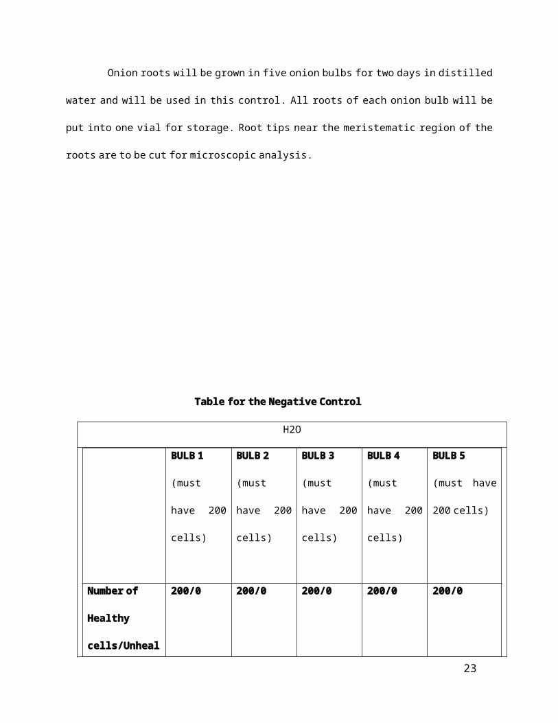
Negative Control
Onion roots will be grown in five onion bulbs for two days in distilled water and will be used in
this control. All roots of each onion bulb will be put into one vial for storage. Root tips near the
meristematic region of the roots are to be cut for microscopic analysis.
17

Table for the Negative Control
H2O
BULB 1
(must have
200 cells)
BULB 2
(must have
200 cells)
BULB 3
(must have
200 cells)
BULB 4
(must have
200 cells)
BULB 5
(must have 200
cells)
Number of
Healthy
cells/Unhealthy
cells
200/0 200/0 200/0 200/0 200/0
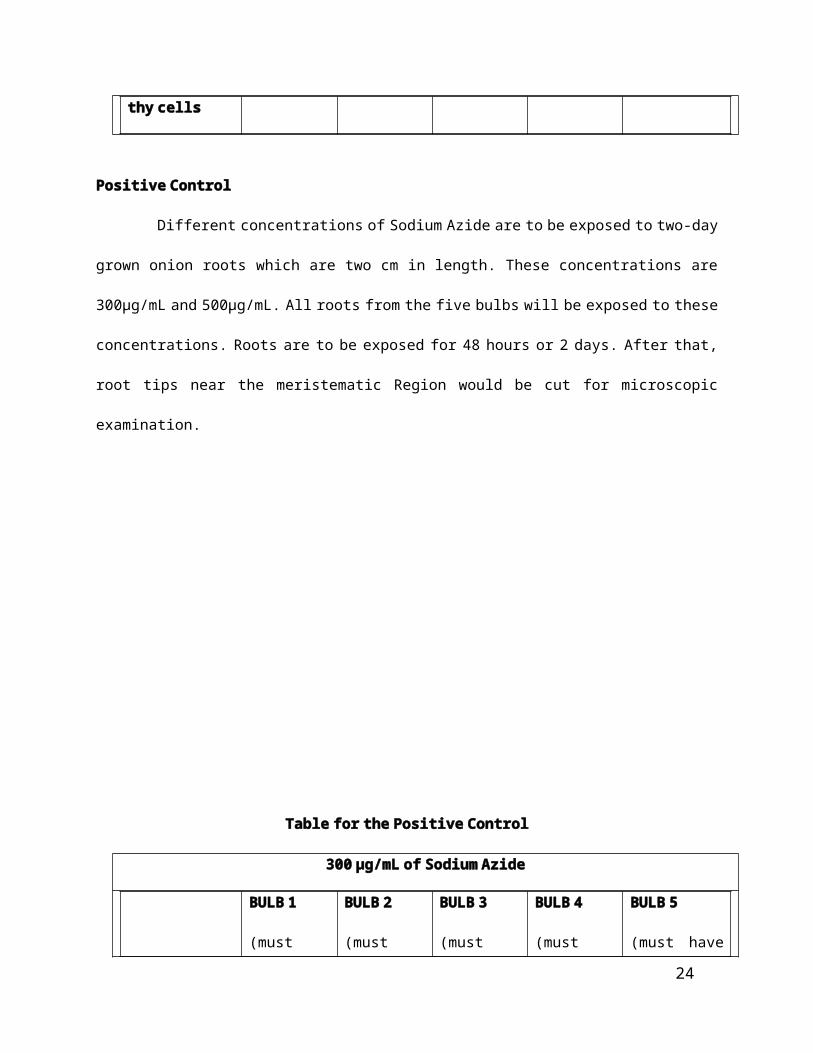
Positive Control
Different concentrations of Sodium Azide are to be exposed to two-day grown onion roots
which are two cm in length. These concentrations are 300µg/mL and 500µg/mL. All roots from the five
bulbs will be exposed to these concentrations. Roots are to be exposed for 48 hours or 2 days. After
that, root tips near the meristematic Region would be cut for microscopic examination.
18
Table for the Positive Control
300 µg/mL of Sodium Azide
BULB 1
(must have
200 cells)
BULB 2
(must have
200 cells)
BULB 3
(must have
200 cells)
BULB 4
(must have
200 cells)
BULB 5
(must have 200
cells)
Number of
Healthy
cells/Unhealthy
cells
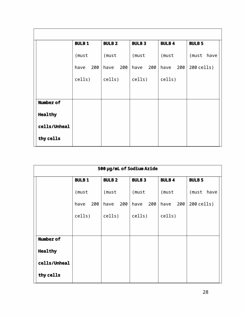
500 µg/mL of Sodium Azide
BULB 1
(must have
200 cells)
BULB 2
(must have
200 cells)
BULB 3
(must have
200 cells)
BULB 4
(must have
200 cells)
BULB 5
(must have 200
cells)
Number of
Healthy
cells/Unhealthy
cells
19

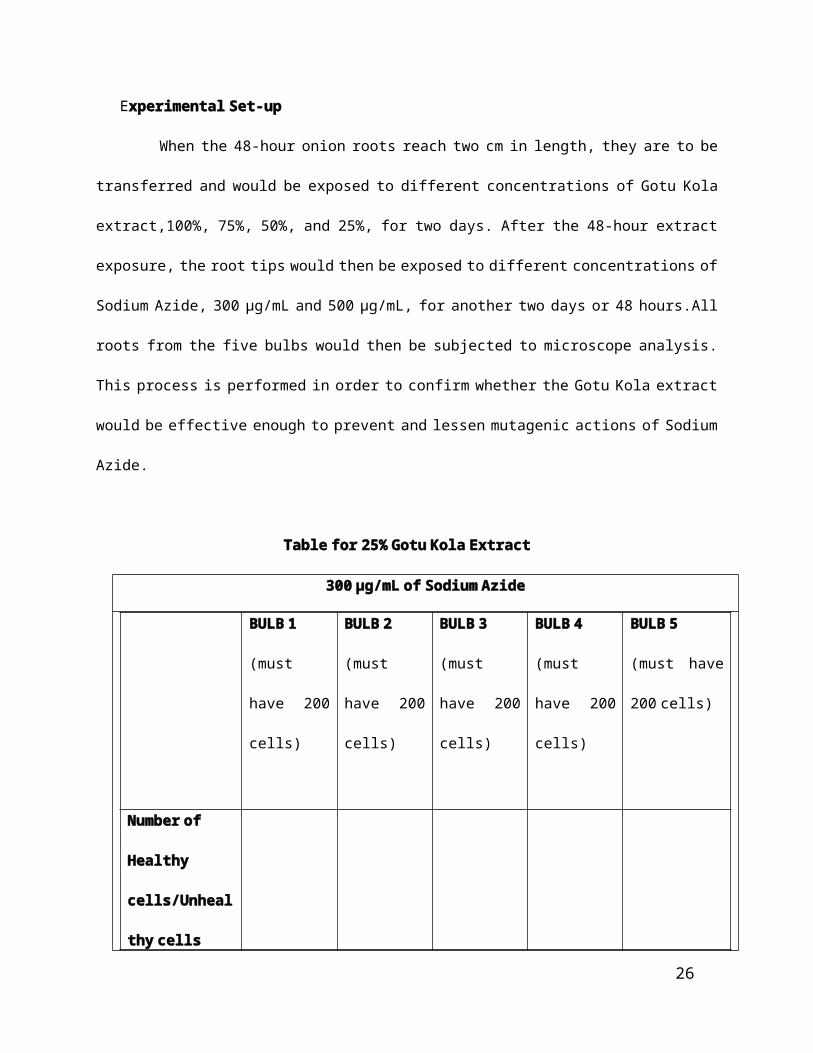
Experimental Set-up
When the 48-hour onion roots reach two cm in length, they are to be transferred and would be
exposed to different concentrations of Gotu Kola extract,100%, 75%, 50%, and 25%, for two days. After
the 48-hour extract exposure, the root tips would then be exposed to different concentrations of
Sodium Azide, 300 µg/mL and 500 µg/mL, for another two days or 48 hours.All roots from the five bulbs
would then be subjected to microscope analysis. This process is performed in order to confirm whether
the Gotu Kola extract would be effective enough to prevent and lessen mutagenic actions of Sodium
Azide.
Table for 25% Gotu Kola Extract
300 µg/mL of Sodium Azide
BULB 1
(must have
200 cells)
BULB 2
(must have
200 cells)
BULB 3
(must have
200 cells)
BULB 4
(must have
200 cells)
BULB 5
(must have 200
cells)
Number of
Healthy
cells/Unhealthy
cells
20

500 µg/mL of Sodium Azide
BULB 1
(must have
200 cells)
BULB 2
(must have
200 cells)
BULB 3
(must have
200 cells)
BULB 4
(must have
200 cells)
BULB 5
(must have 200
cells)
Number of
Healthy
cells/Unhealthy
cells
Table for 50% Gotu Kola Extract
300 µg/mL of Sodium Azide
BULB 1
(must have
200 cells)
BULB 2
(must have
200 cells)
BULB 3
(must have
200 cells)
BULB 4
(must have
200 cells)
BULB 5
(must have 200
cells)
Number of
21
Healthy
cells/Unhealthy
cells
500 µg/mL of Sodium Azide
BULB 1
(must have
200 cells)
BULB 2
(must have
200 cells)
BULB 3
(must have
200 cells)
BULB 4
(must have
200 cells)
BULB 5
(must have 200
cells)
Number of
Healthy
cells/Unhealthy
cells
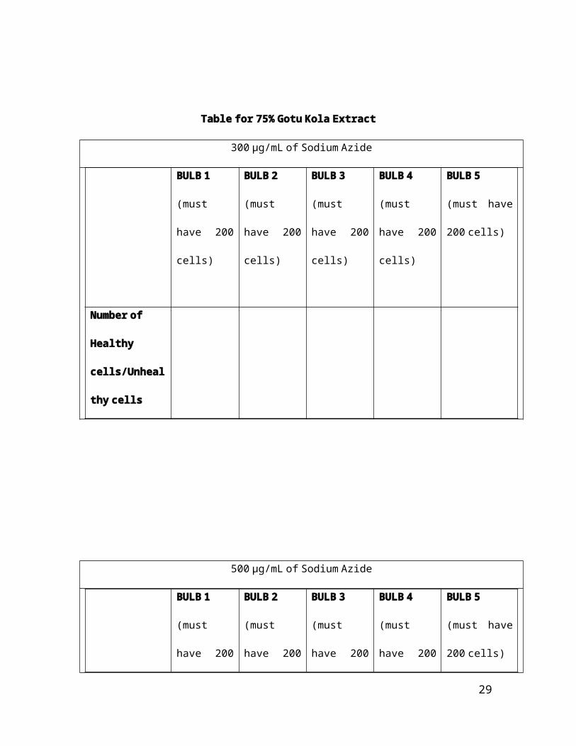
Table for 75% Gotu Kola Extract
300 µg/mL of Sodium Azide
BULB 1
(must have
200 cells)
BULB 2
(must have
200 cells)
BULB 3
(must have
200 cells)
BULB 4
(must have
200 cells)
BULB 5
(must have 200
cells)
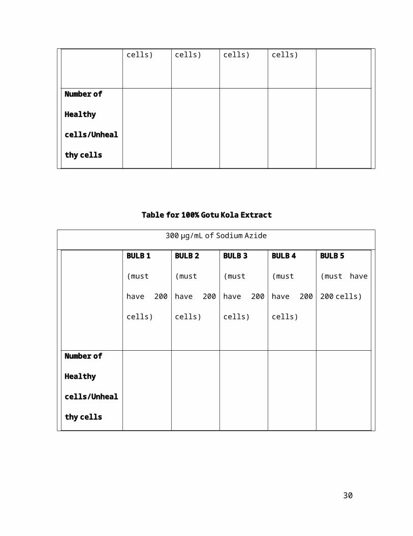
Number of
Healthy
cells/Unhealthy
22
cells
500 µg/mL of Sodium Azide
BULB 1
(must have
200 cells)
BULB 2
(must have
200 cells)
BULB 3
(must have
200 cells)
BULB 4
(must have
200 cells)
BULB 5
(must have 200
cells)
Number of
Healthy
cells/Unhealthy
cells
Table for 100% Gotu Kola Extract
300 µg/mL of Sodium Azide
BULB 1
(must have
200 cells)
BULB 2
(must have
200 cells)
BULB 3
(must have
200 cells)
BULB 4
(must have
200 cells)
BULB 5
(must have 200
cells)
23
Number of
Healthy
cells/Unhealthy
cells
500 µg/mL of Sodium Azide
BULB 1
(must have
200 cells)
BULB 2
(must have
200 cells)
BULB 3
(must have
200 cells)
BULB 4
(must have
200 cells)
BULB 5
(must have 200
cells)
Number of
Healthy
cells/Unhealthy
cells
Preparation of Slides
The cut roots would be put into slides. These slides are placed in vials having 37%
Formalin Solution for 1 hour. Different vials were used for every concentration in the positive
and experimental set-ups. The roots would be then exposed to 2 drops of 1M EDTA Solution for
25 minutes. Excess EDTA solution was removed using tissue paper. Then they were stained with
24
aceto-orcein stain for 30 minutes. After that, the roots would now be macerated, using the
Squash Technique, and were to be labeled accordingly.
Microscopic Examination
For the microscopic examination of the onion root cells, the researchers will set the
parameters to the meristematic cells of the root tip and will only count the first 1000 mitotic
cells that will be identified as normal or abnormal cells. Using the compound microscope at the
PSHS-EVC laboratory, the cells will be manually counted.
II. Anti-mutagenic Potential of Gotu Kola Extract (Centella Asiatica) In Vivo Rodent Erythrocyte
Micronucleus.
Research design
The methods used by Amano, Golo, and Tan are used also in this study with slight modifications.
A dosage of 0.1mL for each Gotu Kola Extract concentration was introduced in mice, which are
preliminarily induced with 0.1mL of Tetracycline Hydrochloride, using the Gavage Method. The Anti-
mutagenic effect of Different Concentrations of Gotu Kola Extract will be measured using the number of
Micronucleated Immature Erythrocytes out of 2000 immature erythrocytes per mouse but instead of
using the bone marrow of these mice, liver cells would be taken and be counted for the presence of
micronuclei
Procedure:
Acquisition of experimental animals
Mice that will be used in this process would be provided by the Leyte Marine Biotoxins Center
from their office in Government Center, Palo, Leyte but only with the factors of age and size taken into
consideration. Fifteen mice will be used.
25
Preparation of crude extracts
Leaves of Gotu Kola would be collected from Tacloban City. These leaves to be collected will be
chosen in such a way that each leaf is free from spots, cuts, tears, and discoloration. The collected leaves
would be then, washed with running water. In order to remove excess water from leaves, and be blot
dryed using tissue paper. After this, all leaves would be placed in a blender with ethanol as the solvent
for the extraction with the ratio 1:2 (mass of leaves is to volume of ethanol) and would be blended. The
blended leaves would be put in a refrigerator overnight. After 24 hours, the mixture would be filtered
with the use of cheesecloth and filter paper, twice.
To separate the extract from the solvent, ethanol, a rotary evaporator would be used. Once
the extract is free from ethanol, it would be prepared with concentrations 25%, 50%, 75%, 100% and
would be stored in three separate vials until further use.
Preparation of Test Animals
Fifteen mice will be used in this study. These mice are separated into 5 groups. 3 groups will be
used for the experimental set-up, one group for the positive control, which would be injected with
0.1mL of tetracycline only, and another group for the negative control, which would not be injected with
anything. Each group has 3 mice. The mice that would be used in the experimental set-up are made to
fast 12 hours prior to the conduct of the Gavage.
Induction of tetracycline
Amano, Golo, conducted preliminary inductions of Tetracycline Hydrochloride and Tan in order to
find out the tolerable dosage since the weight of each mouse is not specifically determined. They found
out that the maximum tolerable amount of tetracycline in mice is 0.1mL. Similarly, the dose of 0.1 mL
26
will be used in this study since factors that were only considered in choosing the mice are the age and
the size. 1cc/mL tuberculine syringes would also be used.
Intraperitoneal injection
The procedures found in the Laboratory Animal Workshop Manual of the Biological Research and
Development Laboratory for Intraperitoneal Injection would be followed.
First the mouse will be restrained and the injection site will be wiped with cotton soaked alcohol
(70% will be used in this research). Then the needle will be inserted ½ to 1 inch midway of the
abdominal cavity. The needle must not hit any visceral organs or blood vessels. Then the plunger of the
syringe will be pushed firmLy to inject the solution. Lastly, the needle will be removed.
This will be repeated to the experimental and the positive control groups using one tuberculine
syringe per mouse.
Induction of Gotu Kola extracts
Twelve hours after the Tetracycline induction, the Gotu Kola extracts would now be induced using
the Gavage Method. The standard procedures that will be followed for the Gavage Method is
mentioned in the Laboratory Animal Workshop Manual of the Biological Research and Development
Laboratory. The mouse must be restrained and be straightened. The Gavage needle would be inserted in
the mouth and throughout its entire length. Then the plunger is pushed to administer the solution in the
stomach and is then removed. 0.1 mL would be the only dosage used for each concentration of extract
in the experimental set-up.
27
Hematoxylin and Eosin Staining
The laboratory mice after it has been subjected to the different tests, will have their livers
removed and washed(following the proper protocol in handling and dissecting). Then samples would
then be soaked in Formalin solutions. Samples would then be cut and be placed in slides for staining.
The slides would then be placed in manual staining racks and would then be air dried in a 60 degrees C
oven for 30 to 40 minutes. Then the following would then be done (in order):
http://www.bcbiolibrary.icapture.ubc.ca/pathologists-researchers/docs/
BL.LAB.GN.006.01%20Haematoxylin%20and%20Eosin%20Staining.pdf
28
Data Analysis:
Micronuclei Scoring
The incidence of micronuclei in liver cells indicates that mutation has occurred. Because of this, the
number of micronucleated liver cells out of 2000 liver cells is determined. (OECD, 1997).
Analysis:
If the average ratio of one experimental set-up is lower compared to that of the Positive control
then the action of anti-mutagenesis has occurred.
Table for Experimental Set-up
0.1 mL of Tetracycline1 mL of 25% Concentration
1 mL of 50% Concentration
1 mL of 75% Concentration
Mice 1st 2nd 3rd 1st 2nd 3rd 1st 2nd 3rd
(number of micronucleus/2000 liver cells)
Table for Positive Control
0.1 mL of TetracyclineMice 1st 2nd 3rd
(number of micronucleus/2000 liver cells)
Table for Negative Control
No induction of tetracycline hydrochlorideMice 1st 2nd 3rd
(number of micronucleus/2000 liver cells)
29
1 mL of 100% Concentration
1st 2nd 3rd
Bibliography
Bhattacharya. (2010, July 17). Science Alert. Retrieved March 04, 2012, from Natural
Antimutagens: A Review: http://scialert.net/fulltext/?doi=rjmp.2011.116.126&org=10
Drugs.com. (n.d.). Tetracycline. Retrieved March 18, 2012, from Drugs.com:
http://www.drugs.com/tetracycline.htmL
Golo, & Amano. (n.d.). Anti-mutagenic effects of Ampalaya plant extracts against
tetracycline-induced mice based on PCE production.
Hawkins, & Ehrlich. (2006). Gotu Kola. University of Maryland Medical Center .
Jun, Kim, & Sung. (2002). Protective effect of soybean saponins and major antioxidants
against aflatoxin B1-induced mutagenicity and DNA-adduct formation.
Krishnaiah, Devi, Bono, & Sabatly. (2009). Studies on Phytochemical constituents of six
Malaysian medicinal Plants. Jounal of Medicinal Plants Research , 067-072.
Matsuda, Morikawa, Ueda, & Yoshikawa. (n.d.). Medicinal foodstuffs. XXVII. Saponin
constituents of Gotu Kola (2): structures of new ursane- and oleanane-type triterpene
oligoglycosides, centellasaponins B, C, and D, from Centella asiatica cultivated in Sri Lanka.
Retrieved March 04, 2012, from http://cpb.pharm.or.jp/cpb/200110/c10_1368.pdf
Ngelangel, & Wang. (2001, August 28). Japanese Journal of Clinical Oncology. Retrieved
March 04, 2012, from http://jjco.oxfordjournals.org/content/32/suppl_1/S52.full
Philippine Medicinal Plants. (n.d.). Takip-kohol. Retrieved February 26, 2012, from
Philippine Medicinal Plants: http://www.stuartxchange.org/TakipKohol.htmL
30

Qari. (n.d.). In vitro evaluation of the anti-mutagenic effect of Origanum majorana
extract . Retrieved march 04, 2012, from
http://jtusci.info/app_content/file/Journal-Files/Manuscript/2.pdf
Ragunathan, & Panneerselvam. (2007). Antimutagenic potential of curcumin on
chromosomal aberrations in Allium cepa. Retrieved March 04, 2012, from
http://www.ncbi.nlm.nih.gov/pmc/articles/PMC1906592/
Sathya, & Ganga, U. (n.d.). Therapeutic uses of Centella Asiatica. Retrieved February 26,
2012, from http://openmed.nic.in/2039/01/Microsoft_Word_-_Centella_asiatica.pdf
Stanton, Dotson, & Somers. (n.d.). Sodium Azide as a tissue culture mutagen. Retrieved
March 04, 2012, from http://www.agron.missouri.edu/mnl/61/148dotson.htmL
Tsutsui, Umeda, Sou, & Maizumi. (1976). Effect of tetracycline on cultured mouse cells.
Mutation Research , 261-8.
http://cshprotocols.cshlp.org/content/2008/5/pdb.prot4986.abstract
31


