

Dept. of Biomed. Eng. BME302: Biomedical Instrumentation Kyung Hee Univ.
http://ejwoo.com 1 Eung Je Woo
Chapter 11 Clinical Laboratory Instrumentation
Analysis of patient specimens: to aid in the diagnosis and evaluate the effectiveness
of therapy
Clinical phathology or clinical laboratory department
Chemistry: blood, urine, cerebrospinal fluid (CSF), and other fluids
Hematology: RBC, WBC, platelets, function of physiological systems in blood
(clotting)
Microbiology: pathological microorganisms in body tissues and fluids
Blood bank: ABO grouping, blood bank
Accuracy and precision are extremely important
Fast response is required
Automation using computer technology
11.1 Spectrophotometry
A general term for a class of instruments including photometers and colorimeters
Enough accuracy and precision, suitable for automation widely used
Substances of clinical interests selectively absorb or emit electromagnetic energy at
different wavelengths: ultraviolet (200 ~ 400 nm), visible (400 ~ 700 nm), and near
infrared (700 ~ 800 nm)
Beer's law: P P aLC 0 10 where
P
P
a
L
C
0
R
S|||
T|||
radiant power arriving at the cuvette
radiant power leaving the cuvette
absorptivity of the sample
length of the path through the sample
concentration of the absorbing substance
Percent transmittance, %TP
PaLC 100
100 100
Absorbance, AP
P TT aLC FHG
IKJ FHGIKJ log log
%log(% )0 100
2
Keep a and L constant and calibrate to get the absorbance, As of the same substance
with a known concentration, Cs . Then, the unknown sample concentration,

Dept. of Biomed. Eng. BME302: Biomedical Instrumentation Kyung Hee Univ.
http://ejwoo.com 2 Eung Je Woo
C CA
Au su
sFHGIKJ
Block diagram in Fig. 11.1
Power sources:
Wavelength selectors
Cuvette
Sample
Photometric system
Dept. of Biomed. Eng. BME302: Biomedical Instrumentation Kyung Hee Univ.
http://ejwoo.com 3 Eung Je Woo
Flame Photometers
Power source and sample-holder function are combined in the flame as in Fig. 11.2
In most cases, measure the sample's emission of light
Only for determining the concentrations of pure metals
Atomic Emission Flame Photometry
Limited use for only Na+, K+, and Li+ (with complicated optical system, Ca2+)
Only 1% of the atoms are raised to an excited level
Only a few elements produce enough power at a single wavelength as they move
back to lower-energy orbits
Fig. 11.2(a): sample combined with a solvent nebulizer flame
Fuel: propane or natural gas mixed with compressed air
Solvent evaporates and particles disintegrate to yield atoms
Atoms emit light as they move back to lower-energy orbits
Parallel determinations of Na+, K+, and Li+ Li+ is used as the internal standard to
correct the errors due to variations in the rate of solution uptake, aerosol production,
and flame characteristics
Good for small variations
Cannot be used for patients receiving Li+ to treat a psychotic disorder
Atomic Absorption Flame Photometry
Very accurate concentration determination for calcium, lead, copper, zinc, iron,
magnesium
Majority of atoms in a flame absorb energy at a characteristic wavelength
Block diagram (Fig. 11.2(b))
Power source: placed in an atmosphere of an inert gas, hollow cathode lamp
constructed from the metal to be determined (or coating)
Heat the cathode atoms leaves the cathode cathode cavity is filled with
atomic vapor atoms are excited due to collisions with electrons and ions
emit light when returning to the ground state this light is directed to the flame
the amount of the light absorption is proportional to the concentration
Monochromator
Detector: PM tube
Rotating-sector disk between the source and the flame pulse output from
power source phase-sensitive demodulator to differentiate the light emitted
Dept. of Biomed. Eng. BME302: Biomedical Instrumentation Kyung Hee Univ.
http://ejwoo.com 4 Eung Je Woo
by the atoms
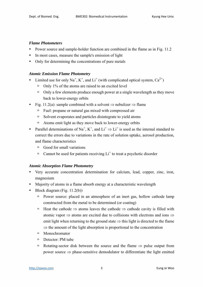
Fluorometry
Molecules: absorption of radiant energy being raised to an excited state emit
light in a characteristic spectrum
Block diagram (Fig. 11.3)
Power source: mercury arc lamp (365, 405, 436, and 546 nm)
Wavelength selector
Detector: PM tube, at the right angle to the power source to avoid direct light
transmission
Higher sensitivity (104 higher than spectrophotometry) and great specificity
picogram can be detected
Only a small number of substances have fluorescence property
Sensitive to pH and temperature
11.2 Automatic Chemical Analyzers
Spectrophotometric methods
Dept. of Biomed. Eng. BME302: Biomedical Instrumentation Kyung Hee Univ.
http://ejwoo.com 5 Eung Je Woo
Specimen aspiration, dilution, combination of sample with reagents, movement of
samples, computation, recording
Enhanced productivity and reduced response time
Synchron CX4
High-capacity specimen processing chemistry analyzer
Microcomputer-controlled discrete random-access clinical analyzer
Automated specimen handling
Performance of a variety of analytical test techniques
Extensive use of microcomputers
Bar code identification technique
End-point and rate assays at 30 and 37 C
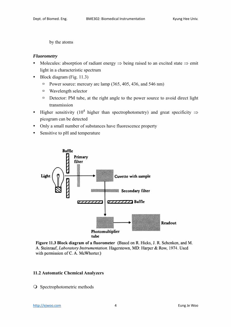
Automatic Clinical Analyzer (ACA)
Flexibility rather than high-capacity
Serial determination of any of 40 tests for each sample
Dept. of Biomed. Eng. BME302: Biomedical Instrumentation Kyung Hee Univ.
http://ejwoo.com 6 Eung Je Woo
11.3 Chromatology
A group of methods for separating a mixture of substances into component parts
Differences in the rate of movement of components of the mixture in the mobile phase
(gas or liquid) due to the interaction of these components with the stationary phase
(liquid or solid) four possible combinations
Liquid stationary phase: partition
Solid stationary phase: adsorption
Detection of complex substances such as drugs or hormones: gas-liquid
chromatographs (GLC) and thin-layer chromatographs (TLC)
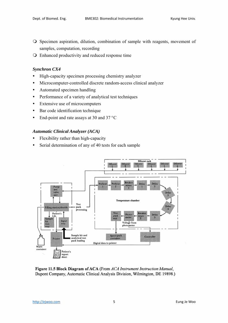
Gas-Liquid Chromatographs (GLC)
Block diagram (Fig. 11.7): fast response (15 min or 1 h), great sensitivity (1 ng), small
amount of sample (a few mL)
Injector
5 mL of sample in the solvent
Temperature is set to flash-evaporate he sample and solvent
Carrier gas: mobile phase
N2 or He (inert gas)
Sweeps the evaporated sample and solvent gas down the column
Column: stationary phase
1 m long, less than 7 mm diameter

Dept. of Biomed. Eng. BME302: Biomedical Instrumentation Kyung Hee Univ.
http://ejwoo.com 7 Eung Je Woo
Packed with solid support material (such as diatomaceous earth)
Solid support is coated with the liquid phase,
Enclosed in a temperature-controlled oven: temperature controller gradually
increases the temperature of the column for the best separation
Detector
At the end of the column
Output electrical signal proportional to the quantity of the compound in the
effluent gas (Fig. 11.8)
Ionization detector, thermal conductivity detector, electron capture detector
Recorder
X-axis (time) distinguishes the components
Y-axis (detector output) determines the quantity of the components
11.4 Electrophoresis
Measure proteins in plasma, urine, and CSF
Separate enzymes into their component isoenzymes
Identify antibodies
Dept. of Biomed. Eng. BME302: Biomedical Instrumentation Kyung Hee Univ.
http://ejwoo.com 8 Eung Je Woo
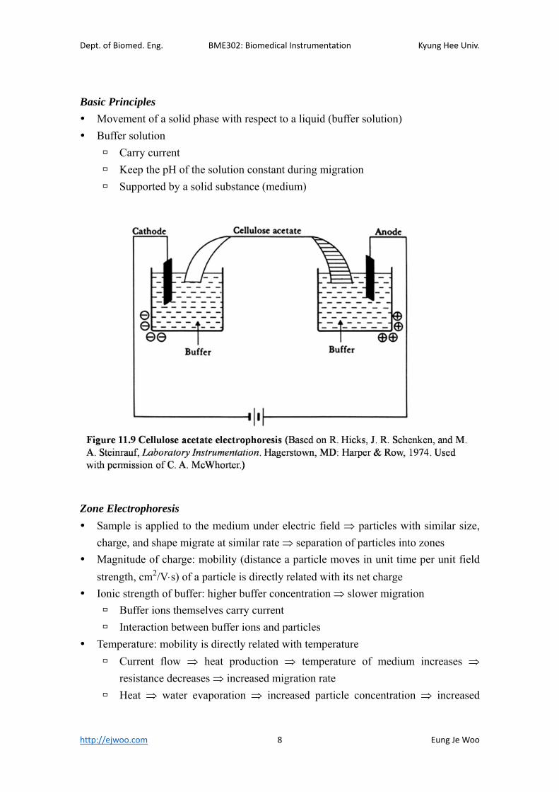
Basic Principles
Movement of a solid phase with respect to a liquid (buffer solution)
Buffer solution
Carry current
Keep the pH of the solution constant during migration
Supported by a solid substance (medium)
Zone Electrophoresis
Sample is applied to the medium under electric field particles with similar size,
charge, and shape migrate at similar rate separation of particles into zones
Magnitude of charge: mobility (distance a particle moves in unit time per unit field
strength, cm2/Vs) of a particle is directly related with its net charge
Ionic strength of buffer: higher buffer concentration slower migration
Buffer ions themselves carry current
Interaction between buffer ions and particles
Temperature: mobility is directly related with temperature
Current flow heat production temperature of medium increases
resistance decreases increased migration rate
Heat water evaporation increased particle concentration increased
Dept. of Biomed. Eng. BME302: Biomedical Instrumentation Kyung Hee Univ.
http://ejwoo.com 9 Eung Je Woo
migration rate
For gel type medium, constant-current source is used to minimize the heat
production
Time: distance of migration is directly related to the time
Types of support media: paper, cellulose acetate, starch gel, agar gel, acrylamide gel,
sucrose
Cellulose acetate (Fig. 11.19) is widely used
Constant voltage of 250 V for 15 ~ 20 min, initial current of 4 ~ 6 mA
Use fixative to fix the migrated protein bands to the buffer
Use dye to stain the bands
Dried for densitometry
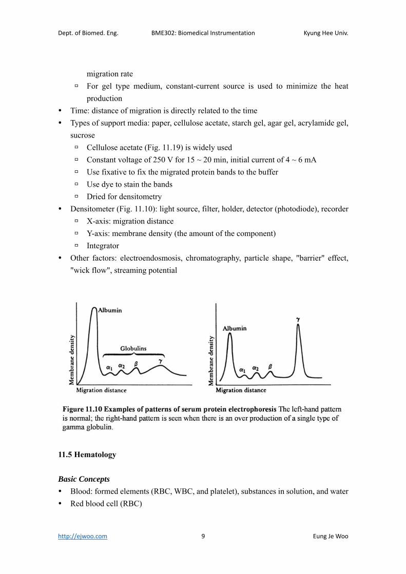
Densitometer (Fig. 11.10): light source, filter, holder, detector (photodiode), recorder
X-axis: migration distance
Y-axis: membrane density (the amount of the component)
Integrator
Other factors: electroendosmosis, chromatography, particle shape, "barrier" effect,
"wick flow", streaming potential
11.5 Hematology
Basic Concepts
Blood: formed elements (RBC, WBC, and platelet), substances in solution, and water
Red blood cell (RBC)

Dept. of Biomed. Eng. BME302: Biomedical Instrumentation Kyung Hee Univ.
http://ejwoo.com 10 Eung Je Woo
Carry oxygen and carbon dioxide
RBC count: 4.6 ~ 6.2106/L (normal adult male), 4.2 ~ 5.4106/L (normal
adult female),
White blood cell (WBC)
Defend the body against infection
Five types (decreasing order): neutrophils, lymphocytes, monocytes, eosinophils,
and basophils
WBC count: 4 500 ~ 11 000/L (normal adult male and female)
Platelet
Plug small breaks in the walls of the blood vessels
Participate in the clotting mechanism
Platelet count: 150 000 ~ 400 000/L (normal adult male and female)
Hematocrit (HCT):
% of the volume of all formed elements to the total volume of blood sample
40 ~ 54 % (normal adult men), 35 ~ 47 % (normal adult female)
Hemoglobin (Hb)
Conjugated protein in RBC
Transports most of O2 and a portion of CO2
13.5 ~ 18 g/dL (normal adult men), 12 ~ 16 g/dL (normal adult female)
RBC indices: characterization of RBC volume and Hb concentration
MCV (mean corpuscular volume): 82 ~ 98 m3
MCV10HCT
RBCcount
MCH (mean corpuscular hemoglobin): 27 ~ 31 pg
MCH10Hb
RBCcount
MCHC (mean corpuscular hemoglobin concentration): 32 ~ 35 %
MCHC100Hb
HCT
RDW (volume distribution width): measure of the spread of the RBC volume
distribution
Electronic Devices for Measuring Blood Characteristics
Detection of changes in electric resistance of a solution when a formed blood element
is passed through an aperture: Coulter, Clay Adams, Lors & Lundberg, Baker
Detection of deflection of light beam caused by the passage of formed blood elements:
Dept. of Biomed. Eng. BME302: Biomedical Instrumentation Kyung Hee Univ.
http://ejwoo.com 11 Eung Je Woo
Technicon
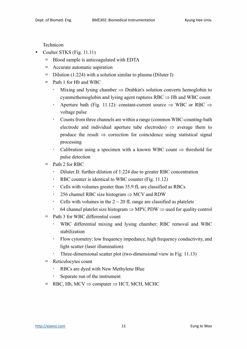
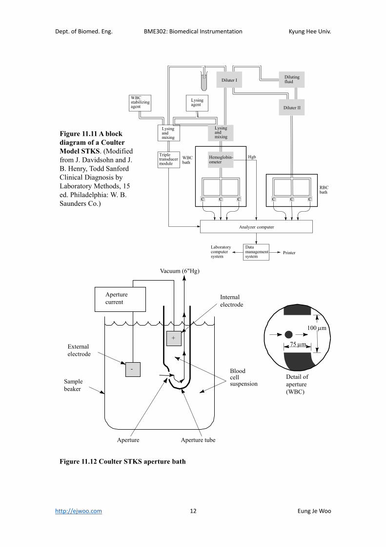
Coulter STKS (Fig. 11.11)
Blood sample is anticoagulated with EDTA
Accurate automatic aspiration
Dilution (1:224) with a solution similar to plasma (Diluter I)
Path 1 for Hb and WBC
Mixing and lysing chamber Drabkin's solution converts hemoglobin to
cyanmethemoglobin and lysing agent ruptures RBC Hb and WBC count
Aperture bath (Fig. 11.12): constant-current source WBC or RBC
voltage pulse
Counts from three channels are within a range (common WBC-counting-bath
electrode and individual aperture tube electrodes) average them to
produce the result correction for coincidence using statistical signal
processing
Calibration using a specimen with a known WBC count threshold for
pulse detection
Path 2 for RBC
Diluter II: further dilution of 1:224 due to greater RBC concentration
RBC counter is identical to WBC counter (Fig. 11.12)
Cells with volumes greater than 35.9 fL are classified as RBCs
256 channel RBC size histogram MCV and RDW
Cells with volumes in the 2 ~ 20 fL range are classified as platelets
64 channel platelet size histogram MPV, PDW used for quality control
Path 3 for WBC differential count
WBC differential mixing and lysing chamber: RBC removal and WBC
stabilization
Flow cytometry: low frequency impedance, high frequency conductivity, and
light scatter (laser illumination)
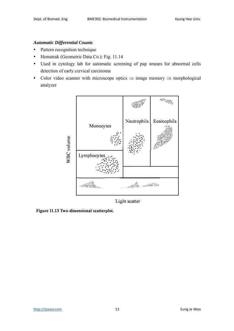
Three-dimensional scatter plot (two-dimensional view in Fig. 11.13)
Reticulocytes count
RBCs are dyed with New Methylene Blue
Separate run of the instrument
RBC, Hb, MCV computer HCT, MCH, MCHC
Dept. of Biomed. Eng. BME302: Biomedical Instrumentation Kyung Hee Univ.
http://ejwoo.com 12 Eung Je Woo
Figure 11.11 A block diagram of a Coulter Model STKS. (Modified from J. Davidsohn and J. B. Henry, Todd Sanford Clinical Diagnosis by Laboratory Methods, 15 ed. Philadelphia: W. B. Saunders Co.)
Analyzer computer
RBCbath
WBCbath
Hgb
Datamanagementsystem
Laboratorycomputersystem
Printer
Lysingagent
WBCstabilizingagent
Lysingandmixing
Tripletransducermodule
Diluter IDilutingfluid
Diluter II
Hemoglobin-ometer
C CC C CC
Lysingandmixing
Figure 11.12 Coulter STKS aperture bath
Internalelectrode
Bloodcellsuspension
Aperture tube
Detail ofaperture(WBC)
Aperture
Samplebeaker
Externalelectrode
Vacuum (6"Hg)
+
-
Aperturecurrent
100 m
75 m
Dept. of Biomed. Eng. BME302: Biomedical Instrumentation Kyung Hee Univ.
http://ejwoo.com 13 Eung Je Woo
Automatic Differential Counts
Pattern recognition technique
Hematrak (Geometric Data Co.): Fig. 11.14
Used in cytology lab for automatic screening of pap smears for abnormal cells
detection of early cervical carcinoma
Color video scanner with microscope optics image memory morphological
analyzer

Dept. of Biomed. Eng. BME302: Biomedical Instrumentation Kyung Hee Univ.
http://ejwoo.com 14 Eung Je Woo


